Báo cáo khoa học: Perturbation of folding and reassociation of lactate dehydrogenase by proline and trimethylamine oxide potx
Bạn đang xem bản rút gọn của tài liệu. Xem và tải ngay bản đầy đủ của tài liệu tại đây (502.19 KB, 12 trang )
Perturbation of folding and reassociation of lactate dehydrogenase
by proline and trimethylamine oxide
Oscar P. Chilson and Anne E. Chilson
Department of Biology, Washington University, St Louis, MO, USA
Investigations of protein–solute interactions typically show
that osmolytes favor native conformations. In this study,
the effects of representative compatible and counteracting
osmolytes on the reactivation of lactate dehydrogenase from
two different conformational states were explored. Contrary
to expectations, proline and trimethylamine oxide inhibited
both the initial time course and the extent of reactivation of
lactate dehydrogenase from bovine heart following dena-
turation in guanidine hydrochloride, as well as following
inactivation at pH 2.3. Reactivation of acid-dissociated
porcine heart lactate dehydrogenase was inhibited by both
proline and trimethylamine oxide (2
M
). In all instances,
trimethylamine oxide was the more effective inhibitor of
reactivation. Analysis of the catalytic properties of the
reactivating enzyme provided evidence that the molecular
species that was enzymatically active during the initial stages
of reactivation of acid-inactivated porcine heart lactate
dehydrogenase reflects a non-native conformation. Proline
and trimethylamine oxide stabilize polypeptides through
exclusion from the polypeptide backbone; the inhibition of
renaturation/reassociation described here is probably due to
attenuation of this stabilizing influence through favorable
interactions of the osmolytes with sidechains of residues that
lie at the interfaces of the monomers and dimers that asso-
ciate to form the active tetramer. In addition, these osmo-
lytes may stabilize non-native intermediates in the folding
pathway. The high viscosity of solutions containing more
than 3
M
proline was a major factor in the inhibition of
reassociation of acid-dissociated porcine heart lactate
dehydrogenase as well as other viscosity-dependent trans-
formations that may occur during reactivation following
unfolding in guanidine hydrochloride.
Keywords: renaturation; osmolytes; proline; trimethylamine
oxide; lactate dehydrogenase.
In order to accommodate environmental water stress (e.g.
salinity, desiccation, freezing), many organisms accumulate
one or more osmotically active solutes (osmolytes) [1]. Two
classes of osmolytes are recognized. Those that stabilize
proteins in vitro without significantly perturbing protein
function are defined as compatible osmolytes [2]. Counter-
acting osmolytes, such as trimethylamine oxide (TMAO),
tend to buffer proteins and other cellular constituents
against elevated concentrations of chaotropic agents such
as urea [1,3,4].
Efforts to delineate the molecular basis of osmolyte
action have generated large amounts of literature on
protein–solute interactions. Virtually all studies of the
effects of osmolytes on protein stability have demonstrated
that these chemical chaperones strongly favor the native
conformation. For example, TMAO protects ribonuclease
T1 against thermal denaturation [4]. There are also several
reports by Bolen and coworkers showing that both proline
and TMAO, as well as other osmolytes, have a propensity
for ÔforcingÕ intrinsically unstable polypeptides to fold into
more compact, native-like, conformations (e.g. [5]).
Interest in protein–osmolyte interactions arises from
several considerations. Inasmuch as unfolded polypeptides
would be expected to be particularly sensitive to environ-
mental stress and protease action, it is reasonable to ask
whether osmolytes may facilitate the folding of nascent
polypeptides; i.e. perhaps one of the functions of osmolytes
is to act as chemical chaperones during the terminal stages
of protein synthesis. Observations showing the effects of
osmolytes on protein conformation in vivo provide support
for this hypothesis [6–10].
Several investigators have noted that some osmolytes are
of potential practical use in the rescue of inclusion bodies
[e.g. 7,11]. It is also conceivable that coexpression of appro-
priate osmolytes may retard or prevent the formation of
such aggregates in expression systems.
With apparently few exceptions [e.g. 1,7,11–14], previous
investigations did not include testing of the possible effects
of osmolytes on the kinetics of reactivation of denatured
polypeptides. Also, study of the effects of osmolytes on the
reactivation of oligomeric, cytosolic proteins seems to have
been somewhat limited (see Discussion).
In the light of these considerations, we chose to
explore the possible effects of osmolytes on renaturation/
Correspondence to O. P. Chilson, Department of Biology, Box1137,
One Brookings Drive, Washington University, St. Louis,
MO, 63130–4899, USA.
Fax: + 314 9354432, Tel.: + 314 9356859,
E-mail: or
Abbreviations: BHLDH, lactate dehydrogenase from bovine heart;
EDTA, ethylenediaminetetraacetic acid; GdnHCl, guanidine hydro-
chloride; LDH, lactate dehydrogenase; NADH, nicotinamide adenine
dinucleotide (reduced); PHLDH, lactate dehydrogenase from porcine
heart; TMAO, trimethylamine oxide.
Enzyme: lactate dehydrogenase (EC 1.1.1.27).
(Received 27 May 2003, revised 18 October 2003,
accepted 20 October 2003)
Eur. J. Biochem. 270, 4823–4834 (2003) Ó FEBS 2003 doi:10.1046/j.1432-1033.2003.03881.x
reassociation of lactate dehydrogenase (LDH; EC 1.1.1.27).
The choice of experimental system was based on the
following considerations. LDH is an oligomer, comprised of
four polypeptides of identical size, and its refolding and
reactivation following denaturation/dissociation in various
solvent media have been extensively investigated. Thus, the
pathway for refolding and reassociation is generally well
established [15]. Reactivation of LDH following denatura-
tion in 6
M
guanidine hydrochloride (GdnHCl) begins with
the fully unfolded polypeptide subunits and the time course
reflects a complex series of molecular events that include
folding, dimerization of monomers and association of the
dimers to form the active tetramer (see Discussion).
However, when acid-dissociated monomers are stabilized
by sodium sulfate, the rate limiting step is restricted to
association of the dimer to produce the active tetrameric
species [15]. Thus, study of renaturation (following unfold-
ing in GdnHCl), as well as reassociation (following inacti-
vation at low pH), allowed exploration of the effect of
osmolytes on the reactivation of the enzyme from two very
different conformational states.
In this investigation we explored the effects of represen-
tative compatible and counteracting osmolytes on the
kinetics and extent of reactivation of LDH from beef heart
following denaturation in 6
M
GdnHCl, as well as following
dissociation at pH 2.3 in the presence of sodium sulfate. In
contrast with expectation, based on results obtained with
other proteins [5,11,16], TMAO and proline were found to
inhibit both the time course and extent of renaturation of
LDH from bovine heart (BHLDH) following unfolding by
the chaotropic agent, as well as reactivation of the enzyme
following inactivation at low pH. Reactivation of acid-
dissociated LDH from porcine heart (PHLDH) was also
sensitive to both osmolytes. Evidence was obtained that the
molecular species that is enzymatically active during the
initial stages of reactivation of acid-inactivated PHLDH
reflects an altered conformation and that this non-
native species is kinetically stabilized by interaction with
osmolytes.
Materials and methods
Dithiothreitol, ethylenediaminetetraacetic acid (EDTA),
GdnHCl, LDH from bovine heart (type III), nicotinamide
adenine dinucleotide (reduced, NADH), sodium pyruvate
and Tris base were obtained from Sigma-Aldrich (St.
Louis, MO, USA). Lactate dehydrogenase from porcine
heart was from Roche Molecular Biochemical (Indiana-
polis, IN, USA).
L
-Proline was from Sigma-Aldrich
(Sigma Ultra) or Fluka (MicroSelect; Milwaukee, WI,
USA). Trimethylamine N-oxide dihydrate (> 99%) was
from Fluka. N,N-Bis(hydroxyethyl)-2-aminoethane sulfon-
ic acid (Bes) was from Research Organics (Cleveland, OH,
USA).
Enzyme stock solutions
Stock solutions of enzyme (% 5–9 mgÆmL
)1
) were prepared
by dialysis (% 5 °C) against 100 m
M
Tris/HCl, 1 m
M
EDTA (pH 7.4; prior to denaturation in GdnHCl) or
100 m
M
sodium phosphate, 100 m
M
EDTA, 1 m
M
dithio-
threitol (pH 7.6; prior to dissociation at low pH).
Enzyme concentration
Enzyme concentration (mg proteinÆmL
)1
) was calculated
from A
0:1%
280
¼ 1.5 for BHLDH [17] and 1.4 for PHLDH
[18]. The preparation of LDH from beef heart was
composed of approximately 70% H
4
and 30% H
3
M[17];
i.e. > 92% H subunits. The preparation from pig heart was
composed of approximately 95% H
4
[19] and a small
fraction of H
3
M; it contained % 98% H subunits.
Assay of enzymatic activity
This was performed at room temperature (21–24 °C) by
measurement of the rate of decrease in absorbance at
340 nm with a Shimadzu 1601PC spectrophotometer.
Reaction mixtures (1.021 mL, in polystyrene cuvettes)
contained 128 l
M
NADH, 350 l
M
sodium pyruvate (unless
stated otherwise) and approximately 1 pmol enzyme (added
last). The buffer for the assay was 100 m
M
potassium Bes
(or Tris/HCl, pH 7.0, for enzyme denatured in GdnHCl)
or 100 m
M
sodium phosphate, 1 m
M
EDTA (pH 7.6, for
enzyme inactivated at acid pH). The specific activities of the
BHLDH and PHLDH were 317 and 310 UÆmg
)1
, respect-
ively, at 22 °C. Molar concentration of enzyme was based
on a tetramer molecular mass of 140 000 Da.
None of the osmolytes tested inhibited enzymatic activity
of the untreated enzyme at the concentrations that were
present during enzyme assays (£ 60 m
M
TMAO, £ 150 m
M
proline; data not shown).
Denaturation and acid-induced dissociation
Apart from where indicated, unfolding in 6
M
GdnHCl was
initiated by the addition of a 10 lL aliquot of stock enzyme
(containing 76–81 lg LDH) to 90 lL6.7
M
GdnHCl in
100 m
M
Tris/HCl , 1 m
M
EDTA (pH 7.4). The inactivation
mixtures were incubated for 10 min at room temperature.
Inactivation at pH 2.3 was initiated by addition of 8–9 lL
LDH (containing 52–54 lg protein) to 91–92 lLcold
100 m
M
sodium phosphate, 800 m
M
sodium sulfate
(pH 2.3); samples were incubated on ice for 60 min. All
incubations, for both inactivation and reactivation, were
performed in polypropylene tubes.
Reactivation
Renaturation following unfolding in GdnHCl was initiated
by 50-fold dilution in 100 m
M
Tris/HCl, 1 m
M
EDTA,
2m
M
dithiothreitol (pH 7.4), with or without the indicated
concentration of the specified osmolytes (proline or
TMAO); stock solutions of osmolytes were adjusted to
pH 7.4. All reactivations were performed at room tempera-
ture. Protein concentration in these reactivation mixtures
was typically 15–16 lgÆmL
)1
.
Reactivation of acid-inactivated enzyme was initiated by
50- or 100-fold dilution in 100 m
M
sodium phosphate, 1 m
M
EDTA, 1 m
M
dithiothreitol (pH 7.6) plus or minus the
indicated osmolytes (TMAO or proline) at room tempera-
ture; stock solutions of osmolytes were adjusted to pH 7.6.
The protein concentrations in reactivation mixtures are
specified in the relevant figure legends. Aliquots were
removed from reactivation mixtures at the indicated times
4824 O. P. Chilson and A. E. Chilson (Eur. J. Biochem. 270) Ó FEBS 2003
after initiation of reactivation and assayed for enzymatic
activity as described above.
Molecular graphics analysis
Two models of the structure of the H
4
isoform of LDH are
found in the RCSB Protein Data Bank (PDB). The PDB
code for porcine H
4
is 5LDH. Analysis of both this model
and the one for the major isoform from human cardiac
muscle (PDB code 1I0Z) by
DEEP VIEW
[20,21] shows that
the latter is the superior model. This assessment was based
on the fact that opening up the model for 5LDH in
DEEP
VIEW
, reveals a lengthy list of missing amino acid sidechains;
this reflects uninterpretable electron density in those areas.
There are no such uncertainties in the model for 1I0Z. As
the primary structures for LDH H
4
from pig and human
heart are 95% identical (97% similar) we chose to base our
analysis of buried and surface residues on the human
enzyme. The model for 1I0Z is for the dimer. We obtained
a model for the tetramer from 1I0Z through
PROTEIN
EXPLORER
[22], using the link to protein quaternary analysis
(PQS [23]).
Results
Effect of osmolytes on the reactivation of bovine LDH
following denaturation in GdnHCl
Proline inhibits the rate of reactivation. The equilibrium
level of reactivation of BHLDH in the absence of osmolytes,
following denaturation in 6
M
GdnHCl (65 ± 2.8%,
relative to the activity of the untreated enzyme; data not
shown), was several-fold greater than that reported for
similar studies of PHLDH under similar conditions [24].
For each experiment, the activity for the control (no
osmolyte in the reactivation mixture), determined 24 h after
initiation of reactivation, was taken as representing the
equilibrium level of reactivation under the experimental
conditions employed and was assigned a value of 1.0. The
enzymatic activity observed at intermediate times (with or
without osmolyte) was expressed as a fraction of this
equilibrium control value and was defined as Ôrelative
reactivationÕ. The initial time course for reactivation of
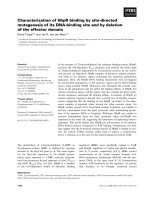
controls was routinely hyperbolic (Fig. 1).
The kinetic profile for reactivation in the presence of 1
M
proline was virtually indistinguishable from that of controls,
but in the presence of increasing concentrations of proline,
the time course became increasingly sigmoidal (Fig. 1). Due
to this sigmoidicity, the effects of intermediate levels of
proline (2
M
and 3
M
) were most pronounced during the
early phase of reactivation; e.g. while inhibition by 2
M
proline was significant during the first hour, by 5 h the
activity approached that of controls. Relative reactivation in
the presence of 4 and 5
M
proline remained at less than 0.1
throughout the indicated time period.
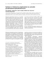
Inhibition of the extent of reactivation of LDH by proline
is correlated with the unusual solution properties of
proline. Relative reactivation, based on activity determined
at presumed equilibrium (24 h after initiation of reactiva-
tion), was taken as a measure of the extent of reactivation.
The effect of proline concentration on the extent of
reactivation of LDH is summarized in Fig. 2A. At 1
M
proline, reactivation was unaffected, and 2
M
proline
diminished the extent of reactivation only slightly, but
inhibition was progressively more significant above 3
M
;at
5
M
proline, inhibition was virtually complete.
The concentration dependence of the viscosity of aqueous
solutions of proline is somewhat unusual relative to that of
compounds of similar molecular weight [26]; values for the
viscosities of proline solutions over the concentration range
from 1 to 6
M
are included in Fig. 2. Inhibition of
reactivation is most pronounced in solutions of proline that
exhibit the greatest viscosity. This relationship is illustrated
more clearly in Fig. 2B.
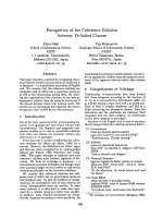
Trimethylamine oxide is a potent inhibitor of the extent of
reactivation of GdnHCl-denatured LDH. The effect of
TMAO on the relative reactivation at equilibrium is
summarized in Fig. 3. The data for proline are included
for comparison. The counteracting osmolyte, TMAO, was
the more potent inhibitor of reactivation. The concentration
of TMAO required to reduce the relative reactivation at
equilibrium to 0.5 was approximately 700 m
M
, while the
concentration of proline that was required to elicit a similar
level of inhibition was 3.2
M
.
Perturbation of the reactivation of acid-dissociated
LDH by proline and TMAO
Reactivation of acid-dissociated BHLDH was inhibited
by proline. Both the initial rate and extent of reactivation
of bovine LDH, following dissociation at pH 2.3, were
significantly greater than for enzyme denatured in GdnHCl.
Fig. 1. Effect of proline on the kinetics of reactivation of LDH from beef
heart after denaturation in GdnHCl. Enzyme was denatured in 6
M
GdnHCl as described in Materials and methods. Reactivation was
initiated by 50-fold dilution (to 15 lgÆmL
)1
)in100m
M
Tris/HCl,
1m
M
EDTA, 2 m
M
dithiothreitol (pH 7.4) in the absence (control) or
presence of the indicated concentrations of proline. The time course
over the first 5 h after initiation of reactivation is illustrated. For each
experiment, the enzymatic activity at each time point was expressed
relative to the activity of the corresponding control, determined 24 h
after initiation of reactivation; the relative reactivation of controls at
24 h was assigned a value of 1.0. Each of the lines shown for proline
represents a single experiment, the points for the line for controls reflect
five independent determinations.
Ó FEBS 2003 Perturbation of protein folding by osmolytes (Eur. J. Biochem. 270) 4825
Similar differences have been reported by Jaenicke and
coworkers in studies of the porcine LDH [27]. The time
required for relative reactivation in the absence of osmolytes
to reach 0.5 during reactivation from GdnHCl was
% 30 min (Fig. 1), but % 5 min for enzyme inactivated at
acid pH in the absence of the chaotropic agent (Fig. 4A).
Activity at apparent equilibrium in the absence of osmolytes
following acid dissociation approached 90% of the activity
of the untreated enzyme (data not shown). The time courses
for reactivation of acid-dissociated enzyme in the absence
of osmolytes, as well as in the presence of 1 or 2
M
proline,
were hyperbolic and virtually indistinguishable (Fig. 4A);
3
M
proline, however, was inhibitory and the time required
to attain a relative reactivation of 0.5 was increased to
% 20 min. At 4
M
proline the time course became slightly
sigmoidal and the time required to reach a relative
reactivation of 0.5 was % 90 min (Fig. 4A); the initial rate
of reactivation in the presence of 5
M
proline was virtually
zero and relative reactivation rose only slightly (to % 0.1)
over the 5 h incubation period (Fig. 4A).
Proline concentrations up to 3
M
did not inhibit the
extent of reactivation (Fig. 4B); at 4 and 5
M
proline,
relative reactivation was reduced to approximately 0.85 and
0.3, respectively. A limited analysis of the effect of high
proline concentrations on the reactivation of acid-inacti-
vated PHLDH yielded similar results (data not shown).
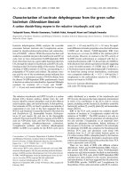
Trimethylamine oxide inhibits the rate of reactivation of
bovine LDH following inactivation at acid pH. The time
required to reach a relative reactivation of 0.5 was increased
from % 5to% 25 min by 1
M
TMAO; at 5 h, relative
reactivation with the osmolyte approached that of controls
(Fig. 5A). When reactivation was performed in the presence
of 2
M
TMAO, relative reactivation was less than 0.1 at 5 h
after initiation of reactivation.
TMAO (1
M
) enhances the initial rate of reactivation of
porcine LDH following inactivation at pH 2.3, while 2
M
TMAO inhibits reactivation. The time course for reacti-
vation of acid-dissociated PHLDH in the absence of
osmolyte was hyperbolic (Fig. 5B), and reactivation
reached apparent equilibrium at approximately 90% of
the activity of the untreated enzyme (data not shown).
The initial rate of reactivation was slower that that of the
beef enzyme; the time required to reach a relative
reactivation of 0.5 was increased from % 5min(Fig.5A,
BHLDH) to % 20 min (Fig. 5B, PHLDH). In marked
contrast to the inhibitory effect of 1
M
TMAO on
reactivation of the bovine enzyme (Fig. 5A), this concen-
tration of the osmolyte enhanced the rate of reactivation
of the pig dehydrogenase; the time required to reach a
relative reactivation of 0.5 was reduced from % 20 min to
% 7 min (Fig. 5B). At 5 h after initiation of reactivation
the activities of controls and the mixture containing 1
M
Fig. 3. Comparison of the effects of TMAO, and proline on the extent of
reactivation of GdnHCl-denatured BHLDH. Enzyme was denatured
and reactivated as described in the legend for Fig. 1 in the absence and
presence of the indicated concentrations of the osmolytes. Relative
reactivation at presumed equilibrium was assessed as described in the
legend for Fig. 2. Each point represents the mean of two independent
determinations; error bars are shown where the magnitude of the error
exceeds the size of the symbol.
Fig. 2. Effect of proline concentration on the extent of reactivation of
LDH from bovine heart and on the viscosity of proline solutions. Enzyme
was denatured in GdnHCl and reactivated as described in the legend
for Fig. 1. (A) Relative reactivation at presumed equilibrium (j)was
taken as a measure of the extent of reactivation at each proline con-
centration. These values were based on measurements of enzymatic
activities of reactivation mixtures (with and without added osmolyte)
24 h after initiation of reactivation. Each point represents the mean of
two independent determinations; error bars are shown where the
magnitude of the error exceeds the size of the symbol. Values of
intrinsic viscosity for proline solutions (.,g) were taken from a pre-
vious report by Schobert and Tschesche [25], with permission from
Elsevier Science. (B) A plot of the values for the extent of reactivation
vs. the viscosity of proline solutions, both taken from (A).
4826 O. P. Chilson and A. E. Chilson (Eur. J. Biochem. 270) Ó FEBS 2003
TMAO reached similar levels. In the presence of 2
M
TMAO, however, the initial rate of reactivation of
PHLDH was strongly inhibited, but slightly less so than
with BHLDH (Fig. 5A,B).
TMAO (2
M
) significantly reduced the extent of reacti-
vation of both bovine and porcine LDH. In the presence of
1
M
TMAO, the relative reactivation at presumed equilib-
rium for the porcine enzyme was slightly greater than that of
controls, while the value for the bovine enzyme was reduced
to % 0.9 (Fig. 6). In 2
M
TMAO, the relative reactivation at
equilibrium was reduced to % 0.4 and % 0.1 for PHLDH
and BHLDH, respectively.
As with reactivation of enzyme denatured in GdnHCl,
TMAO was a more potent inhibitor of reactivation of acid-
denatured BHLDH than proline. The concentration of
TMAO required to reduce relative reactivation at equilib-
rium to 0.5 for enzyme dissociated at pH 2.3 was approxi-
mately 1.5
M
(Fig. 6), whereas for a similar level of
inhibition by proline, the concentration required was
approximately threefold greater (compare Figs 4B and 6).
The time course of reactivation of acid-dissociated LDH
in the presence of proline is dependent on protein
concentration. The initial time course for the reactivation
of PHLDH in the absence of osmolytes was dependent on
the concentration of LDH protein in the reactivation
mixture (Fig. 7A), consistent with a pathway involving a
rate-determining association step (see Discussion). If attenu-
ation, by osmolytes, of the reactivation of enzyme inacti-
vated at low pH involves modulation of a rate-determining
Fig. 5. Effect of TMAO on the kinetics of reactivation of LDH from
bovine and porcine heart after inactivation at pH 2.3. Inactivation at
pH 2.3 was performed as described in Materials and methods. Reac-
tivation was initiated by 100-fold dilution (to 5.4 lgproteinÆmL
)1
)
in 100 m
M
sodium phosphate, 1 m
M
EDTA, 1 m
M
dithiothreitol
(pH 7.6) in the absence (control) and presence of the indicated con-
centrations of TMAO. Relative reactivation was assessed as described
in the legend for Fig. 1. (A) Data obtained with LDH from bovine
heart. (B) Data obtained with LDH from porcine heart. Each point for
the controls represents the mean of four independent determinations;
error bars are shown where the magnitude of the error exceeds the size
of the symbol. The points for the lines for the experiments with TMAO
represent the means of two independent experiments; error bars are
shown where the magnitude of the error exceeds the size of the symbol.
Fig. 4. Effect of proline concentration on the kinetics and extent of
reactivation of LDH from bovine heart after inactivation at pH 2.3.
Inactivation was in 100 m
M
sodium phosphate, 800 m
M
sodium sul-
fate (pH 2.3) as described in Materials and methods. Reactivation was
initiated by 100-fold dilution (to 5.4 lgproteinÆmL
)1
) in 100 m
M
sodium phosphate, 1 m
M
EDTA, 1 m
M
dithiothreitol (pH 7.6) in the
absence (control) and presence of the indicated concentrations of
proline. (A) The time course of reactivation during the first 5 h after
initiation of reactivation. Each of the lines shown for proline represents
a single experiment, the points for the line for controls reflect five
independent determinations. (B) Effect of proline on the extent of
reactivation. The extent of reactivation was assessed as described in the
legend for Fig. 2. Each point represents the mean of two independent
determinations; error bars are shown where the magnitude of the error
exceeds the size of the symbol.
Ó FEBS 2003 Perturbation of protein folding by osmolytes (Eur. J. Biochem. 270) 4827
association step, the kinetics of reactivation in the presence
of an inhibitory concentration of osmolyte should also be
dependent on protein concentration. Proline-inhibited reac-
tivation of acid-dissociated LDH was also protein concen-
tration-dependent (Fig. 7B). TMAO-inhibited reactivation
of acid-dissociated enzyme, however, was independent of
protein concentration (Fig. 7C).
Analysis of interfacial contacts in lactate dehydrogenase
from cardiac muscle. The program
MS
[28] was used to
calculate the surface area buried in each subunit upon
formation of the tetramer, based on the coordinates
provided in PDB file 1I0Z, as modified as described in
Materials and methods. Approximately 55% of these
buried sidechains are nonpolar in nature (Table 1). When
the model for the native tetramer was analyzed for groups
on the surface that are exposed to solvent using
DEEP VIEW
[20,21], % 43% were found to be nonpolar (data not shown).
Effect of osmolytes on the kinetic properties of PHLDH
during reactivation following acid-induced dissoci-
ation. The H
4
isoform of LDH is particularly sensitive to
pyruvic acid [29]. An early study of the reactivation of LDH
from avian cardiac muscle, following unfolding in GdnHCl,
showed that during the initial stage of reactivation there
were one or more enzymatically active species that exhibited
diminished thermal stability and reduced inhibition by
pyruvic acid [30]. It was of interest therefore to determine
whether during reactivation of acid-inactivated PHLDH
there were enzymatically active species that exhibited altered
pyruvate sensitivity and whether concentrations of proline
and/or TMAO that inhibited reactivation kinetically stabil-
ized these non-native molecular species.
Two identical aliquots of PHLDH were inactivated at
pH 2.3; during reactivation, one reactivation mixture was
assayed at 350 l
M
pyruvate and the other at 10 m
M
pyruvate. The ratio of the rate observed at the lower
pyruvate concentration to that at the higher substrate
concentration for the untreated enzyme was typically % 2.6
(data not shown). For reactivating enzyme, however, the
activity observed at 10 m
M
pyruvate during the initial stages
Fig.7. Effect of protein concentration on reactivation following dissoci-
ation at pH 2.3. Enzyme was inactivated by addition of 7 lL PHLDH
(8.36 mgÆmL
)1
;in100m
M
sodium phosphate, 1 m
M
EDTA, 1 m
M
dithiothreitol, pH 7.6) to 93 lL100m
M
sodium phosphate, 800 m
M
sodium sulfate (pH 2.3), followed by incubation on ice for 60 min.
Reactivation was initiated at room temperature by dilution to
5.85 lgproteinÆmL
)1
(j)or11.7lgproteinÆmL
)1
(m) in buffer alone
(A, 100 m
M
sodium phosphate, 1 m
M
EDTA, 1 m
M
dithiothreitol,
pH 7.6), or in buffer containing 3.4
M
proline (B) or in buffer con-
taining 1.6
M
TMAO (C). Each line represents a single experiment.
Fig. 6. Effect of TMAO on the extent of reactivation of LDH after
inactivation at pH 2.3. Acid-induced inactivation and reactivation were
performed as described in the legend for Fig. 5. The extent of reacti-
vation was assessed as described in the legend for Fig. 2. For 1 and 2
M
TMAO, each point represents an individual determination; for 2
M
osmolyte and BHLDH, the two points were superimposed. Points for
[TMAO] at < 1
M
reflect single determinations.
4828 O. P. Chilson and A. E. Chilson (Eur. J. Biochem. 270) Ó FEBS 2003
of reactivation was slightly higher than that with 350 l
M
pyruvate; in the absence of osmolyte, at approximately
2 min after initiation of reactivation, the enzymatic rates
became equivalent (Fig. 8A). Subsequently, the rate
observed at the lower substrate concentration became
increasingly greater than that at the higher substrate
concentration. The addition of the osmolytes to the
reactivation mixtures markedly increased the period during
which the enzymatically active species was less sensitive to
substrate inhibition. In the presence of proline (Fig. 8B;
3.4
M
)orTMAO(Fig.8C;1.6
M
), the activity at the higher
substrate concentration remained greater than that at the
lower substrate concentration until approximately 20 and
10 min in the presence of proline and TMAO, respectively.
At presumed equilibrium (24 h after initiation of reactiva-
tion) the ratio of the rate observed at the lower substrate
concentration to that at the higher substrate concentration
was the same (within 5%) as for the untreated enzyme (data
not shown).
The results that are summarized in Fig. 8 represent a
typical experiment. Three such experiments were performed.
While there was significant variation in absolute values for
points that determine the time courses, all the patterns were
similar to those shown in Fig. 8. This experimental variation
probably reflects the complexity of the molecular events
associated with the generation of the putative non-native
intermediate and its conversion to the native conformation,
together with interaction with the osmolytes. It is significant,
however, that the large differences between the controls (no
osmolyte in the reactivation mixture) and experimental
(with proline or TMAO in the reactivation mixtures)
samples in the time required for the rates observed at the
lower and higher substrate concentrations to become
equivalent were similar among experiments. The results
for these experiments are summarized in Table 2.
Fig. 8. Effect of osmolytes on the kinetic properties of PHLDH during
reactivation following acid-induced dissociation. Two aliquots of enzyme
were inactivated by addition of 7 lL PHLDH (8.27 mgÆmL
)1
;in
100 m
M
sodium phosphate, 1 m
M
EDTA, 1 m
M
dithiothreitol, pH 7.6
buffer) to 93 lL100m
M
sodium phosphate, 800 m
M
sodium sulfate
(pH 2.3), followed by incubation on ice for 60 min. Reactivation was
initiated at room temperature by dilution to 5.8 lgÆmL
)1
in buffer
alone (A, 100 m
M
sodium phosphate, 1 m
M
EDTA, 1 m
M
dithio-
threitol, pH 7.6). One reactivation mixture was assayed at 350 l
M
(j)
and the other at 10 m
M
(m) pyruvic acid. Two similar experiments
were performed in which reactivation mixtures were composed of
buffer containing 3.4
M
proline (B) or 1.6
M
TMAO (C). Each line
represents a single experiment.
Table 1. Analysis of interfacial contacts in lactate dehydrogenase from
cardiac muscle. The program MS [28] was used to calculate the surface
buried in each subunit upon formation of the tetramer, based on the
coordinates provided in PDB file 1I0Z, as modified as described in
Materials and methods. For these calculations, a probe radius of 1.7 A
˚
was used.
Residue type
Surface area
(A
˚
2
) by residue class
Main chain Sidechain
Acidic: D,E 65.668 340.564
Basic: H,K,R 176.551 704.390
Polar: N,Q,S,T 192.795 480.940
Small: A,G 234.482 87.306
Hydrophobic: C,I,L,M,P,V 291.975 1460.52
Aromatic (nonpolar): F 5.249 79.957
Aromatic (polar): W,Y 26.231 311.659
Total area represented by
sidechains
3378 A
˚
2
Area represented by
hydrophobic sidechains
1852 A
˚
2
(54.8%)
Area represented by polar
sidechains
1526 A
˚
2
(45.2%)
Ó FEBS 2003 Perturbation of protein folding by osmolytes (Eur. J. Biochem. 270) 4829
Discussion
Early in the development of concepts regarding the interplay
between the effects on protein structure and function of
perturbants, such as urea, and counteracting osmolytes
(such as TMAO and alkyl amines), it was recognized that
alone, the latter might be harmful [31]. Studies of the levels
and distribution of counteracting osmolytes among various
organisms support this hypothesis. The concentrations of
alkyl amines (mostly TMAO) in muscles of several deep-sea
organisms approach 300 mmolÆkg tissue
)1
[32], but are
elevated only in species in which a perturbant is also present
[33]; TMAO is high in deep-sea animals where pressure is a
perturbant, as well as in all cartilaginous fishes where urea is
a perturbant. It was also demonstrated that several stabil-
izing solutes enhance the formation of abnormal amyloid
structures in vitro [34].
The data summarized in Fig. 3 seem to be consistent with
this hypothesis. While the reduction in relative reactivation
at equilibrium (following denaturation in GdnHCl) by
250 m
M
TMAO was modest, it was significant. This
concentration of the osmolyte approaches the physiological
range for some organisms. Thus, to the extent that refolding
and reassociation of the polypeptides of LDH following
denaturation in GdnHCl mimic the folding of the nascent
protein, TMAO may be a physiologically significant
regulator of protein folding in some deep-sea organisms.
Perhaps shallow-water organisms accumulate less TMAO
because it would interfere with protein folding. Possible
further support for this hypothesis is provided by the
observations indicating that osmolytes may sometimes
stabilize altered protein conformations during folding
(Fig. 8, and see below).
The effects of proline on folding and reassociation of LDH
described here occur over a concentration range that is much
higher than estimates of the level of accumulation of proline
in various organisms under physiological conditions.
It is likely that molecular chaperones are involved in the
folding of LDH in vivo, but results of such investigations
have not been reported. Studies of the interplay among
nascent (or unfolded) polypeptides, molecular chaperones
and osmolytes seem to be limited. An investigation of the
effects of salt and heat stresses on aggregation and
disaggregation of malate dehydrogenase showed that sev-
eral osmolytes modulate the effects of complex chaperone
networks on protein folding [35]. In vitro studies showed
that physiological levels of trehalose stabilized an inactive,
partially folded, conformation of luciferase and inhibited
chaperone-assisted reactivation of luciferase that had been
unfolded in GdnHCl [7,8].
We are aware of only two prior reports of the effect of
osmolytes on the reactivation of denatured LDH. An early
study by Yancey and Somero [1] showed that following
inactivation at low pH, TMAO (200 m
M
) enhanced both
the rate and extent of reactivation of the somewhat unstable
isoform of LDH from rabbit muscle. In addition to the
species and isoform differences, those experiments differed
in two significant respects from the current study; dissoci-
ation was performed in the absence of sodium sulfate to
stabilize the monomers, and reactivation mixtures contained
1.5 m
M
NAD
+
. The results were somewhat similar to the
enhanced rate of reactivation of acid-dissociated PHLDH
by 1
M
TMAO (Fig. 5B). There are apparently no other
reports of the effects of either of the osmolytes employed in
this investigation on the reactivation of lactate dehydro-
genase; however, glycerol was shown to retard the rate of
reactivation of acid-dissociated porcine LDH isoforms [12].
These reports appear to be the first recorded instances of the
effects of osmolytes on the renaturation/reactivation of an
oligomeric, cytosolic protein.
In assessing possible molecular bases of the observations
described in this communication, it is useful to consider some
of what is known about (a) the kinetics and mechanism of
refolding and reactivation of LDH following denaturation/
dissociation in various media; (b)the energetics ofdifferential
interactions of solvent and osmolytes with sidechains and the
polypeptide backbone; (c) the anomalous colligative prop-
erties of proline in aqueous solution; and (d) the effects of
proline and TMAO on the stability, folding and biological
activity of LDH, as well as a few other proteins.
Studies of the time course of folding of several tetrameric
enzymes, following denaturation in various media have led
to the following general pathway for folding and association
[15,27]:
4m ! 4M
Ã
very fast ð1Þ
4M
Ã
! 4M k
1
first order ð2Þ
4M ¼ 2D rapid equilibrium ð3Þ
2D ! Tk
2
second order ð4Þ
where m represents the fully unfolded monomeric polypep-
tide and M* represents the partially folded monomer having
significant secondary structure, while M designates the
monomeric polypeptide having assumed its tertiary struc-
ture; D and T indicate the dimer and tetramer, respectively.
Thus, the model includes the major molecular species in the
transition from random coil to native tetramer. Inasmuch as
the investigations by Jaenicke and coworkers have provided
strong support for the proposition that only the tetramer is
enzymatically active, appearance of activity parallels the
formation of native structure (see below, however).
For the H
4
and M
4
isoforms of LDH from porcine heart
and muscle, the equilibrium constant for the 4M to 2D
conversion is of the order of 10
8
LÆmol
)1
,andtherate
approaches that for a diffusion controlled reaction; the
slow, first order 4M* to 4M conversion is preceded by a very
fast 4m to 4M* transition that occurs before the initial
measurement is performed [27].
Table 2. Effect of proline and TMAO on kinetic properties of PHLDH
following inactivation at pH 2.3. Enzyme was inactivated and reacti-
vated with and without the indicated concentration of osmolytes, and
assays of enzymatic activity were performed at 350 l
M
and 10 m
M
pyruvic acid as described in the legend for Fig. 8. The rates determined
at 350 l
M
and 10 m
M
were designated as L and H, respectively.
Numbers in parentheses indicate the number of independent deter-
minations.
Osmolyte added Time at L/H ¼ 1.0
None 2.3 ± 0.5 min (4)
3.4
M
Proline 16.7 ± 3.1 min (3)
1.6
M
TMAO 12.5 ± 2.3 min (3)
4830 O. P. Chilson and A. E. Chilson (Eur. J. Biochem. 270) Ó FEBS 2003
The time course of reactivation following acid-induced
dissociation in the presence of sodium sulfate reflects a
somewhat simpler sequence of molecular events than that
following unfolding in the presence of a chaotropic agent.
In this instance, the first and second order events in the
mechanism of renaturation are uncoupled; reactivation
begins with structured monomers. Following the rapid
equilibrium of the diffusion-controlled association of
monomers to form the dimer, the rate determining step is
the association of dimers to form the active tetramer; under
these conditions the kinetic profile is second order and
hyperbolic. Experimental support for this reassociation
pathway was provided by studies of the porcine LDH
isoforms by Jaenicke and coworkers [15,27,36].
There have been no similar dissociation/reactivation
studies of LDH from bovine tissues, but given the structural
and functional similarities among the major isoforms of
LDH from heart tissue of various species [37], and the
similarity in kinetic profiles for reactivation, in the absence
of osmolyte, of acid-inactivated PHLDH and BHLDH
observed in this study (Fig. 5), it is probable that the
mechanism proposed for reassociation of the porcine
dehydrogenase also applies to the bovine enzyme. There is
a clear difference, however, in the effect of TMAO
concentration on reactivation. While 2
M
osmolyte inhibits
reactivation of both enzymes, 1
M
TMAO enhances the
initial rate of reactivation of the porcine dehydrogenase but
inhibits initial stages in the reactivation of the bovine
enzyme (Fig. 5). This most likely reflects species differences
in sensitivity of exposed residues to interaction with the
solute (see below), due to conformational variations arising
from differences in primary structure.
Useful insights regarding differential interactions of
sidechains and the polypeptide backbone with osmolytes
were provided in a recent review by Bolen and Baskakov
[5]. Analysis of the free energy of transfer of the sidechains
and polypeptide backbone of ribonuclease T
1
from water to
osmolyte showed that interactions between osmolyte and
sidechains were uniformly favorable (negative DG) but
interactions between osmolyte and the polypeptide back-
bone were unfavorable (positive DG). For both the native
and denatured conformations, the magnitude of the
unfavorable interaction with the polypeptide backbone
was much greater than the favorable interaction with the
sidechains. The principal difference for the two conforma-
tions was that the magnitude of the free energy change for
transfer of the backbone of the denatured conformation
from water to osmolyte solution was much greater than
that for the native conformer. The net result of this
solvophobic effect, which they term ÔosmophobicÕ,isthe
stabilization of the native conformation. Their analysis
further showed that although proline is similar to TMAO
as a stabilizing solute, on a molar basis, it is significantly
less effective. Interaction of both proline and TMAO with
sidechains of amino acids is uniformly favorable, and while
both osmolytes interact more strongly with polar residues,
interaction of these residues with proline is significantly
stronger than with TMAO [38].
The protein concentration dependence of the effect of
proline on reactivation of PHLDH, following inactivation
at pH 2.3 (Fig. 7B), is consistent with inhibition of an
association process, and with the hypothesis that reactiva-
tion in the presence of the osmolyte follows a path similar to
that in buffer alone. The perturbation of reactivation of
acid-dissociated LDH by this osmolyte may be partially
mediated by interactions between proline and sidechains of
amino acid residues. Such interactions could arise from
clustering of sidechains that lie at the interfaces of folded
monomers or dimers that are involved in the stabilization of
quaternary structure, as in the formation of dimers and/or
the enzymatically active tetramer. To the extent that
osmolytes bind preferentially to interfacial domains of
monomers or dimers, and/or a non-native conformation of
the presumed tetramer (see below), formation of the fully
native LDH tetramer would be retarded.
As noted above, analysis of the buried surface area for
each subunit in the LDH tetramer showed that these
interfacial regions are approximately 55% nonpolar
(Table 1), while approximately 57% of those on the surface
of the fully native tetramer that are exposed to solvent were
found to be polar (see Results). Thus, there is not a
differential clustering of polar residues (with which proline
and TMAO interact preferentially [38]) in the regions that
interact to form the tetramer. It is conceivable that the
strength of the interaction of the osmolytes with sidechains
of certain residues (or some combinations of them) is greater
than the interaction with others and that these residues are
distributed preferentially in the interfacial regions.
Efforts to explain the effects of high concentrations of
proline on refolding and/or reassociation of LDH must also
include consideration of the unusual colligative properties of
this osmolyte [11,16,25,26,39]. It is unusually soluble, and
unlike most low molecular weight compounds, the relative
viscosity of aqueous proline solutions increases exponenti-
ally with increasing concentration; the rise is particularly
dramatic above 3.5
M
([25] and Fig. 2).
The rates of second order processes, such as the rate-
determining association of dimers to generate active tetra-
mers in the reactivation of acid-dissociated LDH (see
above), are inversely proportional to the viscosity of the
medium. It should also be noted that if there are motions on
the surface of a monomer, which are large enough to affect
the monomer–monomer (or dimer–dimer) interface, then
they could be viscosity- dependent, irrespective of the
diffusion of the monomer (or dimer) per se. The correlation
between the effect of increasing proline concentration on
viscosity and on reactivation of acid-dissociated enzyme
(Figs 2 and 4B) supports the proposition that much of the
effect of proline on reactivation following dissociation at
low pH is due to the high viscosity of the medium. The
viscosity of glycerol solutions undoubtedly contributed to
the inhibition of reactivation of acid-inactivated LDH that
was previously reported [12].
It was suggested that some of the unusual colligative
properties of proline in aqueous solution are due to its
association to form multimeric species, the size of which is
concentration-dependent [26]. The structure proposed for
these supramolecular assemblies remains somewhat specu-
lative [11,16], but it is plausible that some of the effects of
proline on reassociation following acid dissociation (or
renaturation from GdnHCl) involve association of poly-
peptide intermediates in the reactivation pathway with these
postulated multimeric proline species. It is likely that the
energetics of interaction between exposed sidechains on the
Ó FEBS 2003 Perturbation of protein folding by osmolytes (Eur. J. Biochem. 270) 4831
surface of intermediates in the folding/reassociation path-
way and these proline assemblies differ significantly from
their interaction with proline monomers.
Trimethylamine oxide is a more potent inhibitor of
reactivation of acid-dissociated enzyme than proline; e.g.
while 2
M
proline had virtually no effect on the level of
reactivation at presumed equilibrium, 2
M
TMAO inhibited
the extent of reactivation > 50% (% 60% for PHLDH and
% 90% for BHLDH; Fig. 6). As noted above, evidence
from studies by others supports the hypothesis that in the
presence of sodium sulfate, the acid-dissociated subunits are
stabilized in their native conformation [36]. However, the
absence of protein concentration dependence on inhibition
of reactivation of acid-dissociated enzyme by TMAO
(Fig. 7C) indicates that, unlike proline, inhibition of reac-
tivation by this compound is not due to attenuation of a
rate–determining association step. It is also very unlikely
that the effect of TMAO includes a viscosity component,
but it is probable that this osmolyte inhibits reactivation by
stabilization of non-native conformation(s) of one or more
intermediates in the reactivation pathway, presumably by
favorable interaction between TMAO and exposed clus-
tered sidechains. Perhaps the sodium sulfate-stabilized
monomers are in equilibrium with a partially folded
monomer (non-native) that is stabilized by binding of
exposed residues to TMAO.
While the major isoforms of LDH in skeletal muscle (M
4
)
and cardiac tissue (H
4
) are very similar, there are very
significant differences. For example, H
4
is typically more
stable than M
4
, and is much more sensitive to inhibition by
pyruvic acid [17,29,37,40].
Analysis of substrate inhibition provided additional
insight regarding the basis of osmolyte effects on the time
course of reactivation of acid-inactivated PHLDH. As
outlined above, one interpretation of the inhibitory effects
of osmolytes on the initial rate of reactivation of acid-
dissociated lactate dehydrogenase suggests that proline
and TMAO may stabilize one or more intermediates in
the reactivation pathway. Although previous studies have
shown that the tetramer is the only enzymatically active
molecular species during the reactivation of lactate
dehydrogenase [15,27], in the course of the current studies,
it was found that during the early stages of the
reactivation of PHLDH following acid-induced inactiva-
tion, the enzymatically active species exhibits a kinetic
property (i.e. diminished substrate inhibition) that differs
markedly from that of the untreated enzyme or reactiva-
ted enzyme at presumed equilibrium (see above). The
presence of inhibitory concentrations of osmolytes during
reactivation of acid-inactivated PHLDH prolonged the
lifetime of one or more enzymatically active (presumably
tetrameric) molecular species that was/were less sensitive
to pyruvate inhibition approximately five- to sevenfold
(Fig. 8 and Table 2). These observations are consistent
with the proposition that the molecular species that is
(are) enzymatically active during the initial period of
reactivation has (have) an altered conformation(s) and
that concentrations of proline or TMAO that inhibit
reactivation tend to stabilize this (these) altered confor-
mation(s).
As noted above, the pathway for folding and association
presented above (Eqns 1–4, above), as formulated by
Jaenicke and coworkers [15,27], postulates that the final
step in the pathway is:
2D ! T ð4Þ
where T, the tetramer, is the only enzymatically active
species. In light of the results presented in Fig. 8 and
Table 2, perhaps step four of the pathway should be revised,
and an additional step should be added as follows:
2D ! T
Ã
ð4Þ
T
Ã
! T ð5Þ
where T* represents the non-native tetramer and T repre-
sents the native enzyme.
Compelling evidence for the existence of tetrameric
species having altered conformations during the early stages
of the reassociation of bovine and porcine LDH polypep-
tides was presented by King and Weber [41]. The enzyme
dissociates at high hydrostatic pressure, generating enzy-
matically inactive subunits having diminished affinity for
one another; on decompression the tetramer forms rapidly,
but due to slow reversal of the conformational drift that
occurs upon reassociation, recovery of activity occurs on a
much slower time scale. The results presented in Fig. 8 and
Table 2 are consistent with such a model.
The effects of TMAO and proline on the rate and extent
of reactivation following denaturation in GdnHCl were
qualitatively similar to those observed with acid-dissociated
enzyme. Reactivation following unfolding in the chaotropic
agent, however, was far more sensitive to the osmolytes
(compare Fig. 3 with Figs 4B and 6). For example,
inhibition of the extent of reactivation of the GdnHCl-
treated enzyme by 4
M
proline was approximately 75%, but
only 15% for the enzyme inactivated at low pH. TMAO
was the more potent inhibitor; concentrations of TMAO up
to 1
M
were virtually without effect on the extent of
reactivation of the acid-dissociated enzyme (Fig. 6), but
inhibition of the extent of reactivation following unfolding
in GdnHCl by 500 m
M
TMAO was very significant and was
almost complete in 1
M
TMAO; equivalent inhibition by
proline required 5
M
osmolyte (Fig. 3).
As with acid-dissociated protein, it seems likely that
inhibition of reactivation following unfolding in GdnHCl
by these osmolytes arises from stabilization of non-native
intermediates in the reactivation pathway. Due to extensive
unfolding by the chaotropic agent, the potential for
interaction with sidechains that are not exposed to solvent
in the sodium sulfate stabilized subunits of the acid-
dissociated protein, as well as those that lie at the interfaces
of the subunits, may contribute to the greater osmolyte
sensitivity of reactivation from GdnHCl. Interaction among
one or more of these molecular species and the postulated
multimeric proline species may also contribute to the
inhibitory effects of this osmolyte.
Viscosity undoubtedly also plays a role in inhibition by
proline of reactivation of LDH following denaturation in
GdnHCl. However, the greater complexity of the reactiva-
tion pathway precludes identification of the specific mole-
cular transitions that may be sufficiently large to be affected
by the hydrodynamic properties of the solute; some of these
are likely to be more viscosity-sensitive than others. Thus, it
is perhaps not surprising that with enzyme unfolded by
the chaotropic agent, inhibition becomes significant at
4832 O. P. Chilson and A. E. Chilson (Eur. J. Biochem. 270) Ó FEBS 2003
somewhat lower concentrations of proline than for acid-
dissociated enzyme (compare Figs 2 and 4B). The unfolded
molecular species produced in the chaotropic agent is
perhaps a better model for the nascent polypeptide during
biosynthesis than the acid-dissociated enzyme.
The observations reported here are somewhat similar to
results reported by Singer and Lindquist that established a
direct link between accumulation of trehalose and thermo-
tolerance in yeast [7]. It was further shown that trehalose
both stabilized yeast proteins and attenuated the aggrega-
tion of denatured yeast proteins in vivo.Furthermore,
in vitro studies showed that trehalose (0.5
M
, corresponding
to physiological levels attained in yeast cytoplasm) stabilized
an inactive, partially folded conformation of denatured
luciferase [7,8]. In the current investigation we found that
0.5
M
trehalose slightly inhibited the initial rate of reactiva-
tion of BHLDH following denaturation in GdnHCl (data
not shown).
Several relatively recent reports describe effects of proline
on refolding of monomeric proteins following denaturation
in GdnHCl. Aggregation of lysozyme was largely prevented,
and significant regeneration of biological activity was
observed, when renaturation was performed in the presence
of 2
M
proline [11]. Similar results were obtained with
carbonic anhydrase [16]. In both instances, it was postulated
that a major factor in the prevention of aggregation of
folding intermediates was interaction of exposed residues
with the putative supramolecular assembly of proline
molecules (see above).
A more compact, native-like conformation of reduced
and carboxyamidated ribonuclease A was also strongly
favored in the presence of several osmolytes, including
proline and TMAO; the latter was the most effective [38]
and stabilization of the folded conformation was mediated
by the osmophobic effect (see above).
There are a few reports on the stabilizing effects of
proline on the structure and/or function of LDH (e.g. [42]);
proline stabilizes the somewhat unstable isoform from
rabbit muscle against several stress conditions, including
repeated freezing and thawing, as well as thermal and
GdnHCl-induced inactivation. Although not excluding
involvement of preferential hydration [43], it was suggested
that there are direct interactions between the hydrophobic
portion of the pyrrolidine ring of postulated proline
multimers and exposed nonpolar patches on the surface
of LDH subunits.
With regard to the current study, perhaps the most
relevant reports of the effects of proline and TMAO on the
structure and/or function of lactate dehydrogenase are
based on investigations performed by Bolen and coworkers
[3,44,45]. A study of the effect of proline on the catalytic
activity of LDH from rabbit muscle provided evidence for
interaction between proline and native enzyme [44];
although neither K
m
nor V
m
were altered by proline in the
concentration range up to 2
M
, when enzymatic activity was
determined at 3 and 4
M
proline, activity was significantly
reduced. It should be noted that the diminished enzymatic
activity in solutions containing proline was most
pronounced in the concentration range where the viscosity
of the solvent is sharply increased. We observed a similar
effect of high proline concentration (> 1
M
) on the kinetic
properties of BHLDH (data not shown).
Trimethylamine oxide (¼ 600 m
M
) exhibited modest
effects on the kinetic parameters of LDH from rabbit
muscle that were consistent with its role in counteracting the
effects of urea [3], and offered powerful protection against
urea-induced dissociation and inactivation [45]. There have
been no reports of similar investigations of the major
isoform of LDH from cardiac muscle.
It is to be expected that there are significant differences
between native LDH and the unfolded (GdnHCl), or acid-
dissociated, protein in the nature and number of residues that
are exposed to solute. However, in light of the TMAO-
induced perturbations of protein folding observed in the
current study, it is perhaps significant that in short-term
experiments, TMAO generally stabilized native LDH from
rabbit muscle, but long-term exposure to osmolyte alone
decreased the half time of inactivation by a factor of two [45].
While this manuscript was in preparation, a report
appeared showing that high concentrations of several osm-
olytes, including proline (> 1
M
), inhibited the reactivation
of creatine kinase following denaturation in GdnHCl [14].
Thus, the results with this oligomer (a dimer) are qualitatively
similar to our observations. However, it was also found that
lower concentrations of proline (< 1
M
), enhanced the rate
and extent of reactivation of creatine kinase. In the limited
instances in which wehave examined the effect of low proline
concentrations (< 1
M
) on the reactivation of GdnHCl-
denatured LDH, the effect was insignificant. The observa-
tions of Ou and coworkers [14] are somewhat reminiscent of
our observations showing that the initial rate of reactivation
of acid-dissociated PHLDH (but not BHLDH) was en-
hanced by 1
M
, but inhibited by 2
M
proline (Fig. 5).
Acknowledgements
We are indebted to Dr Carl Frieden for helpful discussions of viscosity-
dependent processes and Dr Tom Smith for analysis (by the program
MS) of the surface buried in each subunit upon formation of the
tetramer. We are grateful to Elsevier Science for permission to include
in Fig. 2 the values for the viscosity of proline solutions that were taken
from Fig. 2 of Schobert B and Tschesche H. (1978) Unusual solution
properties of proline and its interaction with proteins. Biochim. Biophys.
Acta, 541, 270–277.
References
1. Yancey, P.H. & Somero, G.N. (1979) Counteraction of urea
destabilization of protein structure by methylamine osmo-
regulatory compounds of elasmobranch fishes. Biochem. J. 183,
317–323.
2. Wang, A., Robertson, A.D. & Bolen, D.W. (1995) Effects of a
naturally occurring compatible osmolyte on the internal dynamics
of ribonuclease A. Biochemistry 34, 15096–15104.
3. Baskakov, I., Wang, A. & Bolen, D.W. (1998) Trimethylamine-
N-oxide counteracts urea effects on rabbit muscle lactate dehy-
drogenase function: a test of the counteraction hypothesis.
Biophys. J. 74, 2666–2673.
4. Lin, T.Y. & Timasheff, S.N. (1994) Why do some organisms use a
urea–methylamine mixture as osmolyte? Thermodynamic com-
pensation of urea and trimethylamine N-oxide interactions with
protein. Biochemistry 33, 12695–12701.
5. Bolen, D.W. & Baskakov, I.V. (2001) The osmophobic effect:
natural selection of a thermodynamic force in protein folding.
J. Mol. Biol. 310, 955–963.
Ó FEBS 2003 Perturbation of protein folding by osmolytes (Eur. J. Biochem. 270) 4833
6. Brown, C.R., Hong-Brown, L.Q., Biwersi, J., Verkman, A.S. &
Welch, W.J. (1996) Chemical chaperones correct the mutant
phenotype of the delta F508 cystic fibrosis transmembrane con-
ductance regulator protein. Cell Stress Chaperones 1, 117–125.
7. Singer, M.A. & Lindquist, S. (1998) Multiple effects of trehalose
on protein folding in vitro and in vivo. Mol. Cell 1, 639–648.
8. Singer, M.A. & Lindquist, S. (1998) Thermotolerance in Sac-
charomyces cerevisiae: the Yin and Yang of trehalose. Trends
Biotechnol. 16, 460–468.
9. Welch, W.J. & Brown, C.R. (1996) Influence of molecular and
chemical chaperones on protein folding. Cell Stress Chaperones 1,
109–115.
10. Tatzelt, J., Prusiner, S.B. & Welch, W.J. (1996) Chemical cha-
perones interfere with the formation of scrapie prion protein.
EMBO J. 15, 6363–6373.
11. Samuel, D., Kumar, T.K.S., Ganesh, G., Jayaraman, G., Yang,
P.W., Hwang, K.C. & Chang, D.K.C. (2000) Proline inhibits
aggregation during protein folding. Protein Sci. 9, 344–352.
12. Tenenbaum-Bayer, H. & Levitzki, A. (1976) The refolding of
lactate dehydrogenase subunits and their assembly to the func-
tional tetramer. Biochim. Biophys. Acta 445, 261–279.
13. Teschner, W., Rudolph, R. & Garel, J R. (1987) Intermediates in
the folding pathway of octopine dehydrogenase from pectin
jacobaeus. Biochemistry 26, 2791–2796.
14. Ou, W.B., Park, Y.D. & Zhou, H.M. (2002) Effect of osmolytes as
folding aids on creatine kinase refolding pathway. Int. J. Biochem.
Cell Biol. 34, 136–147.
15. Jaenicke, R. & Lilie, H. (2000) Folding and association of
oligomeric and multimeric proteins. Adv. Protein Chem. 53,
329–401.
16. Kumar, T.K., Samuel, D., Jayaraman, G., Srimathi, T. & Yu, C.
(1998) The role of proline in the prevention of aggregation during
protein folding in vitro. Biochem. Mol. Biol. Int. 46, 509–517.
17. Pesce,A.,McKay,R.H.,Stolzenbach,F.,Cahn,R.D.&Kaplan,
N.O. (1964) The comparative enzymology of lactate dehy-
drogenases I. Properties of the crystalline beef and chicken
enzymes. J. Biol. Chem. 239, 1753–1761.
18. Bernhardt, G., Rudolph, R. & Jaenicke, R. (1981) Reassociation
of lactic dehydrogenase from pig heart studied by cross-linking
with glutaraldehyde. Z. Naturforsch. [C] 36, 772–777.
19. Fieldhouse, B. & Masters, C.J. (1966) Developmental redistribu-
tions of porcine lactate dehydrogenase. Biochim. Biophys. Acta
118, 538–548.
20. Guex, N., Diemand, A. & Peitsch, M.C. (1999) Protein modelling
for all. Trends Biochem. Sci. 24, 364–367.
21. Guex, N. & Peitsch, M.C. (1997) SWISS-MODEL and the Swiss-
PdbViewer: an environment for comparative protein modeling.
Electrophoresis 18, 2714–2723.
22. Martz, E. (2002) Protein explorer: easy yet powerful macro-
molecular visualization. Trends Biochem. Sci. 27, 107–109.
23. Henrick, K. & Thornton, J.M. (1998) PQS: a protein quaternary
structure file server. Trends Biochem. Sci. 23, 358–361.
24. Rudolph, R., Heider, I., Westhof, E. & Jaenicke, R. (1977)
Mechanism of refolding and reactivation of lactic dehydrogenase
from pig heart after dissociation in various solvent media.
Biochemistry 16, 3384–3390.
25. Schobert, B. & Tschesche, H. (1978) Unusual solution properties
of proline and its interaction with proteins. Biochim. Biophys. Acta
541, 270–277.
26. Schobert, B. (1977) The anomalous colligative properties of pro-
line. Naturwissenschaften 64,386.
27. Jaenicke, R. (1987) Folding and association of proteins. Prog.
Biophys. Mol. Biol. 49, 117–237.
28. Connolly, M.L. (1981) Protein Surfaces and Interiors.University
of California, Berkeley, CA.
29. Everse, J., Berger, R.L. & Kaplan, N.O. (1970) Physiological
concentrations of lactate dehydrogenases and substrate inhibition.
Science 168, 1236–1238.
30. Chilson, O.P., Kitto, G.B., Pudles, J. & Kaplan, N.O. (1966)
Reversible inactivation of dehydrogenases. J. Biol. Chem. 241,
2431–2445.
31. Yancey, P.H., Clark, M.E., Hand, S.C., Bowlus, R.D. & Somero,
G.N. (1982) Living with water stress: evolution of osmolyte sys-
tems. Science 217, 1214–1222.
32. Kelly, R.H. & Yancey, P.H. (1999) High contents of trimethyl-
amine oxide correlating with depth in deep-sea teleost fishes,
skates, and decapod crustations. Biol. Bull. 196, 18–25.
33. Yancey, P.H., Blake, W.R. & Conley, J. (2002) Unusual organic
osmolytes in deep-sea animals: adaptations to hydrostatic pressure
and other perturbants. Comp. Biochem. Physiol. A Mol. Integr.
Physiol. 133, 667–676.
34. Yancey, P.H., Fyfe-Johnson, A.L., Kelly, R.H., Walker, V.P. &
Aunon, M.T. (2001) Trimethylamine oxide counteracts effects of
hydrostatic pressure on proteins of deep-sea teleosts. J. Exp. Zool.
289, 172–176.
35. Diamant, S., Eliahu, N., Rosenthal, D. & Goloubinoff, P. (2001)
Chemical chaperones regulate molecular chaperones in vitro and
in cells under combined salt and heat stresses. J. Biol. Chem. 276,
39586–39591.
36. Hermann, R., Jaenicke, R. & Rudolph, R. (1981) Analysis of the
reconstitution of oligomeric enzymes by cross-linking with glu-
taraldehyde: kinetics of reassociation of lactic dehydrogenase.
Biochemistry 20, 5195–5201.
37. Kaplan, N.O. (1964) Lactate dehydrogenases – structure and
function. Brookhaven Symp. Biol. 17, 131–153.
38. Qu, Y., Bolen, C.L. & Bolen, D.W. (1998) Osmolyte-driven con-
traction of a random coil protein. Proc.NatlAcad.Sci.USA95,
9268–9273.
39. Samuel, D., Kumar, T.K., Jayaraman, G. & Yang, P.W.C. (1997)
Proline is a protein solubilizing solute. Biochem. Mol. Biol. Int. 41,
235–242.
40. Bloxham, D.P. & Wilton, D.C. (1977) Modification of pig heart
lactate dehydrogenase with methyl methanethiosulphonate to
produce an enzyme with altered catalytic activity. Biochem. J. 161,
643–651.
41. King, L. & Weber, G. (1986) Conformational drift of dissociated
lactate dehydrogenases. Biochemistry 25, 3632–3637.
42. Rajendrakumar, C.S., Reddy, B.V. & Reddy, A.R. (1994) Pro-
line–protein interactions: protection of structural and functional
integrity of M4 lactate dehydrogenase. Biochem. Biophys. Res.
Commun. 201, 957–963.
43. Timasheff, S.N. & Arakawa, T. (1997) Stabilization of protein
structure by osmolytes. In Protein Structure: a Practical Approach
(Creighton, T.E., ed.), pp. 344–364. IRL Press, Oxford.
44. Wang, A. & Bolen, D.W. (1996) Effect of proline on lactate
dehydrogenase activity: testing the generality and scope of the
compatibility paradigm. Biophys. J. 71, 2117–2122.
45. Baskakov, I. & Bolen, D.W. (1998) Time-dependent effects of
trimethylamine-N-oxide/urea on lactate dehydrogenase activity:
an unexplored dimension of the adaptation paradigm. Biophys.
J. 74, 2658–2665.
Supplementary material
The following material is available from http://blackwell
publishing.com/products/journals/suppmat/EJB/EJB3881/
EJB3881sm.htm
Fig. S1. Effect of protein concentration on reactivation with
or without osmolytes following dissociation at pH 2.3.
4834 O. P. Chilson and A. E. Chilson (Eur. J. Biochem. 270) Ó FEBS 2003