Báo cáo khoa học: Dynamic reorganization of the motor domain of myosin subfragment 1 in different nucleotide states docx
Bạn đang xem bản rút gọn của tài liệu. Xem và tải ngay bản đầy đủ của tài liệu tại đây (370.87 KB, 11 trang )
Dynamic reorganization of the motor domain of myosin subfragment 1
in different nucleotide states
Em
}
oke Bo
´
dis
1
, Krisztina Szarka
2
, Miklo
´
s Nyitrai
2
and Be
´
la Somogyi
1,2
1
Department of Biophysics, Faculty of Medicine, University of Pe
´
cs, Hungary;
2
Research Group for Fluorescence Spectroscopy,
Office for Academy Research Groups Attached to Universities and Other Institutions, Department of Biophysics,
Faculty of Medicine, University of Pe
´
cs, Hungary
Atomic models of the myosin motor domain with different
bound nucleotides have revealed the open and closed con-
formations of the switch 2 element [Geeves, M.A. & Holmes,
K.C. (1999) Annu. Rev. Biochem. 68, 687–728]. The two
conformations are in dynamic equilibrium, which is con-
trolled by the bound nucleotide. In the present work we
attempted to characterize the flexibility of the motor domain
in the open and closed conformations in rabbit skeletal
myosin subfragment 1. Three residues (Ser181, Lys553 and
Cys707) were labelled with fluorophores and the probes
identified three fluorescence resonance energy transfer pairs.
The effect of ADP, ADP.BeF
x
, ADP.AlF
–
4
and ADP.V
i
on the conformation of the motor domain was shown by
applying temperature-dependent fluorescence resonance
energy transfer methods. The 50 kDa lower domain was
found to maintain substantial rigidity in both the open and
closed conformations to provide the structural basis of the
interaction of myosin with actin. The flexibility of the
50 kDa upper domain was high in the open conformation
and further increased in the closed conformation. The con-
verter region of subfragment 1 became more rigid during the
open-to-closed transition, the conformational change of
which can provide the mechanical basis of the energy
transduction from the nucleotide-binding pocket to the light-
chain-binding domain.
Keywords: protein dynamics and conformation; myosin;
muscle; nucleotides; fluorescence resonance energy transfer.
The mechanisms underlying the contraction of muscle
involve the cyclic interaction of actin with myosin. The
binding and hydrolysis of ATP by the myosin induces a
series of conformational changes within the motor domain
of myosin, which lead to the sliding of the thick and thin
filaments relative to each other. Some of the intermediate
states of ATP hydrolysis are short-lived and thus stable
structural analogues are required to study these states [1–3].
Recently, the structures of the recombinant truncated
Dictyostelium discoideum myosin subfragment 1 (S1) in
the apo-state [4], or with ATP [4], ADP, ADP.BeF
x
[5],
ADP.AlF
–
4
[5] or ADP.V
i
[6], were shown to provide an
excellent structural framework for using to understand
the mechanism of muscle contraction. According to these
D. discoideum structures, S1.ADP.BeF
x
resembles the
S1.ATP conformation, whereas S1.ADP.AlF
–
4
and S1.
ADP.V
i
resemble the S1.ADP.P
i
conformation. On the
other hand, the smooth muscle myosin S1 atomic structures
with ADP.BeF
x
and ADP.AlF
–
4
were almost identical [7].
Analysis of these atomic models revealed that a key
structural part of the nucleotide induced conformational
changes in the core of the motor domain is the switch 2
(SWII) element, which consists of the SWII helix (residues
475–509) and the SWII loop (residues 511–520). The SWII
element can be in an open or closed conformation in the
individual states of the ATPase cycle [8]. The two confor-
mations are in a dynamic equilibrium, which is controlled
by the bound nucleotide. The open state is dominant in the
pre- and postpower-stroke states, such as the apo-enzyme or
S1 with bound ATP or ADP, or in the nucleotide states
mimicked by b-c-imidoadenosine 5¢-triphosphate or ATPcS
[8]. The closed conformation was attributed to the transition
state and was observed with bound ADP.P
i
analogues,
ADP.V
i
or ADP.AlF
–
4
.IntheADP.BeF
x
bound motor
domain, both the open and closed conformation could be
detected [5,7]. During the open-to-closed transition, the
SWII element moves towards the c-phosphate [8]. This
transition step can be followed by the hydrolysis of ATP
and the closure of the active site through the relative
rotation of the 50 kDa upper domain and the 50 kDa lower
domain. The helix consisting of residues 648–666 is in the
fulcrum of this rotation. In conjunction with this transition,
the converter domain rotates by 60°, which induces the
movement of the C–terminal end of S1 by 12 nm [9].
Tryptophan fluorescence has proved to be a powerful
experimental tool when used to characterize the different
aspects of myosin interaction with nucleotides[10–13]. Rapid
kinetic experiments using tryptophan fluorescence indicated
Correspondence to B. Somogyi, Department of Biophysics,
University of Pe
´
cs, Faculty of Medicine, Pe
´
cs, Szigeti Str. 12,
H-7624, Hungary. Fax: + 36 72 536261, Tel.: + 36 72 536260,
E-mail:
Abbreviations: ANN, 9-anthroylnitrile; FHS, 6-(fluorescein-5-carb-
oxamido)-hexanoic acid succinimidyl ester; FRET, fluorescence
resonance energy transfer; IAEDANS, N-[[(iodoacetyl)amino]ethyl]-
5-naphthylamine-1-sulfonate; IAF, 5-(iodoacetamido)-fluorescein;
S1, myosin subfragment 1.
(Received 13 August 2003, revised 10 October 2003,
accepted 21 October 2003)
Eur. J. Biochem. 270, 4835–4845 (2003) Ó FEBS 2003 doi:10.1046/j.1432-1033.2003.03883.x
that the delicately poised equilibrium between the closed and
open conformations was influenced by temperature changes
in a nucleotide dependent manner [14–16]. The apo-form
and ADP bound form of either a single tryptophan
D. discoideum myosin II motor domain construct [15] or
skeletal muscle myosin S1 [16] were predominantly in the
open conformation, while the ADP.AlF
–
4
bound forms
were predominantly in the closed conformation over the
4–30 °C temperature range. The open/closed equilibrium
was shifted towards the closed conformation by increased
temperature when the motor domain bound ADP.BeF
x
[15,16].
In the work presented here we attempted to characterize
the protein flexibility of the open and closed motor domain
conformations. By applying temperature-dependent fluor-
escence resonance energy transfer (FRET) methods [17,18],
we investigated how the dynamic properties of the rabbit
skeletal S1 motor domain adapted to the biological function
in different nucleotide states. We labelled three residues of
S1 with suitable fluorophores, as follows: in the first case
Ser181 was labelled with 9-anthroylnitrile (ANN) and
Lys553 was labelled with 6-(fluorescein-5-carboxamido)-
hexanoic acid succinimidyl ester (FHS); in the second
case Cys707 (SH1) was labelled with N-[[(iodoace-
tyl)amino]ethyl]-5-naphthylamine-1-sulfonate (IAEDANS)
andLys553waslabelledwithFHS;andinthethirdcase
Ser181 was labelled with ANN and Cys707 (SH1) was
labelled with 5-(iodoacetamido)-fluorescein (IAF). The
effects of ADP, ADP.BeF
x
, ADP.AlF
–
4
and ADP.V
i
on
the flexibility of the motor domain were characterized. The
results suggest that the 50 kDa lower domain of S1
maintains substantial rigidity in both open and closed
conformations, which may be important for the optimal
interaction with actin. The upper 50 kDa domain was
flexible in all nucleotide states, which may be important for
providing the permeability of the back door of the myosin
for surrounding water or for the dissociating phosphate
product. The binding of ADP or ADP.BeF
x
to apo-S1,
which is thought to be an open conformation, had little
effect on the overall flexibility of the motor domain. The
flexibility of the motor domain was different in the
S1.ADP.AlF
–
4
state from either apo-S1 or S1.ADP.V
i
states.
The largest reorganization of the domains was observed in
S1.ADP.V
i
. The observed changes suggest that in the closed
conformation the flexibility of the 50 kDa upper domain is
further increased. The relative internal fluctuation of the
50 kDa upper domain and actin binding domain was
suppressed, which reflected the stiffening of the converter
region between the nucleotide-binding site and the light-
chain-binding domain. The transition to this rigid structure
may be part of the mechanism by which the energy from
ATP hydrolysis is transferred to the lever arm.
Materials and methods
Reagents
Tes, Mops, Tris, Na
2
HPO
4
,MgCl
2
,CaCl
2
, NaCl, KCl,
NaOH, glycine-ethyl-esther, a-chymotrypsin, trypsin,
phenylmethanesulfonyl fluoride, EDTA, EGTA, 2-merca-
ptoethanol, dimethylformamide, dithiothreitol, IAEDANS,
NaF, AlCl
3
,Na
3
VO
4
, NADH, pyruvate kinase, lactate
dehydrogenase and phosphoenol pyruvic acid were
obtained from Sigma Chemical Co.; ADP and ATP were
obtained from Merck; ANN, FHS and IAF were purchased
from Molecular Probes; BeSO
4
was purchased from Fluka;
N,N,N¢,N¢-tetramethylethyliendiamine (TEMED) and the
Coomassie Protein Micro-Assay were purchased from Bio-
Rad;andSDSwasfromUSBiochemical.
Protein preparations and modifications
Both myosin and actin were prepared from rabbit skeletal
muscle according to the methods described by Margossian
& Lowey [19] and Spudich & Watt [20], respectively. S1 was
prepared by a-chymotrypic digestion of myosin [21]. The
labelling of S1 with ANN [22], IAEDANS [23], FHS [24] or
IAF [23] was performed according to previously published
procedures. The concentrations of S1 and G-actin were
determined from absorption data using the extinction
coefficient of A
1%
1cm
¼ 7.45 at 280 nm [25] and
A
1%
1cm
¼ 6.30 at 290 nm [26], respectively. The concentra-
tions of ANN, IAEDANS, FHS and IAF were deter-
mined at pH 7.0 using the absorption coefficients of
8400
M
)1
Æcm
)1
at 361 nm [22], 6100
M
)1
Æcm
)1
at 336 nm
[23], 68 000
M
)1
Æcm
)1
at 495 nm [24] and 55 000
M
)1
Æcm
)1
at 496 nm (determined for pH 7.0 based upon the work of
Takashi [27]), respectively. The labelling ratio was calcula-
ted as the ratio of the dye concentration to protein
concentration. When S1 was labelled with fluorophores,
the absorbance measured for determining the protein
concentration at 280 nm was corrected for the contribution
of the labels using A
280
¼ A
361
for the bound ANN;
A
280
¼ 0.21A
336
for the bound IAEDANS; A
280
¼ 0.3A
495
for the bound FHS; and A
280
¼ 0.3A
496
for the bound IAF.
Relying on the absorption data, the labelling ratios of
different samples were found to be 0.4–1.0, 0.6–0.9, 0.8–1.0
and 0.7–1.0 mol probe per mol S1 for ANN, IAEDANS,
FHS and IAF, respectively.
The complexes of S1 and phosphate analogues, as AlF
–
4
and BeF
x
, were formed by incubating S1 with 0.2 m
M
ADP,
5m
M
NaF and either 0.2 m
M
AlCl
3
or BeSO
4
[28]. The
complex of S1, ADP and the VO
4
anion was formed by
incubating S1 with 0.2 m
M
ADP and 0.2 m
M
VO
4
[29], and
is referred to hereafter as S1.ADP.V
i
. Previously, nucleotide
analogues were used successfully to study S1 labelled on
Ser181 [30], Lys553 [31] or Cys707 [23,32]. In this work, in
order to provide optimal conditions for the formation of
S1–analogue complexes, the ADP and the analogues were
not removed from the samples during the experiments.
Labelled S1 was routinely characterized by determining the
K
+
/EDTA- and Ca
2+
ATPase activities through measur-
ing the release of phosphate [33]. The assays were performed
at room temperature in 50 m
M
Tris/HCl, pH 8.0, 0.6
M
KCl, 2.5 m
M
ATP and either 10 m
M
EDTA or 9 m
M
CaCl
2
. The ATPase activities measured simultaneously for
unlabelled S1 served as a reference. Labelling S1 with either
ANNorIAEDANS,ataratioof0.4(ANN–S1)or0.6
(IAEDANS–S1), modified the Ca
2+
ATPase activity to
47% or to 190%, compared with that of the unlabelled
protein, and decreased the K
+
/EDTA ATPase activity to
53% and 44%, respectively. These observations are in
agreement with previous results [22,23]. Subsequent label-
ling of ANN–S1 with FHS or IAF modified the Ca
2+
4836 E. Bo
´
dis et al. (Eur. J. Biochem. 270) Ó FEBS 2003
ATPase activity to 64% and 20%, respectively, while the
K
+
/EDTA ATPase activity of these samples decreased to
14% or 15% of that of the unlabelled protein, respectively.
The modification of IAEDANS–S1 with FHS increased
the Ca
2+
ATPase activity to 119% and decreased the
K
+
/EDTA ATPase activity to 19% compared with that of
the unlabelled protein, respectively. To characterize the
biological activity of the labelled S1 samples, the Mg
2+
ATPase activities were also measured in the presence or
absence of actin (17 l
M
or 30 l
M
) by using the coupled
enzyme assay [34]. The experiments were carried out in
20 m
M
Mops, pH 7.0, 100 m
M
KCl, 1 m
M
MgCl
2
,0.5m
M
ATP, 1 m
M
PEP, 0.5 m
M
EGTA, 0.15 m
M
NADH,
200 UÆmL
)1
pyruvate kinase and 400 UÆmL
)1
lactate
dehydrogenase. The conversion of NADH to NAD
+
(molar equivalent to the hydrolysis of ATP) was monitored
by measuring the absorbance at 340 nm in a Shimadzu
UV-2100 spectrophotometer. The S1 concentration was
0.5 l
M
in the assays. The Mg
2+
ATPase results are
presented in Table 1 and discussed below, in the Results.
In order to test whether the dyes bound specifically to the
desired residues, limited tryptic cleavage of donor and
donor–acceptor labelled S1 was performed. Labelled S1 in
20 m
M
Tris (pH 8.0), 50 m
M
NaCl, was incubated with
0.02 mgÆmL
)1
trypsin for 10 min at 25 °C [22]. The sample
was added to solubilizing solution and 20 mgÆmL
)1
dithio-
threitol in boiling water for 1 min to prepare for gel
electrophoresis. The tryptic digested samples were analysed
by SDS/PAGE [35] using 12% acrylamide gels. To detect
the fluorescent bands, gels were washed with methanol and
acetic acid and photographed. After photographs had been
taken,thegelswerestainedwithCoomassieBluetoallow
sizing of the digested fragments by comparison with the
molecular mass marker. Analysis of SDS/PAGE gels for the
products of tryptic digestion of donor or donor–acceptor
labelled S1 samples showed that ANN fluorescence
appeared only in the 23 kDa peptide, IAEDANS and
IAF fluorescence appeared only in the 20 kDa peptide, and
FHS fluorescence only in the 50 kDa peptide of S1,
confirming that the labelling sites were, as designed, in
either the single- or double labelled S1 samples.
Fluorescence measurements
Fluorescence was measured using a Perkin Elmer LS50B
luminescence spectrometer. The measurements were carried
out in buffer comprising 25 m
M
Tes, pH 7.0, 80 m
M
KCl,
5m
M
MgCl
2
,2m
M
EGTA and 4 m
M
2-mercaptoethanol,
and the protein concentration was 2 mgÆmL
)1
.Tocalculate
the FRET efficiency, the fluorescence intensities of the
donor (ANN or IAEDANS) were recorded in the presence
and absence of acceptors (FHS or IAF). The excitation
monochromator was set to 350 nm, and both the excitation
and emission slits were set to 5 nm. The corrected fluores-
cence intensity of ANN and IAEDANS were monitored at
400–470 nm with the optical slits adjusted to 5 nm. The
contributions of fluorescence by either of the applied
acceptor molecules to the measured fluorescence intensity
can be excluded over this wavelength range. The fluores-
cence intensities were corrected for inner filter effect. The
FRET efficiency (E
obs
) was calculated as:
E
obs
¼ 1 ÀðF
DA
=F
D
Þð1Þ
where F
DA
and F
D
are the fluorescence integrated intensities
(between 400 and 470 nm) of the donor molecule in the
presence and in the absence of the acceptors, respectively.
As the acceptor labelling ratio was less than 1, the calculated
FRET efficiency (E
obs
) was corrected as:
E ¼ E
obs
=b ð2Þ
where E and E
obs
are the corrected and observed FRET
efficiencies, respectively, and b is the actual acceptor/protein
molar ratio. The distance between the donor and the
acceptor (R) was calculated from:
E ¼ R
6
o
=ðR
6
o
þ R
6
Þð3Þ
where R
o
is Fo
¨
rster’s critical distance, defined as the donor–
acceptor distance at which the FRET efficiency is 0.5. The
value of R
o
, and the overlap integral required to calculate
the donor–acceptor distances, were determined as described
previously [36]. The normalized FRET efficiency, f¢,was
defined as [18]:
f
0
¼ E=F
DA
ffihk
t
i=k
f
%hR
À6
j
2
ið4Þ
where k
t
and k
f
are the rate constants for the energy transfer
and donor emission, j
2
is the orientation factor, and Ææ
denotes the average of the given parameter. This method
[17,18], assumes that the equilibrium distance (ÆRæ) between
the donor and the acceptor does not change with the
temperature, while the R distribution becomes wider with
the increase in temperature. It comes from the nature of the
method [18], that the term ÔflexibilityÕ (owing to normaliza-
tion of the f¢) is not directly related to the width of the
donor–acceptor distance distribution. Instead, this term
is related to how easily the donor–acceptor distance
Table 1. The Mg
2+
ATPase activity of unlabelled and labelled rabbit skeletal muscle myosin subfragment 1 (S1) in the presence and absence of actin
filaments (as given in the left column). The activities were measured using the coupled enzyme assay [34]. The labelling ratios of the fluorophores in
these samples were as follows: ANN, 0.96; IAEDANS, 0.86; IAF, double labelled IAF–ANN–S1, 1.00; double labelled FHS–ANN–S1, 0.95; and
double labelled FHS–IAEDANS–S1, 1.00. All the ATPase data are given in s
)1
.
Actin
Probe(s) and labelled residue(s)
Unlabelled ANN–Ser181
ANN–Ser181/
IAF–Cys707
FHS–Lys553/
ANN–Ser181 IAEDANS–Cys707
IAEDANS–Cys707/
FHS–Lys553
0 l
M
0.05 0.08 0.05 0.04 0.19 0.19
17 l
M
0.25 0.20 0.15 0.09 0.36 0.27
30 l
M
0.45 0.40 0.27 0.17 0.61 0.33
Ó FEBS 2003 Dynamic properties of the myosin motor domain (Eur. J. Biochem. 270) 4837
distribution widens as a response to the additional energy
represented by the increase in temperature. Therefore, the
temperature profile of f¢ is characteristic of the flexibility of
the protein matrix between the fluorophores, provided that
the average orientation of the fluorophores (j
2
)remains
unchanged with the variation of the temperature. Note that
owing to the )6 power dependence of f¢ on R, the
temperature profile of f¢ is dominated by the change of
the R distribution, even in the case of a slight variation of
Æj
2
æ [18]. Comparison of the temperature induced changes
in different forms of the protein therefore provides infor-
mation regarding the differences of protein flexibility
between the forms.
Steady-state anisotropy measurements
Steady-state anisotropy measurements were carried out in a
Perkin Elmer LS50B spectrofluorimeter to characterize the
volume within which the fluorophores could wobble. The
temperature dependence (6–26 °C) of the steady-state
anisotropy in the absence of nucleotides was measured.
The results were analysed using the Perrin equation:
1=r ¼ 1=r
0
½1 þðkT=VgÞsð5Þ
whereristhesteady-stateanisotropy,r
0
is the limiting
anisotropy, k is the Boltzman constant, T is the absolute
temperature, V is the volume of the rotating unit, g is the
viscosity and s is the lifetime of the fluorophore. The
apparent limiting anisotropy (r
0
app
) was determined from
the y-intercept of linear fits to the 1/r vs. T/g plots [36], while
the value of V was determined from the slopes.
Fluorescence lifetime experiments
Fluorescence lifetime experiments were carried out using an
ISS K2 multifrequency phase fluorimeter, as described
previously [37]. The excitation wavelength was 350 nm for
ANN and IAEDANS, and 495 nm for FHS and IAF. The
fluorescence emission was monitored through a WG335
(ANN and IAEDANS) or 550FL07-25 (FHS and IAF)
optical filter. The average fluorescence lifetime was calcu-
lated as:
s
av
¼
X
s
2
n
a
n
=
X
s
n
a
n
ð6Þ
where s
n
is the n
th
component of the lifetime and a
n
is the
amplitude of the n
th
lifetime.
Results
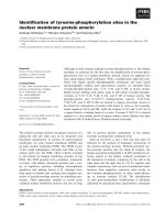
The aim of this study was to characterize the change of
protein flexibility during the nucleotide-induced reorgani-
zation of the motor domain of rabbit skeletal S1. Three
amino acids in the motor domain were labelled with
fluorescent dyes (Fig. 1), as follows (a) Ser181, a conserva-
tive amino acid of the nucleotide-binding pocket [38,39],
was labelled with ANN [22,40,41]; (b) Lys553, in the actin-
binding region, was labelled with FHS [24]; and (c) Cys707
(SH1), the cysteine of S1 with the highest reactivity, was
labelled with either IAEDANS or IAF [23]. The labelled
residues determined three FRET donor–acceptor pairs
(ANN–FHS, ANN–IAF, and IAEDANS–FHS) along
the sides of a triangle, which lay over the protein matrix
of the motor domain (Fig. 1). By using temperature
dependent FRET experiments, we investigated how the
flexibility of the protein matrix between these labels
depended on the binding of nucleotides and nucleotide
analogues such as ADP, ADP.BeF
x
, ADP.AlF
–
4
and
ADP.V
i
.
We attempted to test the biological activity of the labelled
S1 samples by measuring the Mg
2+
ATPase activities in the
absence of actin and in the presence of 17 l
M
or 30 l
M
actin
filaments. The results are summarized in Table 1. The
Mg
2+
ATPase activity of the unlabelled S1 was 0.05 s
)1
,
Fig. 1. Schematic representation of the motor domain of Dictyostelium discoideum myosin in apo-form. The50kDaupperdomainislabelledindark
blue, the 50 kDa lower domain is labelled in green, and the 25 kDa domain and the truncated 20 kDa domain are labelled in grey. The SWII
element (residues 466–500) is labelled in red, and the converter domain (residues 693–759) is labelled in light blue. In this work, the Ser181, Lys553
and Cys707 residues of rabbit skeletal myosin subfragment 1 (S1) were labelled with fluorophores. The corresponding residues (Ser181, Lys546 and
Thr688 [1]) are shown in the D. discoideum motor domain with yellow surfaces. The yellow dashed lines highlight the applied FRET pairs. Atomic
coordinates were obtained from the Protein Data Bank (accession number 1FMV).
4838 E. Bo
´
dis et al. (Eur. J. Biochem. 270) Ó FEBS 2003
similar to that observed previously [42]. For the labelled S1
samples, the basal Mg
2+
ATPase activities were similar to
or greater, and the actin activation lower, than for the
unlabelled S1. The results obtained after the binding of
IAEDANS to Cys707, or of FHS to Lys553, were in
agreement with previously published observations
[24,43,44]. The data show that although the binding of
fluorescence labels modified the physiological Mg
2+
ATPase activity of S1, the fundamental behavior of S1
was preserved. The ATPase cycle was similar in the labelled
samples to that operating in the unlabelled S1. In view of the
fact that, in this study, we stabilized different states of the
ATPase cycle in the absence of nucleotides, or by adding
ADP or nucleotide analogues, we concluded that the
fluorescence experiments reported on the proper character-
istics of the individual ATPase cycle states.
Fluorescence lifetime and anisotropy
The temperature dependence of the steady-state anisotropy
of the fluorophores in the absence of nucleotides was
measured between 6 and 26 °C, and analysed using the
Perrin equation (Eqn 5). For the analyses, the fluorescence
lifetimes were also measured. The average fluorescence
lifetime (Eqn 6) of ANN (Ser181), FHS (Lys553), IAE-
DANS (Cys707) and IAF (Cys707) were 12.0 ns (varied
1.0 ns between 6 °Cand26°C), 3.9 ns (varied 0.1 ns
between 6 °Cand26°C), 17.8 ns (varied 0.3 ns between
6 °Cand26°C) and 3.6 ns (varied < 0.1 ns between 6 °C
and 26 °C), respectively. The temperature dependent
anisotropy data were fitted to Eqn (5) by using the above
average lifetimes and r
0
¼ 0.4 (data not shown) to obtain
estimates for the apparent limiting anisotropy r
0
app
(the
intercept of the straight line with the 1/r axis) and the
volume of the rotating unit (V). The values obtained for r
app
0
were 0.36, 0.37, 0.30 and 0.28 for ANN, FHS, IAEDANS
and IAF, respectively. The V-values were 2.9 · 10
4
A
˚
3
,
1.08 · 10
4
A
˚
3
,5.8· 10
4
A
˚
3
and 6.4 · 10
4
A
˚
3
for ANN,
FHS, IAEDANS and IAF, respectively.
The donor–acceptor distances
The shape of the emission spectra of donors (IAEDANS
and ANN) was nucleotide and temperature independent
except in the case of the S1.ADP.V
i
complex, where the
ANN spectrum was blue shifted compared with those
measured in other nucleotide states. The transfer efficiency
(E), the quantum yield of the donors, the overlap integrals
for each fluorophore pairs and the Fo
¨
rster critical distances
(R
0
) were determined from the experimental data in
different nucleotide states at each temperature. The calcu-
lated R
0
values and the measured FRET efficiencies (E)are
shown in Table 2. The donor–acceptor distances (R) were
determined using Eqn (3), and the results obtained at 6 °C
and 22 °C are shown in Table 3. The distances did not show
sharp temperature induced changes, and the data obtained
at these two temperatures provided appropriate information
regarding the overall effect of temperature. The FRET
distances were 32–36 A
˚
, 44–47 A
˚
and 30–39 A
˚
for the
ANN–FHS, IAEDANS–FHS and ANN–IAF pairs,
Table 2. The nucleotide dependence of the Fo
¨
rster critical distance (R
0
) and the measured FRET efficiencies (E) for the three fluorophore pairs used in
this study. The data presented here were measured at 22 °C. The standard deviations were 0.3–1.1 A
˚
for the R
0
and 0.8–1.5% for the FRET
efficiency data, as determined from the results of experiments on at least three independent preparations.
Nucleotide state
ANN–Ser181/
FHS–Lys553
IAEDANS–Cys707/
FHS–Lys553
ANN–Ser181/
IAF–Cys707
R
0
(A
˚
) E (%) R
0
(A
˚
) E (%) R
0
(A
˚
) E (%)
Apo 38.9 68.6 46.3 55.5 39.7 63.1
ADP.BeF
x
36.6 69.6 44.5 49.8 37.3 74.8
ADP.AlF
–
4
40.7 68.9 46.2 53.0 41.5 72.4
ADP.V
i
37.7 66.2 45.6 45.2 38.4 84.7
ADP 36.3 66.9 45.5 52.3 37.1 76.3
Table 3. The nucleotide dependence of the apparent donor–acceptor distances measured at 6 °C and 22 °C in rabbit skeletal myosin subfragment 1
(S1). The standard errors calculated from at least three independent experiments were smaller than 1 A
˚
, in all cases. Note that these errors provided
the lower limit for the physically veritable errors. For comparison, the distances from the chicken S1 structure [39], corresponding to the apo state,
were determined: Ser181–Lys553, 33.8 A
˚
; Cys707–Lys553, 40.5 A
˚
; and Ser181–Cys707, 28.3 A
˚
. The distances calculated from the Dictyoste-
lium discoideum atomic models [4–6], between the corresponding residues (Ser181, Lys546 and Thr688) [8], are presented in columns labelled D.d.
All distances are given in A
˚
.
Nucleotide state
Ser181–Lys553
D.d.
Cys707–Lys553
D.d.
Ser181–Cys707
D.d.6 °C22°C6°C22°C6°C22°C
Apo 35.9 34.2 36.5 44.9 44.6 44.6 38.9 36.3 29.8
ADP.BeF
x
33.9 31.8 36.1 45.2 44.5 43.4 33.1 31.1 29.7
ADP.AlF
–
4
37.2 35.7 32.6 46.5 45.3 46.3 37.0 35.3 28.3
ADP.V
i
34.2 33.7 33.9 46.6 47.1 45.8 31.7 28.8 28.7
ADP 33.9 32.3 36.2 45.2 44.8 43.2 32.9 30.5 29.4
Ó FEBS 2003 Dynamic properties of the myosin motor domain (Eur. J. Biochem. 270) 4839
respectively. The data indicated that the effect of nucleotides
on these distances was small, with the greatest variation
being 3–4 A
˚
(Table 3), in agreement with the observation
that the position of the lever arm can be modulated with
only minor changes in the motor domain conformation [45].
The FRET distances were close to the distances obtained
from the atomic model of chicken S1 [39] or the
D. discoideum myosin II motor domain [4–6] (Table 3),
which will be discussed further below, in the Discussion. One
possible way to improve the reliability of FRET distances is
to perform the experiments with different fluorophores. In
our experiments, the labelling of Ser181 and Lys553 has only
been shown for the fluorophores used here and therefore
these control experiments were not feasible.
Protein flexibility
The temperature dependence of the f¢ (Figs 2 and 3) was
smooth and showed a monotonic increase with increasing
temperature. Major temperature induced conformational
changes were not detected, except in the case of the ANN–
IAF pair in the ADP.AlF
–
4
state. This exceptional case will
be discussed in more detail below, in the Discussion.The
absence of any major change in donor–acceptor distances
(Table 3) indicates that there is no major conformational
change over the temperature range studied. Accordingly,
the temperature dependence of the normalized transfer
efficiency (f¢; Eqn 4) could be attributed solely to the
flexibility of the protein matrix. In general, the larger change
of the f¢ results from greater flexibility of the protein matrix
[17,18].
Figure 2 shows the results obtained in the absence of
nucleotides or in the presence of ADP. In the nucleotide-free
S1, the temperature induced change in f¢ was substantially
smaller for IAEDANS–FHS–S1 than for either the ANN–
IAF–S1 or the ANN–FHS–S1. ADP binding had only
minor effects on the temperature dependence of f¢ in the case
of ANN–IAF or ANN–FHS pairs. In the case of the
IAEDANS–FHS pair, ADP increased the change of f¢ from
less than 5%, measured in the apo-form, to 15%.
The f¢ data measured in ADP.BeF
x
,ADP.V
i
and
ADP.AlF
–
4
states are presented, for the individual donor–
acceptor pairs, in Fig. 3A (ANN–FHS), Fig. 3B (IAE-
DANS–FHS) and Fig. 3C (ANN–IAF). For comparison,
the results obtained from ADP experiments (Fig. 2) are
shown in the figures as dotted lines. In the ADP.BeF
x
state,
the change of f¢ was only slightly smaller than that of the
ADP states for all three fluorophore pairs. Formation of the
ADP.AlF
–
4
–S1 complex did not change the temperature
dependence of f¢ between ANN and FHS (Fig. 3A). For the
other two fluorophore pairs (ANN–IAF and IAEDANS–
FHS), the change in f¢ was smaller in ADP.AlF
–
4
than in
ADP (Fig. 3B,C). The largest effect of ADP.AlF
–
4
was
observed between the residues labelled with ANN and IAF
(Fig. 3C). In this case, the overall change of f¢ was only
% 10%, much less than in other nucleotide states (60–80%).
The temperature profile of f¢ showed a saturation tendency,
reaching a maximum value between 14 and 18 °C.
The binding of ADP.V
i
to the S1 provided the greatest
effects amongst the nucleotide analogues on the protein
flexibility of the motor domain. The temperature induced
change of f¢ was less than in any other nucleotide states
(Fig. 3), for either the IAEDANS–FHS (< 5%) or the
ANN–FHS (% 15%) pairs (Fig. 3A,B). Between the resi-
dues labelled by ANN and IAF in ADP.V
i
, the overall
change of f¢ was % 70% at a temperature range of 6–26 °C
(Fig. 3C).
Discussion
In this study, the distances determined by the three donor–
acceptor pairs highlighted three structural aspects of the
motor domain of skeletal muscle myosin (Fig. 1). The
protein matrix between Cys707 (IAEDANS) and Lys553
(FHS) is located in the 50 kDa lower domain and is built up
of a-helixes, which are quasi parallel to the direction of this
side of the imaginary triangle (Fig. 1). The data obtained by
measuring the energy transfer between Ser181 (ANN) and
Cys707 (IAF) characterize the part of the 50 kDa upper
domain that is located more closely to the light-chain
binding domain. The third side of the triangle, Ser181
(ANN) and Lys553 (FHS), cross over the nucleotide-
binding pocket. The FRET experiments between ANN and
FHS reported on the relative motion of the 50 kDa upper
and 50 kDa lower domains. Based upon the FRET results,
the effects of nucleotides and nucleotide analogues follow
each other in the order of apo-, ADP, ADP.BeF
x
,
ADP.AlF
–
4
and ADP.V
i
, in agreement with previous
observations [46].
Although the FRET distances were in good agreement
with those obtained from either chicken or D. discoideum
atomic coordinates (Table 3), the results of analysis of the
temperature dependence of steady-state anisotropy data
Fig. 2. Temperature dependence of the normalized FRET efficiency in
the absence of nucleotides (black symbols) and in the presence of ADP
(white symbols). Data are presented for ANN–Ser181 and FHS–
Lys553 (circles), ANN–Ser181 and IAF–Cys707 (triangles), and
IAEDANS–Cys707 and FHS–Lys553 (squares) fluorophore pairs.
The donors ANN or IAEDANS were excited at 350 nm and the
emission was monitored between 400 and 470 nm in buffer comprising
25 m
M
Tes (pH 7.0), 80 m
M
KCl, 5 m
M
MgCl
2
,2m
M
EGTA and
4m
M
2-mercaptoethanol.
4840 E. Bo
´
dis et al. (Eur. J. Biochem. 270) Ó FEBS 2003
suggested that the agreement was coincidental. The distan-
ces from FRET experiments were calculated using j
2
¼ 2/3,
which assumes free rapid probe motion on a nanosecond
timescale. The high values (‡ 0.28) obtained for the r
0
app
indicated that the dyes were rigidly attached to the protein
segments, thus preventing the free rotation of the probes.
Therefore, the j
2
¼ 2/3 assumption is probably not valid
and the calculated donor–acceptor distances can be taken as
apparent distances. The calculated values for the rotating
volumes are approximately two orders of magnitude greater
than the volumes of the spheres with a radius of the length
of the fluorophores (< 10
3
A
˚
3
), indicating that the motion
of the labels reflects the motion of the protein segment to
which they are attached. The results suggested that the
temperature profile of the f¢ is not sensitive to local probe
motions, similarly to the case of actin monomers, where the
IAEDANS on the Cys374 was sensitive to the cation
exchange [37], but the temperature dependent FRET
experiments between IAEDANS and FITC on Lys61
showed no changes in the dynamics of the smaller domain
of actin [47]. The apparent donor–acceptor distances
showed no major change with the temperature (Table 3),
i.e. the equilibrium distances between the donor–acceptor
pairs do not change with the variation of the temperature in
this range, in accordance with the basic assumption of the
method [18]. [The fact that the apparent donor–acceptor
distances do not change with the temperature let us
conclude that the actual distances also remain unchanged.
Otherwise, one would have to use the very unlikely
assumption that any change in the equilibrium donor–
acceptor distance is compensated for by the appropriate
change of j
2
to leave the apparent distance unchanged.] We
concluded that the changes in the f¢ were related to the
increased width of donor–acceptor distance distribution,
and the greater slope of the temperature dependence of f¢
indicated the more flexible protein matrix between the
labels.
The FRET data will be interpreted based upon the
structural model, which assumes that the motor domain can
exist in two conformations – open and closed – defined by
the conformation of the SWII element [8]. The equilibrium
between these conformations is controlled by the bound
nucleotide and was characterized previously for unlabelled
myosins by using temperature and pressure jump experi-
ments [15,16]. In the present study we applied external
labels, which probably modified the open–closed equilib-
rium. The tryptophan fluorescence measured for these
labelled S1 samples would be informative regarding these
undesired effects [15,16]. However, the absorption and
emission spectra of tryptophan overlap with those of the
fluorophores used, which did not allow us to carry out these
control experiments. The results will be discussed therefore
using the equilibrium constants determined previously for
unlabelled myosins.
Comparison of the 50 kDa upper domain
with the 50 kDa lower domain
The temperature induced increase of f¢, along the Cys707–
Lys553 direction, was much smaller than along the other
two sides (Ser181–Lys553 and Ser181–Cys707) (Figs 2
and 3), which raises the possibility that the motor domain
Fig. 3. The temperature dependence of the normalized FRET efficiency
in S1.ADP.BeF
x
(h), S1.ADP.AlF
–
4
(d) and S1.ADP.V
i
(m). Data are
presented for the ANN–Ser181 and FHS–Lys553 pair (A), the IAE-
DANS–Cys707 and FHS–Lys553 pair (B), and the ANN–Ser181 and
IAF–Cys707 pair (C). For comparison, the data obtained in the
presence of ADP (Fig. 2) are also presented in the figures as dotted
lines. The experimental conditions were as described for Fig. 2.
Ó FEBS 2003 Dynamic properties of the myosin motor domain (Eur. J. Biochem. 270) 4841
is heterogeneous from the dynamic point of view. The
sensitivity of the normalized energy transfer (f¢) depends on
the r/R ratio (where r is the amplitude of the donor–
acceptor fluctuation and R is the equilibrium distance),
which is characteristic for the studied protein. The tem-
perature dependence of f¢ canalsodependonthevalueof
the Fo
¨
rster critical distance, which describes the sensitivity
of the fluorophore system applied. In our study, the spectral
properties of the individual donor–acceptor pairs were
similar, giving R
0
data in a relatively narrow range between
36 A
˚
and 48 A
˚
(Table 2). The measured distances were
between % 30 A
˚
and 44 A
˚
. The effect of these spectral and
geometric parameters cannot account for the large devia-
tions of f¢ found between the three sides of the triangle.
Accordingly, the direct juxtaposition of the flexibility data
obtained along the three directions within the motor
domain is reliable.
The smaller temperature induced change of f¢ along the
Cys707–Lys553 direction (as compared to the other two
directions) can only be attributed to the smaller relative
amplitude of the donor–acceptor fluctuations. The structure
of the 50 kDa lower domain in the apo-enzyme is more rigid
than that of the 50 kDa upper domain. The rigidity of the
50 kDa lower domain could be provided by the set of
a-helixes that run quasi parallel to the Cys707–Lys553
direction. The binding of either ADP or ADP.P
i
or ATP
analogues had little effect on the flexibility of the protein
matrix between Cys707 and Lys553, which implies that the
50 kDa lower domain behaves as a rigid body during the
nucleotide induced reorganizations of the S1. The rigidity of
this protein region can provide the structural stability for the
proper interactions with actin. This conclusion agrees with
the observation that the protein matrix between Cys707 in
S1 and the actin (labelled on Cys374) is rigid [48], and the
width of the positional distribution of Cys707 is narrow in
the absence of nucleotides [49], which suggests that the
rigidity of the actin binding region is maintained during the
interaction of S1 with actin.
In the apo-enzyme, the flexibility of the protein matrix
along the Ser181–Cys707 direction was the greatest of the
three directions. This large flexibility was maintained in the
ADP and ADP.BeF
x
states, although to differing extents,
and further increased in ADP.V
i
. In ADP.AlF
–
4
,the
temperature dependence of the f¢ is more complex and will
be discussed below. The large flexibility along Ser181–
Cys707, i.e. in the 50 kDa upper domain, may be important
in providing the structural frame for the motion and
reorientation of the phosphate group and for its interaction
with surrounding water molecules. Oxygen exchange studies
have shown that the cleavage of the myosin bound ATP is
reversible, the equilibrium between myosin bound ATP and
myosin-products complexes is rapid and the bound nucleo-
tide is able to undergo a fast and reversible reaction with
water to exchange all three oxygens [50,51]. Such inter-
actions require the rapid rotation and reorientation of the
phosphate group. Based on crystal structures it is assumed
that the phosphate is coordinated by three strong bonds, in
addition to the covalent bond in the strong conformation,
with no indication of how would it rotate rapidly after
hydrolysis [8]. We assume that the amplitude and frequency
of local protein fluctuations in this region should be
sufficiently large to provide the motional freedom for the
phosphate. The flexibility of the 50 kDa upper domain is
important in permitting such large-scale fluctuations. In the
back door enzyme model [52], it is believed that the
dissociation of the phosphate product occurs through
the back door of the motor domain on the opposite side
of the head to the one where the ATP enters. The atomic
structures suggest that access to the back door, however, is
partially blocked in either the open or closed conformations
[4–6]. In the absence of actin, the phosphate product is
trapped in the nucleotide binding pocket and its dissociation
from the motor domain is slow (% 0.05 s
)1
). The binding of
actin to myosin can accelerate the phosphate release. With
the lack of data in the presence of actin we can only
speculate that the large-scale breathing motion of the
flexible upper 50 kDa domain may become important in the
actin–myosin complex for the dissociation of the phosphate
product.
The effect of nucleotides on the flexibility
of the motor domain
The binding of ADP to the apo-S1 influenced the protein
dynamics only marginally. The flexibility slightly increased
between the Cys707 and Lys553. The atomic structures
[4,5], and the results of rapid kinetic experiments [15,16],
indicated that the motor domain is predominantly in the
open conformation in either the apo-S1 or when ADP is
bound, which suggests that the small ADP-induced change
in the flexibility between Cys707 and Lys553 may not be
directly related to the open-to-close transition. It has been
shown previously, by EPR [53,54], FRET [23,55,56] and
covalent cross-linking [57] assays, that the binding of
nucleotides loosens the structure of the essential SH/hinge
region (involving Cys707) where the donor IAEDANS was
located. It is probable that melting of the SH helix was
reflected by the slightly more flexible structure detected in
our FRET experiments along the Cys707–Lys553 direction.
Accordingly, the small effect of ADP on the flexibility of the
motor domain is attributed to local conformational changes
around the Cys707 residue, and the binding of ADP did not
change the overall structure and dynamics of the motor
domain. Recent results from electron microscopy experi-
ments showed that the release of ADP from the acto–S1
complex is accompanied with a 35 A
˚
swing of the lever arm
in the case of smooth muscle myosin [58]. In accordance
with these results, it was shown recently, by pressure-jump
experiments, that the increase in molar volume for skeletal
muscle S1 binding to ADP was half of that observed for
smooth muscle S1 [59]. ADP-induced movement of the
light-chain binding domain was also found in brush border
myosin-I [60], but was not detected in myosins from skeletal
muscle. The lack of ADP-induced swinging of the lever arm
in skeletal muscle S1 agrees with our observation that the
binding of ADP did not alter the dynamic properties of the
motor domain.
The binding of BeF
x
to ADP–S1 slightly decreased the
change in the normalized transfer efficiency measured for
the three the donor–acceptor pairs between 6 °Cand26°C.
The interpretation of the temperature dependent FRET
data, however, is complex in the case of ADP.BeF
x
.The
small decrease of the change in the normalized energy
transfer efficiency could be a local conformational effect
4842 E. Bo
´
dis et al. (Eur. J. Biochem. 270) Ó FEBS 2003
induced by the binding of BeF
x
, or could reflect the
temperature-induced shift of the open/closed equilibrium.
In skeletal S1 [16], or in the D. discoideum myosin II motor
domain [15], an increase in temperature shifted the equilib-
rium towards the closed conformation in the ADP.BeF
x
state. The FRET data indicate that the motor domain
adapts a more rigid conformation in the closed conforma-
tion than in the open state. However, the observed changes
of the FRET parameters were small and the overall
structure of the motor domain was similar in the
S1.ADP.BeF
x
to that observed in apo-S1 or S1.ADP. As,
in these latter two states, the open conformation is
dominant, the FRET results suggest that the open–closed
equilibrium was shifted towards the open conformation in
S1.ADP.BeF
x
.
ADP.AlF
–
4
is thought to mimic the ADP.P
i
state of S1. In
S1.ADP.AlF
–
4
, the temperature profile of f¢ showed a
saturation curve between Ser181 and Cys707 (Fig. 3C).
The intramolecular events behind this observation can
involve either temperature-induced changes in the protein
structure, which alters the distance or average orientation
between the donor and the acceptor, or steric constraints
which limit the fluctuations of the protein segments where
the donor or acceptor is located. The presence of such an
effect in S1.ADP.AlF
–
4
, and the lack of it in the other
nucleotide states (Fig. 3), implies that the conformation of
the motor domain is different in ADP.AlF
–
4
than in the apo-,
ADP or ADP.BeF
x
conformations. Accordingly, after the
binding of AlF
–
4
, the open conformation of S1 no longer
dominated. On the other hand, the binding of AlF
4
could
only partially reproduce the V
i
effects (Fig. 3).
In the atomic models, the SWII element was in the closed
conformation in both S1.ADP.V
i
and S1.ADP.AlF
–
4
[5,6].
However, according to the FRET results, the conforma-
tions observed for S1 with bound ADP.V
i
and ADP.AlF
–
4
were different. The interpretation of the FRET data,
measured between Ser181 and Cys707 in S1.ADP.AlF
–
4
,is
not clear. In the other two directions (Ser181–Lys553 and
Cys707–Lys553), the results for the ADP.AlF
–
4
state were
intermediate between the ADP.V
i
and apo states, which
suggests that in S1.ADP.AlF
–
4
, the contribution of the open
conformation of the motor domain was substantial. This
conclusion is in conflict with the temperature and pressure
jump results showing that in S1.ADP.AlF
–
4
, the closed
conformation dominated between 4 and 30 °C [15,16]. It is
possible that the tryptophan fluorescence, which was
monitored in the cited studies and the FRET pairs, applied
here, reported on different structural aspects of the S1
motor domain, which could account for the different
conclusions reached regarding the ADP.AlF
–
4
state. Alter-
natively, the shift towards the open conformation may have
appeared in the present work owing to the application of
external labels. Our conclusion, that the S1 population
is different with bound ADP.V
i
from that with
bound ADP.AlF
–
4
, agrees with the observation that the
nucleotide-binding cleft is only half closed in the ADP.AlF
–
4
X-ray structure [5,9] as compared to the ADP.V
i
structure.
In this work, ADP.V
i
was used to mimic the transition
state as an alternative ADP.P
i
analogue. The atomic
models suggested that S1 was predominantly in the closed
conformation when ADP.V
i
-bound [6]. The effect of
binding of ADP.V
i
on the dynamic properties of S1 was
the largest amongst the nucleotides investigated, and we
interpret these observations as characteristic for the closed
conformation. The steady-state fluorescence experiments
showed that the binding of V
i
shifted the emission spectra
of ANN to the blue by 5 nm, indicating that the solvent
accessibility of the ANN on Ser181 was reduced. These
observations suggest that the 50 kDa domain became more
compact in the closed conformation of the motor domain.
The FRET results suggest that in the closed conformation
the protein matrix between Ser181 and Cys707 became
more flexible than in the open conformation, which could
further accommodate the breathing motion of the 50 kDa
upper domain. In contrast, in the Ser181–Lys553 direction,
the temperature induced increase of f¢ was substantially
smaller in the closed conformation than in the open one,
which suggests that the amplitude of the relative fluctu-
ation of the 50 kDa upper and 50 kDa lower domains was
suppressed. The 50 kDa upper and 50 kDa lower domains
are connected by the end of the nucleotide-binding cleft
through the protein matrix that links the 50 kDa fragment
to the light-chain binding domain. Our results suggest that
this protein region becomes more rigid in the closed
conformation. The conformational transition underlying
the change in the dynamic properties could reflect the
relocation of the converter domain and probably plays a
role in transferring the energy from the catalytic site to the
lever arm.
Conclusions
The structural basis for the interaction of skeletal S1 with
actin is provided, at least partly, by the 50 kDa lower
domain, which was found to maintain substantial rigidity in
the different nucleotide states (Figs 2 and 3). The confor-
mation of the S1 in the apo-enzyme and in S1.ADP.V
i
set
the two extremes amongst the nucleotide states studied here.
Considering the atomic structures and the results of rapid
kinetic experiments, we assume that S1 was predominantly
in the open conformation in the apo-form and in the closed
conformation in S1.ADP.V
i
. The changes in the flexibility
of the S1 during the open-to-closed transition are complex;
we observed contrasting tendencies on comparison of
different protein regions. This complexity is probably
attributed to the different roles played by the protein
regions in the function of S1. In the open conformation, the
flexibility of the 50 kDa upper domain was the greatest of
the three directions studied here and this large flexibility
further increased during the open-to-closed transition. The
flexible nature of this protein region can be essential in
providing the structural conditions for the rapid motion and
reorientation of the phosphate group and for its interaction
with surrounding water molecules, and may become
important in the actin–myosin complex for the dissociation
of the phosphate product. The solvent accessibility of the
Ser181 was reduced, and the amplitude of the relative
fluctuations of the upper 50 kDa and lower 50 kDa
domains was suppressed in the closed conformation as
compared to that of the open one. The suppressed
amplitude suggests that the protein region near the bottom
of the nucleotide-binding cleft, which links the two domains
together, becomes more rigid. The more rigid conformation
adapted in the closed conformation can provide the
Ó FEBS 2003 Dynamic properties of the myosin motor domain (Eur. J. Biochem. 270) 4843
mechanical basis of the transfer of the information or energy
from the catalytic site to the light-chain binding domain.
Acknowledgements
The authors gratefully acknowledge Dr Michael A. Geeves’s continu-
ous support and suggestions during the preparation of the manuscript,
and the insightful comments from Andra
´
s Luka
´
cs and from Dr Jo
´
zsef
Bela
´
gyi during the course of this work. This work was supported by
grants from the National Research Foundation (OTKA grants:
T32700, T34442, T43103), from the Ministry of Education (0252/
2000), and from the Hungarian Academy of Sciences NKFP 1/026/
2001. M. Nyitrai is an EMBO/HHMI Scientist.
References
1. Goodno, C.C. (1979) Inhibition of myosin ATPase by vanadate
ion. Proc. Natl Acad. Sci. USA 76, 2620–2624.
2. Maruta, S., Henry, G.D., Sykes, B.D. & Ikebe, M. (1993) For-
mation of the stable myosin-ADP-aluminum fluoride and myosin-
ADP-beryllium fluoride complexes and their analysis using 19F
NMR. J. Biol. Chem. 268, 7093–7100.
3. Phan, B. & Reisler, E. (1992) Inhibition of myosin ATPase by
beryllium fluoride. Biochemistry 31, 4787–4793.
4. Bauer,C.B.,Holden,H.M.,Thoden,J.B.,Smith,R.&Rayment,I.
(2000) X-ray structures of the apo and MgATP-bound states of
Dictyostelium discoideum myosin motor domain. J. Biol. Chem.
275, 38494–38499.
5. Fisher, A.J., Smith, C.A., Thoden, J.B., Smith, R., Sutoh, K.,
Holden, H.M. & Rayment, I. (1995) X-ray structures of the
myosin motor domain of Dictyostelium discoideum complexed
with MgADP.BeFx and MgADP.AlF4. Biochemistry 34, 8960–
8972.
6. Smith, C.A. & Rayment, I. (1996) X-ray structure of the magne-
sium (II) ADP.vanadate complex of the Dictyostelium discoideum
myosin motor domain to 1.9 A
˚
resolution. Biochemistry 35, 5404–
5417.
7. Dominguez, R., Freyzon, Y., Trybus, K.M. & Cohen, C. (1998)
Crystal structure of a vertebrate smooth muscle myosin motor
domain and its complex with the essential light chain: visualization
of the pre-power stroke state. Cell 94, 559–571.
8. Geeves, M.A. & Holmes, K.C. (1999) Structural mechanism of
muscle contraction. Annu. Rev. Biochem. 68, 687–728.
9. Holmes, K.C. (1997) The swinging lever-arm hypothesis of muscle
contraction. Curr. Biol. 7, R112–R118.
10. Ritchie, M.D., Geeves, M.A., Woodward, S.K. & Manstein, D.J.
(1993) Kinetic characterization of a cytoplasmic myosin motor
domain expressed in Dictyostelium discoideum. Proc. Natl Acad.
Sci. USA 90, 8619–8623.
11. Burghardt, T.P., Garamszegi, S.P., Park, S. & Ajtai, K. (1998)
Tertiary structural changes in the cleft containing the ATP
sensitive tryptophan and reactive thiol are consistent with
pivoting of the myosin heavy chain at Gly699. Biochemistry 37,
8035–8047.
12. Malnasi-Csizmadia, A., Hegyi, G., Tolgyesi, F., Szent-Gyorgyi,
A.G. & Nyitray, L. (1999) Fluorescence measurements detect
changes in scallop myosin regulatory domain. Eur. J. Biochem.
261, 452–458.
13. Yengo,C.M.,Chrin,L.R.,Rovner,A.S.&Berger,C.L.(2000)
Tryptophan 512 is sensitive to conformational changes in the rigid
relay loop of smooth muscle myosin during the MgATPase cycle.
J. Biol. Chem. 275, 25481–25487.
14. Malnasi-Csizmadia, A., Woolley, R.J. & Bagshaw, C.R. (2000)
Resolution of conformational states of Dictyostelium myosin II
motor domain using tryptophan (W501) mutants: implications
for the open/closed transition identified by crystallography.
Biochemistry 39, 16135–16146.
15. Malnasi-Csizmadia, A., Pearson, D.S., Kovacs, M., Woolley,
R.J., Geeves, M.A. & Bagshaw, C.R. (2001) Kinetic resolution of
a conformational transition and the ATP hydrolysis step using
relaxation methods with a Dictyostelium myosin II mutant con-
taining a single tryptophan residue. Biochemistry 40, 12727–12737.
16. Urbanke, C. & Wray, J. (2001) A fluorescence temperature-jump
study of conformational transitions in myosin subfragment 1.
Biochem. J. 358, 165–173.
17. Somogyi, B., Lakos, Z., Szarka, A. & Nyitrai, M. (2000) Protein
flexibility as revealed by fluorescence resonance energy transfer:
an extension of the method for systems with multiple labels.
J. Photochem. Photobiol. B 59, 26–32.
18. Somogyi, B., Matko, J., Papp, S., Hevessy, J., Welch, G.R. &
Damjanovich, S. (1984) Forster-type energy transfer as a probe for
changes in local fluctuations of the protein matrix. Biochemistry
23, 3403–3411.
19. Margossian, S.S. & Lowey, S. (1982) Preparation of myosin and
its subfragments from rabbit skeletal muscle. Methods Enzymol.
85, 55–71.
20. Spudich, J.A. & Watt, S. (1971) The regulation of rabbit skeletal
muscle contraction. I. Biochemical studies of the interaction of the
tropomyosin-troponin complex with actin and the proteolytic
fragments of myosin. J. Biol. Chem. 246, 4866–4871.
21. Weeds, A.G. & Taylor, R.S. (1975) Separation of subfragment-1
isoenzymes from rabbit skeletal muscle myosin. Nature 257,
54–56.
22. Hiratsuka, T. (1989) Nucleotide-induced specific fluorescent
labeling of the 23-kDa NH2- terminal tryptic peptide of myosin
ATPase by the serine-reactive reagent 9-anthroylnitrile. J. Biol.
Chem. 264, 18188–18194.
23. Xing, J. & Cheung, H.C. (1995) Internal movement in myosin
subfragment 1 detected by fluorescence resonance energy transfer.
Biochemistry 34, 6475–6487.
24. Bertrand, R., Derancourt, J. & Kassab, R. (1995) Production and
properties of skeletal myosin subfragment 1 selectively labeled
with fluorescein at lysine-553 proximal to the strong actin-binding
site. Biochemistry 34, 9500–9507.
25. Wagner, P.D. (1977) Fractionation of heavy meromyosin by
affinity chromatography. FEBS Lett. 81, 81–85.
26. Houk, T.W. Jr (1974) The measurement of actin concentration in
solution: a comparison of methods. Anal. Biochem. 62, 66–74.
27. Takashi, R. (1979) Fluorescence energy transfer between sub-
fragment-1 and actin points in the rigor complex of actosubfrag-
ment-1. Biochemistry 18, 5164–5169.
28. Peyser, Y.M., Ajtai, K., Burghardt, T.P. & Muhlrad, A. (2001)
Effect of ionic strength on the conformation of myosin subfrag-
ment 1- nucleotide complexes. Biophys. J. 81, 1101–1114.
29. Goodno, C.C. (1982) Myosin active-site trapping with vanadate
ion. Methods Enzymol. 85, 116–123.
30. Hiratsuka, T. (1990) Transmission of ADP.vanadate-induced
conformational changes to three peptide segments of myosin
subfragment-1. J. Biol. Chem. 265, 18791–18796.
31. Peyser, Y.M. & Muhlrad, A. (1999) Actin and nucleotide induced
conformational changes in the vicinity of Lys553 in myosin sub-
fragment 1. Eur. J. Biochem. 263, 511–517.
32. Hiratsuka, Y., Eto, M., Yazawa, M. & Morita, F. (1998)
Reactivities of Cys707 (SH1) in intermediate states of myosin
subfragment-1 ATPase. J. Biochem. (Tokyo) 124, 609–614.
33. Fiske, C.H. & Subbarow, Y. (1925) The colorimetric determina-
tion of phosphorus. J. Biol. Chem. 66, 375–400.
34. Norby, J.G. (1971) Studies on a coupled enzyme assay for rate
measurements of ATPase reactions. Acta Chem. Scand. 25, 2717–
2726.
4844 E. Bo
´
dis et al. (Eur. J. Biochem. 270) Ó FEBS 2003
35. Laemmli, U.K. (1970) Cleavage of structural proteins during the
assembly of the head of bacteriophage T4. Nature 227, 680–685.
36. Lakowicz, J. (1986) Time-dependent decay of fluorescence aniso-
tropy. In Principles of Fluorescence Spectroscopy (Lakowicz, J.R.,
ed), pp. 305–339. Plenum Press, New York.
37. Nyitrai, M., Hild, G., Belagyi, J. & Somogyi, B. (1997) Spectro-
scopic study of conformational changes in subdomain 1 of
G-actin: influence of divalent cations. Biophys. J. 73, 2023–2032.
38. Andreev, O.A., Takashi, R. & Borejdo, J. (1995) Fluorescence
polarization study of the rigor complexes formed at different
degrees of saturation of actin filaments with myosin subfragment-1.
J. Muscle Res. Cell Motil. 16, 353–367.
39. Rayment, I., Rypniewski, W.R., Schmidt-Base, K., Smith, R.,
Tomchick, D.R., Benning, M.M., Winkelmann, D.A., Wesen-
berg, G. & Holden, H.M. (1993) Three-dimensional structure of
myosin subfragment-1: a molecular motor. Science 261, 50–58.
40. Szarka, K., Bodis, E., Visegrady, B., Nyitrai, M., Kilar, F. &
Somogyi, B. (2001) 9-Anthroylnitrile binding to serine-181 in
myosin subfragment 1 as revealed by FRET spectroscopy and
molecular modeling. Biochemistry 40, 14806–14811.
41. Hiratsuka, T.K.T. (2003) Chemical identification of serine 181 at
the ATP-binding site of myosin as a residue esterified selectively by
the fluorescent reagent 9-anthroylnitrile. J. Biol. Chem. 278,
31891–31894.
42. Trentham, D.R., Eccleston, J.F. & Bagshaw, C.R. (1976) Kinetic
analysis of ATPase mechanisms. Q. Rev. Biophys. 9, 217–281.
43. Sleep, J.A. (1981) Single turnovers of adenosine 5¢-triphosphate
by myofibrils and actomyosin subfragment 1. Biochemistry 20,
5043–5051.
44. Mulhern, S.A. & Eisenberg, E. (1978) Interaction of spin-labeled
and N-(iodacetylaminoethyl)-5-naphthylamine-1-sulfonic acid
SH1-blocked heavy meromyosin and myosin with actin and
adenosine triphosphate. Biochemistry 17, 4419–4425.
45. Houdusse, A., Szent-Gyorgyi, A.G. & Cohen, C. (2000) Three
conformational states of scallop myosin S1. Proc. Natl Acad. Sci.
USA 97, 11238–11243.
46. Park, S., Ajtai, K. & Burghardt, T.P. (1997) Mechanism for
coupling free energy in ATPase to the myosin active site. Bio-
chemistry 36, 3368–3372.
47. Nyitrai, M., Hild, G., Lakos, Z. & Somogyi, B. (1998) Effect of
Ca
2+
-Mg
2+
exchange on the flexibility and/or conformation of
the small domain in monomeric actin. Biophys. J. 74, 2474–2481.
48. Nyitrai, M., Hild, G., Bodis, E., Lukacs, A. & Somogyi, B. (2000)
Flexibility of myosin-subfragment-1 in its complex with actin as
revealed by fluorescence resonance energy transfer. Eur. J.
Biochem. 267, 4334–4338.
49. Nyitrai,M.,Hild,G.,Lukacs,A.,Bodis,E.&Somogyi,B.(2000)
Conformational distributions and proximity relationships in the
rigor complex of actin and myosin subfragment-1. J. Biol. Chem.
275, 2404–2409.
50. Bagshaw,C.R.,Trentham,D.R.,Wolcott,R.G.&Boyer,P.D.
(1975) Oxygen exchange in the gamma-phosphoryl group of
protein-bound ATP during Mg
2+
-dependent adenosine triphos-
phatase activity of myosin. Proc. Natl Acad. Sci. USA 72, 2592–
2596.
51. Webb, M.R. & Trentham, D.R. (1981) The mechanism of ATP
hydrolysis catalyzed by myosin and actomyosin, using rapid
reaction techniques to study oxygen exchange. J. Biol. Chem. 256,
10910–10916.
52. Yount, R.G., Lawson, D. & Rayment, I. (1995) Is myosin a Ôback
doorÕ enzyme? Biophys. J. 68, 44S–47S [discussion 47S)49S].
53. Raucher, D., Sar, C.P., Hideg, K. & Fajer, P.G. (1994) Myosin
catalytic domain flexibility in MgADP. Biochemistry 33, 14317–
14323.
54. Belagyi, J. & Lorinczy, D. (1996) Internal motions in myosin head:
effectofADPandATP.Biochem. Biophys. Res. Commun. 219,
936–940.
55. Garland, F., Gonsoulin, F. & Cheung, H.C. (1988) The MgADP-
induced decrease of the SH1-SH2 fluorescence resonance energy
transfer distance of myosin subfragment 1 occurs in two kinetic
steps. J. Biol. Chem. 263, 11621–11623.
56. Cheung, H.C., Gryczynski, I., Malak, H., Wiczk, W., Johnson,
M.L. & Lakowicz, J.R. (1991) Conformational flexibility of the
Cys 697-Cys 707 segment of myosin subfragment-1: distance dis-
tributions by frequency-domain fluorometry. Biophys. Chem. 40,
1–17.
57. Rajasekharan, K.N., Mayadevi, M., Agarwal, R. & Burke, M.
(1990) MgADP-induced changes in the structure of myosin S1
near the ATPase-related thiol SH1 probed by cross-linking. Bio-
chemistry 29, 3006–3013.
58. Whittaker, M., Wilson-Kubalek, E.M., Smith, J.E., Faust, L.,
Milligan, R.A. & Sweeney, H.L. (1995) A 35-A
˚
movement of
smooth muscle myosin on ADP release. Nature 378, 748–751.
59.Pearson,D.S.,Holtermann,G.,Ellison,P.,Cremo,C.&
Geeves, M.A. (2002) A novel pressure-jump apparatus for the
microvolume analysis of protein–ligand and protein–protein
interactions: its application to nucleotide binding to skeletal-
muscle and smooth-muscle myosin subfragment-1. Biochem. J.
366, 643–651.
60. Jontes, J.D., Wilson-Kubalek, E.M. & Milligan, R.A. (1995) A 32
degree tail swing in brush border myosin I on ADP release. Nature
378, 751–753.
Ó FEBS 2003 Dynamic properties of the myosin motor domain (Eur. J. Biochem. 270) 4845