Báo cáo khoa học: Human ATP-dependent RNA ⁄ DNA helicase hSuv3p interacts with the cofactor of survivin HBXIP ppt
Bạn đang xem bản rút gọn của tài liệu. Xem và tải ngay bản đầy đủ của tài liệu tại đây (474.57 KB, 12 trang )
Human ATP-dependent RNA ⁄ DNA helicase hSuv3p
interacts with the cofactor of survivin HBXIP
Michal Minczuk
1
, Seweryn Mroczek
1
, Sebastian D. Pawlak
1,
* and Piotr P. Stepien
1,2
1 Department of Genetics, University of Warsaw, Pawinskiego 5A, Warsaw, Poland
2 Institute of Biochemistry and Biophysics, Polish Academy of Sciences, Pawinskiego 5A, Warsaw, Poland
The NTP-dependent RNA ⁄ DNA helicase Suv3p
belongs to the Ski2 class of DExH-box RNA helicases
and its orthologues have been found in bacteria, yeast,
plants and animals [1]. The product of the SUV3 gene
was described for the first time in Saccharomyces cere-
visiae [2], where it functions in mitochondrial RNA
surveillance. Yeast Suv3p is one of the two subunits of
a protein complex called mitochondrial degradosome
or MtEXO, which displays an NTP-dependent exo-
ribonucleolytic activity [3–5]. The second component
of the degradosome is Dss1p, a single-strand specific
exoribonuclease with motifs similar to bacterial RN-
ase II [6]. The RNA-degrading activity of the degrado-
some complex is necessary for maintaining proper
mitochondrial RNA metabolism in yeast. Mutations in
either of the two degradosome subunits result in over-
accumulation of excised group I introns [7], distur-
bances in processing at 5¢ and 3¢ ends of mtRNA
precursors, lack of mitochondrial translation and in
changes in steady-state levels of mature mt mRNAs;
yeast strains bearing deletions of SUV3 or DSS1 genes
are respiratory incompetent but are viable on ferment-
able carbon sources [4,8,9].
In contrast to yeast, much less is known about the
human SUV3 and its physiological functions. Our
recent report indicated that the hSUV3 exhibits typical
characteristics for a nuclear-encoded mitochondrial
gene, which is constitutively expressed [10]. The human
Keywords
apoptosis; helicase; intracellular localization;
mitochondria; mitochondrial import
Correspondence
M. Minczuk, Department of Genetics,
University of Warsaw, Pawinskiego 5A,
02-106 Warsaw, Poland
Fax: +48 22 5922244
Tel: +48 22 5922240
E-mail:
*Present address
Laboratory of Bioinformatics and Protein
Engineering, International Institute of
Molecular and Cell Biology, Ks. Trojdena 4a,
02-109 Warsaw, Poland
(Received 4 July 2005, revised 5 August
2005, accepted 10 August 2005)
doi:10.1111/j.1742-4658.2005.04910.x
The human SUV3 gene encodes an NTP-dependent DNA ⁄ RNA DExH
box helicase predominantly localized in mitochondria. Its orthologue in
yeast is a component of the mitochondrial degradosome complex involved
in the mtRNA decay pathway. In contrast to this, the physiological func-
tion of human SUV3 remains to be elucidated. In this report we demon-
strate that the hSuv3 protein interacts with HBXIP, previously identified as
a cofactor of survivin in suppression of apoptosis and as a protein that
binds the HBx protein encoded by the hepatitis B virus. Using deletion
analysis we identified the region within the hSuv3 protein, which is respon-
sible for binding to HBXIP. The HBXIP binding domain was found to be
important for mitochondrial import and stability of the Suv3 protein
in vivo. We discuss the possible involvement of the hSuv3p–HBXIP inter-
action in the survivin-dependent antiapoptotic pathway.
Abbreviations
aa, amino acids; IAP, inhibitor of apoptosis; BIR, baculovirus IAP repeat; FITC, fluorescein isothiocyanate; GFP, green fluorescence protein;
HA, hemagglutinin; hSUV3, human SUV3; IVT, in vitro translation; TAP, tandem affinity purification.
5008 FEBS Journal 272 (2005) 5008–5019 ª 2005 FEBS
Suv3p enzyme expressed in Escherichia coli had a
strong preference for a double stranded DNA, while
also displaying an NTP-dependent RNA helicase activ-
ity [11]. Recently, Shu et al. [12] confirmed the multiple
substrate unwinding activity of the human Suv3 pro-
tein, being able to unwind DNA, RNA and hetero-
duplex substrates. The unwinding reaction was found
to depend on conformational change of the protein
induced by pH. The human Suv3p has a bona fide
mitochondrial leader sequence and using immunofluo-
rescence analysis, an in vitro mitochondrial uptake
assay and subfractionation of human mitochondria we
showed that hSuv3p is a soluble protein localized in
the mitrochondrial matrix [11]. However, intracellular
localization studies using polyclonal antibodies, raised
against the heterologously expressed protein, revealed
that in HeLa cells endogenous hSuv3p also exhibits a
faint nuclear localization signal in addition to a strong
mitochondrial signal [11].
Interestingly, recent data have shown that a fraction
of the Suv3p helicase may indeed be localized in the
nucleus. Such a suggestion was made by Bader [13],
who employed in silico analysis of the network of
yeast protein–protein interactions. Employing this
method he identified yeast Suv3p as a potential mem-
ber of proliferating cell nuclear antigen-like complex.
In addition, high-throughput analysis of yeast pro-
tein–protein interactions has revealed several nuclear
protein partners of the yeast Suv3p, most of them
being involved in DNA replication, repair and recom-
bination [14]. Among the identified Suv3p interactors
the following proteins have been reported: (a) the
SGS1 helicase, involved in maintaining genome stabil-
ity, homologous to E. coli RecQ and human WRN
helicase (defective WRN helicase leads to premature
aging disorder Werner syndrome); (b) the RFC4 pro-
tein, a DNA binding ATPase that acts as a processivity
factor for DNA polymerase delta and epsilon and
loads proliferating cell nuclear antigen; (c) MEC3 pro-
tein, involved in checkpoint control and DNA repair;
and (d) DDC1 protein, involved in the DNA damage
checkpoint. In agreement with above observations Shu
et al. [12] have recently suggested the nuclear localiza-
tion of a fraction of cellular human Suv3p, but no
data were presented. The authors proposed that
hSuv3p has multiple physiological roles in the cell,
including telomere maintenance, DNA repair and cell
cycle checkpoint control.
In this paper we show the results of the yeast two-
hybrid system in screening for interactors of the
human SUV3 gene product. We demonstrate that
hSuv3p interacts with HBXIP, which was previously
identified as a cofactor of survivin in apoptosis
suppression and as a protein binding to the hepatitis B
viral protein X.
Results
Identification and characterization of the HBXIP–
hSuv3p interaction in the two-hybrid system
In order to screen the cDNA library derived from
HeLa cells in two-hybrid system as described by Finley
& Brent [15] we constructed baits by linking N-ter-
minal or C-terminal part of hSuv3p (residues 1–479
and 380–786, respectively) to the LexA DNA binding
domain. LexA-hSuv3p 380–786 fusion was chosen for
further two-hybrid experiments after our initial tests
have shown the lack of its self-activation ability,
proper nuclear import and operator binding ability in
yeast cells (supplementary Appendix S1, Fig. S1). The
6 · 10
6
library clones were screened and 57 positive
yeast colonies were identified and subjected to sequen-
cing. Among 57 positive colonies 18 appeared to be
independent clones and HBXIP (HBx interacting pro-
tein) proved to be the most frequently occurring
cDNA among the isolates (seven out of 18 of the
hSuv3p interacting clones; sequence characterization of
the clones is shown in supplementary Appendix S1,
Fig. S2). In order to rule out the possibility of nonspe-
cific interaction of HBXIP, different nonrelevant baits
including bicoid, CD4 and IC-LexA fusion proteins
[15] were tested with all positive interacting cDNA
clones. In order to identify and partially characterize
the hSuv3p domain interacting with HBXIP prey
clones, several deletion mutants of the C-terminal
hSuv3p bait were used in the two-hybrid test. As
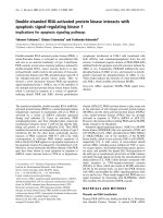
depicted in Fig. 1A, the hSuv3p fragment necessary
for interaction with HBXIP is contained within the
136 C-terminal amino acids of hSuv3p (amino acids
650–786).
HBXIP interacts with hSuv3p in vitro
We employed an in vitro binding test in order to
exclude the possibility that the interaction between the
HBXIP and hSuv3p proteins occurred through a
yeast-derived bridging protein(s) and to provide evi-
dence of direct binding of the proteins in a different
system. We constructed HBXIP fusion protein contain-
ing TAP tag [16] at the C-terminus. We purified
HBXIP-TAP fusion on IgG-agarose resin after hetero-
logous expression in E. coli and studied the interaction
with an in vitro translated (IVT) [
35
S]methionine labe-
led hSuv3p. In this assay two versions of hSuv3p were
used: full-length protein (hSuv3p 1–786) and a protein
M. Minczuk et al. Human helicase hSuv3p interacts with HBXIP
FEBS Journal 272 (2005) 5008–5019 ª 2005 FEBS 5009
lacking the 136 C-terminal amino acids (hSuv3p 1–
650); both of them contained a c-myc epitope-tag at
the C-terminus. As illustrated in Fig. 1B only IVT of
full-length hSuv3p interacted with HBXIP-TAP immo-
bilized on IgG-agarose. No interaction was detected
in the case of heterologously purified TAP-tag or the
IgG-agarose resin only (Fig. 1B). This result is consis-
tent with the finding that the 136 amino acid-long
C-terminal part of hSuv3p is responsible for forming a
complex with the HBXIP protein.
HBXIP shows nucleo-cytosolic localization
in human cells
The exact subcellular localization of HBXIP has not
been studied up to date. To address this issue we tried
to develop the anti-HBXIP polyvalent antibodies. We
purified HBXIP-TAP fusion in the two step procedure
as described by Rigaut et al. [16] after heterologous
expression in E. coli but we failed to obtain high-affin-
ity antibodies against the small hydrophobic HBXIP
protein in rabbit. Therefore, to determine the subcellu-
lar distribution of HBXIP, the protein was C-termin-
ally tagged with c-myc or HA epitope and its
subcellular localization was studied in transiently
transfected HeLa cells. The cells were stained with the
primary anti-myc (or anti-HA) monoclonal antibodies
and visualized with fluorescein isothiocyanate (FITC)-
conjugated secondary antibodies. In addition, the cells
were stained with nuclear marker (DAPI) and mitoch-
ondrial marker (MitoTracker CMXRos). As presented
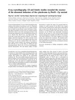
in Fig. 2A,B for HeLa cells the HBXIP-myc fusion
showed double nucleo-cytosolic localization and practi-
cally no colocalization with mitochondria was
observed. The same result was obtained for the
HBXIP-HA fusion and in the case of simian COS-1
cells (data not shown).
Next, in order to provide further evidence on intra-
cellular localization of HBXIP, a subcellular fraction-
ation experiment was performed. As depicted in
Fig. 2C the HBXIP protein was confirmed to reside in
the cytosolic fraction of HeLa cells and it was not
found in the mitochondrial fraction.
The C-terminal fragment of hSuv3p that interacts
with HBXIP is important for hSuv3p mitochondrial
import
In order to verify whether deletion of the C-terminal
fragment of hSuv3p could have an effect on protein
function in vivo we transiently expressed C-terminally
truncated (136 amino acids) hSuv3p (hSuv3p-myc 1–
650) in COS-1 cells. First, we analyzed the subcellular
distribution of the truncated mutant protein using
anti-myc monoclonal antibodies visualized with anti-
mouse secondary antibodies conjugated with FITC. In
the case of hSuv3p-myc 1–650 fusion colocalization
with the mitochondrial marker MitoTracker was
substantially reduced, as compared to the wildtype
A
B
Fig. 1. Interaction of hSuv3p with HBXIP (A) Schematic representa-
tion of the hSuv3p baits used in this study with the relative binding
affinities to HBXIP prey clone. In order to screen the HeLa cell-
derived cDNA library by the yeast two-hybrid screening, stable bait
was generated by fusing the LexA DNA binding domain with the
C-terminal part of hSuv3p (hSuv3p 380–786). The yeast bait vector
carrying the full-length hSuv3p was also tested under the same
experimental conditions. Several hSuv3p deletion mutants were
used to specifically identify and partially characterize the hSuv3p
domain that is necessary for interaction with HBXIP. (B) In vitro
interaction of hSuv3p with HBXIP. The SDS ⁄ PAGE analysis of the
binding of wildtype hSuv3p or its mutant devoid of the C-terminal
136 amino acids (hSuv3p 1–650) to IgG-agarose-HBXIP is shown.
Both variants of hSuv3p have been synthesized using IVT in the
presence of [
35
S]methionine. Lanes 1 and 2 show hSuv3p (WT)
and the hSuv3p 1–650 mutant, respectively, bound to HBXIP-TAP
immobilized on IgG-agarose. Lanes 3 and 4 show the binding of
hSuv3p (WT) and the hSuv3p 1–650 mutant, respectively, bound to
IgG-agarose resin alone. Lanes 5 and 6 show the binding of hSuv3p
(WT) and the hSuv3p 1–650 mutant, respectively, to heterologically
expressed TAP-tag immobilized on IgG-agarose. Lanes 7 and 8
show 20% of the input of the IVT [
35
S]methionine labeled hSuv3p
(WT) and the hSuv3p 1–650 mutant, respectively.
Human helicase hSuv3p interacts with HBXIP M. Minczuk et al.
5010 FEBS Journal 272 (2005) 5008–5019 ª 2005 FEBS
hSuv3-myc protein expressed under exactly the same
conditions, and a significant fraction of the protein
was retained in the cytosol (Fig. 3). It therefore
appears that, in addition to the N-terminal mitochond-
rial leader peptide, the 136 amino acid-long C-terminal
part of hSuv3p is involved in targeting of the protein
to mitochondria. It is worth mentioning, however, that
the 136 amino acid-long C-terminal part of hSuv3p
alone cannot serve as a bona fide mitochondrial target-
ing signal because hSuv3p 650–786-TAP protein con-
struct (see below) is not localized in mitochondria
(data not shown).
The C-terminal fragment of hSuv3p that binds
HBXIP is important for hSuv3p stability
The experiment described above also showed that the
truncated form of hSuv3p, lacking the domain respon-
sible for HBXIP binding, was expressed in a signifi-
cantly lower number of cells as compared to the
wildtype form of the protein. The transfection effi-
ciency was 21% and 1.5% for full-length and trun-
cated hSuv3p, respectively (although the transfection
conditions and plasmid DNA preparations were the
same in both cases). Such a difference could be the
result of mRNA instability, accelerated protein degra-
dation or cell toxicity of the truncated hSuv3 protein.
In order to discriminate between those possibilities we
measured the mRNA steady-state levels by Northern
hybridization. As illustrated in Fig. 4A there was no
significant difference in mRNA level for hSuv3p wild-
type and truncated construct. In order to exclude that
lowered expression frequency observed for the trun-
cated hSuv3p results from cellular toxicity, the wild-
type and 1–650 forms were coexpressed with green
fluorescence protein (GFP) as a internal marker (GFP
was encoded within the vector backbone, therefore all
cells expressing either form of hSuv3p expressed GFP
as well). First, we measured the transfection efficiency
for the constructs encoding either version of hSuv3p
by counting GFP-positive cells; in both cases the effi-
ciency was 17%. Then, after transient expression,
wildtype and truncated forms of hSuv3p, the proteins
were visualized by anti-myc monoclonal antibodies and
anti-mouse Texas Red conjugated secondary antibod-
ies. Furthermore, we studied the correlation between
red (hSuv3p-derived) and green (GFP-derived) fluores-
cence for full-length and truncated forms of hSuv3p as
described in Experimental procedures. As presented in
Fig. 4B,C the correlation observed was 84 ± 12% and
9 ± 6% for wildtype hSuv3p and hSuv3p 1–650 trun-
cated protein, respectively. This result indicated that
the hSuv3p version lacking HBXIP binding domain
A
B
C
Fig. 2. Intracellular localization of HBXIP in mammalian cells. (A)
HeLa cells were grown on coverslips and transiently transfected
with cDNA encoding c-myc tagged HBXIP (HBXIPmyc). After incu-
bation with MitoTracker red, fixation and permeabilization as des-
cribed in Experimental procedures the cells were immunostained
with anti-(c-myc) monoclonal antibody (9E10), which was then visu-
alized with fluorescein isothiocyanate-conjugated antibody. At the
final stage, nuclei were stained with DAPI present in the mounting
medium. The figure shows representative fluorescent image of
cells stained with DAPI (blue), c-myc-tagged HBXIP (green) and
MitoTracker (red) taken by a confocal microscope. Similar results
were obtained for cDNA encoding HA tagged HBXIP expressed in
HeLa cells as wells as for the HBXIPmyc and HBXIP-HA expressed
in COS-1 cells. (B) Magnified confocal microscope images prepared
as in (A) for HeLa cells transfected with cDNA encoding HBXIPmyc
are shown. (C) HeLa cells were transiently transfected with cDNA
encoding c-myc tagged HBXIP (HBXIPmyc). Cell lysates from
unfractionated HeLa cells (T), the cytoplasmic (C) and the mitoch-
ondrial fraction (M) were immunoblotted with anti-myc monoclonal
antibodies. The fractions were verified using anti-hSuv3p serum
used here as a marker for the mitochondrial fraction.
M. Minczuk et al. Human helicase hSuv3p interacts with HBXIP
FEBS Journal 272 (2005) 5008–5019 ª 2005 FEBS 5011
might be less stable in comparison to wildtype hSuv3p
protein.
In order to address the question of whether the
hSuv3p 1–650 truncated protein is less stable than the
wildtype form in vivo, protein synthesis was inhibited in
transfected cells by cycloheximide and protein decay
rates were measured at various time points thereafter.
In these studies, COS-1 cells were transiently transfected
with expression vectors encoding either TAP-tagged
wildtype hSuv3p or the 1–650 truncated form of
hSuv3p. At 24 h following the transfection, the protein
synthesis was inhibited with cyclohexamide treatment
and total cell extracts were prepared at 0, 2, 4, 6 or 8 h
after cycloheximide addition. The protein levels at var-
ious time points were analyzed by western blot using
the PAP antibody (i.e. antibody against protein A
which is encompassed within the TAP tag) to measure
protein decay rates. As presented in Figure 5A the
TAP-taged hSuv3p 1–650 form lacking the protein frag-
ment responsible for hSuv3p–HBXIP interaction was
significantly less stable as compared to the wildtype
hSuv3TAP. The absence of HBXIP binding domain
resulted in 50% decline in the truncated protein levels
within 4 h following inhibition of protein synthesis by
cycloheximide. We next wanted to examine whether the
presence of HBXIP binding protein fragment of the
hSuv3 protein by itself can increase the protein stability.
For these studies the protein decay rates were measured
for the COS-1 cells transfected with the expression vec-
tors encoding the HBXIP binding domain of hSuv3p
fused to TAP (construct hSuv3TAP 650–786; Fig. 5B)
or the TAP protein alone. Figure 5B illustrates that the
protein stability of TAP fused to the HBXIP binding
domain of the hSuv3p protein is much higher in com-
parison to TAP alone. These results suggest that the
hSuv3p C-terminal fragment (residues 650–786), which
was also found to bind HBXIP, plays an important role
in regulation of the hSuv3 protein stability.
Discussion
The data presented in this paper indicate that human
hSuv3p helicase interacts with HBXIP protein. The
human HBXIP is a small protein of 91 amino acids that
was discovered by Melegari et al. [17], as the result of
yeast two-hybrid screening for the interactors of hepati-
tis B virus-encoded protein HBx. The viral HBx protein
seems to be the major cause of hepatocarcinogenesis
Fig. 3. Localization of the truncated form of hSuv3p lacking the HBXIP interacting C-terminal domainCOS-1 were grown on coverslips and
transiently transfected with cDNA encoding c-myc tagged hSuv3p (hSUV3myc WT) or the hSuv3p mutant lacking the C-terminal 136 amino
acids (hSuv3myc 1–650). After incubation with MitoTracker red, fixation and permeabilization the cells were immunostained with anti-(c-myc)
monoclonal antibody, which was then visualized with fluorescein isothiocyanate-conjugated antibody (9E10). Fluorescent images of mito-
chondria stained with MitoTracker (red) and c-myc-tagged variants of hSuv3p (green) were taken by a confocal microscope. Colocalization of
either forms of hSuv3p with mitochondria appears in yellow ⁄ orange in digitally overlaid images.
Human helicase hSuv3p interacts with HBXIP M. Minczuk et al.
5012 FEBS Journal 272 (2005) 5008–5019 ª 2005 FEBS
[18] and is a multifuctional regulator of transcription,
cell responses to genotoxic stress, protein degradation
and signaling pathways [19]. Recent data indicate that
HBx localizes in mitochondria, and its overexpression
induces a perinuclear mitochondrial distribution and
loss of a mitochondrial membrane potential [20,21].
Studies with the mutant HBx proteins revealed that its
mitochondrial targeting sequences are important for
mitochondrial localization, mitochondrial membrane
potential disruption and cell death [21,22]. Further-
more, it has been reported that HBx interacts with at
least two mitochondrial proteins: (a) VDAC3, which is
confined to the outer mitochondrial membrane [22];
and (b) the heat shock protein 60 predominately locali-
zed in the mitochondrial matrix [23]. However, the
exact physiological significance of the intramitochond-
rial localization of HBx and of the HBx–HBXIP inter-
action remains unknown.
Recently it has been shown that HBXIP is a neces-
sary cofactor of survivin in the process of suppression
of apoptosis in cancer cells. Survivin is a small protein
(16.5 kDa) that contains N-terminal zinc binding bacu-
lovirus inhibitor of apoptosis repeat (BIR) domain
linked to a C-terminal amphipathic helix [24]. Under
normal physiological conditions survivin is involved in
coordinating the chromosomal and cytoskeletal events
of mitosis [25]. In most cancer cells survivin is strongly
up-regulated, forms a complex with HBXIP and inhibits
apoptosis. The mechanism of the inhibition is mediated
by binding of the survivin-HBXIP to Apaf1 and pre-
venting the activation of procaspase 9 [26]. Thus,
HBXIP has an important function in apoptosis suppres-
sion. The siRNA inhibition of either survivin or HBXIP
results in restoration of apoptotic ability of cancer cells.
To the best of our knowledge no other interactors
of the HBXIP have been reported, nor has its exact
A
B
C
Fig. 4. Expression of hSuv3p lacking the HBXIP interacting domain A. Northern blot analysis of the steady-state levels of the hSuv3myc and
hSuv3myc 1–650 mRNA. COS-1 were transiently transfected with the pcDNA3.1(–) vector (lane 1), cDNA encoding c-myc tagged, wildtype
form of hSuv3p (lane 2, expected transcript length 2690 nt) or the hSuv3p mutant lacking the C-terminal 136 amino acids (lane 3, expected
transcript length 2282 nucleotides). Total RNA from the cells was isolated and subjected to the northern blot analysis as described in the
Experimental procedures. In the conditions applied the hybridization signal corresponding to the endogenous hSUV3 mRNA is not visible
(lane 1). (B) Coexpression of GFP with either the c-myc tagged wildtype hSuv3p or the hSuv3p mutant lacking the C-terminal 136 amino
acids. COS-1 were grown on coverslips and transiently transfected with cDNA encoding c-myc tagged hSuv3p (hSUV3myc WT) or the
hSuv3p mutant (hSuv3myc 1–650). After fixation and permeabilization the cells were immunostained with anti-(c-myc) monoclonal antibody,
which was then visualized with TexasRed-conjugated antibody. The panel shows representative fluorescent images of c-myc-tagged variants
of hSuv3p (red) coexpressed with GFP (green). (C) Quantitative analysis of the correlation between the expression of the hSuv3p variants
and GFP. Black columns represent the correlation between hSuv3p-derived (red column) and GFP-derived (green column) fluorescence in
the cells coexpressing GFP with either the wildtype form of hSuv3p (hSuv3myc WT) or the truncated version lacking of the C-terminal 136
amino acids (hSuv3p 1–650) calculated as described in Experimental procedures from the three independent experiments.
M. Minczuk et al. Human helicase hSuv3p interacts with HBXIP
FEBS Journal 272 (2005) 5008–5019 ª 2005 FEBS 5013
subcellular localization been analyzed. In this work the
fragment of the hSuv3 protein encompassing 380–786
of its total 786 amino acids has been used as the bait
in a yeast two-hybrid system. Out of 18 bona fide
clones representing interacting human proteins, seven
clones were found to encode the HBXIP protein. The
results of our two-hybrid screen have been confirmed
by pull-down assays.
We constructed a series of deletions of the hSUV3
cDNA and demonstrated that only the 136 amino acid
long C-terminal domain of the hSuv3 protein is
responsible for the observed interaction with HBXIP.
This domain has no obvious homology to the domain
within the hepatitis B virus protein X, which is known
to bind HBXIP as well [17]. Nevertheless, the 136
amino acid domain seems to be of importance for
functioning of the hSuv3 protein. Its deletion not only
abolishes interactions with HBXIP, but leads to delo-
calization of the hSuv3 protein: a significant portion of
it has been found in the cytosol. In addition, the trun-
cated hSuv3 protein is less stable.
Initially we assumed that because hSuv3p was
shown to be a mitochondrial protein, the site of the
discovered hSuv3p–HBXIP interaction should be a
mitochondrion. In contrast to this, our data on
subcellular distribution of HBXIP indicated mainly
cytosolic or nuclear localization. Therefore, the inter-
action of hSuv3p with HBXIP in vivo might occur out-
side mitochondria, for instance: (a) in the cytosol
before Suv3p translocates through mitochondrial mem-
branes or (b) in the nucleus, where a fraction of
hSuv3p has been recently suggested to reside [12]. This
result is in agreement with studies of Marusawa et al.
[26], which suggested that the HBXIP–survivin interac-
tion is not localized in mitochondria. On the other
hand, owing to limited detection limits of the methods
used by both us and others, it cannot be excluded that
a vary small amount of HBXIP is localized in mito-
chondria. Another possibility is that HBXIP may
change its cellular localization and, for example, could
be translocated to mitochondria in certain physiologi-
cal conditions.
What could be the physiological significance of the
observed hSuv3p–HBXIP interaction? Two hypotheses
can be put forward. First, HBXIP can serve as a chap-
erone for Suv3p, necessary for its proper import into
mitochondria after being translated on cytosolic ribo-
somes. Because a fraction of truncated Suv3p, i.e. lack-
ing the C-terminal HBXIP binding domain, can be
found in the cytosol, the binding of HBXIP may
AB
Fig. 5. The role of the C-terminal, HBXIP interacting, domain of hSuv3p in protein stability. (A) The protein stability of the wildtype hSuv3TAP
and the hSuv3TAP variant lacking the C-terminal 136 amino acids (hSuv3TAP 1–650) in mammalian cells. COS-1 cells were transfected with
pcSUVTAP or pcSUVTAP-1–650 and after 24 h the protein synthesis was inhibited by addition of cycloheximide to the culture medium. The
protein levels were examined by Western blot in the indicated time points. (B) The protein stability of the fusion protein containing the C-ter-
minal 136 amino acids of hSuv3p fused to TAP (hSuv3TAP-650–786) and TAP alone in mammalian cells. COS-1 cells were transfected with
pcSUVTAP-650–786 or pcTAP and treated as described in (A). The graphs illustrate the densitometric quantification of the amounts of pro-
tein for a representative experiment presented as a percentage of protein in the time point 0 h ‘P’ and ‘M’ indicates the precursor and
mature mitochondrial form of hSuv3p, respectively. The asterisk indicates an unidentified degradation product.
Human helicase hSuv3p interacts with HBXIP M. Minczuk et al.
5014 FEBS Journal 272 (2005) 5008–5019 ª 2005 FEBS
promote mitochondrial localization of hSuv3p. Analy-
sis of recent reports on mitochondrial localization ⁄
function of HBx [27,28] suggests that the interaction of
HBx and HBXIP might also be necessary for the
import of HBx into mitochondria. The region within
HBx responsible for the binding of HBXIP [17] coin-
cides with the domain necessary for the mitochondrial
localization of HBx (supplementary Appendix S1,
Fig. S3). Therefore, this hypothesis would assume
similarity with the functions of HBXIP in transport of
viral hepatitis B protein X into mitochondria. It
should be stressed, however, that the HBx terminal
domain is only one of the determinants of mitochond-
rial localization: similarly as for hSuv3p, the N-term-
inal leader sequence is required for the import as well.
The chaperone hypothesis also seems to be in agree-
ment with our data that the hSuv3 protein devoid of
the 136 amino acid C-terminal domain is significantly
less stable. This in turn is consistent with the report by
Zhao et al. [29], which shows that mutations of critical
amino acid residues within the BIR domain of survi-
vin, which is responsible for interaction with HBXIP
[26], sensitize survivin to degradation.
Our second hypothesis assumes that the interaction
of hSuv3p and HBXIP plays a role in the suppression
of apoptosis by the survivin–HBXIP complex. Accord-
ingly, hSuv3p would interact with this complex, which
prevents binding of Apaf1 to procaspase 9 [26].
Because HBXIP was shown to be a necessary cofactor
in this process, by binding to survivin, the ability of
hSuv3p to interact with HBXIP would constitute an
important regulatory mechanism in apoptosis suppres-
sion in cancer cells. Our preliminary data indicate that
this possibility cannot be excluded, as siRNA inhibi-
tion of hSUV3 in HeLa cells resulted in apoptosis (A.
Dmochowska, unpublished data, Warsaw, Poland).
Interestingly, recent reports have shown that survivin
also localizes in mitochondria, and in response to cell
death stimulation, the mitochondrial pool of survivin
is displaced into cytosol, where it prevents casapase
activation [30,31]. Such trafficking of the proteins in
and out of mitochondria might constitute an important
element in apoptosis control. It is clear that more
research is needed to test the involvement of hSUV3 in
this pathway and our experiments are in progress.
Experimental procedures
Plasmid construction
The bait plasmids, used in the two-hybrid screen, encoded
LexA DNA binding domain fused to various fragments
of hSuv3p and were constructed using pEG202 [15] as
described below. The schematic representation of all the
bait fusion proteins is shown in Fig. 1A. Numbers in the
LexA fusion names correspond to amino acid positions in
the hSuv3 protein fragments. All enzymes used for cloning
were purchased from Fermentas (Vilnius, Lithuania).
The pEGhSUV3-1–479 plasmid encoding the LexA-
hSuv3p 1–479 fusion was constructed by PCR amplification
of the appropriate hSuv3p cDNA fragment using the fol-
lowing primers: CCG
GAATTCTCGATGTCCTTCTCCC
GTGC (forward; incorporating EcoRI site, underlined)
and GCG
GGATCCGAAACCGTGAGCTGAATCTGCC
(reverse, incorporating BamHI site, underlined). The result-
ing fragment was cloned into pEG202 using EcoRI and
BamHI.
The pEGhSUV3-380–786 plasmid encoding the LexA-
hSuv3p 380–786 fusion was constructed by PCR amplifica-
tion of the appropriate hSuv3p cDNA fragment using the
following primers: GCG
GAATTCTCTGTGAGTCGGCA
GATTGAA (forward; incorporating EcoRI site, under-
lined) and CATG
CCATGGCTAGTCCGAATCAGGTTC
CT (reverse, incorporating NcoI site, underlined). The
resulting fragment was cloned into pEG202 using EcoRI
and NcoI.
The pEGhSUV3-1–786 plasmid encoding the LexA-
hSuv3p 1–786 fusion was constructed by PCR amplification
of the appropriate hSuv3p cDNA fragment using the for-
ward primer as in case of pEGhSUV3-1–479 and the
reverse primer as in case of pEGhSUV3-380–786. The
resulting fragment was cloned into pEG202 using EcoRI
and NcoI.
The pEGhSUV3-380–786D393–506 plasmid encoding
LexA-hSuv3p 380–786D393–506 fusion was constructed by
excision of the PvuII-PvuII form pEGhSUV3-380–786 and
religation.
The pEGhSUV3-380–735, pEGhSUV3-380–650 and
pEGhSUV3-380–580 plasmids encoding the LexA-hSuv3p
380–735, 380–650 and 380–580 fusions, respectively, were
constructed by PCR amplification of the appropriate
hSuv3p cDNA fragments using the forward primer as in
case of pEGhSUV3-380–786 and the following reverse
primers: CCAT
CCATGGCTAGGAAGCAAGGGACAGC
TCTCC, GGAT
CCATGGTCATGGAAACATATCCATA
AATCGG and CCAT
CCATGGTCAGTTGATAGGAGC
TGTGAAGAAAAC, respectively (all incorporating NcoI
site, underlined). The resulting fragments were cloned into
pEG202 using EcoRI and NcoI.
The pEGhSUV3-650–786 and pEGhSUV3-650–735 plas-
mids encoding LexA-hSuv3p 650–786 and 650–735 fusions,
respectively, were constructed by PCR amplification of the
appropriate hSuv3p cDNA fragments using the following
forward primer CCT
GAATTCGATGCCAGCCTTATTCG
AGATCTCC (EcoRI site underlined) and the reverse prim-
ers as in the case of pEGhSUV3-380–786 and pEGhSUV3-
380–735, respectively. The resulting fragments were cloned
into pEG202 using EcoRI and NcoI.
M. Minczuk et al. Human helicase hSuv3p interacts with HBXIP
FEBS Journal 272 (2005) 5008–5019 ª 2005 FEBS 5015
The pcHBXIPmyc and pcHBXIP-HA constructs used for
immunoflourescence analysis that encode HBXIP fused to
C-terminal epitope tags c-myc and HA, respectively, were
constructed as follows: the cDNA fragment encoding
HBXIP of full-length was PCR amplified using the follow-
ing reverse primers: for pcHBXIPmyc CCAT
AAGCTTCA
CAGGTCCTCCTCGGAGATCAGCTTCTGCTCAGAGGC
CATTTTGTGCACTGCC introducing c-myc epitope cod-
ing sequence (italic) and Hind III site (underlined); for
pcHBXIP-HA CCAT
AAGCTTCAGAGGCTAGCGTAATC
CGGAACATCGTATGGGTAAGAGGCCATTTTGTGCAC
TGCC introducing HA epitope coding sequence (italic) and
HindIII site (underlined). In both cases the forward BCO1
primer (CCAGCCTCTTGCTGAGTGGAGATG) was used,
which binds upstream of the multiple cloning site (MCS) in
the cDNA library pJG4-5 plasmid [15]. The 3–54 clone
(supplementary Appendix S1, Fig. S2) selected from the
cDNA library in the yeast two-hybrid system was used as a
template for both constructs. The resulting fragment was
cloned into EcoRI and HindIII sites of pcDNA3.1(–) vector
(Invitrogen, Carlsbad, CA, USA).
The pET15HBXIP-TAP construct used for overexpres-
sion of the HBXIP-TAP fusion in E. coli was constructed
as follows: the BamHI and NcoI fragment encoding TAP-
tag was excised from pBS1539 [16] and inserted into the
pET15b bacterial expression vector (Novagen, Madison,
WI, USA). The resulting plasmid was named pET15TAP
and expressed TAP-tag only. Then the fragment encoding
HBXIP was PCR-amplified using the 3–54 clone template
(supplementary Appendix S1, Fig. S2) and the following
primers: CGAT
CCATGGAGGCGACCTTGGAGCAG
(forward) and GACT
CCATGGAGGCCATTTTGTGC
ACTG (reverse), both incorporating NcoI site. The
obtained PCR fragment was cloned into pET15TAP using
NcoI site.
The pchSUV3myc plasmid used for expression of wild-
type hSuv3p in a c-myc-tagged form in mammalian cells
was as described previously [11]. The pchSUV3-1–650myc
construct encoding hSuv3p lacking the 136 C-terminal
amino acids with a c-myc epitope (named hSuv3p 1–650)
was constructed as follows: a DNA fragment encoding the
first 650 amino acids of hSuv3p was PCR amplified using
the following primers: GCA
TCTAGACACGATGGCCTT
CTCCCGTGCCCTATTGTGG (forward) introducing XbaI
site (underlined) and CGT
GAATTCACAGGTCCTCCTCG
GAGATCAGCTTCTGCTCTGGAAACATATCCATAAAT
CGGTAGC (reverse) introducing c-myc epitope coding
sequence (italic) and EcoRI site (underlined). The
pchSUV3myc (see above) plasmid served as a template. The
resulting fragment was cloned into XbaI and EcoRI sites of
the pcDNA3.1(–) vector (Invitrogen).
The pTRhSUV3myc and pTRhSUV3-1–650myc con-
structs used for coexpression of the wildtype form of
hSuv3p or the hSuv3p 1–650 mutant with GFP were con-
structed by subcloning of the NheI-EcoRI fragments from
pchSUV3myc and pchSUV3-1-650myc, respectively, into
pTRACER CMV ⁄ Bsd (Invitrogen).
In order to obtain the pchSUV3TAP plasmid, encoding
the full-length hSuv3p as a C-terminal fusion with TAP-
tag, the hSUV3 cDNA was amplified using the following
primers: TAC
CCATGGGCATCTGCTCTGCCCTTCG –
forward and CATG
CCATGGCTAGTCCGAATCAGGT
TCCT – reverse, both incorporating NcoI site (underlined).
The resulting fragment was cloned into NcoI site of the
pET15TAP plasmid (see above). Then the Bam HI-BamHI
fragment was subcloned into the pchSUV3myc vector.
The pchSUV3TAP-1–650 plasmid encoding the truncated
form of hSuv3p (lacking 136 C-terminal amino acids) fused
to TAP-tag was constructed as described for pchSUV3TAP
with the exception of using the following reverse pri-
mer: GGAT
CCATGGTCATGGAA ACATATCCA TAAA
TCGG.
The pchSUV3TAP-650–786 plasmid encoding the C-ter-
minal part of hSuv3p as a fusion with TAP-tag was
obtained by the PCR amplification of the appropriate
cDNA fragment from the pchSUV3TAP plasmid with the
following primers: CCT
CTCGAGATGGATGCCAGCCTT
ATTCGAGATCTCC – forward (incorporating XhoI site,
underlined) and GCT
GAATTCTCAGGTTGACTTCCCC
GCGGAGTTCG – reverse (incorporating EcoRI site,
underlined). The resulting fragment was cloned into the
pcDNA3.1(–) vector. Please note: letter in boldtype in the
reverse primer represents the AfiG mutation introduced in
order to disrupt the EcoRI site present in the original TAP
sequence.
The pcTAP plasmid encoding TAP-tag only was gener-
ated similarly to pchSUV3-650–786-TAP with the exception
that the forward primer had the following sequence:
CGT
CTCGAGATGGAAA AGAGAAGA TGGAAA AAG
AATTTC (XhoI site is underlined).
Two-hybrid screening
Before conducting the two-hybrid screening, the LexA-
hSuv3p 1–479 and LexA-hSuv3p 380–735 baits were tested
in order to verify whether the fusion proteins are able to
enter the nucleus, bind LexA operators, and not activate
transcription of the reporter genes by themselves. The test
was performed as described previously [15]. Additionally, it
was verified by immunoblotting with the anti-hSuv3p serum
described in [11] whether the full-length bait proteins were
made by the yeast cells transformed with the pEGhSUV3-
1–479 or pEGhSUV3-380–786 bait plasmids.
The yeast two-hybrid screening was performed according
to the sequential method described previously [15] with a
HeLa-derived cDNA library cloned into the yeast pJG4-5
shuttle vector [32]. Briefly, the EGY48 yeast strain (contain-
ing LEU2 reporter gene) was transformed with the HIS
pSH18-34 LacZ reporter plasmid [15] and the URA pEG-
hSUV3-380–786 bait plasmid (this work). Then the TRP
Human helicase hSuv3p interacts with HBXIP M. Minczuk et al.
5016 FEBS Journal 272 (2005) 5008–5019 ª 2005 FEBS
library plasmids were transformed into this strain using
high-efficiency transformation as described previously [15]
and the transformants ( 3 · 10
7
) were counted, harvested
from the 24 · 24 cm plates (Nunc, Wiesbaden, Germany)
and frozen for storage at )70 °C. Aliquots of the library-
transformed pellets were thawed and plated onto selective
medium (containing galactose and lacking leucine –Gal ⁄ Raf
ura-his-trp-leu-) following 4 h of amplification. In the next
step, single yeast resistant colonies were replicated onto the
following media: Gal ⁄ Raf ura-his-trp-leu-, Gal ⁄ Raf ura-his-
trp- X-gal, Glu ura-his-trp-leu- and Glu ura-his-trp-X-gal.
Galactose dependent LEU+ blue colonies were identified
and subjected to a plasmid DNA isolation procedure as
described previously [15]. Library cDNA inserts were then
PCR amplified with the BCO1 and BCO2 primers [15] and
subjected to restriction analysis in order to identify repetit-
ive clones, and then sequenced. Human proteins encoded
by the library inserts were identified by blastx [33]. Plas-
mids of independent clones encoding different clones of
HBXIP (supplementary Appendix S1, Fig. S2) were rescued
using the E. coli KC8 strain [34]. Next, the HBXIP prey
plasmids were retransformed into the yeast EGY48 strain
carrying plasmids encoding nonspecific baits, i.e. fusions of
LexA with either bicoid, CD4, CD4D85 or IC [15]. Addi-
tionally, one of the HBXIP clones (supplementary Appen-
dix S1, Fig. S2, 3–54) was transformed into the EGY48
strain harbouring several deletion mutants of the hSUV3-
380–786 C-terminal bait (Fig. 1A). The resulting strains
were tested on the selective media as described above.
Protein purification
In order to overexpress the HBXIP-TAP fusion or the
TAP-tag control the E. coli BL21-CodonPlus(DE3)-RP
strain (Stratagene, Kirkland, WA, USA) transformed with
pET15HBXIP-TAP or pET15TAP, respectively, was grown
to D
600
¼ 0.6 and induced for 20 h in 16 °C with 1 mm iso-
propyl thio-b-d-galactoside. Bacterial pellets were incubated
in the IPP150 buffer without NP40 (10 mm Tris ⁄ HCl
pH 8.0, 150 mm NaCl, 1 mm EDTA) supplemented with
proteinase inhibitor cocktail (Roche, Mannheim, Germany),
1mm phenylmethanesulfonyl fluoride and 100 mm lyso-
syme. After the incubation, NP40 was added to 0.1% (w ⁄ v)
and the samples were sonicated. Insoluble material was pel-
leted (26 000 g), the supernatant was loaded on the IgG-
agarose column equilibrated with IPP150 and the sample
was rotated for 2 h at 4 °C. Following the incubation,
unbound proteins were eluted with IPP150 and a portion of
the resin with bound HB-XIP or TAP-tag was mixed with
the SDS loading buffer and boiled for 5 min. The IgG-
agarose immobilized proteins were then resolved using
SDS ⁄ PAGE, stained with Coomassie and subjected to den-
sitometry in order to assay the purity of the sample. In
addition, the purified proteins were immunobloted and
probed with PAP antibodies (Sigma, Steinheim, Germany).
In vitro protein–protein interaction
The in vitro interaction between HBXIP and hSuv3p was
studied as follows: the wildtype form of hSuv3p or the
hSuv3p 1–650 mutant lacking the C-terminal 136 amino
acids were in vitro translated (IVT) in the presence of
[
35
S]Met using the TNT Quick coupled transcription ⁄ trans-
lation system (Promega, Madison, WI, USA) and the
pchSUV3myc or pchSUV3-1–650myc plasmid as a tem-
plate. The [
35
S]Met labeled proteins were incubated with
purified and IgG-agarose immobilized HBXIP-TAP (or
TAP-tag) in 0.1 m phosphate buffer (pH ¼ 8.1) for 1 h at
4 °C. After the incubation, the IgG-agarose resin was inten-
sively washed with 0.1 m phosphate buffer, mixed with the
SDS loading buffer, boiled for 5 min and resolved in the
SDS ⁄ PAGE gel. After electrophoresis the gel was dried and
subjected to autoradiography.
Immunofluorescence experiments and cell
fractionation
For the immunofluorescence studies of HBXIP, hSuv3myc
and its hSuv3myc 1–650 mutant form in HeLa or COS-1
the cells were plated in 6-well cluster dishes with a cover
slip placed at the bottom of the well and grown overnight
in DMEM (Sigma, St Louis, MO, USA) supplemented with
10% FCS and 4 mm glutamine. The cells were then trans-
fected using FuGene6 reagent (Roche, Indianapolis, IN,
USA). At 24 h after the transfection staining of the mito-
chondria and the immunodetection of the tagged proteins
was carried out as described previously [11]. The primary
antibodies against c-myc and HA as well as the secondary
anti-mouse antibodies conjugated with fluorescein isothio-
cyanate or TexasRed were purchased form Santa Cruz Bio-
technology (Santa Cruz, CA, USA). In some experiments,
where indicated, cell nuclei were stained with DAPI present
in the Vectashield mounting medium (Vector Laboratories,
Burlingame, CA, USA).
In order to study the correlation between expression levels
of the hSuv3p variants and GFP cells were transfected with
pTRhSUV3myc or pTRhSUV3-1–650myc and prepared for
immunofluorescence analysis as described above. Then, for
100 randomly selected cells intensity of red fluorescence,
derived form TexasRed conjugated secondary antibody
bound to either form of hSuv3p, and green fluorescence,
derived from GFP, were calculated using imagej software
(W. Rosband and expressed as rel-
ative units. The ‘correlation TexasRed vs. GFP’ as presen-
ted on Fig. 4C was obtained by dividing the sum of the red
fluorescence by the sum of green fluorescence.
For cellular fractionation experiments three to four 6-well
cluster dishes of HeLa cells were transfected with pcHBXIP-
myc as described above. At 24 h after the transfection cell
fractionation and isolation of mitochondria on sucrose gra-
dient were performed as described previously [11]. The
M. Minczuk et al. Human helicase hSuv3p interacts with HBXIP
FEBS Journal 272 (2005) 5008–5019 ª 2005 FEBS 5017
subcellular fractions normalized for protein contents were
analyzed with anti- myc monoclonal antibody. Blotting using
anti-hSuv3p serum described previously [11] was also per-
formed as a marker for a mitochondrial protein.
Northern blots
The total RNA from mammalian cells was isolated using
TRIzol reagent (Gibco, Paisley, UK) according to the
instruction manuals. For northern blots 5–10 lg of total
RNA were dissolved in 1· NBC buffer (50 mm boric acid,
1mm sodium acetate, 5 mm NaOH), containing 5.6% (v ⁄ v)
formaldehyde and 50% (v ⁄ v) formamide, heat-denatured
for 5 min at 65 °C, mixed with the appropriate volume of
10· loading dye [15% (w ⁄ v) Ficoll, 0.25% (w ⁄ v) bromo-
phenol blue, 0.25% (w ⁄ v) xylenecyanol in 0.1 m EDTA,
pH ¼ 8.0] and run on 1% denaturing agarose ⁄ formalde-
hyde gel in 1· NBC. Following electrophoresis, RNA was
blotted onto Protran membrane (Schleicher & Schuell,
Dassel, Germany) by overnight capillary transfer in 20·
NaCl ⁄ Cit (3 m sodium chloride, 0.3 m sodium citrate). The
membrane was then washed with 2· NaCl ⁄ Cit and the
RNA was immobilized by UV crosslinking. Transfer effi-
ciency was monitored by staining the filter with 0.03%
methylene blue in 0.3 m sodium acetate, pH ¼ 5.2. The
hybridization was performed in PerfectHybTM Plus buffer
(Sigma). The PCR product corresponding to internal region
of hSUV3 was labeled with [
32
P]ATP using HexaLabel
DNA Labeling Kit (Fermentas) and used as a probe. Fol-
lowing hybridization, filters were exposed to the Phosphor-
Imager screens and scanned using storm scanner
(Amersham Bioscience, Little Chalfont, UK).
Protein stability assay
In order to study the protein stability of various variants of
hSuv3p fused to TAP-tag COS-1 cells were plated in six-
well cluster dishes and transfected using FuGene6 reagent
(Roche). At 24 h after transfection, cycloheximide (Sigma)
was added to the cell medium at a final concentration of
40 lgÆmL
)1
. The cells were then harvested at 0, 1, 2, 4, 6 or
8 h following cycloheximide treatment, and protein extracts
were prepared at the indicated times with SDS loading buf-
fer. The extracts were then subjected to SDS ⁄ PAGE and
western blot analysis with PAP antibodies (Sigma). The
densitometric analysis of the protein levels was calculated
using imagequant software (Amersham Bioscience) and
the amounts of the protein in each time-point were presen-
ted as percentage of time
0
on the graphs in Fig. 5.
Acknowledgements
This work was supported by Polish Committee for
Scientific Research (KBN) grants PBZ-KBN 091 ⁄ P05 ⁄
2003. M.M. was supported by the Annual Stipend for
Young Scientists of the Foundation for Polish Science.
Financial support of the Centre of Excellence for
Multi-scale Biomolecular Modelling, Bioinformatics
and Applications, Poland No. QLRI-CT-2002-90383
and the Centre of Excellence in Molecular Biotechno-
logy, Poland No ICA1-CT-2000-70010 is also acknow-
ledged. We would like to thank Monika Papworth for
her careful reading of the manuscript and helpful sug-
gestions. M.M. would like to thank Andreas Tzakos
for his help and patience during the preparation of the
figures.
References
1 Dmochowska A, Kalita K, Krawczyk M, Golik P,
Mroczek K, Lazowska J, Stepien PP & Bartnik E
(1999) A human putative Suv3-like RNA helicase is
conserved between Rhodobacter and all eukaryotes.
Acta Biochim Pol 46, 155–162.
2 Stepien PP, Margossian SP, Landsman D & Butow RA
(1992) The yeast nuclear gene suv3 affecting mitochon-
drial post-transcriptional processes encodes a putative
ATP-dependent RNA helicase. Proc Natl Acad Sci USA
89, 6813–6817.
3 Dziembowski A & Stepien PP (2001) Genetic and bio-
chemical approaches for analysis of mitochondrial
degradosome from Saccharomyces cerevisiae. Methods
Enzymol 342, 367–378.
4 Dziembowski A, Piwowarski J, Hoser R, Minczuk M,
Dmochowska A, Siep M et al. (2003) The yeast mito-
chondrial degradosome. Its composition, interplay
between RNA helicase and RNase activities and the role
in mitochondrial RNA metabolism. J Biol Chem 278,
1603–1611.
5 Gagliardi D, Stepien PP, Temperley RJ, Lightowlers
RN & Chrzanowska-Lightowlers ZM (2004) Messenger
RNA stability in mitochondria: different means to an
end. Trends Genet 20, 260–267.
6 Dmochowska A, Golik P & Stepien PP (1995) The
novel nuclear gene DSS-1 of Saccharomyces cerevisiae is
necessary for mitochondrial biogenesis. Curr Genet 28,
108–112.
7 Stepien PP, Kokot L, Leski T & Bartnik E (1995) The
suv3 nuclear gene product is required for the in vivo
processing of the yeast mitochondrial 21s rRNA tran-
scripts containing the r1 intron. Curr Genet 27, 234–238.
8 Minczuk M, Dmochowska A, Palczewska M & Stepien
PP (2002) Overexpressed yeast mitochondrial putative
RNA helicase Mss116 partially restores proper mtRNA
metabolism in strains lacking the Suv3 mtRNA helicase.
Yeast 19, 1285–1293.
9 Dziembowski A, Malewicz M, Minczuk M, Golik P,
Dmochowska A & Stepien PP (1998) The yeast nuclear
Human helicase hSuv3p interacts with HBXIP M. Minczuk et al.
5018 FEBS Journal 272 (2005) 5008–5019 ª 2005 FEBS
gene DSS1, which codes for a putative RNase II, is
necessary for the function of the mitochondrial degrado-
some in processing and turnover of RNA. Mol Gen
Genet 260, 108–114.
10 Minczuk M, Lilpop J, Boros J & Stepien PP (2005) The
5¢ region of the human hSUV3 gene encoding mitoch-
ondrial DNA and RNA helicase: Promoter characteriza-
tion and alternative pre-mRNA splicing. Biochim
Biophys Acta 1729, 81–87.
11 Minczuk M, Piwowarski J, Papworth MA, Awiszus K,
Schalinski S, Dziembowski A, Dmochowska A, Bartnik
E, Tokatlidis K, Stepien PP & Borowski P (2002) Loca-
lisation of the human hSuv3p helicase in the mitochon-
drial matrix and its preferential unwinding of dsDNA.
Nucleic Acids Res 30, 5074–5086.
12 Shu Z, Vijayakumar S, Chen CF, Chen PL & Lee WH
(2004) Purified human SUV3p exhibits multiple-sub-
strate unwinding activity upon conformational change.
Biochemistry 43, 4781–4790.
13 Bader JS (2003) Greedily building protein networks with
confidence. Bioinformatics 19, 1869–1874.
14 Ho Y, Gruhler A, Heilbut A, Bader GD, Moore L,
Adams SL et al. (2002) Systematic identification of pro-
tein complexes in Saccharomyces cerevisiae by mass
spectrometry. Nature 415, 180–183.
15 Finley R & Brent R (1995) Interaction Trap Cloning
with Yeast (Glower B & Hames BD, eds) Oxford Uni-
versity Press, Oxford, England.
16 Rigaut G, Shevchenko A, Rutz B, Wilm M, Mann M &
Seraphin B (1999) A generic protein purification method
for protein complex characterization and proteome
exploration. Nat Biotechnol 17, 1030–1032.
17 Melegari M, Scaglioni PP & Wands JR (1998) Cloning
and characterization of a novel hepatitis B virus X bind-
ing protein that inhibits viral replication. J Virol 72,
1737–1743.
18 Murakami S (1999) Hepatitis B virus X protein: struc-
ture, function and biology. Intervirology 42, 81–99.
19 Murakami S (2001) Hepatitis B virus X protein: a multi-
functional viral regulator. J Gastroenterol 36, 651–660.
20 Henkler F, Hoare J, Waseem N, Goldin RD, McGarvey
MJ, Koshy R & King IA (2001) Intracellular localiza-
tion of the hepatitis B virus HBx protein. J Gen Virol
82, 871–882.
21 Takada S, Shirakata Y, Kaneniwa N & Koike K (1999)
Association of hepatitis B virus X protein with mito-
chondria causes mitochondrial aggregation at the
nuclear periphery, leading to cell death. Oncogene 18,
6965–6973.
22 Rahmani Z, Huh KW, Lasher R & Siddiqui A (2000)
Hepatitis B virus X protein colocalizes to mitochondria
with a human voltage-dependent anion channel,
HVDAC3, and alters its transmembrane potential.
J Virol 74, 2840–2846.
23 Tanaka Y, Kanai F, Kawakami T, Tateishi K, Ijichi H,
Kawabe T et al. (2004) Interaction of the hepatitis B
virus X protein (HBx) with heat shock protein 60
enhances HBx-mediated apoptosis. Biochem Biophys Res
Commun 318, 461–469.
24 Verdecia MA, Huang H, Dutil E, Kaiser DA, Hunter T
& Noel JP (2000) Structure of the human anti-apoptotic
protein survivin reveals a dimeric arrangement. Nat
Struct Biol 7, 602–608.
25 Vagnarelli P & Earnshaw WC (2004) Chromosomal
passengers: the four-dimensional regulation of mitotic
events. Chromosoma 113, 211–222.
26 Marusawa H, Matsuzawa S, Welsh K, Zou H, Arm-
strong R, Tamm I & Reed JC (2003) HBXIP functions
as a cofactor of survivin in apoptosis suppression.
EMBO J 22, 2729–2740.
27 Shirakata Y & Koike K (2003) Hepatitis B virus X pro-
tein induces cell death by causing loss of mitochondrial
membrane potential. J Biol Chem 278, 22071–22078.
28 Huh KW & Siddiqui A (2002) Characterization of the
mitochondrial association of hepatitis B virus X protein,
HBx. Mitochondrion 1, 349–359.
29 Zhao J, Tenev T, Martins LM, Downward J & Lemoine
NR (2000) The ubiquitin-proteasome pathway regulates
survivin degradation in a cell cycle-dependent manner.
J Cell Sci 113, 4363–4371.
30 Dohi T, Beltrami E, Wall NR, Plescia J & Altieri DC
(2004) Mitochondrial survivin inhibits apoptosis and
promotes tumorigenesis. J Clin Invest 114, 1117–1127.
31 Dohi T & Altieri DC (2005) Mitochondrial dynamics of
survivin and ‘four dimensional’ control of tumor cell
apoptosis. Cell Cycle 4, 21–23.
32 Gyuris J, Golemis E, Chertkov H & Brent R (1993)
Cdi1, a human G1 and S phase protein phosphatase
that associates with Cdk2. Cell 75, 791–803.
33 Altschul SF, Madden TL, Schaffer AA, Zhang J, Zhang
Z, Miller W & Lipman DJ (1997) Gapped BLAST and
PSI-BLAST: a new generation of protein database
search programs. Nucleic Acids Res 25, 3389–3402.
34 Zervos AS, Gyuris J & Brent R (1993) Mxi1, a protein
that specifically interacts with Max to bind Myc-Max
recognition sites. Cell 72, 223–232.
Supplementary material
The following material is available online for this
article:
Appendix 1.
M. Minczuk et al. Human helicase hSuv3p interacts with HBXIP
FEBS Journal 272 (2005) 5008–5019 ª 2005 FEBS 5019