Báo cáo khoa học: Advances in functional protein microarray technology Paul Bertone and Michael Snyder doc
Bạn đang xem bản rút gọn của tài liệu. Xem và tải ngay bản đầy đủ của tài liệu tại đây (652.12 KB, 12 trang )
MINIREVIEW
Advances in functional protein microarray technology
Paul Bertone and Michael Snyder
Department of Molecular, Cellular and Developmental Biology, Yale University, New Haven, CT, USA
DNA microarrays have become ubiquitous in genomic
research, evident by their widespread use in profiling
gene expression patterns, mapping novel transcripts,
detecting sequence mutations and deletions, and loca-
ting transcription factor binding sites. Although micro-
array experiments are invaluable for large-scale
sequence analyses, little can be inferred from these
studies about the functions of gene products. In con-
trast to the high-throughput (HT) experiments affor-
ded by DNA arrays, those designed to elucidate the
biochemical activities of encoded proteins have tradi-
tionally been carried out on single molecules. Recently,
significant effort has been made towards adapting
proteomic analytical methods for use with DNA
microarray technologies to enable the elucidation of
proteome-wide biochemical activities and interactions.
The large-scale characterization of protein complexes
generally involves (a) the separation of complex pro-
tein samples and (b) the subsequent identification of
individual proteins. Among the methods currently
available for proteome analysis are 1D gel electrophor-
esis (1D-GE) and 2D gel electrophoresis (2D-GE),
MS, affinity chromatography, N-terminal Edman pro-
tein sequencing, metal affinity shift assays,
15
N isotope
labeling, and tandem affinity purification (TAP) tag-
ging [1] to isolate protein complexes from cell extracts.
Of these, protein separation by 2D-GE and subsequent
identification using MS have remained the two core
technologies for large-scale proteomics. The 2D-GE
method entails the separation of complex protein mix-
tures by molecular charge in the first dimension and
by mass in the second dimension. Although recent
advances in 2D-GE have improved the resolution and
reproducibility, the technique remains difficult to auto-
mate in a HT setting. For this reason, alternative
approaches that obviate the need for gel separation,
such as multidimensional protein identification tech-
nology [2], have gained popularity for large-scale
proteomics efforts and are able to generate a compre-
hensive catalog of proteins present in complex cell
extracts. HT protein analysis is expected to accel-
erate with the introduction of new robotic liquid
Keywords
biochemical interactions; high-throughput
proteomics; large-scale protein production;
microarray analysis; protein arrays
Correspondence
P. Bertone, Department of Molecular,
Cellular and Developmental Biology, Yale
University, New Haven, CT 06520, USA
Fax: +1 203 432 3597
Tel: +1 203 432 6139
E-mail:
(Received 28 June 2005, revised 9 September
2005, accepted 14 September 2005)
doi:10.1111/j.1742-4658.2005.04970.x
Numerous innovations in high-throughput protein production and micro-
array surface technologies have enabled the development of addressable
formats for proteins ordered at high spatial density. Protein array imple-
mentations have largely focused on antibody arrays for high-throughput
protein profiling. However, it is also possible to construct arrays of full-
length, functional proteins from a library of expression clones. The advent
of protein-based microarrays allows the global observation of biochemical
activities on an unprecedented scale, where hundreds or thousands of pro-
teins can be simultaneously screened for protein–protein, protein–nucleic
acid, and small molecule interactions. This technology holds great potential
for basic molecular biology research, disease marker identification, toxico-
logical reponse profiling and pharmaceutical target screening.
Abbreviations
2D-GE, 2D gel electrophoresis; CV, coefficient of variation; ESI, electrospray ionization; HT, high-throughput; RCA, rolling circle amplification;
SELDI, surface-enhanced laser desorption ⁄ ionization; TAP, tandem affinity purification.
5400 FEBS Journal 272 (2005) 5400–5411 ª 2005 FEBS
chromatography systems and high-resolution analysis
methods such as top-down Fourier transform mass
spectrometry [3].
The use of MS for protein identification has come
into wider use with the advent of soft ionization tech-
niques, such as electrospray ionization (ESI) [4] and
MALDI [5,6]. Additionally, the emergence of hybrid
methods incorporating electrospray technology [7] with
quadrupole time-of-flight mass spectrometer tandem
mass analysis (ESI Q-TOF MS ⁄ MS) allows more accu-
rate identification of specific proteins through the gen-
eration of collision-induced dissociation (CID) spectra
that yield accurate sequence tags from protonated
peptide ions. Recently, the range of mass spectrometric
applications has been extended by other tandem
approaches such as as MALDI TOF–TOF MS⁄ MS
[8,9] and MALDI Q-TOF MS ⁄ MS [10].
Beyond the identification of individual proteins,
quantitative analysis of complex samples can be
accomplished through the use of surface-enhanced
laser desorption⁄ ionization (SELDI) [11,12]. This
approach incorporates standard ionization techniques
on different surfaces, comprising a solid support modi-
fied with various chemical or biological bait molecules.
These may include antibodies, proteins, nucleic acids
and metal ions. The differential surface capture of sol-
ubilized protein samples provides a unique signature
that varies depending on protein composition. Unlike
MS techniques, SELDI is not able to identify specific
proteins in a complex sample. This is expected to
change in the near future through the combined use of
SELDI technology with tandem mass spectrometers.
Regardless of the methods used to measure and cata-
log an organism’s proteome, the majority of detection
and quantification methods result in denaturing of the
protein samples and thus functional characterization is
not possible. To obtain detailed functional informa-
tion, proteins must be cloned and expressed in recom-
binant form and subjected to systematic biochemical
analyses. Martzen et al. [13] developed a multiplexed
assay to characterize protein function on a large scale
through a divide-and-conquer strategy. The approach
entails the generation of pooled purified protein sam-
ples, which are then assayed in parallel for various bio-
chemical activities. Individual proteins that exhibit
specific activities in a pooled sample are identified
through a series of recurrent analyses of subpools. This
procedure allows the rapid identification of proteins
that participate in various biochemical pathways via a
divisive search through subpopulations of functional
proteins. An advantage of this method is that bio-
chemically active, multimeric protein complexes may
be identified in vitro; however, this does not represent
an exhaustive combinatorial search as only proteins
that happen to be present in a given pool may inter-
act.
Development of addressable protein
arrays
The ultimate goal of protein microarray development
is to construct ordered arrays of individual proteins to
assess biochemical activities on a single-molecule basis.
One solution has entailed the use of analytical arrays
for the purpose of protein profiling. These typically
comprise a library of peptides or antibodies arrayed on
the surface of a glass microscope slide [14]. In general,
protein profiling entails the measurement of binding
specificity, affinity, or abundance of proteins in a bio-
logical sample. To address this with microarrays
involves the construction of an array whose elements
are designed to capture, and thereby measure the bind-
ing specificity of, proteins present in a complex mixture
such as a cell lysate or serum sample. Array features
are typically antibodies that are mechanically printed
as independent purified samples, but may also be pep-
tides that are synthesized in situ. The latter technology
holds promise for the rapid screening of high-affinity
binding sequences and the identification of potential
drug targets.
The synthesis of individual peptides in situ can
be accomplished via photolithography, originally
described by Fodor et al. [15] and later applied to
oligonucleotide-based microarray fabrication [16].
Photolithographic peptide arrays involve the use of
photolabile phosphoramitides that enable deprotection
of the Boc group from an amino moiety, allowing poly-
mer synthesis to proceed at discrete array locations
when illuminated by a laser. Using this method, an
array of 1024 peptides was constructed and probed with
a mAb. Pellois et al. [17] developed an alternative tech-
nique that can make use of natural amino acids for
peptide synthesis, using a photogenerated acid to chemi-
cally deprotect the growing polymer chain upon expo-
sure to light.
A simpler approach to photolithography is described
by the SPOT protocol [18], in which a series of activa-
ted amino acids is mechanically deposited onto a por-
ous surface, thereby building the desired peptides
sequentially. SPOT is based on conventional solid-
phase synthesis chemistry, and may therefore be more
accessible in terms of implementation.
Recently, Li et al. [19] described a novel approach
to homogeneous in situ peptide synthesis based on a
common cyclic peptide scaffold. The procedure
involves the deprotection of NPPOC phosphoramitide
P. Bertone and M. Snyder Protein array technology
FEBS Journal 272 (2005) 5400–5411 ª 2005 FEBS 5401
groups to affect the addition of side-chains to a univer-
sal core molecule, which is presythesized and applied
to a silanized glass slide in a uniform manner. A lib-
rary of individual peptides can then be synthesized in
situ using maskless photolithography [20], in which a
spatially addressable array is fabricated through suc-
cessive photodeprotection using a bank of digitally
controlled micromirrors.
To date, most protein microarray systems have been
based on contact-printed antibody libraries that are
used for profiling complex analyte mixtures. The most
widely adopted strategy consists of a multiplex adapta-
tion of the classical antibody sandwich assay, where a
pair of antibodies binds two discrete recognition surfa-
ces on each protein [21,22]. In this procedure, an array
of antibodies, which have been immobilized through
covalent bonding to a silanized glass surface, is probed
with various analytes. A second biotinylated antibody
is then applied which binds to captured analytes, form-
ing an immune complex. The second antibody is finally
detected with a universal antibiotin antibody conju-
gated to a fluorophore. This approach has been used
to great effect for the simultaneous detection of mul-
tiple cytokine or chemokine levels in biological samples
[23]. The highly specific antibody–complex recognition
is ideal for detecting low-abundance cytokines and
holds much potential for clinical diagnostic applica-
tions and discovery of therapeutic drug targets [24].
A variation on this experiment utilizes rolling circle
amplification (RCA) to enhance the fluorescence signals
emitted from the immune complex [25]. In this method,
the detection antibody is not fluorescence-labeled but is
instead conjugated to oligonucleotides that serve to
prime the RCA reaction. Complementary circular oligo-
nucleotides are extended with DNA polymerase, produ-
cing RCA products that consist of tandem repeats.
Because these repeat sequences provide many redundant
hybridization targets, an amplification in fluorescence
signal is achieved when the RCA products are detected
with fluorescence-labeled complementary DNA probes.
An impediment to the further development of anti-
body array technology lies in the availability of high-
quality antibodies against the individual proteins in a
complex sample. At present it remains unfeasible to
obtain hundreds or thousands of different antibodies
that can recognize and capture various proteins with
high affinity and specificity – factors that are essential
for preventing cross-reactivity. The problem is com-
pounded when considering multiplex sandwich immu-
noassays, where two highly specific antibodies must be
obtained for each protein captured. These must also
recognize two different regions of the protein, each
without masking the other binding domain.
Although this issue may eventually be addressed by
new methods of HT antibody generation, several alter-
natives to protein capture have been developed that do
not rely on antibody recognition. Among the most
innovative of these involves imprinting technology to
create artificial molecular recognition surfaces [26,27].
Peptides that correspond to signal sequences in various
target proteins are used as a structural scaffold,
around which polymerizable monomers are allowed to
self-assemble. The monomers are crosslinked in place
and the template molecule is stably removed from the
complex. The cavity or imprint that remains is shape-
complementary to the original template and will there-
fore bind identical structures with high affinity. This
technology promises to accelerate antibody array
development by increasing the throughput of artificial
epitope production, although at present imprinting is
unable to mimic larger functional proteins or other
macromolecular structures.
The antibody array represents an excellent platform
for HT protein profiling. However, the large-scale
study of protein biochemistry using the microarray for-
mat requires the development of arrays of full-length,
functional proteins. HT protein production, combined
with technologies shared with the proven DNA micro-
array format, allow the simultaneous analysis of thou-
sands of protein activities in a single experiment. In
addition to enabling the inverse profiling experiment
where functional proteins can be interrogated with
individual antibodies [28], protein microarrays enable a
wide range of biochemical assays in response to any
solution-phase binder.
Array technologies for functional
protein analysis
The principal challenges in functional protein array
development comprise (a) creation of a comprehensive
expression clone library, (b) HT protein production,
including expression, isolation and purification, (c)
adaptation of DNA microarray technology to accom-
modate protein substrates, (d) ensuring the stability of
arrayed proteins, and (e) reduction of inter- and intra-
slide variability of protein concentration between
deposited samples.
By far the greatest obstacle in developing functional
protein microarrays is the construction of a compre-
hensive expression clone library from which a large
number of distinct protein samples can be produced
(Fig. 1). In building a clone library, it is desirable to
construct recombinant genes where fusion proteins can
be produced for the purpose of affinity purification
and ⁄ or slide surface attachment. Cloning the genes of
Protein array technology P. Bertone and M. Snyder
5402 FEBS Journal 272 (2005) 5400–5411 ª 2005 FEBS
interest with an inducible promoter allows individual
proteins to be expressed in high abundance. HT purifi-
cation can be accomplished with the addition of C- or
N-terminal tags, such as glutathione-S-transferase or
the IgG-binding domain of Protein A. The incorpor-
ation of fusion tags also facilitates the verification of
clone inserts by sequencing across the vector–insert
junction. It is highly desirable to transform the expres-
sion vector into a homologous or related cell type,
ensuring the proper delivery of the protein product to
the secretory pathway and hence correct folding and
post-translational modification of each recombinant
protein.
Prototype formats for functional
protein arrays
A critical aspect of the development of arrays of func-
tional proteins is the selection of an experimental sup-
port and its associated method of surface attachment
and immobilization of proteins. If proteins can be
immobilized without disrupting their native conforma-
tions, they are likely to remain biologically active
in vitro. The method of immobilization will also greatly
influence the orientation in which proteins are attached
to the support surface. Engineering a common point
of attachment for all samples, typically through the
inclusion of affinity tags, ensures that at least a
subset of proteins maintain a uniform presentation to
solution-phase binders. The array format must be
compatible with appropriate detection instruments and
exhibit a wide dynamic range of intensity values asso-
ciated with protein binding or catalysis events. Addi-
tionally, nonspecific binding of labeled samples should
be minimized in order to reduce the background and
increase the signal-to-noise ratio of the experimental
platform.
The materials traditionally associated with conven-
tional protein assays are often incompatible with
robotic arrayers, cannot provide the sensitivity or
dynamic range expected from microarray experiments,
or contribute high fluorescence background resulting
in low signal-to-noise ratios. To circumvent these
problems, a number of innovative platforms have been
explored for prototyping the protein array format.
Among these, two make use of an agarose or acryl-
amide gel situated on glass slides, thereby combining
the utility of a solid support with the loading and
binding capacity of a porous gel matrix. These meth-
ods were originally devised to increase the potential
loading capacity of DNA samples on a planar micro-
array surface, but are generally applicable to a variety
of substrates, including nucleic acids, proteins and
small molecules.
Guschin et al. [29] developed arrays of polyacryl-
amide gel pads on a hydrophobic glass surface using a
combination of gel photopolymerization and manual
contact pin deposition. Three test proteins were loaded
onto the arrays – mouse IgG1, rabbit IgG and
BSA. Immunoanalysis of fluorescein isothiocyanate
A
BC
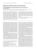
Fig. 1. Example of high-throughput cloning strategies for expression library construction. (A) The construction of in-frame gene fusions used
by Zhu et al. [35,38] relies on gap repair-mediated recombination in yeast. Amplified ORFs are mixed with a linearized vector with ends that
are identical to those of the amplified DNA, and the mixture is transformed into yeast cells where the DNA is integrated. (B) Recombination
cloning based on the Gateway k-phage integration ⁄ excision system (Invitrogen, Carlsbad, CA, USA). Recombination between the k att Pand
host att B sites yields an integrated phage with ends att L and att R. During excision, recombination between att Landatt R sites regener-
ates att Pandatt B. Amplified ORFs with att B1 and att B2 sites undergo recombination with a donor vector containing att P1 and att P2
sites, producing the entry vector with att L1 and att L2 sites. Subsequent recombination with a destination vector containing att R1 and
att R2 sites allows transfer of the ORF into the expression vector, regenerating att B sites. (C) Expression clones are rescued into Escheri-
chia coli and verified by DNA sequencing.
P. Bertone and M. Snyder Protein array technology
FEBS Journal 272 (2005) 5400–5411 ª 2005 FEBS 5403
(FITC)-labeled IgG against mouse IgG1 demonstrated
selective binding when the arrays were imaged with
a fluorescence microscope. In a related study,
Mirzabekov and coworkers [30,31] experimented with
a copolymerized acrylamide–bisacrylamide substrate,
producing arrays of discrete gel pads of between 10
and 100 lm in diameter. In a later study, Kiyonaka
et al. [32] developed a method of supramolecular gel
formation as a spontaneous process that does not
require additional polymerization steps, using the
approach to develop a sensor array of fluorescent
metal anion and cation receptors in a glycosylated
amino acetate hydrogel matrix [33].
A study by Afanassiev et al. [34] explored the use of
a thin, uniform layer of agarose film on a glass surface
to achieve a similar effect. Activated agarose contain-
ing NaIO
4
was applied to silanized slides, and samples
were mechanically deposited using a robotic micro-
spotter after the gel had solidified. A limited applica-
tion of this platform to protein-based assays
demonstrated the binding of mAbs to immobilized
recombinant human BAD protein, and reciprocally, of
recombinant human (rh)BAD to immobilized anti-
bodies in a sandwich immunoassay. Another approach
involved the use of a liquid silica compound to create
flexible sheets of microwell arrays in which biochemical
reactions are performed en masse. Zhu et al. [35]
devised a system of casting a silicone elastomer (poly-
dimethylsiloxane) onto a reusable mold of laser-milled
acrylic. After the microwell sheets had cured, various
molecules were immobilized to the interior surface of
the wells using the chemical crosslinking agent, 3-glyci-
doxypropyltrimethoxysilane.
Aside from demonstrating the technical feasibility of
protein immobilization and binding to various mole-
cules in vitro, it is essential to conduct biologically rel-
event experiments if protein microarrays are to become
an established research platform. To explore the utility
of microwell arrays for the detection of enzymatic
activities, Zhu et al. [35] focused on 119 protein kinases
from the budding yeast Saccharomyces cerevisiae
(95 known and 24 uncharacterized). Each of the pro-
tein kinases was expressed in recombinant form and
purified, then assayed for the ability to phosphorylate
17 substrate proteins. A total of 17 arrays were fabrica-
ted and one of the substrate proteins was immobilized
in every well; a different protein kinase was delivered
into each well in the presence of c-ATP to determine
which kinases were capable of phosphorylating the
given substrate protein. The arrays were incubated and
then washed such that all free enzyme was removed,
and the signal from the radiolabeled proteins in
each well were quantified using a phosphoimager. The
possibility that measurements originated from the
autophosphorylation of the kinases themselves was
discounted, as in each case the substrates were bound
in the wells while the enzymes remained free in solu-
tion; the wells were cleared of this reaction mixture
and washed prior to imaging.
In addition to detecting expected phosphorylation
activities, 27 protein kinases were found to be capable
of phosphorylating tyrosine after incubation with a
synthesized poly(tyrosine-glutamate) peptide. This find-
ing was significant as yeast protein kinases are gener-
ally known to phosphorylate only serine or threonine.
Phylogenetic analysis revealed sequence similarities
among the tyrosine-phosphorylating kinases that were
specific to several amino acids oriented in the catalytic
cleft and substrate-binding domain of the enzyme.
These appeared exclusively in the kinases that phos-
phorylated tyrosine and not in those that phosphoryl-
ated serine or threonine alone. Although it is likely
that most yeast protein kinases will preferentially phos-
phorylate serine or threonine in vivo, this study demon-
strated that protein arrays are sensitive enough to
reveal previously uncharacterized biochemical proper-
ties in a HT assay.
On balance, each of these formats retains a number
of important properties for proteomic experiments.
Microwells provide the ability to preserve native pro-
tein function by carrying out reactions in an aqueous
environment [35], while the hydrated gel matrix
approach [29–34] also immobilizes the proteins to
some degree. Any potential cross-contamination
between arrayed proteins is eliminated owing to phys-
ical barriers between them [35] or the spatial separ-
ation of discrete gel pads [29–33]. Each platform
affords a far greater loading capacity than the planar
surface of a glass slide, allowing experiments to be per-
formed at varying substrate concentrations. Finally,
the microwell format offers the potential to perform
complex, multistage experiments in solution if reaction
mixtures are exchanged using microfluidic robotics.
Contact-printed functional protein
microarrays
Although these approaches offer numerous technical
advantages, increasing attention has been paid to
adapting existing glass-slide microarray technologies for
use with proteins. As a result, the practice of printing
directly onto chemically treated glass surfaces, apart
from their indirect function as a solid support, is now in
wider use for protein arrays. The principal motivating
factor for using glass slides is to take advantage of
robotic microspotting arrayers and laser scanners that
Protein array technology P. Bertone and M. Snyder
5404 FEBS Journal 272 (2005) 5400–5411 ª 2005 FEBS
have become commonplace for DNA microarray fabri-
cation and image acquisition, respectively. The standard
microarray format therefore affords the opportunity for
a broader range of investigators to adopt HT proteomic
formats using existing DNA microarray construction
and analysis equipment.
A number of different chemical treatments are
suitable for the immobilization of proteins on glass,
including poly(l-lysine), aldehyde, nickel, epoxy, avidin
and nitrocellulose (Table 1). The choice of surface
chemistry will determine the method of protein attach-
ment to the functionalized support, which in turn will
influence whether the proteins require modification
prior to arraying. Amine-reactive coatings (e.g. 3-amino-
propyltriethoxysilane) do not require proteins to be
modified for effective immobilization, as covalent
attachment is achieved through the random crosslinking
of amine groups. Protein attachment to either nickel- or
avidin-coated slides is made through affinity binding to
histidine residues or biotin, respectively, and therefore
requires a recombinant approach to construct fusion
proteins [36].
The advantage of noncovalent immobilization is
that because attachment takes place exclusively at the
affinity tag, a significant subset of proteins will be
immobilized in a presumably uniform orientation
where their functional domains are exposed to solution
and available to interact with the labeled sample.
Other membrane-based solutions, such as fluorescent
array surface technology slides (Schleicher & Schuell
BioScience, Dassel, Germany), facilitate protein immo-
bilization through passive adsorption on a nitro-
cellulose surface, and in many cases yield superior
signal-to-noise ratios in comparison with poly(l-lysine)
or aldehyde-treated slides (Fig. 2).
Robotic printing of protein microarrays
Constructing microarrays from purified proteins is
more challenging than building their DNA counter-
parts. Aside from the complexities of preparing hun-
dreds or thousands of individual protein samples, the
proteins must remain active during the manufacture
and probing of microarrays. This entails keeping pro-
tein samples in cold storage for as long as possible,
and arraying them in a glycerol solution to maintain
their native state on the slide surface. The procedures
used for printing protein arrays are similar to those
developed for DNA arrays [37], but with several
important differences, discussed below.
DNA samples are arrayed in a high-humidity cham-
ber by a robotic microspotter. Although the same
equipment can be used to print protein arrays, individ-
ual samples are typically handled in a 30–35% glycerol
solution to prevent denaturing, and arrayed at concen-
trations in the range of 0.3–1 mgÆmL
)1
. As glycerol is
hygroscopic, care must be taken to reduce the ambient
humidity to 28–30%; this differs from the 45% humid-
ity environment in which DNA arrays are usually prin-
ted. Printed spots of protein–glycerol solution will
absorb moisture from the air in high-humidity environ-
ments and begin to expand on the slide surface. This
can lead to cross-contamination of samples if the pro-
teins are arrayed at high spatial density.
The diameter and pitch (the spacing between sam-
ples) of spots can remain similar to the parameters used
for printing DNA microarrays, although this will
depend on the type of slides used and the viscosity of
the buffer. For example, epoxy-coated slides provide
more efficient protein attachment, which generally
allows for higher-density printing. Pilot studies should
be carried out to determine the appropriate printing
density for a given sample preparation and surface
chemistry, adjusting the pitch from a more conservative
spacing of 1000 lm down to a minimum of 300 lm.
Depending on the number of pins used (typically
between 16 and 48), a 300 lm pitch allows up to 20 000
individual protein samples to be printed on a standard
microscope slide, yielding 1600 features per cm
2
where each feature is 150 lm in diameter. (Fig. 3C).
As with DNA arrays, fabrication precision should
be assessed relative to the printing consistency across
Table 1. Surface chemistries for glass slide protein microarrays.
Surface chemistry Protein attachment
Protein
orientation
Modifications
required
Epoxy Covalent crosslinking Random None
Aldehyde Covalent crosslinking Random None
Poly(
L-lysine) Adsorption Random None
Nitrocellulose Adsorption, absorption Random None
Poly(vinylidene difluoride) Adsorption, absorption Random None
Avidin Affinity binding Random Biotinylation
Nickel-nitrilotriacetic acid Affinity binding Uniform His
6
fusion
P. Bertone and M. Snyder Protein array technology
FEBS Journal 272 (2005) 5400–5411 ª 2005 FEBS 5405
all of the features on the array, consistency between
features from multiple arrays, and the uniformity of
sample concentrations. This is typically measured as
the coefficient of variation (CV), which estimates the
reproducibility of printing as the standard deviation
over the mean fluorescence signal intensity. For func-
tional protein arrays prepared from individual purified
samples, intra-array CV measurements fall in the range
of 15–20%, and are a measurement of both the variab-
ility in protein concentrations and the pin-to-pin con-
sistency of sample deposition during the printing cycle.
Inter-array CVs over all protein samples from replicate
slides are typically lower, in the range of 12–15%.
Much of the variation in sample concentration is
attributed to the expression and purification of low-
abundance proteins, whereas printing variation can
arise as a function of the order in which samples are
arrayed during long print runs.
The incorporation of an epitope tag, such as gluta-
thione-S-transferase, is invaluable for HT affinity puri-
fication, and also allows the global quantification of
protein signals by probing the array with a fluor-conju-
gated antibody [38]. This measurement is used to cal-
culate the ratio of the probe-binding signal relative to
the baseline fluorescence intensity of each spot on the
array. As the relative concentrations and deposited
amounts of protein samples will vary slightly, compu-
ting the fluorescence values in terms of intensity ratios
enables the application of statistical methods similar to
those used for DNA microarray data analysis [39].
Detecting biochemical interactions
Depending on the type of assay being performed, pro-
tein–substrate binding or catalysis can be detected via
fluorescence labeling, chemiluminescence, radioisotope
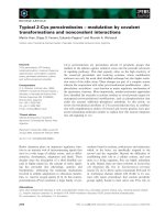
Fig. 2. Contact-printed functional protein microarrays. (A) Control proteins are spotted in a dilution series and illuminated with fluorescein-
labeled antibodies. (B) Protein–protein interaction assays reveal specific binding targets. (C) Detection of lectin modifications across the yeast
proteome.
Protein array technology P. Bertone and M. Snyder
5406 FEBS Journal 272 (2005) 5400–5411 ª 2005 FEBS
labeling, or label-free methods, such as surface plas-
mon resonance imaging, atomic force microscopy or
reflectometric interference spectroscopy. Fluorescence
labeling is generally preferred as a safe and efficient
method for HT analysis that is compatible with cur-
rent microarray laser scanners. The detection of pro-
tein binding, as described in Zhu et al. [38], involves a
two-step process similar to that used for multiplex
sandwich immunoassays. First, the sample used to
probe the array is biotinylated using a simple cros-
slinking method. To perform the experiment, the
microarray is incubated with a solution containing the
biotinylated sample, washed, then incubated a second
time in the presence of a fluorescent streptavidin mole-
cule. The actual protein interactions are formed during
the first incubation; subsequent binding of the strept-
avidin–fluor conjugate to the biotin label then allows
specific interactions to be detected using a standard
microarray scanner.
A simpler approach can be employed by attaching a
fluorophore directly to the solution-phase binder used
to probe the array, provided that the label does not
A
B
C
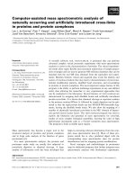
Fig. 3. Detecting biochemical interactions on protein arrays. (A and C) Protein–protein interactions were detected with fluorescein-labeled
antibodies to specific proteins. In this case, BODIPYFL–IgG, Cy3–IjBa and Cy5–FKBP12 identify the presence of protein G, p50 and FKBP–
rapamycin-associated protein, respectively [40]. (B) Phosphoinositide-binding specificity to functional proteins is determined in a phospholip id
probing assay. The detection of protein–protein and protein–lipid binding [38] involves a two-step process. First, the sample used to probe
the array is biotinylated using a simple crosslinking method. To perform the experiment, the microarray is incubated with a solution contain-
ing the biotinylated sample, washed, then incubated a second time in the presence of a fluorescent streptavidin molecule. The actual protein
interactions are formed during the first incubation; subsequent binding of the streptavidin–fluor conjugate to the biotin label then allows
these interactions to be detected using a microarray scanner. A simpler approach can also be employed by attaching a fluorophore directly
to the solution-phase binder used to probe the array. Proteins can be labeled using amine-reactive dyes, where the succinimidyl ester moiet-
ies react with the primary amines of the protein to form stable conjugates.
P. Bertone and M. Snyder Protein array technology
FEBS Journal 272 (2005) 5400–5411 ª 2005 FEBS 5407
mask an active site or binding domain of the sample
molecule. Proteins can be labeled directly using amine-
reactive dyes, where the succinimidyl ester moieties
react with the primary amines of the protein to form
stable conjugates. The labeled protein can then be
purified using size-exclusion spin columns and applied
to the array for the detection of protein–protein inter-
actions. Of course, other molecules can be applied to
functional protein arrays to assess different molecular
interactions, such as protein–DNA binding. Fluores-
cence labeling of DNA can be achieved in a variety of
ways, such as through direct incorporation of fluores-
cent-labeled nucleotides during reverse transcription,
or by secondary conjugation of amine-reactive succin-
imidyl esters to 5-(3-aminoallyl)-dUTPs.
Functional protein arrays in use
The first study reporting the use of contact-printed,
glass-slide protein arrays was described by MacBeath
& Schreiber [40], who investigated the binding activit-
ies of three known pairs of interacting proteins
(Fig. 3A). One protein of each pair was printed in
quadruplicate onto aldehyde slides, and the arrays
were probed with the labeled partners. The group also
explored various feature densities of printed protein
samples, successfully arraying a single protein as
10 800 discrete features on a standard microarray slide
(Fig. 3C). Importantly, the researchers were able to
quantify the concentrations of the bound and solution-
phase proteins necessary to carry out the experiments.
Sample concentrations between 100 lgÆmL
)1
and 1 mgÆ
mL
)1
were found to be suitable for protein immobi-
lization and detection, whereas solution-phase proteins
at concentrations of 12.5 pm yielded fluorescence sig-
nals that scaled linearly over four orders of magnitude.
Thus, these experiments demonstrated the feasibility of
arraying proteins in a standard microarray format and
at feature densities comparable with those of contact-
printed DNA arrays.
A subsequent study by Zhu et al. [38] described the
development of a yeast proteome microarray contain-
ing the full-length, purified expression products of over
93% of the organism’s complement of 6280 protein-
coding genes. A total of 5800 ORFs were cloned as
glutathione-S-transferase::His
6
fusions, and expressed
in their native cells under a Gal-inducible promoter.
Following HT purification, each protein sample was
printed in duplicate onto glass slides using a standard
robotic microspotter, at a feature density of 13 000
samples per array. This work represented the first sys-
tematic cloning and purification of an entire eukaryotic
proteome, as well as the first large-scale functional
protein array comprising discrete functional proteins.
A number of protein attachment chemistries were eval-
uated, including aldehyde and nickel surface treatment.
Aldehyde surfaces promote the covalent attachment of
proteins by their N termini, although a concern with
this method is that the random crosslinking of primary
amines may disrupt the tertiary structures of some pro-
teins. An alternative approach employed nickel-coated
slides, attaching proteins via the incorporated His
6
tag.
This yielded higher signal-to-noise ratios, presumably
because fewer proteins were denatured after nickel
attachment and their functional domains were more
likely to be oriented uniformly away from the slide
surface. Over 90% of the samples were found to yield
significant fluorescence signals over background levels,
in the range of 10–950 fg of protein.
Several different experiments were performed with
the arrays, including a calmodulin-binding survey to
assess protein–protein interactions and a large-scale
screen for phospholipid-binding specificity. In the
latter analysis, self-assembling phosphatidylcholine
liposomes were incubated with five different phos-
phoinositide species and 1% biotin to form a series
of biotinylated phospholipid probes (Fig. 3B). Proteo-
me-wide microarray experiments identified 150 lipid-
binding proteins, of which 52 were uncharacterized.
In particular, the array-based interaction assays iden-
tified sets of proteins that demonstrated preferential
binding to one or more phosphoinositides, apart
from those that bound all of the phospholipids with
equal affinity. As phosphoinositides are important
components of membrane structures and second-
messenger pathways, these types of protein–lipid
interactions constitute a significant class of biochemi-
cal functionality.
Further applications of protein arrays
In many cases, complex proteomic experiments may
benefit from the combined application of multiple ana-
lytical techniques. In a study by Huang et al. [41], a
chemical genetic approach was used for the develop-
ment of a screen for small-molecule inhibitors and
enhancers of rapamycin, a peptide exhibiting a variety
of functional roles in nutrient metabolism and cell
cycle progression in eukaryotes. Following the identifi-
cation of candidates, yeast proteome arrays [38] were
interrogated with two biotinylated rapamycin-inhibi-
tory molecules to identify their potential target pro-
teins. Each was observed to bind multiple target
proteins in addition to phosphatidylinositides. Analysis
of various phenotypes revealed which binding events
represented biologically meaningful interactions.
Protein array technology P. Bertone and M. Snyder
5408 FEBS Journal 272 (2005) 5400–5411 ª 2005 FEBS
A combined experiment involving the use of protein
and DNA microarrays was recently described for the
systematic identification and location analysis of
transcription factor proteins. Hall et al. [42] assayed
the yeast proteome in search of novel DNA-binding
proteins by probing the protein microarrays with labe-
led yeast genomic DNA. A total of 200 DNA-binding
proteins were identified, half of which were known, or
expected, to bind DNA. Of these, eight candidates
were subjected to chromatin profiling via the ChIP-
chip method [43], which is designed to identify tran-
scription factor binding sites in genomic DNA. This
technique entails the immunoprecipitation of specific
protein–DNA complexes using antibodies against a
native transcription factor protein or epitope tag. The
immunoselected DNA is then sonicated, labeled and
used to interrogate a DNA microarray representing
intergenic or total genomic regions, thereby revealing
the locations of transcription factor binding along a
chromosome. ChIP-chip analysis on yeast intergenic
arrays revealed Arg5,6, a mitochondrial enzyme
involved in ornithine biosynthesis, to bind DNA at
specific nuclear and mitochondrial loci. Altered gene
expression levels were observed in Arg5,6 deletion
mutants, further indicating its role as a transcriptional
regulator.
Conclusions
The advent of protein-based microarrays allows the
global observation of biochemical activities on an
unprecedented scale, where hundreds or thousands of
proteins can be simultaneously screened for protein–
protein, protein–nucleic acid, and small molecule inter-
actions, as well as post-translational modifications.
Advances in HT separation techniques offer the poten-
tial for arraying proteins directly, although these tech-
nologies are at an early stage of development. Ouyang
et al. [44] explored the separation, deposition and ana-
lysis of individual proteins from complex samples
using ion soft landing and MS. In this study, a mixture
of four proteins was introduced into a mass spectro-
meter by ESI. The proteins were then separated by
their respective mass-to-charge ratios and independ-
ently deposited onto a gold substrate via ion beam
focusing. Two methods of analysis were explored. Ini-
tially, the arrays were rinsed with a methanol⁄ water
solution and the mixture analyzed by ESI-MS. It was
later found that the deposited proteins could be ana-
lyzed in situ by MALDI-MS.
Although automated mass-spectrometric separation
and deposition remains a promising technology for
protein microarray assembly, it is unclear whether soft-
landing techniques can be applied en masse to the wide
range of proteins constituting the entire proteome of an
organism. At present, it seems clear that a well-curated
expression clone library allows researchers to maintain
the quality and identity of every protein under investi-
gation. Additionally, MS may disrupt low-affinity bio-
molecular interactions, or even denature individual
proteins, prior to experimental analysis. Ramachandran
et al. [45] recently described an in vitro transcription
and translation system that can generate proteins from
a PCR product. The reactions are carried out in paral-
lel directly on a solid support, through the use of affin-
ity tags to anchor the products to the surface. The
main drawback of this approach is that some proteins
may not be properly folded and modified outside their
native cellular environment. Ultimately, both of these
developments offer the potential to reduce the time and
complexity involved with the cloning and purification
of individual proteins as a prerequisite to constructing
functional protein microarrays.
In terms of assessing protein–protein interactions,
protein microarray experiments can be qualitatively
compared with the two-hybrid assay [46,47]. However,
microarray experiments afford the ability to control
the environmental parameters of an experiment, such
as ion concentration, buffer pH, and the addition of
reaction cofactors, in a precise and reproducible man-
ner. Additionally, because microarrays exploit parallel
interrogation to acquire many individual measurements
on the same physical platform, the resulting data can
be subjected to rigorous statistical analyses and tested
for accuracy and reproducibility. Thus, protein-based
microarrays provide the ability to characterize the bio-
chemical functions of thousands of proteins in a paral-
lel, quantitative format.
References
1 Rigaut G, Shevchenko A, Rutz B, Wilm M, Mann M &
Seraphin B (1999) A generic protein purification method
for protein complex characterization and proteome
exploration. Nat Biotechnol 17, 1030–1032.
2 Washburn MP, Wolters D & Yates JR (2001) Large-
scale analysis of the yeast proteome by multidimen-
sional protein identification technology. Nat Biotechnol
19, 242–247.
3 Li Y, McIver RT Jr & Hunter RL (1994) High-accuracy
molecular mass determination for peptides and proteins
by Fourier transform mass spectrometry. Anal Chem 66,
2077–2083.
4 Fenn JB, Mann M, Meng CK, Wong SF & Whitehouse
CM (1989) Electrospray ionization for mass spectro-
metry of large biomolecules. Science 246, 64–71.
P. Bertone and M. Snyder Protein array technology
FEBS Journal 272 (2005) 5400–5411 ª 2005 FEBS 5409
5 Karas M & Hillenkamp F (1988) Laser desorption
ionization of proteins with molecular masses exceeding
10,000 daltons. Anal Chem 60, 2299–2301.
6 Hillenkamp F & Karas M (1990) Mass spectrometry
of peptides and proteins by matrix-assisted ultraviolet
laser desorption ⁄ ionization. Methods Enzymol 193,
280–295.
7 Shevchenko A, Keller P, Scheiffele P, Mann M &
Simons K (1997) Identification of components of trans-
Golgi network-derived transport vesicles and detergent-
insoluble complexes by nanoelectrospray tandem mass
spectrometry. Electrophoresis 18, 2591–2600.
8 Medzihradszky KF, Campbell JM, Baldwin MA,
Falick AM, Juhasz P, Vestal ML & Burlingame AL
(2000) The characteristics of peptide collision-induced
dissociation using a high-performance MALDI-TOF ⁄
TOF tandem mass spectrometer. Anal Chem 2, 552–
558.
9 Baldwin MA, Medzihradszky KF, Lock CM, Fisher B,
Settineri TA & Burlingame AL (2001) Matrix-assisted
laser desorption ⁄ ionization coupled with quadru-
pole ⁄ orthogonal acceleration time-of-flight mass spectro-
metry for protein discovery, identification, and
structural analysis. Anal Chem 73, 1707–1720.
10 Shevchenko A, Loboda A, Shevchenko A, Ens W &
Standing KG (2000) MALDI quadrupole time-of-flight
mass spectrometry: a powerful tool for proteomic
research. Anal Chem 72, 2132–2141.
11 Hutchens TW & Yip TT (1993) New desorption strate-
gies for the mass spectrometric analysis of macromole-
cules. Rapid Commun Mass Spectrom 7, 576–580.
12 Tang N, Tornatore P & Weinberger SR (2004) Current
developments in SELDI affinity technology. Mass
Spectrom Rev 23, 34–44.
13 Martzen MR, McCraith SM, Spinelli SL, Torres M,
Fields S, Grayhack EJ & Phizicky EM (1999) A bio-
chemical genomics approach for identifying genes by
the activity of their products. Science 286 , 1153–1155.
14 Angenendt P, Glokler J, Murphy D, Lehrach H &
Cahill DJ (2002) Toward optimized antibody micro-
arrays: a comparison of current microarray support
materials. Anal Biochem 309, 253–260.
15 Fodor SP, Read JL, Pirrung MC, Stryer L, Lu AT &
Solas D (1991) Light-directed, spatially addressable par-
allel chemical synthesis. Science 251, 767–773.
16 Lipshutz RJ, Fodor SP, Gingeras TR & Lockhart DJ
(1999) High density synthetic oligonucleotide arrays.
Nat Genet 21, 20–24.
17 Pellois JP, Zhou X, Srivannavit O, Zhou T, Gulari E &
Gao X (2002) Individually addressable parallel peptide
synthesis on microchips. Nat Biotechnol 20, 922–926.
18 Frank R (2002) The SPOT-synthesis technique. Syn-
thetic peptide arrays on membrane supports–principles
and applications. J Immunol Methods 267, 13–26.
19 Li S, Marthandan N, Bowerman D, Garner HR &
Kodadek T (2005) Photolithographic synthesis of cyclic
peptide arrays using a differential deprotection strategy.
Chem Commun 5, 581–583.
20 Singh-Gasson S, Green RD, Yue Y, Nelson C, Blattner
F, Sussman MR & Cerrina F (1999) Nat Biotechnol 17,
974–978.
21 Nielsen UB & Geierstanger BH (2004) Multiplexed
sandwich assays in microarray format. J Immunol
Methods 290, 107–120.
22 Saviranta P, Okon R, Brinker A, Warashina M, Eppin-
ger J & Geierstanger BH (2004) Evaluating sandwich
immunoassays in microarray format in terms of the
ambient analyte regime. Clin Chem 50, 1907–1920.
23 Huang RP (2004) Cytokine protein arrays. Methods
Mol Biol 264, 215–231.
24 Huang RP, Yang W, Yang D, Flowers L, Horowitz IR,
Cao X & Huang R (2005) The promise of cytokine anti-
body arrays in the drug discovery process. Expert Opin
Ther Targets 9, 601–615.
25 Schweitzer B, Roberts S, Grimwade B, Shao W, Wang
M, Fu Q, Shu Q, Laroche I, Zhou Z, Tchernev VT
et al. (2002) Multiplexed protein profiling on micro-
arrays by rolling-circle amplification. Nat Biotechnol 20,
359–365.
26 Ansell RJ, Ramstrom O & Mosbach K (1996) Towards
artificial antibodies prepared by molecular imprinting.
Clin Chem 42, 1506–1512.
27 Titirici MM & Sellergren B (2004) Peptide recognition
via hierarchical imprinting. Anal Bioanal Chem 378,
1913–1921.
28 Michaud GA, Salcius M, Zhou F, Bangham R, Bonin
J, Guo H, Snyder M, Predki PF & Schweitzer BI (2003)
Analyzing antibody specificity with whole proteome
microarrays. Nat Biotechnol 21, 1509–1512.
29 Guschin D, Yershov G, Zaslavsky A, Gemmell A, Shick
V, Proudnikov D, Arenkov P & Mirzabekov A (1997)
Manual manufacturing of oligonucleotide, DNA, and
protein microchips. Anal Biochem 250, 203–211.
30 Vasiliskov AV, Timofeev EN, Surzhikov SA, Drobyshev
AL, Shick VV & Mirzabekov AD (1999) Fabrication of
microarray of gel-immobilized compounds on a chip by
copolymerization. Biotechniques 27, 592–594,596–
598,600.
31 Rubina AY, Dementieva EI, Stomakhin AA, Darii EL,
Pan’kov SV, Barsky VE, Ivanov SM, Konovalova EV
& Mirzabekov AD (2003) Hydrogel-based protein
microchips: manufacturing, properties, and applications.
Biotechniques 34, 1008–1014,1016–1020,1022.
32 Kiyonaka S, Sada K, Yoshimura I, Shinkai S, Kato N
& Hamachi I (2004) Semi-wet peptide ⁄ protein array
using supramolecular hydrogel. Nat Mater 3, 58–64.
33 Yoshimura I, Miyahara Y, Kasagi N, Yamane H, Ojida
A & Hamachi I (2004) Molecular recognition in a
Protein array technology P. Bertone and M. Snyder
5410 FEBS Journal 272 (2005) 5400–5411 ª 2005 FEBS
supramolecular hydrogel to afford a semi-wet sensor
chip. J Am Chem Soc 126, 12204–12205.
34 Afanassiev V, Hanemann V & Wolfl S (2000) Prepara-
tion of DNA and protein micro arrays on glass slides
coated with an agarose film. Nucleic Acids Res 28, E66.
35 Zhu H, Klemic JF, Chang S, Bertone P, Casamayor A,
Klemic KG, Smith D, Gerstein M, Reed MA & Snyder
M (2000) Analysis of yeast protein kinases using protein
chips. Nat Genet 26, 283–289.
36 Nasoff M, Bergseid M, Hoeffler JP & Heyman JA
(2000) High-throughput expression of fusion proteins.
Methods Enzymol 328, 515–529.
37 Shalon D, Smith SJ & Brown PO (1996) A DNA micro-
array system for analyzing complex DNA samples using
two-color fluorescent probe hybridization. Genome Res
6, 639–645.
38 Zhu H, Bilgin M, Bangham R, Hall D, Casamayor A,
Bertone P, Lan N, Jansen R, Bidlingmaier S, Houfek T
et al. (2001) Global analysis of protein activities using
proteome chips. Science 293, 2101–2105.
39 Luscombe NM, Royce TE, Bertone P, Echols N, Horak
CE, Chang JT, Snyder M & Gerstein M (2003) Express-
Yourself: a modular platform for processing and visua-
lizing microarray data. Nucleic Acids Res 31, 3477–3482.
40 MacBeath G & Schreiber SL (2000) Printing proteins as
microarrays for high-throughput function determina-
tion. Science 289, 1760–1763.
41 Huang J, Zhu H, Haggarty SJ, Spring DR, Hwang H,
Jin F, Snyder M & Schreiber SL (2004) Finding new
components of the target of rapamycin (TOR) signaling
network through chemical genetics and proteome chips.
Proc Natl Acad Sci USA 101, 16594–16599.
42 Hall DA, Zhu H, Zhu X, Royce T, Gerstein M &
Snyder M (2004) Regulation of gene expression by a
metabolic enzyme. Science 306, 482–484.
43 Horak CE & Snyder M (2002) ChIP-chip: a genomic
approach for identifying transcription factor binding
sites. Methods Enzymol 350, 469–483.
44 Ouyang Z, Takats Z, Blake TA, Gologan B, Guymon
AJ, Wiseman JM, Oliver JC, Davisson VJ & Cooks RG
(2003) Preparing protein microarrays by soft-landing of
mass-selected ions. Science 301, 1351–1354.
45 Ramachandran N, Hainsworth E, Bhullar B, Eisenstein
S, Rosen B, Lau AY, Walter JC & LaBaer J (2004)
Self-assembling protein microarrays. Science 305,
86–90.
46 Ito T, Tashiro K, Muta S, Ozawa R, Chiba T,
Nishizawa M, Yamamoto K, Kuhara S & Sakaki Y
(2000) Toward a protein–protein interaction map of the
budding yeast: a comprehensive system to examine two–
hybrid interactions in all possible combinations between
the yeast proteins. Proc Natl Acad Sci USA 97, 1143–
1147.
47 Uetz P, Giot L, Cagney G, Mansfield TA, Judson RS,
Knight JR, Lockshon D, Narayan V, Srinivasan M,
Pochart P et al. (2000) A comprehensive analysis of
protein–protein interactions in Saccharomyces cere-
visiae. Nature 403, 623–627.
P. Bertone and M. Snyder Protein array technology
FEBS Journal 272 (2005) 5400–5411 ª 2005 FEBS 5411