Báo cáo khoa học: Analysis of the stability of the spermadhesin PSP-I ⁄ PSP-II heterodimer Effects of Zn 2+ and acidic pH pdf
Bạn đang xem bản rút gọn của tài liệu. Xem và tải ngay bản đầy đủ của tài liệu tại đây (228.45 KB, 8 trang )
Analysis of the stability of the spermadhesin PSP-I ⁄ PSP-II
heterodimer
Effects of Zn
2+
and acidic pH
Marı
´a
Asuncio
´
n Campanero-Rhodes
1
, Margarita Mene
´
ndez
1
, Jose
´
Luis Sa
´
iz
1
, Libia Sanz
2
,
Juan Jose
´
Calvete
2
and Dolores Solı
´
s
1
1 Instituto de Quı
´
mica Fı
´
sica ‘Rocasolano’, CSIC, Madrid, Spain
2 Instituto de Biomedicina de Valencia, CSIC, Valencia, Spain
Proteins are designed to have a particular activity in a
specific environment, and their fold and assembly are
intimately related to this physiological function. Infor-
mation on the organization of the protein structure,
however, is usually acquired in simple buffer systems,
far removed from the complex conditions encountered
in intracellular and extracellular spaces and fluids.
Besides the crucial influence of the local concentration
of macromolecules, the presence of co-solutes may
have a decisive effect on protein conformation and sta-
bility [1].
Seminal plasma is a composite fluid, comprising
secretions from the testes, epididymis and accessory
sex glands. It is not merely a vehicle for the ejaculated
sperm but it is also involved in numerous activities in
the male and female reproductive tract, ensuring the
viability and fertilizing capacity of spermatozoa. The
seminal plasma contains abundant concentrations of
different amino acids, peptides, lipids, fatty acids and
various osmolytes, and it is an important source
of cations [2]. In boar seminal plasma, for example,
the concentration of Zn
2+
is surprisingly high
Keywords
heterodimer dissociation; PSP-I ⁄ PSP-II;
spermadhesins; thermal stability; Zn
2+
Correspondence
D. Solı
´
s, Instituto de Quı
´
mica Fı
´
sica
Rocasolano, Serrano 119, 28006 Madrid,
Spain
Fax: +34 91 564 24 31
Tel: +34 91 561 94 00
E-mail:
(Received 20 June 2005, revised 7 September
2005, accepted 14 September 2005)
doi:10.1111/j.1742-4658.2005.04974.x
Spermadhesins are a family of 12–16 kDa proteins with a single CUB
domain. PSP-I and PSP-II, the most abundant boar spermadhesins, are
present in seminal plasma as a noncovalent heterodimer. Dimerization
markedly affects the binding ability of the subunits. Notably, heparin and
mannose 6-phosphate binding abilities of PSP-II are abolished, indicating
that the corresponding binding sites may be located at (or near) the dimer
interface. Pursuing the hypothesis that cryptic binding sites in PSP-I ⁄ PSP-II
may be exposed in specific physiological environments, we examined the
influence of Zn
2+
and acidic pH on the heterodimer stability. According to
near-UV CD spectra, the core native fold is preserved in the presence of
physiological concentrations of Zn
2+
, a cation unusually abundant in boar
seminal plasma. However, the thermostability of the heterodimer decreases
significantly, as observed by CD and differential scanning calorimetry. The
effect is Zn
2+
-specific and is reversed by EDTA. Destabilization is also
observed at acidic pH. Gel filtration analysis using radioiodinated PSP-I ⁄
PSP-II reveals that dissociation of the heterodimer at low (nanomolar)
protein concentrations is promoted by both Zn
2+
and acidic pH. Although
the integrity of the heterodimer in seminal plasma seems to be guaranteed by
its high concentration, dissociation may be facilitated in the female genital
tract because of dilution of the protein in the intraluminal fluids of the cervix
and the uterus, and the acidic fluid of the uterotubal junction. Such a
mechanism may be relevant in the regulation of uterine immune reactions.
Abbreviations
DSC, differential scanning calorimetry.
FEBS Journal 272 (2005) 5663–5670 ª 2005 FEBS 5663
(0.3–0.7 mm) [3,4], reaching the spermatozoa at ejac-
ulation [5]. Seminal plasma also contains a large num-
ber of different proteins that exert multiple effects on
sperm function, including a diversity of enzymes,
hormones, growth factors and transport proteins [6].
However, the precise role of most of the seminal
plasma proteins in sperm physiology remains obscure.
Spermadhesins are a family of 12–16 kDa proteins
found in seminal plasma and ⁄ or attached to the sper-
matozoal surface of a variety of mammalian species
(e.g. boar, bull and horse) [7]. These proteins are
composed of 109–133 amino acids, show a 40–60%
sequence identity, and contain a single CUB domain
[8]. Members of the spermadhesin family have been
shown to bind zona pellucida glycoproteins, serine
proteinase inhibitors, phospholipids and ⁄ or sulfated
glycosaminoglycans [9], suggesting that they may be
involved in different steps of the complex fertilization
process. In the boar, spermadhesins represent about
75% of the total protein content of seminal plasma,
their concentration ranging from 0.6 to 7 mgÆmL
)1
[10]. PSP-I and PSP-II, the most abundant boar
spermadhesins, occur as a noncovalent heterodimer [11].
The secondary structure and stability of the PSP-I⁄
PSP-II heterodimer in solution has been investigated
[12], and the crystal structure solved at 2.4 A
˚
resolution
[13]. Both subunits consists of a compact ellipsoidal
b-sandwich structure organized into two five-stranded
(parallel and antiparallel) b-sheets.
Accumulating evidence points to a role for PSP-I ⁄
PSP-II as an exogenous modulator of both sperm
function and uterine immune activity, thus ensuring
reproductive success. The PSP-I ⁄ PSP-II complex con-
tributes to maintaining sperm with high viability,
motility, and mitochondrial activity [14]. In addition,
PSP-I and PSP-II are immunostimulatory for lympho-
cyte activity in vitro [15]. Lymphocyte binding of PSP-I
has been demonstrated [16]. Furthermore, the PSP-I ⁄
PSP-II heterodimer and its isolated subunits induce the
recruitment of neutrophils into the peritoneal cavity
of rats [17] and pigs [18]. The neutrophil migration-
inducing activity of PSP-I ⁄ PSP-II, and possibly of the
PSP-II subunit, is mediated by the stimulation of resi-
dent macrophages, which release a neutrophil chemo-
tactic substance [19]. In contrast, PSP-I appears to act
directly on neutrophils [17]. The purpose of these
immunostimulatory activities would be to prevent
possible infections of the lower reproductive tract and
to provide a foreign-cell-free uterine environment for
the descending early embryos.
The ligand-binding capabilities of the isolated sub-
units have been investigated thoroughly. The PSP-II
subunit exhibits mannose 6-phosphate and heparin
binding abilities [20], whereas conflicting results on the
heparin-binding ability of the PSP-I subunit have been
reported [11,21,22]. These binding sites are nonetheless
cryptic in the heterodimer, which is typically isolated
from the nonheparin-binding fraction of boar seminal
plasma [11], raising the question of their biological sig-
nificance. In this context, it is noteworthy that the
stimulatory activity of PSP-II on macrophages is selec-
tively inhibited by mannose 6-phosphate [17].
Here we show that, in the presence of physiological
concentrations of Zn
2+
, the stability of the hetero-
dimer is significantly lowered, promoting at low pro-
tein concentrations dissociation of the PSP-I and
PSP-II subunits. Similar behaviour is induced by acidic
pH. The results point to the possibility that the cryptic
binding sites in the PSP-I ⁄ PSP-II heterodimer are
exposed in the female genital tract environment.
Results
CD spectroscopy
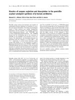
The far-UV CD spectrum of PSP-I ⁄ PSP-II exhibits a
large positive band at % 202 nm and a negative region
at 215 nm [12], as expected for the b-sandwich topol-
ogy of the CUB domain [13]. In addition, the near-UV
CD spectrum was dominated by the presence of a
sharp positive band at 291 nm, in the tryptophan
region (Fig. 1A). Furthermore, the spectrum showed
a large negative region with minima around 287 and
268 nm. Thermal denaturation of PSP-I ⁄ PSP-II led to
a decrease in the intensity of both the positive and
negative bands (Fig. 1A) along with an increase in
ellipticity below 250 nm. These changes reflect the loss
of tertiary structure of the protein. Monitoring of the
decrease with temperature of the ellipticity at 268 nm
facilitated tracing of the denaturalization process. PSP-
I ⁄ PSP-II thermal denaturation was irreversible [12],
but the thermal denaturation profiles were practically
scan-rate independent. Experimental curves were there-
fore phenomenologically analyzed using a sigmoidal
function (see Experimental procedures) from which a
T
1 ⁄ 2
(temperature at which 50% of the protein is dena-
tured) of 62.2 °C can be estimated (Table 1).
The far-UV and near-UV CD spectra of PSP-I ⁄ PSP-
II were not affected by the presence of ZnCl
2
in the
medium at concentrations up to 4 mm (data not
shown). However, the stability of the heterodimer
against thermal denaturation was significantly reduced,
as evidenced by monitoring the variation with tem-
perature of the ellipticity at 268 nm (Fig. 1B). At
0.5 mm ZnCl
2
, a concentration of Zn
2+
in the range
of those reported for porcine seminal plasma, T
1 ⁄ 2
falls
Stability of PSP-I ⁄ PSP-II heterodimer M. A. Campanero-Rhodes et al.
5664 FEBS Journal 272 (2005) 5663–5670 ª 2005 FEBS
to 53.2 °C, and a further decrease was observed at
higher Zn
2+
concentrations (Table 1).
Differential scanning calorimetry (DSC)
In a former study [12], the thermal stability of the
PSP-I ⁄ PSP-II heterodimer was analysed by DSC,
showing that the entire dimer constituted the cooper-
ative unfolding unit. Thermal denaturation curves of
PSP-I ⁄ PSP-II presented a single peak with a maximum
at 60.5 °C and an apparent enthalpy change of
439 kJÆ(mol dimer)
)1
[12]. We have since observed
some differences among protein batches in the calori-
metric enthalpy changes, with a mean ± SD DH
cal
of
405 ± 17 kJÆmol
)1
(r, n ¼ 8). These variations are
not related to the protein concentration or the scan
rate used in the analysis. However, the T
m
values of
the DSC transitions were quite reproducible from
batch to batch (60.7 ± 0.3 °C), thus serving as a use-
ful gauge of the heterodimer thermostability.
DSC data confirmed that, in the presence of ZnCl
2
,
the thermal stability of PSP-I ⁄ PSP-II was substantially
reduced (Fig. 2A). As the Zn
2+
concentration was
increased, a concomitant decrease in both the trans-
ition temperature and the apparent enthalpy of dena-
turation was observed (Table 1), and, at 4 mm ZnCl
2
,
protein precipitation occurred above 65 °C. The desta-
bilization induced by Zn
2+
was reversed by the addi-
tion of EDTA to the sample (Fig. 2A). On the other
hand, no significant decrease in the heterodimer stabil-
ity was observed in the presence of 4 mm CaCl
2
(Table 1), emphasizing the specificity of the effect of
Zn
2+
.
Thermal destabilization of PSP-I ⁄ PSP-II was also
noticed at acidic pH (Fig. 2B) in the absence of Zn
2+
cations. At pH 3.8 the apparent enthalpy of denatura-
tion decreased % 75 kJÆmol
)1
and the transition tem-
perature was 8 °C lower (Table 1).
Ultracentrifugation and chromatographic
behaviour
The sedimentation equilibrium data for PSP-I ⁄ PSP-II
(0.25–0.5 mgÆmL
)1
) could be fitted to a single-ideal-
component model with a weight-average molecular
mass of 27 933 Da, confirming that PSP-I ⁄ PSP-II
behaved in solution as a dimer. No influence of Zn
2+
at concentrations up to 4 mm on the average mole-
cular mass of PSP-I ⁄ PSP-II was observed at this
protein concentration range.
On gel filtration chromatography, the elution time
of PSP-I ⁄ PSP-II at concentrations of, or above,
0.01 mgÆmL
)1
was 26 min, consistent with the time
predicted for the dimer. However, analysis of the gel
filtration behaviour using
125
I-labelled PSP-I ⁄ PSP-II
revealed a broadening of the peak at lower protein
concentrations (Fig. 3A), with the appearance of minor
species at the elution volume of the isolated subunits.
Fig. 1. Near-UV CD of PSP-I ⁄ PSP-II. Variation with temperature (A) and effect of Zn
2+
on the thermal denaturation (B) of the heterodimer.
Spectra were obtained for 1 mgÆmL
)1
PSP-I ⁄ PSP-II solutions in 20 mM Hepes, pH 7.0. (A) Representative spectra acquired at 25 °C(h), 50 °C
(n), 56 °C(n), 62 °C(m), 70 °C(s)and77°C(d) °C. (B) Variation in ellipticity at 268 nm with temperature monitored in the absence (s)orin
the presence of 0.5 (n)or4(h)m
M Zn
2+
. The continuous lines correspond to the fit of the experimental data to a sigmoidal function.
Table 1. Thermodynamic parameters of the thermal denaturation
of PSP-I ⁄ PSP-II as determined by CD (T
1 ⁄ 2
) and DSC (T
m
, DH
cal
).
ND, Not determined.
pH
Additive
(m
M)
T
1 ⁄ 2
(°C)
T
m
(°C)
DH
CAL
(kJÆmol
)1
)
7 None 62.2 ± 0.5 60.7 ± 0.3 405 ± 17
ZnCl
2
(0.5) 53.2 ± 0.2 59.8 ± 0.1 260 ± 20
ZnCl
2
(0.5)
+EDTA (1)
ND 60.8 ± 0.1 440 ± 40
ZnCl
2
(4) 46.8 ± 0.2 51.8 ± 0.3 240 ± 10
CaCl
2
(5) ND 61.6 ± 0.1 460 ± 30
3.8 None ND 52.9 ± 0.6 330 ± 20
M. A. Campanero-Rhodes et al. Stability of PSP-I ⁄ PSP-II heterodimer
FEBS Journal 272 (2005) 5663–5670 ª 2005 FEBS 5665
This behaviour was not related to the radioiodination
of the protein because a 0.75 lgÆmL
)1
solution of
125
I-labelled PSP-I ⁄ PSP-II was eluted as a single sharp
peak at 26 min when it was chromatographed in the
presence of unlabelled protein (Fig. 3A). In contrast,
the results suggested the existence of an association-
dissociation equilibrium leading to dissociation of the
heterodimer at protein concentrations in the low nano-
molar range.
The presence of 3 mm CaCl
2
did not modify the
chromatographic behaviour of PSP-I⁄ PSP-II. In con-
trast, the addition of 2 mm Zn
2+
intensified the
deviation of the elution profile at low protein
concentrations from that of the dimer. Thus, at PSP-I ⁄
PSP-II concentrations below 0.06 mgÆmL
)1
, the radio-
iodinated protein was eluted as a broadened peak,
with a displacement of the maximum towards longer
elution times and a decrease in the total area of the
peak (Fig. 3B). At a given protein concentration,
the changes in the profile became more intense when
the sample was preincubated with Zn
2+
before the
chromatography, as shown in Fig. 4A for a 6 lgÆmL
)1
solution of
125
I-labelled PSP-I ⁄ PSP-II analysed imme-
diately after the addition of 2 mm ZnCl
2
or after an
incubation period of either 2 h or 16 h. The composi-
tion of the fractions eluted from the column was ana-
lysed by RP-HPLC, using a protocol designed for the
separation of the PSP-I and PSP-II subunits [11].
When a mixture of unlabelled and
125
I-labelled PSP-
I ⁄ PSP-II was chromatographed under the above condi-
tions, two radioactivity peaks were co-eluted with the
unlabelled PSP-I and PSP-II subunits, together with a
third radioactive peak, appearing at the void volume,
which corresponded to free
125
I (Fig. 4B). A similar
analysis of the material eluted from the gel filtration
column revealed that the first fractions of the sample
eluted immediately after the addition of Zn
2+
con-
tained both PSP-I and PSP-II subunits, whereas the
fractions eluted later were mainly composed of PSP-II,
supporting the dissociation of the heterodimer
(Fig. 4B). Preincubation of the
125
I-labelled PSP-I ⁄
PSP-II sample with Zn
2+
resulted in a gradual
decrease in the amount of PSP-I eluted from the gel
filtration column, so that, after incubation for 16 h,
only the PSP-II subunit was detected by HPLC analy-
sis. The
125
I-labelled PSP-I subunit became partially
adsorbed to the vials used for preincubation, as
revealed by radioactivity monitoring and SDS ⁄ PAGE
followed by autoradiography of the material eluted
Fig. 2. DSC profiles of the thermal denaturation of PSP-I ⁄ PSP-II.
Effect of Zn
2+
(A) and pH (B). The excess heat capacity function
(DC
p
) of PSP-I ⁄ PSP-II was determined at a scanning rate of
20 °CÆh
)1
in 20 mM Hepes, pH 7 (thick solid line in A and B) or (A)
in the same buffer containing 0.5 m
M Zn
2+
(thin solid line), 0.5 mM
Zn
2+
plus 1 mM EDTA (dash line), 1 mM Zn
2+
(dash-dot line) or
4m
M Zn
2+
(dot line) or (B) in 10 mM citric acid ⁄ sodium citrate,
pH 3.8 (dot line).
Fig. 3. Dependence on protein concentration of the gel filtration
chromatographic behaviour of PSP-I ⁄ PSP-II. Effects of Zn
2+
(B) and
acidic pH (C). A 0.75 lgÆmL
)1
solution of
125
I-labelled PSP-I ⁄ PSP-II
alone (dot lines) or in the presence of 5.5 mgÆmL
)1
unlabelled PSP-
I ⁄ PSP-II (continuous lines) was chromatographed on a Superose
12 column equilibrated with 10 m
M Tris ⁄ HCl (pH 7.8) ⁄ 0.15 M
NaCl ⁄ 0.02% NaN
3
(Tris ⁄ NaCl), in the absence (A) or presence of
2m
M ZnCl
2
(Tris ⁄ NaCl-Zn
2+
) (B), or with 50 mM sodium acet-
ate ⁄ acetic acid buffer (pH 4) ⁄ 0.15
M NaCl ⁄ 0.02% NaN
3
(C). In (B),
the elution profile of a 0.06 mgÆmL
)1
solution of
125
I-labelled PSP-
I ⁄ PSP-II in Tris ⁄ NaCl containing 2 m
M Zn
2+
is also shown (dashed
line).
Stability of PSP-I ⁄ PSP-II heterodimer M. A. Campanero-Rhodes et al.
5666 FEBS Journal 272 (2005) 5663–5670 ª 2005 FEBS
from the vial with SDS⁄ PAGE sample buffer. The
remaining
125
I-labelled PSP-I was nonspecifically
retained on the FPLC column (results not shown).
Overall, the results show Zn
2+
-enhanced dissociation
of the PSP-I and PSP-II subunits at low heterodimer
concentrations. No enhancing effect of Mg
2+
on the
dissociation of
125
I-labelled PSP-I ⁄ PSP-II samples was
observed at concentrations up to 30 mm.
The heterodimer dissociation was also enhanced at
acidic pH. Gel filtration of a 0.75 lgÆmL
)1
solution of
125
I-labelled PSP-I ⁄ PSP-II at pH 4 resulted in broaden-
ing of the peak and the appearance of species at the
elution volume of the isolated subunits (Fig. 3C). The
addition of Zn
2+
at this pH did not induce additional
changes in the chromatographic behaviour.
Discussion
The near-UV CD spectrum of PSP-I ⁄ PSP-II reflects
the specific environment of chiral aromatic side chains
in the tertiary structure of the folded protein, and the
band intensities decrease in a sigmoidal way as ther-
mal denaturation occurs. In particular, the spectrum
is characterized by the presence of a sharp positive
band in the tryptophan absorption region (Fig. 1A).
Both PSP-I and PSP-II subunits contain a single
tryptophan residue, which is accommodated within
the hydrophobic core of the CUB domain. This core
is conserved in the X-ray structures of proteins con-
taining the CUB signature, including the mannan-
binding lectin-associated protease-2 (MASP-2) [23], its
alternative splicing product Map19 [24], and the C1s
protease of the C1 complex of complement [25]. Thus,
the Trp band can be regarded as a characteristic
fingerprint of the native fold of PSP-I and PSP-II.
The near-UV CD spectra of the isolated PSP-I and
PSP-II subunits are also characterized by the presence
of this band (data not shown), strongly suggesting that
they preserve the overall fold of the CUB domain.
In the presence of Zn
2+
concentrations resembling
physiological total amounts in seminal plasma, the ter-
tiary structure of native PSP-I ⁄ PSP-II is preserved.
However, the thermal stability of the heterodimer is
significantly lower than in the absence of this cation,
as evidenced by a lower apparent enthalpy and trans-
ition temperature of the thermal denaturation. This
destabilization occurs with the dissociation of the het-
erodimer at low protein concentrations. Nevertheless,
the concentration of PSP-I⁄ PSP-II in seminal plasma is
clearly high enough to guarantee the integrity of the
dimer. In addition, it should not be overlooked that
complexation by other Zn
2+
-binding molecules in sem-
inal plasma definitely limits the level of free zinc avail-
able. The neutral to alkaline pH of normal boar
seminal plasma also prevents dissociation of the PSP-
I ⁄ PSP-II heterodimer, and perhaps contributes to the
reported protective action of this spermadhesin com-
plex on sperm viability [14]. In fact, whereas free PSP-
I has also been found in the heparin-binding fraction
of boar seminal plasma [26], no free PSP-II has been
detected, indicating that PSP-I is synthesized in excess
over PSP-II, and that the PSP-II subunit is quantita-
tively engaged in complex formation with PSP-I.
Therefore, the heparin and mannose 6-phosphate bind-
ing sites of PSP-II, which have been proposed to be
located at the heterodimer interface [20], may not be
exposed in the male genital tract.
On the other hand, an acidic pH, such as that found
in seminal vesicle dysfunction, may decrease the ther-
mal stability of PSP-I⁄ PSP-II and favours its dissoci-
ation at low protein concentrations. Previous DSC
studies on the thermal denaturation of PSP-I⁄ PSP-II
[12] showed that the whole dimer constituted the
cooperative unfolding unit, suggesting that inter-
subunit interactions may contribute critically to the
thermal stability. The heterodimer interface is largely
hydrophobic, consisting of a central, solvent-inacces-
sible hydrophobic core flanked at both sides by a clus-
ter of polar ⁄ charged residues and a solvent-exposed
aromatic amino acid (Fig. 5) [13]. In addition to
Fig. 4. Effect of incubation of PSP-I ⁄ PSP-II heterodimer with Zn
2+
at low protein concentration. Gel filtration behaviour (A) and analy-
sis by RP-HPLC (B) of the composition of the fractions derived
from the gel filtration column. (A) A 6 lgÆmL
)1
solution of
125
I-labelled PSP-I ⁄ PSP-II was chromatographed at 0.5 mLÆmin
)1
on
a Superose 12 column equilibrated with Tris ⁄ NaCl-Zn
2+
immediately
after the addition of 2 m
M ZnCl
2
(continuous line) or after incuba-
tion for either 2 h (dash line) or 16 h (dot line) with the cation. Then
1-mL fractions were collected. The composition of selected frac-
tions of 0 h (d, s) and 16 h (m, n)
125
I-labelled PSP-I ⁄ PSP-II-Zn
2
was subsequently analysed by RP-HPLC (B) on a C
18
column eluted
with an acetonitrile gradient (indicated by the line), as described in
Experimental procedures. Control
125
I-labelled PSP-I ⁄ PSP-II (h).
M. A. Campanero-Rhodes et al. Stability of PSP-I ⁄ PSP-II heterodimer
FEBS Journal 272 (2005) 5663–5670 ª 2005 FEBS 5667
hydrophobic contacts and van der Waals interactions,
a salt bridge and a number of hydrogen bonds contrib-
ute to stabilization of the heterodimeric association.
Weakening of these polar interactions, substantiated
by the increased tendency of PSP-I ⁄ PSP-II to dissoci-
ate at low protein concentrations, because of protona-
tion of the groups involved or as a result of Zn
2+
complexation undoubtedly plays a part in the decrease
in heterodimer thermal stability. For example, proto-
nation and ⁄ or the potential involvement of Asp2 in
Zn
2+
coordination by PSP-I would prevent the forma-
tion of two strong hydrogen bonds with residues
Tyr108 and Ser110 from PSP-II [13].
The entry of semen into the female genital tract is
associated with dilution of the PSP-I ⁄ PSP-II heterodi-
mer, and the acidic environment of the cervical, uterine
and intraluminal sperm reservoir fluids [18] may eventu-
ally contribute to pH-induced destabilization of the qua-
ternary structure of the spermadhesin complex. These
changes, possibly in conjunction with other factors or
conditions encountered in the female tract, may give rise
to separation of the PSP-I ⁄ PSP-II subunits. As a conse-
quence, the heparin and mannose 6-phosphate binding
sites on PSP-II would be exposed. It is important to
emphasize that the reported stimulatory activity of PSP-
II on macrophages is selectively inhibited by mannose
6-phosphate [17], suggesting the involvement of this
binding site in the proposed activity of PSP-II as a
post-mating inflammation mediator. The neutrophil
recruitment induced by PSP-I appears to use a different
mechanism, acting directly on neutrophils [17]. Thus,
the dissociation of the PSP-I ⁄ PSP-II heterodimer in the
female genital tract may be of physiological significance.
It may be of relevance for the regulation of the duration
and magnitude of uterine immune reactions, particularly
in the search of strategies to optimize fecundity in artifi-
cial insemination.
Experimental procedures
Isolation and radioiodination of PSP-I ⁄ PSP-II
The PSP-I⁄ PSP-II heterodimer was isolated from the non-
heparin-binding fraction of boar seminal plasma by gel
filtration chromatography as described [11]. The protein
(300 lg) was labelled with 0.2 mCi
125
I using Iodogen
(Pierce, Rockford, IL, USA), according to the manufac-
turer’s recommendations. The radioiodinated protein was
indistinguishable from the corresponding unlabelled one on
SDS ⁄ PAGE and autoradiography.
CD spectra
PSP-I ⁄ PSP-II samples were dialyzed extensively against
20 mm Hepes buffer, pH 7, in the absence or presence of
different concentrations of ZnCl
2
. CD spectra were recor-
ded in a JASCO J-720 spectropolarimeter (Jasco Corp.,
Tokyo, Japan), fitted with a water bath thermostatted cell
holder, or in a J-810 spectropolarimeter, equipped with a
peltier temperature control system, using a band width of
0.2 nm and a response time of 2 s. Far-UV spectra were
recorded in 0.02 and 0.1 cm pathlength quartz cells at a
protein concentration of 1 and 0.2 mgÆmL
)1
, respectively.
Near-UV spectra were acquired at 1.0 mgÆmL
)1
protein
concentration in 1 cm pathlength cells. At least three differ-
ent scans were acquired and averaged for each sample. For
all CD spectra, the corresponding buffer baseline was sub-
tracted. The observed ellipticities were converted into mean
residue ellipticities using a mean molecular mass per residue
of 127.4. This value was calculated by dividing the average
molecular mass obtained by MALDI MS (28 664 Da) by
the number of amino-acid residues of the mature protein
sequence (225 residues).
Thermal denaturation experiments were carried out by
increasing the temperature from 15 to 85 °C at a heating
rate of 0.33 °CÆ min
)1
, allowing the temperature to equili-
brate for 5 min before recording the spectrum. Variations
in ellipticity were monitored every 0.2 °C at 268 nm, and
the complete spectrum was recorded every 5–15 °C, after
an equilibration time of 1–5 min at the selected tempera-
ture. No differences between the ellipticity values acquired
at a given wavelength and those obtained from the spectra
Fig. 5. Ribbon diagram of the PSP-I ⁄ PSP-II heterodimer showing
the characteristics of the dimer interface. Residues of the hydro-
phobic core are coloured in yellow, and hydrogen bonds formed at
both sides by main-chain or side-chain atoms (coloured in CPK) of
flanking polar residues are represented by dotted lines. The lateral
chains of PSP-I Glu101 and PSP-II Arg43, which are involved in a
salt bridge, are also shown. Residues are numbered according to
the amino-acid sequence of the mature protein. In the lower part of
the figure, PSP-I Asp2, a potential zinc ligand, forms two strong
hydrogen bonds with residues Tyr108 and Ser110 from PSP-II.
Stability of PSP-I ⁄ PSP-II heterodimer M. A. Campanero-Rhodes et al.
5668 FEBS Journal 272 (2005) 5663–5670 ª 2005 FEBS
were observed. Thermal denaturation profiles were des-
cribed in terms of the following sigmoidal function:
HðTÞ¼H
D
ðTÞÀ½H
D
ðTÞÀH
N
ðTÞ=f1 À exp½AðT À T
1=2
Þ=
RTT
1=2
g
where T is the absolute temperature, T
1 ⁄ 2
is the half transition
temperature, R is the gas constant, A is the temperature con-
stant accounting for the ratio between the native and dena-
tured states, and Q
D
(T)andQ
N
(T) are the ellipticity of the
denatured and native states at temperature T. Q
D
and Q
N
were
approximated as linear functions of temperature [Q
i
(T) ¼
Q
i
(T
0
)+m
i
(T ) T
0
), where T
0
is the reference temperature
and m
i
is temperature dependence of Q
i
for i ¼ N or D].
DSC
For DSC, samples were dialyzed extensively against 20 mm
Hepes buffer, pH 7, in the absence or presence of different
concentrations of ZnCl
2
or CaCl
2
, unless otherwise stated.
DSC measurements were performed using a Microcal MCS
instrument (Microcal, Inc., Northampton, MA, USA) at a
heating rate of 0.33 KÆmin
)1
and under an extra constant
pressure of 2 atm. The standard Microcal origin software
was used for data acquisition and analysis. The excess heat
capacity functions were obtained after subtraction of the
buffer baseline. Reversibility of the transitions was checked
by performing a second analysis after the first scan.
Gel filtration chromatography
Gel filtration was carried out on a Superose 12 HR 10 ⁄ 30
column (Pharmacia LKB Biotechnology, Uppsala, Sweden)
equilibrated with 10 mm Tris ⁄ HCl (pH 7.8) ⁄ 0.15 m NaCl
(Tris ⁄ NaCl), containing 0.02% (w ⁄ v) NaN
3
and, where sta-
ted, ZnCl
2
or CaCl
2
at the indicated concentration. Alter-
natively, the column was equilibrated with 50 mm sodium
acetate ⁄ acetic acid buffer (pH 4) ⁄ 0.15 m NaCl ⁄ 0.02%
(w ⁄ v) NaN
3
. The flow rate was 0.5 mLÆmin
)1
, and the
elution was monitored at 280 nm. Control proteins were
chromatographed under similar conditions.
For loading radioiodinated PSP-I ⁄ PSP-II on to the col-
umn, the injection syringe was previously blocked for 3 h at
20 ° C with 10% (v ⁄ v) Tween 20 (Sigma, St Louis, MO,
USA). Then 1-mL fractions were collected into vapex sam-
ple tubes (PerkinElmer, Turku, Finland), similarly blocked
with 0.5% (v ⁄ v) Tween 20 for 16 h at 20 °C, and their
radioactivity was measured in an LKB MiniGamma counter
(LKB Wallac, Turku, Finland). Composition of the frac-
tions was monitored by HPLC analysis, as described below.
RP-HPLC
Fractions collected from the gel filtration chromatography
of
125
I-labelled PSP-I ⁄ PSP-II were mixed with 250 lg unla-
belled PSP-I⁄ PSP-II, and 500 lL of this mixture was ana-
lysed by RP-HPLC on a 5-lm Hypersil ODS C
18
column
(Sugelabor, Madrid, Spain), eluted at 1 mLÆmin
)1
with an
acetonitrile gradient in 0.1% (v ⁄ v) trifluoroacetic acid as
follows: (a) 35% acetonitrile isocratically for 5 min; (b)
35–40% (v ⁄ v) for 5 min; (c) 40–50% for 80 min; (d)
50–70% (v ⁄ v) acetonitrile for 10 min. The column was
re-equilibrated with 35% (v ⁄ v) acetonitrile for 20 min
before application of a new sample. The elution was moni-
tored at 280 nm, and 3 mL fractions were collected. The
elution position of the radioiodinated PSP-I and PSP-II
subunits was checked by analysing control
125
I-labelled
PSP-I ⁄ PSP-II under the same conditions.
Analytical ultracentrifugation
Sedimentation equilibrium experiments were performed
by centrifugation of 80-lL samples of concentration
0.5 mgÆmL
)1
, at 30 000 g and 20 °C, in an Optima XL-A
analytical ultracentrifuge (Beckman Coulter Instruments,
Inc., Richmond, CA, USA) equipped with UV-Vis optics
and An50Ti analytical rotor. Data were collected using
12 mm pathlength double-sector six-channel centre pieces
with quartz windows. Under these conditions, equilibrium
was reached before 12 h of centrifugation. Baseline offsets
were determined from radial scans of the samples run for
6 h at 160 000 g. Weight-average molecular masses, M
w
,
were calculated with the xlaeq program, using the signal
conservation algorithm [27].
Acknowledgements
We thank DGICYT (BQU2000-1501-C02-02, BQU2003-
03550-C03-03, BIO2003-01952 and BFU2004-1432) for
financial support. We also thank Professor Heriberto
Rodrı
´
guez-Martı
´
nez (Faculty of Veterinary Medicine,
Clinical Centre Ultuna, Uppsala, Sweden) for critical
reading of the manuscript and helpful discussions.
References
1 Minton AP (2005) Influence of macromolecular crowd-
ing upon the stability and state of association of pro-
teins: predictions and observations. J Pharm Sci 94,
1668–1675.
2 Mann T & Lutwak-Mann C (1981) Biochemistry of semi-
nal plasma and male accessory fluids: application to andro-
logical problems. In Male Reproductive Function and
Semen. Springer-Verlag, Berlin, Heidelberg, New York.
3 Boursnell JC, Baronos S, Briggs PA & Butler EJ (1972)
The concentrations of zinc in boar seminal plasma and
vesicular secretion in relation to those of nitrogenous
substances, citrate, galactose and fructose. J Reprod
Fertil 29, 215–227.
M. A. Campanero-Rhodes et al. Stability of PSP-I ⁄ PSP-II heterodimer
FEBS Journal 272 (2005) 5663–5670 ª 2005 FEBS 5669
4 Arver S & Eliasson R (1980) Zinc and magnesium in
bull and boar spermatozoa. J Reprod Fertil 60, 481–484.
5 Rodrı
´
guez-Martı
´
nez H, Ekwall H, Kvist U, Malmgren
L & Plo
¨
en L (1987) X-ray microanalysis of boar sper-
matozoa: changes in the elemental composition at ejacu-
lation. Proceedings of the 40th Annu. Meet. SCANDEM,
Bergen 1 , p. 34.
6 Shivaji S, Scheit K-H & Bhargava PM (1990) Proteins
of Seminal Plasma. Wiley & Sons, New York.
7 Calvete JJ, Sanz L, Dosta
`
lova
`
Z&To
¨
pfer-Petersen E
(1995) Spermadhesins: sperm-coating proteins involved
in capacitation and zona pellucida binding. Fertilita
¨
t 11,
35–40.
8 Romero A, Roma
˜
o MJ, Varela PF, Ko
¨
lln I, Dias JM,
Carvalho AL, Sanz L, To
¨
pfer-Petersen E & Calvete JJ
(1997) The crystal structures of two spermadhesins reveal
the CUB domain fold. Nat Struct Biol 4, 783–788.
9To
¨
pfer-Petersen E, Romero A, Varela PF, Ekhlasi-
Hundrieser M, Dosta
`
lova
`
Z, Sanz L & Calvete JJ
(1998) Spermadhesins: a new protein family. Facts,
hypotheses and perspectives. Andrologia 30, 217–224.
10 Dosta
`
lova
`
Z, Calvete JJ, Sanz L & To
¨
pfer-Petersen E
(1994) Quantitation of boar spermadhesins in accessory
sex gland fluids and on the surface of epididymal, ejacu-
lated and capacitated spermatozoa. Biochim Biophys
Acta 1200, 48–54.
11 Calvete JJ, Mann K, Scha
¨
fer W, Raida M, Sanz L &
To
¨
pfer-Petersen E (1995) Boar spermadhesin PSP-II:
location of posttranslational modifications, heterodimer
formation with PSP-I glycoforms and effect of dimeriza-
tion on the ligand-binding capabilities of the subunits.
FEBS Lett 365, 179–182.
12 Mene
´
ndez M, Gasset M, Laynez J, Lo
´
pez-Zumel C,
Usobiaga P, To
¨
pfer-Petersen E & Calvete JJ (1995)
Analysis of the structural organization and thermal
stability of two spermadhesins. Calorimetric, circular
dichroic and Fourier-transform infrared spectroscopic
studies. Eur J Biochem 234, 887–896.
13 Varela PF, Romero A, Sanz L, Roma
˜
o MJ, To
¨
pfer-
Petersen E & Calvete JJ (1997) The 2.4 A
˚
resolution
crystal structure of boar seminal plasma PSP-I ⁄ PSP-II:
a zona pellucida-binding glycoprotein heterodimer of
the spermadhesin family built by a CUB domain archi-
tecture. J Mol Biol 274, 635–649.
14 Centurio
´
nF,Va
´
zquez JM, Calvete JJ, Roca J, Sanz L,
Parrilla I, Garcı
´
a EM & Martı
´
nez EA (2003) Influence
of porcine spermadhesins on the susceptibility of boar
spermatozoa to high dilution. Biol Reprod 69, 640–646.
15 Leshin LS, Raj SM, Smith CK, Kwok SC, Kraeling RR
& Li WI (1998) Immunostimulatory effects of pig semi-
nal proteins on pig lymphocytes. J Reprod Fertil 114,
77–84.
16 Yang WC, Kwok SCM, Leshin S, Bollo E & Li WI
(1998) Purified porcine seminal plasma protein enhances
in vitro immune activities of porcine peripheral lympho-
cytes. Biol Reprod 59, 202–207.
17 Assreuy AMS, Calvete JJ, Alencar NMN, Cavada BS,
Rocha-Filho DR, Melo SC, Cunha FQ & Ribeiro RA
(2002) Spermadhesin PSP-I ⁄ PSP-II heterodimer and its
isolated subunits induced neutrophil migration into the
peritoneal cavity of rats. Biol Reprod 67, 1796–1803.
18 Rodriguez-Martinez H, Saravia F, Wallgren M, Tien-
thai P, Johannisson A, Va
´
zquez JM, Martı
´
nez E, Roca
J, Sanz L & Calvete JJ (2005) Boar spermatozoa in the
oviduct. Theriogenology 63, 514–535.
19 Assreuy AMS, Alencar NMN, Cavada BS, Rocha-Filho
DR, Feitosa RFG, Cunha FQ, Calvete JJ & Ribeiro
RA (2003) Porcine spermadhesin PSP-I ⁄ PSP-II stimu-
lates macrophages to release a neutrophil chemotactic
substance: modulation by mast cells. Biol Reprod 68,
1836–1841.
20 Solı
´
s D, Romero A, Jime
´
nez M, Dı
´
az-Maurin
˜
oT&
Calvete JJ (1998) Binding of mannose-6-phosphate and
heparin by boar seminal plasma PSP-II, a member of the
spermadhesin protein family. FEBS Lett 431, 273–278.
21 Kwok SCM, Soares MJ, McMurtry JP & Yurewicz EC
(1993) Binding characteristics and immunolocalization
of porcine seminal protein, PSP-I. Mol Reprod Dev 35,
244–250.
22 Jona
´
kova
´
V, Man
ˇ
a
´
skova
´
P, Graus M, Liberda J &
Ticha
´
M (2000) Sperm surface proteins in mammalian
fertilization. Mol Reprod Dev 56, 275–277.
23 Feinberg H, Uitdehaag JC, Davies JM, Wallis R, Drick-
amer K & Weis WI (2003) Crystal structure of the
CUB1-EGF-CUB2 region of mannose-binding protein
associated serine protease-2. EMBO J 22, 2348–2359.
24 Gregory LA, Thielens NM, Matsushita M, Sorensen R,
Arlaud GJ, Fontecilla-Camps JC & Gaboriaud C (2004)
The X-ray structure of human mannan-binding lectin-
associated protein 19 (MAp19) and its interaction site
with mannan-binding lectin and 1-ficolin. J Biol Chem
279, 29391–29397.
25 Gregory LA, Thielens NM, Arlaud GJ, Fontecilla-
Camps JC & Gaboriaud C (2003) X-ray structure of the
Ca
2+
-binding interaction domain of C1s. Insights into
the assembly of the C1 complex of complement. J Biol
Chem 278, 32157–32164.
26 Sanz L, Calvete JJ, Mann K, Gabius HJ & To
¨
pfer-
Petersen E (1993) Isolation and biochemical characteri-
zation of heparin-binding proteins from boar seminal
plasma: a dual role for spermadhesins in fertilization.
Mol Reprod Dev 35, 37–43.
27 Minton AP (1994) Conservation of signal: a new algo-
rithm for the elimination of the reference concentration
as an independently variable parameter in the analysis
of sedimentation equilibrium. In Modern Analytical
Ultracentrifugation (Schuster TH & Laue TH, eds),
pp. 81–93. Birkha
¨
user, Boston.
Stability of PSP-I ⁄ PSP-II heterodimer M. A. Campanero-Rhodes et al.
5670 FEBS Journal 272 (2005) 5663–5670 ª 2005 FEBS