Proteomics analysis of host cell proteins after immobilized metal affinity chromatography: Influence of ligand and metal ions
Bạn đang xem bản rút gọn của tài liệu. Xem và tải ngay bản đầy đủ của tài liệu tại đây (1.92 MB, 10 trang )
Journal of Chromatography A 1633 (2020) 461649
Contents lists available at ScienceDirect
Journal of Chromatography A
journal homepage: www.elsevier.com/locate/chroma
Proteomics analysis of host cell proteins after immobilized metal
affinity chromatography: Influence of ligand and metal ions
Nico Lingg a,b, Christoph Öhlknecht a,c, Andreas Fischer a, Markus Mozgovicz a,
Theresa Scharl a,d, Chris Oostenbrink a,c, Alois Jungbauer a,b,∗
a
Austrian Centre of Industrial Biotechnology, Muthgasse 18, A-1190 Vienna, Austria
Department of Biotechnology, University of Natural Resources and Life Sciences, Vienna, Muthgasse 18, A-1190 Vienna, Austria
Institute of Molecular Modeling and Simulation, University of Natural Resources and Life Sciences, Vienna, Muthgasse 18, A-1190 Vienna, Austria
d
Institute of Statistics, University of Natural Resources and Life Sciences, Vienna, Peter-Jordan-Straße 82, A-1190 Vienna, Austria
b
c
a r t i c l e
i n f o
Article history:
Received 28 August 2020
Revised 20 October 2020
Accepted 26 October 2020
Available online 29 October 2020
Keywords:
Metal chelate
Nitrilotriacetic acid
Iminodiacetic acid
E. coli
Green fluorescent protein
a b s t r a c t
Different degrees of protein purity have been observed in immobilized metal affinity chromatography
ranging from extremely high purity to moderate and low purity. It has been hypothesized that the host
cell protein composition and the metal ligands are factors governing the purity of a protein obtained
after immobilized metal affinity chromatography (IMAC). Ni nitrilotriacetic acid (NTA) has become the
first choice for facile His-tagged protein purification, but alternative ligands such as iminodiacetic acid
(IDA) with other immobilized metal ions such as Zn, Cu and Co are valuable options when the expected
purity or binding capacity is not reached. Especially Cu and Zn are very attractive, due to their reduced
environmental and safety concerns compared to Ni. Co and Zn are more selective than Ni and Cu. This
increased selectivity comes at the cost of weaker binding. In this work, the influence of ligand choice
on protein purity after IMAC was evaluated by several methods, including peptide mapping. His-tagged
GFP was used as model protein. We found that host cell protein (HCP) content varies drastically between
ligands, as IDA eluates generally showing higher HCP concentrations than NTA. The relative content of the
key amino acids His, Cys and Trp in the sequence of the co-eluted protein does not suffice to explain coeluting propensity. The co-elution of HCPs is mostly influenced by metal binding clusters on the protein
surface and not by total content or surface concentration of metal interacting amino acids. Prediction
of co-elution is not dependent on these clusters alone, due to protein-protein interactions, indicted by
a relative low metal binding cluster score but high co-elution propensity and in a lot of cases these
proteins are often part of complex such as ribosome and chaperones. The different co-eluting proteins
were presented by a heatmap with a dendrogram. Ward’s linkage method was used to calculate the
distance between groups of co-eluting proteins. Clustering of co-eluting HCPs was observed according to
ligand and by metal ions, with Zn and Co forming one cluster and Ni and Cu another. The co-elution of
host cell proteins can be explained by clusters of metal interacting amino acids on the protein surface
and by protein-protein interactions. While Ni NTA still appears to be highly advantageous, it might not
be the cure-all for all applications.
© 2020 The Authors. Published by Elsevier B.V.
This is an open access article under the CC BY license ( />
1. Introduction
Immobilized metal affinity chromatography (IMAC) has long
been established as a prime candidate for facile purification of
fusion-tag proteins [1–4] but also for natively metal binding proteins [5–7]. It offers many advantages when combined with a His-
∗
Corresponding author at: Department of Biotechnology, University of Natural
Resources and Life Sciences, Vienna, Muthgasse 18, A-1190 Vienna, Austria.
E-mail address: (A. Jungbauer).
tag on the protein of interest, such as affordable stationary phases,
mild elution conditions, high capacity, high selectivity, and a large
knowledge base. The biggest advantage is the plug-and-play like
ease with which purification can be achieved. Compared to other
types of affinity chromatography though, the purity can be inferior.
As such, an IMAC capture step is often combined with additional
purification steps to achieve a desirable purity of the protein of
interest. The type of downstream processing unit operations will
depend on the type and quantity of impurities still present after
IMAC capture.
/>0021-9673/© 2020 The Authors. Published by Elsevier B.V. This is an open access article under the CC BY license ( />
N. Lingg, C. Öhlknecht, A. Fischer et al.
Journal of Chromatography A 1633 (2020) 461649
It has been hypothesized that the host cell protein composition
and the metal ligands are the important factors governing the purity of a protein obtained after immobilized metal affinity chromatography. A systematic evaluation of the influence of ligands
and metals on the selectivity was done mainly for human serum
proteins and oligo- and polynucleotides [8,9]. Jerker Porath and
others established serum fractions based on metal chelate chromatography [10–12]. The vast body of information in this field can
only restrictively be applied to purification of recombinant proteins
expressed in E. coli.
Within IMAC, the use of nitrilotriacetic acid (NTA) as the ligand and Ni2+ ions as the immobilized metal has become standard [13]. Other ligands, such as iminodiacetic acid (IDA) or 1,4,7triazacyclononane (TACN) [14] are chemically similar but differ in
complexation sites. Consequently, this affects the binding and interaction with the immobilized metal ions. Other metal ions, such
as Cu2+ , Zn2+ and Co2+ can offer varying binding strengths and
selectivity. Cu2+ and Zn2+ in particular would be valuable alternatives to Ni, owing to their lower toxicity compared to Ni2+ and
Co2+ . Both Ni2+ and Co2+ are assigned to class 2A of the ICH Q3D
guideline, with permitted daily exposure (PDE) of 22 μg/day and
5 μg/day respectively [15]. Ni2+ is considered genotoxic and carcinogenic, while Co2+ is a possible carcinogen. Cu2+ is assigned to
class 3 with 340 μg/day PDE and can produce adverse effects in
gastrointestinal tract, liver, and kidney when daily limits are exceeded. These PDE values refer to parenteral exposure, with oral
exposure limits being ten times higher. Zn2+ is not classed at
all, with no PDE established, but with a recommended dietary allowance of 8–11 mg/day [16]. Zn can be a concern in patients with
reduced hepatic function [15]. It is clear that for large scale production, Zn would be preferable over Ni, considering the lack of
adverse effects that residual Zn ions would cause in a drug product.
A deeper insight into co-elution of other impurities can be
made by identification of the individual proteins by mass spectrometry. The collected fractions can be enzymatically digested and
the present proteins can be identified through data bank search for
the peptide fragments. Then, purely quantitative host cell protein
concentration gained through ELISA can be enhanced by qualitative information about those host cell proteins [17]. The proteins
can be grouped for certain properties such as metal binding domains or other features.
Green fluorescence protein is a popular model protein, because
it can be readily overexpressed in soluble form in E.coli [18,19]. It
has been also shown that the his-tagged form can be easily purified by immobilized metal chelate chromatography.
In order to judge the applicability of other metal/ligand combinations, a methodical investigation of critical process parameters,
such as yield, and impurity profile is needed. Here, we attempt a
systematic characterization of yield and impurity pattern of a histagged protein on agarose based stationary phases with either IDA
or NTA ligands with Cu, Zn, Co, and Ni as the immobilized metal
ions. His-tagged green fluorescent protein (GFP) produced in E. coli
was used, as it has well documented compatibility with a variety
of metals [18,20,21].
2.2. Chromatography
GFP mut3b carrying the mutations S65G, S72A [22] and carrying an N-terminal 6-His-tagwas produced in fed-batch fermentation with E. coli strain BL21(DE3) (New England Biolabs, Ipswich,
USA). For protein purification runs, 205 g of frozen cells were
re-suspended in homogenization buffer (50 mM sodium phosphate, 300 mM NaCl, pH 8.0) to a final concentration of 200 g
cell wet mass/L. Cells were lysed in a high-pressure homogenizer (Panda Plus 20 0 0, GEA, Düsseldorf, Germany) in two passages at 10 0 0/10 0 bar. The homogenate was centrifuged for 2 h
at 10,0 0 0 rpm, the pellet was discarded. The supernatant was supplemented with imidazole (8 M) to a final concentration of 10 mM.
Prior to chromatographic capture, samples were 0.22 μm filtered
(Millipore Millex-GV, Merck KGaA, Darmstadt, Germany).
All chromatographic experiments were performed on an Äkta
Pure 25 system (Cytiva, Uppsala, Sweden), with fraction collector
F9-C (and sample pump S9). The stationary phases were WorkBeads IDA or NTA (BioWorks, Uppsala, Sweden) loaded with Ni,
Cu, Zn or Co ions according to the manufacturer’s recommendation
packed in Tricorn 10 columns (Cytiva). The columns had a column
volume of approximately 1.5 mL and were operated with a constant residence time of 2 min.
The mobile phases were 10 mM imidazole, 50 mM sodium
phosphate, 300 mM sodium chloride and pH 8.0 as equilibration
buffer and 500 mM imidazole, 50 mM sodium phosphate, 300 mM
sodium chloride and pH 8.0 as elution buffer. The column was
equilibrated with 5 column volumes and loaded with 3 mL of the
clarified homogenate. The resin was washed in 15 column volumes using equilibration buffer. Elution was performed in a 5 column volumes step-gradient of 100% elution buffer. The column was
cleaned in place using 10 mM NaOH for 30 min.
2.3. Protein quantification
All quantification experiments were performed on the Infinite 200Pro plate reader (Tecan Trading AG, Männedorf, Switzerland). For establishing an internal standard curve, His-tagged GFP,
which was purified using a nickel-affinity and subsequently an
ion exchange chromatography, was used. The protein concentration of the standard, 10.3 g/L, was measured using UV–Vis spectroscopy at 280 nm. The dilution series with GFP quantities ranged
from 515 mg/L to 4 mg/L. Regression analysis were performed to
associate GFP quantity and fluorescent intensity. A slope of 92
(RFU × L/mg) was obtained for the standard curves. For each resin,
the flow-through, the wash and the elution fractions were pooled
according to the volumetric content respectively.
2.4. Peptide mapping
The elution fractions of the eight chromatography runs were digested in solution according to the manufacturer’s instructions. The
proteins were S-alkylated with iodoacetamide and digested with
Trypsin (Promega, Madison, WI, USA). 30 μl of each sample was
transferred into a 1.5 ml screw cap micro-tube and cysteines were
reduced by the addition of 30 μl 15 mM dithiothreitol in 100 mM
ammonium bicarbonate buffer pH 7.8 for 45 min at 56 °C. 30 μl of
55 mM iodoacetamide in 100 mM ammonium bicarbonate buffer
pH 7.8 were added and in 100 mM ammonium bicarbonate and
incubated for 30 min at room temperature in the dark. Proteins
were subsequently precipitated with 360 μl acetone (30 min incubation at −20 °C) and dried in a speed vac concentrator. The samples were re-dissolved in 30 μl 100 mM ammonium bicarbonate
and digested with 6.5 μl (=0.65 μg) trypsin on 37 °C over night.
The digested samples were loaded on a BioBasic C18 column
(BioBasic-18, 150 × 0.32 mm, 5 μm, Thermo Scientific, Waltham,
2. Material and methods
2.1. Chemicals
Imidazole, copper (II) sulfate and cobalt (II) sulfate heptahydrate
were purchased in analytical grade from Sigma-Aldrich (Missouri,
USA). NaCl, sodium dihydrogen phosphate, nickel (II) sulfate and
zinc chloride were purchased in analytical grade from Merck KGaA
(Darmstadt, Germany).
2
N. Lingg, C. Öhlknecht, A. Fischer et al.
Journal of Chromatography A 1633 (2020) 461649
MA, USA) using 80 mM ammonium formate buffer as the aqueous solvent. A gradient from 5% B (B: 80% acetonitrile) to 40% B
in 45 min was applied, followed by a 15 min gradient from 40% B
to 90% B that facilitates elution of large peptides, at a flow rate of
6 μL/min. Detection was performed with QTOF MS (Bruker maXis
4 G, Bruker, Billerica, MA, USA) equipped with the standard ESI
source in positive ion, DDA mode and switching to MSMS mode for
eluting peaks. MS-scans were recorded (range: 150–2200 Da) and
the 6 highest peaks were selected for fragmentation. Instrument
calibration was performed using ESI calibration mixture (Agilent,
Santa Clara, CA, USA). The analysis files were converted (using Data
Analysis, Bruker) to mgf files, which are suitable for performing a
MS/MS ion search with ProteinScape (Bruker, MASCOT embedded)
and/or GPM. The files were searched against a Uniprot database for
the proteome of E. coli (strain B / BL21-DE3) (Proteinscape).
where m(i) is the number of clusters of i key amino acids within
a single protein. imax is the biggest cluster that was found within
the respective protein. In this MBCS, clusters with higher i score
with higher weight. This was done to take cooperative effects into
account: if one key amino acid is bound, another key amino acid
in the vicinity has a higher probability of binding. Next to the
proteins that were found in the peptide mapping, further cytosolic HCPs were evaluated if structural information was available in
the Swiss-Model Repository. This information was available for 102
proteins in total.
3. Results
3.1. Chromatography
GFP capture chromatography was performed with eight different stationary phases. The ligands NTA and IDA were used with
the metals Co, Cu, Ni and Zn. Clarified cell lysis supernatant was
loaded, washed with equilibration buffer containing a low concentration of imidazole and eluted using a step gradient to high
imidazole concentration. The chromatograms with IDA stationary
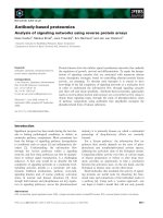
phases generally showed larger wash peaks, but in general all chromatograms were highly similar, as shown in Fig. 1. Only the Cu
NTA stationary phase showed a very small elution peak at the conditions tested.
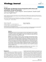
In order to compare the resulting eluates, SDS-PAGE was performed for all eight runs. Fig. 2 shows the load material compared
to pooled flow-through and wash fractions (FT+W) and eluate.
While all eight stationary phases can produce pure and concentrated GFP, the endogenous E. coli protein pattern co-eluting with
the different in all eight cases. NTA ligands appear to have a higher
specificity, owing to their lower number of available complexation
sites, which requires a higher interaction affinity for a protein to
adsorb. Compared to the load material, the elution fractions of all
eight conditions have high purity, with NTA resins, generally leading to less co-elution of impurities, while IDA resins leading to
lower protein losses in the flow-through and wash fractions. The
impurity pattern itself also differs for the four different metal ions
on the IDA resins, where host cell proteins (HCP) are visible in the
SDS-PAGE. No differences in purity can be determined from SDSPAGE analysis for the NTA resins. In order to get more meaningful
impurity data, more sensitive analytical methods were performed.
Table 1 shows the yield and recovery of the chromatography
runs. Interestingly, some columns exhibited irreversible binding,
that led to a total recovery of below 100%. This strong binding
could be confirmed visually, since GFP was seen in some columns
after elution. ELISA was used to quantify the HCP concentration in
all eight eluates, as shown in Table 1. The ELISA results confirm
that the IDA eluates are less pure than the NTA eluates. Interestingly, even though HCPs can be detected in the SDS-PAGE of the Zn
IDA eluate, the HCP ELISA determined this fraction to have a relatively low concentration of 48 ng/mL. Since the total number of
HCPs is relatively low, it is possible that the ELISA underestimated
the actual HCP concentration due to the nature of the assay [26].
Since the ELISA is using a pool of antibodies against various HCPs,
the measured concentration can suffer from bias if only a small
number of HCPs are present. The quantification of dsDNA was difficult to perform experimentally, since the most commonly used
assays all rely on fluorescence measurements at the same wavelength as GFP for detection. This required a different quantitative
method for our samples, namely qPCR. Table 1 shows the dsDNA
concentrations found in the eight eluate samples. Only Ni and Co
NTA eluates had dsDNA over the lower limit of quantification for
the assay. The endotoxin concentration was measured using a recombinant assay. The eluate from all four IDA columns showed endotoxin concentrations over the upper limit of quantification. For
2.5. Host cell protein (HCP) ELISA and endotoxin assay
The E. coli ELISA was purchased from Cygnus (Southport, North
Carolina, USA) and performed according to Sauer et al. [23]. In
brief, 96-well Nunc MaxiSorp Immuno plates (Thermo Fisher) were
coated with anti-E. coli HCP capture antibody and blocked with
BSA. E. coli HCP antigen was used as a standard and 8 concentrations from 0.4 to 50 ng/mL were transferred to the microtiter plate.
Samples were pre-diluted with sample buffer to be in the calibration range and serial dilutions were transferred to the plate. After
incubation and detection with horseradish peroxidase conjugated
anti-E. coli HCP antibody, the absorbance change after addition of
TMB substrate was used to quantify the HCP concentration with an
Infinite M200 Pro plate reader (Tecan).
Endotoxin was quantified using EndoZyme R II recombinant
Factor C (rFC) assay (Hyglos, Bernried, Germany) according to Sauer
et al. [23].
2.6. qPCR
The DNA content was quantified using resDNASEQTM Quantitative E. coli DNA Kit (Thermo Scientific) based on qPCR according to
the manufacturer’s instructions. Due to interaction with the matrix,
the GFP eluate samples had to be buffer exchanged into PBS using
5 kDa cut-off in Amicon Ultra spin vials (Merck).
2.7. Computational analysis
Sequence for the proteome of E. coli (strain B / BL21-DE3)
were downloaded from the Uniprot Knowledgebase [24]. Structures
from homology modeling were downloaded from the Swiss-Model
Repository [25]. Calculations on structural information were performed using Pymol 2.3.4 (The PyMOL Molecular Graphics System,
Version 2.0 Schrödinger, LLC). Three different analyses were performed to link information based on sequence or structure to protein selectivity of binding of immobilized metal ions. The amino
acids His, Cys and Trp were selected as key amino acids for metalion binding. Calculated were a) the relative occurrence of the key
amino acids in the sequence; b) the relative surface area of the
key amino acids compared to the total surface area of the protein;
c) the occurrence of clusters of key amino acids. The occurrence
of clusters that are made up of one or more different types of
key amino acids were counted. A cluster was defined as a minimum of key amino acids of the same or different kind within
a sphere of 1 nm diameter. The total occurrence of metal binding clusters within a protein was transformed into a metal binding
cluster score (MBCS) that was defined as
imax
i2 × m
MBCS =
(1)
i=2
3
N. Lingg, C. Öhlknecht, A. Fischer et al.
Journal of Chromatography A 1633 (2020) 461649
Fig. 1. Chromatograms of all eight runs, with metal ion and ligand denoted in the chromatogram. The blue trace is the absorbance at 280 nm, the dashed green trace is
the specific absorbance for GFP at 488 nm, and the gray dashed trace is the elution buffer concentration. Elution profiles of His-tagged GFP look very similar across all
metal/ligand combinations, while the behavior of host cell proteins during the wash step varies for each column. (For interpretation of the references to color in this figure
legend, the reader is referred to the web version of this article.)
Table 1
Mass balance of chromatographic runs by ligand and immobilized metal-ion. Yield refers to the amount of GFP
in the elution fraction relative to the loaded amount, whereas recovery FTW refers to the amount of GFP lost in
the non-binding flow through and wash steps. HCP, dsDNA and Endotoxin concentrations were measured for the
elution fractions.
Ligand
Metal-ion
Yield
Recovery FTW
HCP (ng/mL)
dsDNA (ng/mL)
Endotoxin (103 EU/mL)
IDA
Co
Cu
Ni
Zn
Co
Cu
Ni
Zn
72%
81%
80%
84%
36%
51%
97%
57%
6%
3%
13%
2%
37%
17%
4%
23%
323 ± 69
247 ± 74
400 ± 108
48 ± 14
171 ± 18
20.3 ± 0.5
265 ± 33
27.1 ± 0.5
< 0.03
< 0.03
< 0.03
< 0.03
0.56
< 0.03
12.3
< 0.03
> 35
> 35
> 35
> 35
5.8 ± 0.7
0.5 ± 0.2
21 ± 2
3.1 ± 0.3
NTA
the NTA stationary phases, the concentration depended strongly
on the immobilized metal ion, with Ni showing the highest concentration and Cu the lowest, and Co and Zn in between. These
endotoxin results somewhat mirror the behavior in regard to HCP
and dsDNA concentration.
by ELISA for comparison. The qualitative and quantitative HCP results are similar, but not identical.
The ligand appears to have a larger influence on the specificity
of the interaction, with IDA being less specific than NTA. The immobilized metal has a lower influence on specificity: Co and Zn
appear to be more specific in their protein interaction, since only
around 50% of the total proteins identified were found on either Co
or Zn. Immobilized Cu and Ni on the other hand lead to co-elution
of 72% and 65% of total HCPs, respectively.
3.2. Peptide mapping
The identity of the specific HCPs co-eluting with the His-tagged
GFP was determined via peptide mapping. In total 381 E. coli proteins were identified, in addition to GFP, trypsin (from the digest)
and human keratin (from the operator). A full list can be found in
the supplementary information. In total 360 HCPs were identified
in at least one of the IDA eluates (94% of the total HCPs) and 109
HCPs were identified in at least one of the NTA eluates (29% of total HCPs), with 88 HCPs (23%) identified eluting from both ligands.
A total of 13 HCPs were identified in all 8 conditions, which can
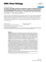
be found in Table 2. The HCPs found co-eluting from 7 or 6 stationary phases can be found in Tables 3 and 4. Fig. 3A shows the
number of identified HCPs from the IDA and NTA eluates depending on the immobilized metal. Fig. 3B shows the effect of immobilized metal on the number of HCPs co-eluting for IDA and NTA
ligands. Fig. 3C shows the quantitative HCP results as determined
3.3. Computational analysis
We hypothesized that metal interacting amino acids play an important role in the co-elution behavior in IMAC. In order to test
this hypothesis, we compared the characteristics of the co-eluting
proteins with a random sample of cytosolic E. coli proteins that
were not found in any of our eluates. We have evaluated 8 systematic experiments varying the ligand (IDA or NTA) and immobilized metal ions (Co2+ , Cu2+ , Ni2+ or Zn2+ ). In total we have
found 381 proteins which have co-eluted in at least one experiment. We refer to the co-elution propensity as n, defined as the
number of metal/ligand combinations in which co-elution was observed. When n = 8 a particular protein is found in all eluates and
4
N. Lingg, C. Öhlknecht, A. Fischer et al.
Journal of Chromatography A 1633 (2020) 461649
Fig. 2. Coomassie stained SDS-PAGE results of ligand (NTA or IDA) and immobilized metal ion (Ni, Co, Cu or Zn) combinations. The FT + W lane corresponds to the pooled
flow-through and wash fractions. GFP with a 6-His-tag was overexpressed in E. coli and cell lysate was loaded to WorkBeads columns.
when n = 1 the respective protein could only be found in a single experiment. For 99 different cytosolic proteins, which have not
been found in the eluates, the co-elution propensity n = 0. This
served as a control group.
Three computational analysis methods were compared regarding their ability to explain differences in interaction specificities
among E. coli HCPs. 1) the relative content of the key amino acids
in the primary sequence, 2) the relative surface area of the key
amino acids, and 3) a metal binding cluster score describing the
presence and size of key amino acid clusters.
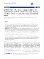
The relative content of the key amino acids His, Cys and Trp in
the sequence does not suffice to explain differences between the
individual groups, as shown by the lack of correlation in Fig. 4A.
Moreover, no correlation of co-elution and the relative surface area
of the three key amino acids could be found (Fig. 4B). An alternative metal binding cluster score (MBCS) was constructed (see
methods) that represents the fact that cooperative effects may occur when multiple key residues are in close vicinity. The MBCS
are available in the supplementary information for each protein
and shown in Tables 2, 3 and 4 for the proteins were n = 8, 7,
6. Scoring surface clusters of the key amino acids can be used to
explain a part of the variability between different HCPs. A protein
with a higher MBCS has a higher probability to be co-eluting in
a larger number of conditions (Fig. 4C). In other words, a higher
amount of different cluster sizes and differently orientated clusters increases the proteins tendency to bind to different immobi5
N. Lingg, C. Öhlknecht, A. Fischer et al.
Journal of Chromatography A 1633 (2020) 461649
Table 2
List of host cell proteins found co-eluting under all conditions.
Accession #
Name
MBCS
pI
Molecular mass (kDa)
A0A140N3N3
A0A140N7Y4
A0A140N3D6
A0A140ND61
A0A140NHM8
A0A140N6W0
A0A140N6E5
A0A140NE13
A0A140N7J1
A0A140NFK2
A0A140N548
A0A140NE25
A0A140N587
tRNA (guanine-N(7)-)-methyltransferase
Pseudouridine synthase
Transcriptional regulator Crp/Fnr family
Chaperone protein HtpG
Soluble pyridine nucleotide transhydrogenase
Elongation factor Tu
D-tagatose-1 6-bisphosphate aldolase subunit GatZ
Ferric uptake regulation protein
50S ribosomal protein L2
30S ribosomal protein S2
30S ribosomal protein S4
Glutamine–fructose-6-phosphate aminotransferase [isomerizing]
Bifunctional polymyxin resistance protein ArnA
33
37
77
84
151
198
214
258
430
446
449
512
667
6.6
5.9
8.2
5.0
6.2
5.2
5.5
5.8
11.2
6.8
10.3
5.6
6.5
27.3
25.8
23.6
71.4
51.6
43.3
47.0
16.8
29.9
26.7
23.5
66.9
74.3
Table 3
List of host cell proteins found co-eluting under seven conditions.
Accession #
Name
MBCS
pI
Molecular mass (kDa)
Not found
A0A140N6Z9
A0A140NBL1
A0A140N319
A0A140N598
A0A140N4M0
A0A140N811
A0A140NAY3
30S ribosomal protein S5
HAD-superfamily hydrolase subfamily IIA
RNase adapter protein RapZ
50S ribosomal protein L13
50S ribosomal protein L17
30S ribosomal protein S15
Histidine biosynthesis bifunctional protein HisB
0
54
118
431
432
449
1105
10.5
5.1
6.9
10.2
11.3
10.7
5.9
17.5
27.1
32.5
16.0
14.4
10.3
40.2
Zn NTA
Ni NTA
Ni NTA
Co NTA
Cu NTA
Zn IDA
Co NTA
Table 4
List of host cell proteins found co-eluting under six conditions.
Accession #
Name
MBCS
pI
Molecular mass (kDa)
Not found
A0A140NFV3
A0A140SS47
A0A140NGK1
A0A140NF03
A0A140NHS0
A0A140NC35
A0A140NBE7
A0A140NB96
A0A140N6V1
A0A140N8K1
A0A140SS84
A0A140N4K1
A0A140NA80
Chaperone protein DnaK
Uncharacterized protein
RNA-binding protein Hfq
Transcriptional regulator IclR family
ATP synthase subunit beta
Bifunctional aspartokinase/ homoserine dehydrogenase
Formate acetyltransferase
Transcriptional regulator LacI family
Peptidyl-prolyl cis-trans isomerase
Transcriptional regulator LysR family
Acetylornithine deacetylase
30S ribosomal protein S3
Succinate dehydrogenase flavoprotein subunit
39
77
77
84
95
95
97
97
183
209
310
452
620
4.7
6.7
7.6
7.9
4.8
5.5
5.7
6.6
4.8
6.2
5.6
10.6
6.0
69.1
15.6
11.2
29.7
50.3
89.0
85.3
38.9
20.8
32.7
42.3
26.0
64.4
Co
Cu
Cu
Cu
Co
Co
Co
Co
Zn
Cu
Co
Co
Co
lized metal ions. However, this MBCS is not sufficient to explain
the entire variability between the different groups. Other effects
such as protein-protein binding may have a significant role too:
proteins that do not bind the matrix directly but are believed to
have high binding affinities towards other proteins. Among the
lower scoring proteins in n > 5 in Fig. 4C, chaperones and ribosomal subunits were found. The most drastic examples of which
is 30S ribosomal protein S5 (A0A140N6Z9) that was found coeluting in 7 out of 8 conditions but has an MBCS of 0. Since other
30S ribosomal sub-units were found to have high MBCS (e.g. S2,
A0A140NFK2, score 446 and S4, A0A140N548, score 449) and coeluting with all eight metal/ligand combinations, it seems plausible that sub-unit S5 was bound merely through protein-protein interaction. Another interesting protein is chaperone protein HtpG
(A0A140ND61), which was found in all 8 conditions, but has a
relatively low MBCS of 50. It is likely that the main mechanism
for chaperones is through protein-protein interaction, instead of
direct metal binding. Indeed, a variety of chaperones were identified (DnaK, DnaJ, ClpB, OmpH, ProQ) with varying MBCS, including 0. Electrostatic interaction of the co-eluted proteins can
be excluded because the experiments were performed at 0.3 M
NaCl and electrostatic shielding can be expected at this high salt
concentration.
IDA, Zn NTA
IDA, Ni IDA
IDA, Ni IDA
NTA, Cu IDA
NTA, Zn NTA
NTA, Zn NTA
NTA, Zn NTA
NTA, Zn NTA
NTA, Zn IDA
IDA, Ni IDA
NTA, Zn NTA
NTA, Zn NTA
NTA, Zn NTA
For the visualization of the output of the experiments a
heatmap was chosen (Fig. 5). It is a two-dimensional representation of the data in which the outcomes of the experiments are
color-coded (light gray – no co-elution, black – co-elution). Additionally, the different experiments as well as the proteins were
reordered by hierarchical clustering. The binary distance was computed to group similar objects next to each other. Ward’s linkage
method was used to calculate the distance between groups of objects. On top of the heatmap a dendrogram of the experiments is
given. The dendrogram is a tree visualizing in which order groups
are merged. Experiments with similar outcome are grouped next to
each other. The dendrogram on the left of the heatmap gives the
similarity between the proteins. Instead of the names of the proteins a color key was used which is a combination of metal binding
cluster score (MBCS), molecular mass and pI.
The co-eluting HCPs can be grouped by ligand first, and within
those groups, the metals Cu and Ni make up a group that co-elute
similar clusters of HCPs and Co and Zn forming a second group
with their own cluster of HCPs, as shown in the dendrogram at
the top of Fig. 5A. These clusters are not exclusive, and many overlaps exist. This clustering is already apparent in Fig. 3. The proteins
with a high MBCS are mostly found co-eluting under multiple conditions (green bars in Fig. 5B), indicating that the MBCS has some
6
N. Lingg, C. Öhlknecht, A. Fischer et al.
Journal of Chromatography A 1633 (2020) 461649
Fig. 3. Venn diagram of E. coli HCPs found in IMAC eluates, depending on metal and ligand. The area of the ellipses is relative to the number of identified HCPs. Numbers
denote the number of HCPs that were identified in each sample. Panel A is grouped by ligand (IDA and NTA) and panel B is grouped by immobilized metal ion (Co, Cu, Ni
and Zn). 381 proteins were identified in total. Panel C shows the quantitative HCP ELISA results (in ng/mL) in the same style.
predictive power for IMAC co-elution. The factors molecular mass
and isoelectric point do not seem to influence the clustering.
ity comes at the cost of weaker binding as described previously
[20] and further confirmed by the data in this work. HCP content
varies drastically between ligands, as IDA eluates generally showing higher HCP concentrations than NTA. Additionally, the choice
of metal ion also having an impact. We have shown that HCP coelution cannot be explained simply by content or surface concentration of metal interacting amino acids (His, Cys, Trp), but depends on the presence of clusters of these proteins on the surfaces.
When using a MBCS for all proteins that were found co-eluting
and comparing it to a sample of HCPs that were not found coeluting, the score correlated with the number of co-eluting conditions. For some HCPs though, co-elution appears to be affected
by protein-protein interactions, as is the case for ribosomal sub-
4. Discussion
While the combination of Ni2+ and NTA reigns supreme in the
world of IMAC, other metal/ligand combinations can be viable alternatives. From the peptide mapping data, it can clearly be deduced that the choice of ligand has an immense impact on the
number of co-purified HCPs, with NTA generally resulting in a
lower number of unique HCPs. The choice of immobilized metal
ion also affects the number of unique HCPs, with Co2+ and Zn2+
being more selective than Ni2+ and Cu2+ . This increased selectiv7
N. Lingg, C. Öhlknecht, A. Fischer et al.
Journal of Chromatography A 1633 (2020) 461649
Fig. 4. Results of the computation analyses on the key amino acids His, Cys and Trp. Several analyses were used to compare differences in the co-elution propensity
(n = 0…8). A) Relative content of the key amino acids based on primary sequence information for all HCPs versus co-elution propensity n. B) Relative surface area of the
key amino acids compared to the total surface area for all HCPs versus co-elution propensity n. C) MBCS of the key amino acids for all HCPs versus co-elution propensity n.
Red spheres mark the median and the vertical red bars mark the standard deviation within the individual groups. (For interpretation of the references to color in this figure
legend, the reader is referred to the web version of this article.)
units and chaperone. Quantitatively, stationary phases with immobilized Ni2+ and Co2+ leads to strong co-elution of HCP, with concentrations in the hundreds of ng/mL. Immobilized Zn2+ on the
other hand leads to lower HCP concentrations co-eluting with histagged GFP. In the case of immobilized Cu2+ , the effect of ligand
choice is strong, with NTA leading to ten times less HCP co-elution
than IDA. The effect on dsDNA depletion was very difficult to study,
since the most common DNA assays rely on fluorescence measurements where GFP directly interferes. Quantitative PCR was used
to quantify the dsDNA concentration in the samples, but the results were much lower than expected, with only Co and Ni2+ NTA
being quantifiable. As such, it is uncertain if the measured concentrations are accurate. Endotoxin concentrations varied widely
between samples, with the IDA samples all being over the upper
limit of quantification of the assay used. Out of the NTA eluates,
the ranking in purity is Cu2+ , Zn2+ , Co2+ and Ni2+ with a 40-fold
range of endotoxin concentrations. A wash step with organic solvent during IMAC can reduce this concentration [27], but an anion
exchange step might be necessary for products where endotoxins
are of concern.
When IMAC is sought as the sole purification step, considering
our results it seems reasonable to choose Ni2+ NTA as the capture adsorbent. Ni2+ NTA exhibits the highest yield and a relatively
high purity, compared to other ligand and metal combinations. The
biggest downside of using Ni, is its inherent toxicity, which necessitates its removal for products intended for administration to humans. Even if Ni2+ leakage can be quite low, in the range of 1 ppm
[28], the successful removal still has to be validated. High concentrations of Ni2+ ions in wastewater for column cleaning further
increase costs. Implementing a downstream process without relying on Ni as part of a two-step process with removal of the affinity tag could be practicable. Such a two-step process consists of
IMAC capture, enzymatic tag removal and subsequent subtractive
IMAC in which the product is in the flow-through fraction and the
previously co-eluting HCPs can be removed along with the affinity tagged enzyme. An important prerequisite of such a process is
the absence of endogenous proteases, that could otherwise digest
the protein of interest. Indeed, of all 381 E. coli proteins, only two
proteases were identified: ATP-dependent zinc metalloprotease and
ATP-dependent Clp protease, both of which should be inactive after
capture due to their ATP dependence.
One effect not studied here, is the potential of displacement of
HCPs by the protein of interest, especially in the case of very high
titers and/or high binding affinity. The exposure of the His-tag may
vary with the protein of interest. The co-elution of proteins can
be explained by clusters of metal interacting amino acids, and by
protein-protein interaction, where one protein binds to the metal
ions and other proteins interact with the bound protein. A total
8
N. Lingg, C. Öhlknecht, A. Fischer et al.
Journal of Chromatography A 1633 (2020) 461649
Fig. 5. Heat map of E. coli HCP co-elution depending on stationary phase. In the central graph, black denotes co-elution and gray denotes no co-elution. The proteins are
color coded by their MBCS (1 < 50, 2 > 50, <325, 3 > 325), by molecular mass (1 < 42.2 kDa, 2 > 42.2 kDa), and by isoelectric point (1 < 7, 2 > 7). Panel A shows all 381
proteins that were found co-eluting from at least one stationary phase, whereas panel B is zoomed in on those proteins co-eluting on at least one NTA stationary phase.
9
N. Lingg, C. Öhlknecht, A. Fischer et al.
Journal of Chromatography A 1633 (2020) 461649
of 13 E. coli HCPs (Table 2) were found co-eluting from all eight
investigated stationary phases. Our database may serve as a reference for others.
[4] W. Jiang, M. Prescott, R.J. Devenish, L. Spiccia, M.T.W. Hearn, Separation of hexahistidine fusion proteins with immobilized metal ion affinity chromatographic
(IMAC) sorbents derived from MN+ -tacn and its derivatives, Biotechnol. Bioeng. 103 (4) (2009) 747–756, doi:10.1002/bit.22302.
[5] D.M. Jandhyala, R.C. Willson, B.T. Sewell, M.J. Benedik, Comparison of cyanidedegrading nitrilases, Appl. Microbiol. Biotechnol. 68 (3) (2005) 327–335,
doi:10.10 07/s0 0253-0 05-1903-8.
[6] B. Xu, U. Hellman, B. Ersson, J.C. Janson, Purification, characterization and
amino-acid sequence analysis of a thermostable, low molecular mass endoβ -1,4-glucanase from blue mussel, Mytilus edulis, Eur. J. Biochem. 267 (16)
(20 0 0) 4970–4977, doi:10.1046/j.1432-1327.20 0 0.01533.x.
[7] Z. Zhang, K.T. Tong, M. Belew, T. Pettersson, J.C. Janson, Production, purification
and characterization of recombinant human interferon γ , J. Chromatogr. A 604
(1) (1992) 143–155, doi:10.1016/0021- 9673(92)85539- 6.
[8] J. Porath, B. Olin, Immobilized metal ion affinity adsorption and immobilized
metal ion affinity chromatography of biomaterials. Serum protein affinities for
gel-immobilized iron and nickel ions, Biochemistry 22 (7) (1983) 1621–1630,
doi:10.1021/bi00276a015.
[9] P. Hubert, J. Porath, Metal Chelate Affinity-Chromatography .2. Group Separation of Mononucleotides and Dinucleotides, J. Chromatogr. 206 (1) (1981) 164–
168, doi:10.1016/S0 021-9673(0 0)82620-5.
[10] J. Porath, J. Carlsson, I. Olsson, G. Belfrage, Metal chelate affinity chromatography, a new approach to protein fractionation, Nature 258 (5536) (1975) 598–
599, doi:10.1038/258598a0.
[11] K. Karkra, K.K.R. Tetala, M.A. Vijayalakshmi, A structure based plasma protein
pre-fractionation using conjoint immobilized metal/chelate affinity (IMA) system, J. Chromatogr. B 1052 (2017) 1–9, doi:10.1016/j.jchromb.2017.02.032.
[12] R.R. Prasanna, M.A. Vijayalakshmi, Characterization of metal chelate methacrylate monolithic disk for purification of polyclonal and monoclonal immunoglobulin G, J. Chromatogr. A 1217 (23) (2010) 3660–3667, doi:10.1016/j.
chroma.2010.03.058.
[13] E. Hochuli, H. Döbeli, A. Schacher, New metal chelate adsorbent selective
for proteins and peptides containing neighbouring histidine residues, J. Chromatogr. A 411 (C) (1987) 177–184, doi:10.1016/S0 021-9673(0 0)93969-4.
[14] W. Jiang, B. Graham, L. Spiccia, M.T.W. Hearn, Protein selectivity with immobilized metal ion-tacn sorbents: chromatographic studies with human serum
proteins and several other globular proteins, Anal. Biochem. 255 (1) (1998) 47–
58, doi:10.1006/abio.1997.2395.
[15] ICH, ICH Q3D elemental impurities, 2019. />ich- q3d- elemental- impurities. (Accessed 2020-02-11).
[16] L.M. Plum, L. Rink, H. Haase, The essential toxin: impact of zinc on human
health, Int. J. Environ. Res. Public Health 7 (4) (2010) 1342–1365, doi:10.3390/
ijerph7041342.
[17] A.L. Tscheliessnig, J. Konrath, R. Bates, A. Jungbauer, Host cell protein analysis
in therapeutic protein bioprocessing - methods and applications, Biotechnol. J.
8 (6) (2013) 655–670, doi:10.10 02/biot.20120 0 018.
[18] M. Zimmer, Green fluorescent protein (GFP): applications, structure, and related photophysical behavior, Chem. Rev. 102 (3) (2002) 759–781, doi:10.1021/
cr010142r.
[19] M. Chalfie, Y. Tu, G. Euskirchen, W.W. Ward, D.C. Prasher, Green fluorescent
protein as a marker for gene expression, Science 263 (5148) (1994) 802–805,
doi:10.1126/science.8303295.
[20] L. KågedalJ.-C. Janson (Ed.), Immobilized metal ion affinity chromatography,
Prot. Purif. (2011) 183–201, doi:10.1002/9780470939932.ch7.
[21] M. Peterka, M. Jarc, M. Banjac, V. Frankovicˇ , K. Bencˇ ina, M. Merhar, V. GabercPorekar, V. Menart, A. Štrancar, A. Podgornik, Characterisation of metal-chelate
methacrylate monoliths, J. Chromatogr. A 1109 (1) (2006) 80–85, doi:10.1016/
j.chroma.2005.08.057.
[22] B.P. Cormack, R.H. Valdivia, S. Falkow, FACS-optimized mutants of the
green fluorescent protein (GFP), Gene 173 (1) (1996) 33–38, doi:10.1016/
0378-1119(95)00685-0.
[23] D.G. Sauer, M. Mosor, A.C. Frank, F. Weiss, A. Christler, N. Walch, A. Jungbauer,
A. Durauer, A two-step process for capture and purification of human basic fibroblast growth factor from E. coli homogenate: yield versus endotoxin clearance, Protein Expr. Purif. 153 (2019) 70–82, doi:10.1016/j.pep.2018.08.009.
[24] C. UniProt, UniProt: a worldwide hub of protein knowledge, Nucleic Acids Res.
47 (D1) (2019) D506–D515, doi:10.1093/nar/gky1049.
[25] A. Waterhouse, M. Bertoni, S. Bienert, G. Studer, G. Tauriello, R. Gumienny,
F.T. Heer, T.A.P. de Beer, C. Rempfer, L. Bordoli, R. Lepore, T. Schwede, SWISSMODEL: homology modelling of protein structures and complexes, Nucleic
Acids Res. 46 (W1) (2018) W296–W303, doi:10.1093/nar/gky427.
[26] J.Y. Baik, J. Guo, K.H. Lee, Host cell proteins during biomanufacturing, Cell Culture Eng. (2019) 295–311, doi:10.1002/9783527811410.ch12.
[27] K.L. Franken, H.S. Hiemstra, K.E. van Meijgaarden, Y. Subronto, J. den Hartigh,
T.H. Ottenhoff, J.W. Drijfhout, Purification of his-tagged proteins by immobilized chelate affinity chromatography: the benefits from the use of organic solvent, Protein Expr. Purif. 18 (1) (20 0 0) 95–99, doi:10.1006/prep.1999.1162.
[28] V. Gaberc-Porekar, V. Menart, Potential for using histidine tags in purification of proteins at large scale, Chem. Eng. Technol. 28 (11) (2005) 1306–1314,
doi:10.10 02/ceat.20 050 0167.
[29] V.M. Bolanos-Garcia, O.R. Davies, Structural analysis and classification of native
proteins from E. coli commonly co-purified by immobilised metal affinity chromatography, Biochim. Biophys. Acta 1760 (9) (2006) 1304–1313, doi:10.1016/j.
bbagen.2006.03.027.
5. Conclusion
In general, we can conclude that NTA ligands result in lower
co-elution of proteins irrespective of the chelated metal ion. In
case of single step purification, NTA is still preferable. In the case
of a more complex downstream process the lower purity in the
capture step can be compensated in subsequent unit operations.
For large scale manufacturing the toxic metal ion Ni2+ can be replaced by other metal ions. Zn2+ IDA, in particular had a similar HCP, DNA, and endotoxin profile as Ni2+ NTA. Unfortunately,
there is no simple prediction of co-elution with proteomics tools.
Co-elution appears to be determined by either clusters of metal
interacting amino acids on the surface of the protein or through
protein-protein interaction of proteins adsorbed on the stationary
phase. The limitation of the prediction is knowledge about the interactome and knowledge about the 3D structure of proteins. Our
results match closely with the results from Bolanos-Garcia et al.
[29] who identified 18 commonly co-eluting proteins from E. coli in
2006. We were able to identify 15 out of the 18 proteins described.
Out of those, 14 had an MBCS higher than 75. Future studies using
other model proteins than GFP might be able to elucidate which
protein-protein interactions are specific to the protein of interest
and which are host specific.
Declaration of Competing Interest
The authors declare that they have no known competing financial interests or personal relationships that could have appeared to
influence the work reported in this paper.
Acknowledgements
We thank Clemens Grünwald-Gruber for performing the MS
analysis. The MS equipment was kindly provided by the EQ-BOKU
VIBT GmbH and the BOKU Core Facility for mass spectrometry. We
thank our company partners at Boehringer-Ingelheim RCV Process
Bioscience for their collaboration and fruitful discussions.
This work has been supported by the Federal Ministry for Digital and Economic Affairs (bmwd), the Federal Ministry for Transport, Innovation and Technology (bmvit), the Styrian Business Promotion Agency SFG, the Standortagentur Tirol, Government of
Lower Austria and ZIT - Technology Agency of the City of Vienna
through the COMET-Funding Program managed by the Austrian Research Promotion Agency FFG. The funding agencies had no influence on the conduct of this research.
Supplementary materials
Supplementary material associated with this article can be
found, in the online version, at doi:10.1016/j.chroma.2020.461649.
References
[1] H. Block, B. Maertens, A. Spriestersbach, N. Brinker, J. Kubicek, R. Fabis,
J. Labahn, F. Schäfer, Chapter 27 immobilized-metal affinity chromatography
(IMAC): a review, in: R.R. Burgess, M.P. Deutscher (Eds.), Methods in Enzymology, Academic Press, 2009, pp. 439–473, doi:10.1016/S0076- 6879(09)63027- 5.
[2] L. Fanou-Ayi, M. Vijayalakshmi, Metal-chelate affinity chromatography as a
separation tool, Ann. N. Y. Acad. Sci. (1983) 300–306.
[3] M. Kenig, S. Peternel, V. Gaberc-Porekar, V. Menart, Influence of the protein
oligomericity on final yield after affinity tag removal in purification of recombinant proteins, J. Chromatogr. A 1101 (1–2) (2006) 293–306, doi:10.1016/j.
chroma.2005.09.089.
10