Negative gradient slope methods to improve the separation of closely eluting proteins
Bạn đang xem bản rút gọn của tài liệu. Xem và tải ngay bản đầy đủ của tài liệu tại đây (1.59 MB, 9 trang )
Journal of Chromatography A 1635 (2021) 461743
Contents lists available at ScienceDirect
Journal of Chromatography A
journal homepage: www.elsevier.com/locate/chroma
Negative gradient slope methods to improve the separation of closely
eluting proteins
Szabolcs Fekete a,b,∗, Amarande Murisier a,b, Jennifer M. Nguyen c, Matthew A. Lauber c,
Davy Guillarme a,b
a
b
c
School of Pharmaceutical Sciences, University of Geneva, CMU-Rue Michel Servet 1, 1211 Geneva 4, Switzerland
Institute of Pharmaceutical Sciences of Western Switzerland, University of Geneva, CMU-Rue Michel Servet 1, 1211 Geneva 4, Switzerland
Waters Corporation, 34 Maple Street, Milford, MA 01757-3696, United States
a r t i c l e
i n f o
Article history:
Received 26 August 2020
Revised 16 November 2020
Accepted 19 November 2020
Available online 23 November 2020
Keywords:
therapeutic proteins
monoclonal antibody
method development
gradient elution
negative gradient step
negative segmented multi-isocratic mode
a b s t r a c t
In the present work, we describe the fundamental and practical advantages of a new strategy to improve
the resolution of very closely eluting peaks within therapeutic protein samples.
This approach involves the use of multiple isocratic steps, together with the addition of a steep negative
gradient segment (with a decrease in mobile phase strength) to "park" a slightly more retained peak
somewhere along the column (at a given migration distance), while a slightly less retained compound
can be eluted.
First, some model calculations were performed to highlight the potential of this innovative approach. For
this purpose, the retention parameters (logk0 and S) for two case studies were considered, namely the
analysis of a mixture of two therapeutic mAbs (simple to resolve sample) and separation of a therapeutic
mAb from its main variant (challenging to resolve sample). The results confirm that the insertion of a
negative segment into a multi-isocratic elution program can be a good tool to improve selectivity between
critical peak pairs. However, it is also important to keep in mind that this approach only works with large
solutes, which more or less follow an “on-off” type elution behavior.
Two real applications were successfully developed to illustrate the practical advantage of this new approach, including the separation of a therapeutic mAb from its main variant possessing very close elution
behavior, and the separation of a carrier protein from an intact mAb as might be encountered in a quantitative bioanalysis assay. These two examples demonstrate that improved selectivity can be achieved for
protein RPLC through the inclusion of a negative gradient slope that selectively bifurcates the elution of
two or more peaks of interest.
© 2020 The Author(s). Published by Elsevier B.V.
This is an open access article under the CC BY-NC-ND license
( />
1. Introduction
Liquid chromatographic separations of proteins are performed
in gradient elution mode. In general, simple linear gradients are
performed since they are easy to generate and control, and consequently those methods can be easily transferred [1]. However, a
simple linear gradient is often unable to provide sufficient chromatographic resolution. Therefore, segmented gradients can be applied to improve separation quality [2]. Two-segment (“bi-linear”)
gradients are often used to shorten the analysis time when a sep-
∗
Corresponding author.
E-mail address: (S. Fekete).
aration includes a few well-resolved late-eluting peaks. Then, a
steeper gradient segment can be set for the late eluting peaks
[1]. For more complex samples, multiple gradient segments can
be combined to attain suitable separation. In order to facilitate the
elution of the peaks, it is common knowledge that gradient slopes
should always be positive; however occasionally, one or more isocratic steps can be inserted to obtain the most optimal separation
[2].
Besides multi-linear gradients, non-linear gradients might also
provide some benefits. Power function based gradients have been
successfully applied for therapeutic protein separations for both reversed phase (RPLC) and ion-exchange (IEX) chromatography [3,4].
Another type of non-linear gradient can also be useful when the
/>0021-9673/© 2020 The Author(s). Published by Elsevier B.V. This is an open access article under the CC BY-NC-ND license
( />
S. Fekete, A. Murisier, J.M. Nguyen et al.
Journal of Chromatography A 1635 (2021) 461743
compounds of interest belong to a series of increasingly more retained analytes (e.g. members of homologues series). In this case,
a logarithmic shape gradient profile provides the best overall selectivity [5]. Customized non-linear gradients (including both concave and convex segments) were also developed by Kall et al. to
separate complex peptide mixtures and were successfully applied
in shotgun proteomics [6]. Some other complex gradient profiles
were also applied based on considering either the elution of only
the first and last eluting peaks [7] or the elution of each individual
peak [8].
For a preparative scale separation (purification) of peptides and
proteins, step or step-wise gradients have been extensively used in
flash chromatography and counter-current chromatography [9,10].
The idea behind such step gradients is that only one or a very few
number of components have to be separated, while the other sample components are just washed out from the column.
Timperman and co-workers demonstrated that a “saw-tooth”
gradient program allows small subsets of proteins to be eluted
from the column intermittently by using short gradient steps separated by a negative slope and isocratic holding segments. The isocratic holding periods can be used to perform additional sample
processing (e.g. online fractioning for a second dimension analysis) [11]. The saw-tooth gradient was found superior to a common
segmented linear gradient/isocratic mode, since the negative steps
prevents band broadening that takes place during isocratic elution
steps. This saw-tooth gradient was set to achieve complete sample
transfer between the first- and second-dimension for protein and
peptide identification [12]. Saw-tooth gradients are also applied for
polymer separations [13].
Armstrong et al. discussed the possibility to run a simple negative linear gradient in RPLC for protein separations [14]. The authors referred to their separation as “non-traditional reverse gradient”. Unusual convex logk–ϕ plots with global minima were reported for ribonuclease, insulin and myoglobin, and the authors
explained that those observations were not in agreement with previously reported results. Nevertheless, it was pointed out that if
there is a minima on a logk–ϕ plot, then retention and elution can
be attained either with positive or negative gradients. The authors
also explained that such unusual behavior was probably related to
solubility-based phenomenon. Despite that the conditions used in
˚
the study were not ideal (narrow pores of 60 A–RPLC
phase, 1%
TFA as mobile phase additive and ambient temperature), the three
proteins were successfully separated with a linear reverse gradient.
Recently, an innovative strategy termed as “multi-isocratic” elution has been shown to provide exponential increase in selectivity between protein variants. It was demonstrated that the combination of multi-isocratic steps and very short, yet steep gradient
segments (with steepness close to infinity) at solute elution allows
one to set the selectivity as desired while maintaining sharp peaks
due to significant band compression effects [15]. This method was
successfully applied for the analytical scale separation of intact
and subunit digested samples of monoclonal antibodies (mAbs) as
well as antibody-drug conjugates (ADCs). Uniform peak distribution (equidistant band spacing) and much higher resolution could
be achieved than with common linear, multilinear, or nonlinear
gradients. In a following study, this approach was combined with
column coupling to further improve separation power. In such a
setup, if a protein peak is trapped at the inlet of a later column
segment - of a serially coupled system - then its band is refocused
and it elutes as an unprecedented sharp peak [16]. Furthermore, it
became possible to perform online on-column fractioning of protein species within a very short analysis time (~1 min) and without
sample dilution. Similar idea has already been reported to sharpen
peaks utilizing post-column refocusing and remobilization on trapping columns [17,18,19].
In the present work, the goal was to further improve the resolution of RPLC separations of very closely eluting peaks of therapeutic proteins. Therefore, the recently developed “multi-isocratic”
elution mode was upgraded by adding a steep negative gradient
segment between the “eluting” and “non-eluting” isocratic segments. As a result of this negative slope segment, it becomes possible to "park" a slightly more retained peak along the column bed
(at a given migration distance) with a condition that still allows
the slightly less retained compound to be eluted. Therefore, this
approach has the potential to resolve compounds possessing very
similar retention properties, which are difficult to separate, even
with the multi-isocratic technique. Here, we present some theoretical considerations and illustrate the capabilities of this approach
for large solutes. The expected benefit of inserting negative gradient steps (short segments) into a “multi-isocratic” program is also
discussed. Two applications were developed to show the practical
advantage of this new approach. These two separations were not
feasible by applying the multi-isocratic elution mode (let alone linear or multi-linear gradients).
2. Materials and methods
2.1. Equipment and software
Chromatographic experiments were performed on a Waters Acquity UPLC I-Class system equipped with a binary solvent delivery pump, an autosampler, a fluorescence (FL) detector and a flow
through needle injection system with 15 μL needle and a 2 μL FL
flow-cell. The overall extra-column volume was about 8.5 μL as
measured from the injection seat of the auto-sampler to the detector cell. The dwell volume was measured as Vd = 0.09 mL. Data acquisition and instrument control were performed by Empower Pro
2 software (Waters). Calculation and data processing were done by
using Drylab (4.2) and Excel (Microsoft) software.
2.2. Chemicals and columns
LC-MS grade acetonitrile (ACN) and LC-MS grade water were
purchased from Fisher Scientific (Reinach, Switzerland). ULC/MS
grade formic acid (FA) and ULC/MS grade trifluoroacetic acid (TFA)
were purchased from Biosolve (Dieuze, France).
Intact mAb Mass Check Standard (murine anticitrinin IgG1)
was obtained from Waters. Bovine Serum Albumin (BSA) was purchased from Sigma-Aldrich (Buchs, Switzerland). Therapeutic mAb
(eculizumab) was obtained as European Union pharmaceuticalgrade drug product from its respective manufacturer.
Prototype columns (15 × 2.1 mm) packed with 3 μm 20 0 0 A˚
polystyrene divinylbenzene (PS-DVB) particles as well as a commercial BioResolve RP mAb Polyphenyl column (50 × 2.1 mm, 2.7
˚ were provided by Waters (Milford, USA). To prepare the
μm, 450 A)
prototype PS-DVB column, a specialized guard column was constructed with a low clearance endnut and a low dispersion coupler
to give a standard female inlet/female outlet configuration.
2.3. Sample and mobile phase preparation
Intact eculizumab was diluted to 1 mg/mL with water and injected without further preparation. Waters Intact mAb Mass Check
Standard was diluted to 0.025 mg/mL with water and mixed with
BSA diluted to 0.25 mg/mL with water.
For the separation of intact eculizumab variants, the mobile
phase A was 0.1% TFA (v/v) in water and B was 0.1% TFA (v/v) in
ACN.
For the separation of anti-citrinin mAb (Waters Intact mAb
Mass Check Standard) and carrier protein (BSA), the mobile phase
A was 0.1% FA (v/v) in water and B was 0.1% FA (v/v) in ACN.
2
S. Fekete, A. Murisier, J.M. Nguyen et al.
Journal of Chromatography A 1635 (2021) 461743
2.4. Chromatographic conditions
ϕ > 0.5, then other non-linear models can be used - e.g. NeueKuss or quadratic models - instead of the LSS model [22]. In our
practice, for peptides and proteins, less than 5% deviation is observed in reversed phase chromatography. However here in this
study, very narrow ϕ ranges were set (< 0.1) and LSS models
were found to be appropriate.)
To improve the selectivity of closely eluting proteins, a linear
gradient separation was compared to separations achieved with
a multi-isocratic elution technique that either contained or did
not contain a negative gradient segment (“negative segmented
multi-isocratic elution”). First, the parameters of the linear solvent
strength (LSS) model were determined from two linear gradients.
For eculizumab, the flow rate was set at 0.5 mL/min, column
temperature was set at 85°C, and 0.5 μL of intact eculizumab sample was injected. For the initial linear gradient experiments, the
gradient times were set as tG1 = 4 and tG2 = 10 minutes and a
25–50%B gradient was run.
For the separation of the anti-citrinin mAb and carrier protein
(BSA), the flow rate was set at 0.4 mL/min and the column temperature was set at 80°C. Sample injection volume was 0.1 μL. For
the initial linear gradient experiments, the gradient times were set
as tG1 = 4 and tG2 = 10 minutes and a 20–50%B gradient was run.
For all measurements, data were acquired at 280 nm excitation
and 350 nm emission wavelengths (FL).
The optimized conditions for multi-isocratic and “negative segmented” programs are detailed in the Results and Discussion section.
2.5.1. Studying the evolution of selectivity
Then, from logk0 and S, the retention factor (k) was estimated
for a given mobile phase composition (ϕ ) for any set of %B program (e.g. linear-, multi-isocratic or negative segmented multiisocratic program). The solute relative velocity and the travelled
distance can be calculated for any time point of a given %B program. To illustrate solute migration and study selectivity, plots
of (1) B fraction vs tG , (2) u/u0 vs z and (3) distance travelled
vs t were constructed. Simulated chromatograms were also plotted
√ assuming the common gradient band compression factor; G =
1+ p+ p2/3
,
1+ p
with p = 2.3b [23]. Please note that this factor G is only
valid for linear gradient. For our calculations for multi-isocratic
separations, we assumed consecutive linear gradient and isocratic
segments.
2.5.2. Optimization of multi-isocratic and negative segmented
separations
It is known that the retention of large solutes such as therapeutic proteins is very sensitive to the mobile phase composition.
A minor change in the mobile phase composition can indeed drastically affect their retention (very high S value in the LSS model).
Snyder explained this phenomenon by the fact that large solutes
are either fully captured at the column inlet or completely released
from the column [20,24,25]. This behavior is today often termed as
an “on-off” or “bind-and-elute” mechanism. Very recently, it was
indeed shown that the retention of large proteins can only be controlled in a very narrow mobile phase composition range (e.g. with
gradients applying only 3.5–5 % B for intact mAbs) [15,16]. Their
relative migration speed varies within the 0 < urel < 1 range only
in this very narrow %B window, otherwise it is either 0 or 1 (corresponding to “on”–fully captured–state or to “off”–released–state).
Therefore, the mobile phase composition required to start the migration of a large molecule (ϕ (urel= 0.01); when the solute starts
traveling with only 1% of the mobile phase velocity, u/u0 = 0.01)
can be estimated as:
2.5. Calculations
In LC, the LSS model - sometimes called exponential model - is
commonly used to describe the relationship between solute retention (k) and mobile phase composition (ϕ ) [20]:
logk = logk0 − Sϕ
(1)
where k is the solute retention factor, ϕ is the volume fraction
of mobile phase “B” (stronger eluent), S is a constant for a given
solute (it describes how sensitive is the solute retention to mobile phase composition) and k0 is the (extrapolated) value of k for
ϕ = 0 (i.e., the retention factor observed in pure mobile phase “A”).
The migration velocity (u) of a solute along the column (measured at a given ϕ ) depends on the interstitial mobile phase velocity (u0 ) and retention factor:
u=
u0
1+k
(2)
Expressing k from Eq. (1) and substituting to Eq. (2) enables one
to describe the relative solute migration speed (urel ) as:
urel =
u
1
=
u0
1 + k0 10(−Sϕ )
ϕ(urel =0.01) = −
(3)
The time spent to reach position z can be expressed as [21]:
t ( z ) = t0
z
1
z
+ log 1 + kin b
L
b
L
(ϕ
tr = t ( L ) = t0 1 +
1
log(1 + kin b)
b
1
0.01 −1
k0
S
(6)
Similarly, the mobile phase composition to reach the “off” state
unbound state with u/u0 = 0.99) can be written as:
(urel= 0.99);
(4)
ϕ(urel =0.99) = −
Where kin is the retention factor at the starting mobile phase composition (inlet retention factor), b is the gradient steepness (b =
t
S · ϕ · t0 ), t0 is the column dead time, L is the column length and
G
z is the solute position along the column. Then, the time to travel
the entire column (z = L) corresponds to the retention time (tr )
and can be written as:
log
log
1
0.99 −1
k0
S
(7)
On the other hand, eluting mobile phase composition with very
low retention factor (ϕ k< 0.1 ) and binding mobile phase composition with high retention factor (ϕ k> 100 ) for a multi-isocratic elution separation can be estimated as [15]:
(5)
Please note that Eqs. (4) and (5) assume linear gradient. For our
calculations, the parameters logk0 and S were derived from experimentally measured retention times data of two preliminary linear
gradient experiments–performed with different gradient times (tG )
(corresponding to different gradient steepness) - using DryLab 4
software. (Please note that, deviations from the LSS model might
be observed, especially when working in a broad %B range–e.g.
ϕk<0.1 >
logk0 + 1
S
(8)
ϕk>100 <
logk0 − 2
S
(9)
The ϕ k< 0.1 and ϕ k> 100 can be good starting points for the
optimization of a multi-isocratic protein separation. However, in
practice, there is sometimes only a minor difference between the
model parameters of closely related proteins (e.g. variants of intact
mAbs) and thus it is hard to predict whether they can be separated
3
S. Fekete, A. Murisier, J.M. Nguyen et al.
Journal of Chromatography A 1635 (2021) 461743
or not. For such a situation, we found that performing a “screening multi-isocratic gradient” can be very helpful. To realize such,
a 5-segmented multi-isocratic condition was set (please note that
any number of segments can be set). The mobile phase composition for the initial step (ϕ in ) was set to be retentive enough (eg.
ϕ k> 100 ), while the composition of the last segment (ϕ last ) was set
to be able to elute all compounds (eg. ϕ k< 0.1 ). Then, five equidistant segments (2 minute long intervals) were set between the initial and final compositions. The difference ( %Bsegment ) between
the mobile phase compositions of the consecutive segments for the
case of n isocratic segments can be determined as:
%Bsegment =
ϕlast − ϕin
n−1
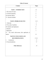
Fig. 1 A-D shows the evolution of solute migration, the travelled distance along a column and the calculated chromatograms
for the simple-sample when performing linear gradient and multiisocratic separations. Based on Fig. 1 B and 1C, it is clear
that once the two compounds start their migration (switching
to “off” mode), their relative migration speed and acceleration
are nearly the same along the entire column. However, peak 1
starts migrating (ϕ (urel= 0.01) = 0.335) far earlier than peak 2 does
(ϕ (urel= 0.01) = 0.366). With a 10 min long linear gradient (32 to
42 %B), peak 1 starts migrating after a parking time (tpark ) of 1.5
min, while peak 2 parks at the column inlet until 4.6 min. After their release from the head of the column, they travel through
the chromatographic bed within nearly the same amount of time
(ttrav = tr –tpark , gives 2.67 and 2.52 min, respectively). Therefore,
the selectivity is mostly determined by the difference of their parking times ( tpark = +3.1 min) and not by their travelling time
( ttrav = -0.15 min). Due to the large difference between the parking times, the separation of those peaks is easy to achieve with
a linear gradient. Figs. 1 E-H show the case of a multi-isocratic
separation. Setting 28%B for the starting isocratic binding segment resulted in very high initial retention for both compounds
(k1 = 2.03∗ 107 and k2 = 1.11∗ 1011 ). Over a 2 min isocratic segment, the two peaks practically do not move from the head of the
column. Changing to 36.8%B mobile phase composition resulted in
the immediate elution of the less retained peak (k1 = 0.07). Meanwhile, peak 2 remained to be strongly retained (k2 = 61.15), albeit with an indication of some very slow migration (urel = 0.016).
Holding this second isocratic segment for 4 min (6 min of total
run time) resulted in z = 0.9 cm travelled distance for peak 2
(Figs. 1 F and G, red curve). Subsequently setting the mobile phase
composition to 45%B resulted in the immediate elution of peak 2
(k2 = 1.44∗ 10−7 ). In conclusion, due to the large difference of retention between the two mAbs (determined by logk0 values), either linear gradient or multi-isocratic separations are easy to implement. In the end, the latter technique has the advantage to
drastically improve selectivity. With the conditions set in this example (36.8%B for the second isocratic segment), the elution distance (selectivity) between the two peaks can be increased up to
~40 min. (With 36.8%B, it takes about 44 min for peak 2 - migrating with urel = 0.016 - to travel the entire column length of 10
cm.)
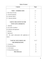
Figs. 2 A-D illustrate the challenge to separate intact atezolizumab and its main hydrophobic variant by applying a linear
gradient. Peak 1 starts migrating at ϕ (urel= 0.01) = 0.335 (33.5%B),
while peak 2 begins travelling at ϕ (urel= 0.01) = 0.338 (33.8%B). By
setting a 10 min long linear gradient (32 to 42 %B), peaks 1 and
2 will start migrating after tpark = 1.50 and 1.76 min, respectively.
Following their release from the column inlet, their travelling times
are also nearly the same (ttrav = 2.66 and 2.58 min, respectively).
Since both their parking times ( tpark = +0.26 min) and travelling times ( ttrav = -0.08 min) are almost identical, it is hardly
possible to afford selectivity through the application of linear gradients. However, by running a multi-isocratic program, the selectivity can be slightly increased (Figs. 2 E-H). By setting 33%B as
initial isocratic segment, both compounds were found to be highly
retained (k1 = 312 and k2 = 590). After two minutes of holding
time and a switch to 34.5% B, both compounds started migrating
with urel = 0.09 and 0.05 (Fig. 2 F). At the end of the second segment, peak 1 traveled z = 2.5 cm while peak 2 traveled z = 1.6 cm
(Fig. 2 G). Then, during the third segment (35.3%B hold for 2 min),
peak 1 accelerated and left the column, while peak 2 approached
z = 9.1 cm. Finally, the last segment (35.9%B) quickly eluted peak 2
from the column. It is important to notice that once peak 1 started
migrating (switches to “off” mode) so too did peak 2, albeit with
a slightly lower velocity. Since the difference between their migration speed is limited (a factor of 1.2–1.6 difference can be real-
(10)
After performing the first screening run, one can fine-tune
the number of steps and the mobile phase composition of the
isocratic steps to improve the separation by performing a socalled “stretched” multi-isocratic run. (Supplementary fig. 1 shows
a schematic view of the optimization procedure, including (1) a
preliminary linear gradient run, (2) a “screening” multi-isocratic
run and (3) a “stretched” multi-isocratic run). As a generic suggestion, for intact mAbs, an effective screening run may consider
a 5% B difference between ϕ last and ϕ in (which is due to the high
S value, typically ranging between 90 and 150 under RPLC conditions).
4. Results and discussion
4.1. Model calculations - potential of inserting a negative gradient
step
It was recently shown that a so-called multi-isocratic elution
technique could produce uniform peak distribution (equidistant
band spacing) for a separation of protein species (assuming they
obey an on-off type elution mechanism) [15]. Ideally, the elution
distance between peaks can be adjusted arbitrarily by changing the
length of the holding isocratic segments. However, this is only feasible if just one of the peaks of interest starts migrating within
a given elution gradient–or isocratic hold - segment. In practice,
it may happen that not only one, but two or more compounds
start to migrate along the column at a given segment (because of
a lack of selectivity, their retention behavior and thus model parameters are very similar). Such cases represent the limits of the
multi-isocratic approach. In case of co-migration (even if the solutes migration speed is slightly different), the selectivity cannot be
increased anymore without boundaries [15]. Accordingly, we were
motivated to find a better solution to resolve such closely eluting
protein compounds. The idea was to park (“freeze”) a migrating
- more retained - compound somewhere along the column, while
letting a less retained compound complete its elution through the
column. For this, we explored the use of a negative gradient segment along with a “holding” isocratic segment immediately after
the elution of the less retained compound.
First, some model calculations were performed to highlight the
potential of a multi-isocratic separation and the effect of inserting
a negative gradient segment into a multi-isocratic elution program.
Two sets of compounds were studied, namely a “simple to resolve
sample” and a “challenging to resolve sample”. For the simplesample, a mixture of intact rituximab and ramucirumab was considered, since there is enough difference between their retention
to separate them either with a linear gradient or multi-isocratic
elution technique. For the challenging-sample, intact atezolizumab
and its main variant were chosen since this sample already faced
the limits of the multi-isocratic elution mode in a former study
[15]. (The parameters of retention models used for these calculations were taken from our previous studies [15,16].)
4
S. Fekete, A. Murisier, J.M. Nguyen et al.
Journal of Chromatography A 1635 (2021) 461743
Fig. 1. Evolution of selectivity (“simple-sample”) with linear gradient 32–42 %B in 10 min (A,B,C,D), and with multi-isocratic (E,F,G,H) elution. The %B program for the
multi-isocratic run was 28%B (0–2 min), 36.8%B (2.01–6 min) and 45%B (6.01–10 min). F = 0.3 mL/min, 100 × 2.1 mm column, ε = 0.62, rituximab peak (1): S = 96.4, log
k0 = 36.3, ramucirumab peak (2): S = 105.2, log k0 = 40.5.
ized), it was not possible to find conditions that simultaneously
yielded high velocity for one compound (“off” mode) and low velocity for the other (close to “on” mode). In contrast, for the case
of the simple-sample, a factor of 58 was obtained between the migration speeds of the two solutes during the second segment of
the multi-isocratic program, as shown on Fig. 1 F.
The challenging-sample can be used to illustrate the limitations
of the multi-isocratic elution technique and the beneficial effects
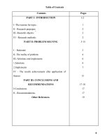
of inserting a negative gradient segment. Fig. 2 G shows an example wherein a short, negative and steep gradient segment was
inserted just as peak 1 left the column (tG = 5.5 min, grey dashed
line in Fig. 2 G). With this, the migration of the more retained
compound was stopped at the position to which it had traveled
up until that point in time(z = 7.2 cm, the crossing point of the
grey dashed line and red curve on Fig. 2 G). Fig. 3 illustrates the
separation for the case when at 5.5 min, the mobile phase composition was set back from 35.3 to 33%B and held until 8 min (to
give a 2.5 min holding (parking) time). As suggested by Figs. 3 B
and 3 C, peak 2 did not move to any appreciable extent during this
negative step. Upon setting a stronger mobile phase composition
(e.g. 38%B) peak 2 was made to immediately elute (k = 5.2∗ 10−3 ).
Based on these model calculations, it is proposed that a negative
segment (decrease of mobile phase strength) can be inserted into
a multi-isocratic elution program to improve selectivity between
critical peak pairs found in large biopharmaceutical drug products.
To the best of our knowledge, this is the first time that such a
combination of positive and negative gradients (and isocratic holding) segments are combined and applied.
5
S. Fekete, A. Murisier, J.M. Nguyen et al.
Journal of Chromatography A 1635 (2021) 461743
Fig. 2. Evolution of selectivity (“challenging-sample”) with linear gradient 32–42 %B in 10 min (A,B,C,D), and with multi-isocratic (E,F,G,H) elution. The %B program for the
multi-isocratic run was 33%B (0–2 min), 34.5%B (2.01–4 min), 35.3%B (4.01–6 min) and 35.9%B (6.01–10 min). F = 0.3 mL/min, 100 × 2.1 mm column, ε = 0.62, main peak
(1, atezolizumab): S = 97.53, log k0 = 34.68, minor peak (2): S = 101.2, log k0 = 36.17.
4.2. Application to the separation of intact mAb variants
performed to estimate the binding (parking) and eluting compositions (Supplementary Figure 2). It was found that 33.8%B mobile phase provided sufficiently high retention for both compounds
to be parked at the column inlet (k1 = 856, k2 = 1062). Then,
four different %B compositions were employed as eluting compositions (37.3, 36.8, 36.3 and 35.8 %B). Whatever the composition,
the two peaks co-migrated with only a minor difference between
their velocities (Supplementary Figure 3, left panel). The lower the
%B - during the elution step/hold - the higher the selectivity was,
but sensitivity decreased drastically due to band broadening of the
macromolecules during isocratic migration with k ≥ 1. Baseline
separation was therefore not feasible with a multi-isocratic elution
mechanism. A mobile phase composition of 36.3%B was selected
as the first eluting segment of the program - as it showed a good
One of the most challenging tasks in the field of therapeutic
protein analysis is the RPLC separation of protein variants at the
intact level. Hence, we tried our new approach for such challenging sample. Eculizumab (humanized therapeutic mAb product) has
been selected as an example, since it contains hydrophobic variants. We have already made several attempts to separate the two
main variants of this mAb by linear gradient separations but have
always failed. Our new method development approach (see description in Section 2.5.2.) has been applied to define the optimal
conditions for a multi-isocratic or negative step inserted multiisocratic separation. LSS parameters were derived from two initial linear gradients and then a screening multi-isocratic run was
6
S. Fekete, A. Murisier, J.M. Nguyen et al.
Journal of Chromatography A 1635 (2021) 461743
Fig. 4. Separation of intact eculizumab variants with a linear gradient (A), multiisocratic elution mode technique (B) and multi-isocratic mode including negative
gradient step technique (C). F = 0.5 mL/min, column: BioResolve RP 50 × 2.1 mm,
mobile phase A: water + 0.1% TFA, mobile phase B: acetonitrile + 0.1% TFA, temperature = 85 0 C, main peak (1, eculizumab): S = 129.46, log k0 = 46.69, minor
peak (2): S = 124.42, log k0 = 45.08. The %B program for the negative step inserted
multi-isocratic run (panel C) was 33.8%B (0–2 min), 36.3%B (2.01–2.4 min), 33.8%B
(2.4–3.5 min) and 36.3%B (3.51–6 min). Red dashed lines correspond to the sum of
column dead time and gradient delay time. The blue curves (%ACN) were corrected
for the total (system + column) delay time.
mentary Figure 3, middle panel). In the case of holding times that
were too short, peak 1 was not completely eluted, while in case of
a too long holding time, a fraction of peak 2 eluted together with
peak 1. At the end, 0.4 min was found to be the optimal holding time since the entire peak of the first compound was eluted
without allowing through a fraction of the second compound. For
one last step of optimization, the selectivity between peaks 1 and
2 was changed by adjusting the length of the negative isocratic
parking segment (Supplementary Figure 3, right panel). Ultimately,
adding a negative gradient and holding step into a multi-isocratic
program enabled us to achieve arbitrary selectivity between critical peak pairs, which was not possible with a linear gradient or
multi-isocratic elution technique. Fig. 4 shows the comparison of
experimentally measured chromatograms obtained by performing
an optimized linear gradient (A), multi-isocratic elution (B) and
negative step inserted multi-isocratic elution mode (C) separation.
It is worth mentioning that the shape of peak 1 is more fronted
with the optimized negative step gradient compared to the linear
gradient elution. The reason is probably that the pre-peak variant
(minor peak eluting in the front part of the main peak 1) is better
separated from peak 1 (Fig. 4 A vs C) since both peaks elute isocratically with very small k values. What is important to say is that
Fig. 3. Evolution of selectivity (“difficult-sample”) with multi-isocratic elution,
when inserting a negative (“parking”) gradient step. Conditions and samples as described in Fig. 2, except the %B program was: 33%B (0–2 min), 34.5%B (2.01–4 min),
35.3%B (4.01–5.5 min), 33%B (5.51–8 min) and 38%B (8.01–10 min).
compromise between selectivity and sensitivity–and then negative
gradient and holding segments were added with an attempt to improve the separation. After the eluting segment, a 0.01 min long
steep negative ramp - from 36.3%B to 33.8%B - was added to stop
the migration of the second peak and this composition (33.8%B)
was held until 4 min. Then at 4.01 min, a positive step was added
to reset to 36.3%B and resume elution of the more retained peak.
The purpose was to elute the entire peak of the less retained compound during the first eluting step while parking the more retained compound at a given migration distance (negative step), and
then finally to elute the parked compound by returning to the elution condition (last positive step). To realize this, the length of the
first eluting isocratic step (36.3%B) has to be optimized. Various
holding times were tried ranging from 0.25 to 0.5 min (Supple7
S. Fekete, A. Murisier, J.M. Nguyen et al.
Journal of Chromatography A 1635 (2021) 461743
the two proteins, they possess very similar retention model parameters (anti-citrinin mAb: S = 54.48, logk0 = 18.87, BSA: S = 50.87,
logk0 = 18.29) (as measured for the utilized PS-DVB stationary
phase and selected mobile phase). The only chance to separate
these compounds was to add a negative segment immediately after
the elution of the mAb peak in order to stop the migration of the
BSA, and thereby improve overall selectivity. The same optimization procedure was applied as in Section 4.2. For the initial binding step, mobile phase composition was set to 28%B (k1 = 4.1∗ 103 ,
k2 = 1.1∗ 104 ) and held for 0.5 min. Switching to 34.6%B eluted both
the mAb peak (k1 ~ 0.9and BSA (k1 ~ 3.9) with noticeably broadened peak shape. Adding another positive step (37%B) at 1 min resulted in the prompt elution of the remaining portion of BSA in
a sharp (compressed) peak. This interesting behavior is portrayed
in Fig. 5 B, where BSA was split into two peaks; the first fraction
experienced isocratic elution, while the second fraction eluted by
a very steep gradient segment (24%B/min). Finally, going back to
28%B after holding the elution segment (34.6%B) until 0.15 min
(0.65 min in the elution program) made it possible to completely
elute the mAb peak and to park the entirety of BSA. At the 1.05
minute mark, the mobile phase was then changed to 37%B to yield
immediate elution of BSA in a single sharp peak (Fig. 5 C).
4.4. Robustness of the measurements
Since very minor changes need to be set in the %B program
when running multi-isocratic and negative step inserted multiisocratic separations, it is essential to consider the repeatability
of the technique. To this end, five consecutive replicates of the
eculizumab sample were injected and the same replicates were
re-injected again on the next day. The relative standard deviations (RSD) of the retention times obtained for the two peaks
over 2 days were lower than 0.1%. Consequently, the results suggest that the accuracy and precision of current modern UHPLC
instrumentation–at least in terms of mobile phase delivery - are
sufficient to perform these negative step inserted multi-isocratic
separations.
In many cases, protein samples need only be separated with a
change in organic modifier content of no greater than 5–10%. As
a result, it might be preferred to prepare mobile phases A and B
as premixed solvents (e.g. A: 70% aqueous + 30% organic solvent,
and B: 50% aqueous + 50% organic solvent). The multi-isocratic
separations (including those with negative slope segments) can be
performed with broader absolute ranges for pump operation. This
contributes to improve the repeatability of the measurements.
Fig. 5. Separation of anti-citrinin mAb and carrier protein (BSA) with a linear gradient (A), multi-isocratic elution mode technique (B) and with multi-isocratic mode
including negative gradient step technique (C). F = 0.4 mL/min, column: PS-DVB,
15 × 2.1 mm, mobile phase A: water + 0.1% FA, mobile phase B: acetonitrile + 0.1%
FA, temperature = 80 0 C, peak 1 (mAb): S = 54.48, log k0 = 18.87, peak 2 (BSA):
S = 50.87, log k0 = 18.29. The %B program for the negative step inserted multiisocratic run (panel C) was 28%B (0–0.5 min), 34.6%B (0.51–0.65 min), 28%B (0.66–
1.05 min), 37%B (1.06–1.5 min) and 50%B (1.51–2 min). Red dashed lines correspond
to the sum of column dead time and gradient delay time. The blue curves (%ACN)
were corrected for the total (system + column) delay time.
no signal loss is observed, the entire quantity of solute 1 elutes in
peak 1–it is supported by Supplementary Figure 3.
5. Conclusion
4.3. Application to the separation of an intact mAb from a carrier
protein
. Separation of therapeutic proteins by RPLC is most commonly
performed by using linear gradients. However, in many cases, common linear gradients do not offer sufficient selectivity and resolving power. For this reason, a recently developed multi-isocratic
elution mode should be considered to enhance the separation of
challenging samples. Even still, unsatisfactory levels of resolution
might be encountered in protein RPLC separations. In this study,
we explored the use of a negative gradient segment along with
a “holding” isocratic segment to beneficially affect critical pairs
of peaks within a protein sample. With the insertion of a negative gradient, the migration of the more retained compound was
stopped (“parked”) somewhere along the column, while a less retained compound was successfully eluted. Note that this approach
only works for large solutes, which approach an “on-off” type elution behavior. For the separation of challenging protein samples,
we suggest the combination of a so-called (1) binding isocratic
segment with (2) eluting short steep gradients and holding segments along with (3) “parking” segments consisting of short steep
RPLC analysis of mAbs often suffers from the loss of recovery and, in addition, solutes may undergo some non-desired
on-column aggregation or degradation [26]. To prevent intact
mAbs from self-aggregation and non-specific binding, albumin (like
bovine serum albumin, BSA) can be added into the sample as a
so-called carrier protein. The carrier protein helps to improve protein recovery and can be especially useful when very low concentrations of proteins need to be analyzed [27,28]. However, it may
happen that the separation of the mAb of interest and the carrier
protein is challenging.
In the example reported in Figs. 5 A and 5 B, we could not
achieve appropriate resolution between an anti-citrinin mAb and
BSA peak, neither with linear gradient nor with multi-isocratic elution separation techniques. We again found that once the slightly
less retained mAb starts its migration, BSA also begins to migrate.
Despite the large difference between the molecular structures of
8
S. Fekete, A. Murisier, J.M. Nguyen et al.
Journal of Chromatography A 1635 (2021) 461743
negative gradients and holding steps. Please note that true “onoff” mechanism does not exist, large solutes just approach this behavior. In our practice, we saw that solutes possessing molecular
weights of MW > 20 - 25 kDa are close enough to a retention
behavior which can benefit a lot from the multi-isocratic and the
negative gradient slope methods.
The theoretical benefits of the negative segmented multiisocratic elution mode have been demonstrated in comparison
with common linear and optimized multi-isocratic separations.
Two real applications have also been developed and we have
proven their utility and the significance of this new separation
mode.
We have also reported a fast and efficient optimization procedure to develop multi-isocratic and negative segmented multiisocratic separations. The proposed procedure includes (1) two initial linear gradients, followed by a (2) “screening” multi-isocratic
run and one (or few) (3) “stretched” multi-isocratic runs. In the
end, the length of the eluting and parking isocratic segments need
to be empirically determined. The total time of method development only takes a few hours.
This negative segmented multi-isocratic elution mode can potentially be applied to improve the separation of notoriously
heterogeneous biopharmaceutical samples (e.g. intact mAb variants, Fc-fusion proteins, bispecific-mAb, antibody mixtures, or ADC
species). Moreover, a uniform peak distribution (equidistant band
spacing) can be achieved if so desired.
[5] B. Bobály, G.M. Randazzo, S. Rudaz, D. Guillarme, S. Fekete, Optimization of
non-linear gradient in hydrophobic interaction chromatography for the analytical characterization of antibody-drug conjugates, J. Chromatogr. A 1481 (2017)
82–91 />[6] L. Moruz, P. Pichler, T. Stranzl, K. Mechtler, L. Kall, Optimized Nonlinear gradients for reversed-phase liquid chromatography in shotgun proteomics, Anal.
Chem. 85 (2013) 7777–7785 />[7] E. Tyteca, A. Liekens, D. Clicq, A. Fanigliulo, B. Debrus, S. Rudaz, D. Guillarme,
G. Desmet, Predictive elution window stretching and shifting as a generic
search strategy for automated method development for liquid chromatography,
Anal. Chem. 84 (2012) 7823–7830 />[8] E. Tyteca, K. Vanderlinden, M. Favier, D. Clicq, D. Cabooter, G. Desmet, Enhanced selectivity and search speed for method development using onesegment-per-component optimization strategies, J. Chromatogr. A 1358 (2014)
145–154 />[9] S. He, Li S, J. Yang, H. Ye, S. Zhong, H. Song, Y. Zhang, C. Peng, A. Peng,
L. Chen, Application of step-wise gradient high-performance counter-current
chromatography for rapid preparative separation and purification of diterpene
components from Pseudolarix kaempferi Gordon, J. Chromatogr. A 1235 (2012)
34–38 />[10] D.V. Camper, R.E. Viola, Fully automated protein purification, Anal. Biochem.
393 (2009) 176–181 />[11] D.L. Morris, J.N. Sutton, R.G. Harper, A.T. Timperman, Reversed-phase HPLC
separation of human serum employing a novel saw-tooth gradient: toward
multidimensional proteome analysis, J. Proteome Res. 3 (2004) 1149–1154
/>[12] C. Guimei, "Evaluating the effective peak capacity of a saw-tooth gradient for
reverse-phase high performance liquid chromatography separation of proteins
and peptides" (2007). Graduate theses, dissertations, and problem reports.
1819. />[13] B. Durner, T. Ehmann, F.M. Matysik, High-resolution polymer high performance
liquid chromatography: Application of a saw tooth gradient for the separation
of various polymers, J. Chromatogr. A 1587 (2019) 88–100 />1016/j.chroma.2018.11.075.
[14] R.S. Blanquet, K.H. Bui, D.W. Armstrong, Mechanistic considerations on the reversed phase liquid chromatographic separation of proteins, J. Liq. Chrom. 9
(1986) 1933–1949 />[15] S. Fekete, A. Beck, J.L. Veuthey, D. Guillarme, Proof of concept to achieve infinite selectivity for the chromatographic separation of therapeutic proteins,
Anal. Chem. 91 (2019) 12954–12961 />9b03005.
[16] S. Fekete, H. Ritchie, J. Lawhorn, J.L. Veuthey, D. Guillarme, Improving selectivity and performing online on-column fractioning in liquid chromatography
for the separation of therapeutic biopharmaceutical products, J. Chromatogr. A
1618 (2020) 460901 />[17] V. Pepermans, J. De Vos, S. Eeltink, G. Desmet, J. Chromatogr. A 1586 (2019)
52–61 />[18] J. De Vos, G. Desmet, S. Eeltink, J. Chromatogr. A 1360 (2014) 164–171 https:
//doi.org/10.1016/j.chroma.2014.07.072.
[19] J. De Vos, G. Desmet, S. Eeltink, J. Chromatogr. A 1455 (2016) 86–92 https:
//doi.org/10.1016/j.chroma.2016.05.046.
[20] M.A. Stadalius, M.A. Quarry, L.R. Snyder, Optimization model for the gradient
elution separation of peptide mixtures by reversed-phase high-performance
liquid chromatography: Application to method development and the choice
of column configuration, J. Chromatogr. 327 (1985) 93–113 />1016/S0021-9673(01)81640-X.
[21] S. Fekete, S. Codesido, S. Rudaz, D. Guillarme, K. Horváth, Apparent efficiency
of serially coupled columns in isocratic and gradient elution modes, J. Chromatogr. A 1571 (2018) 121–131 />[22] A. Vaast, E. Tyteca, G. Desmet, P.J. Schoenmakers, S. Eeltink, Gradient-elution
parameters in capillary liquid chromatography for high-speed separations of
peptides and intact proteins, J. Chromatogr. A 1355 (2014) 149–157 https://doi.
org/10.1016/j.chroma.2014.06.010.
[23] L.R. Snyder, Gradient elution, in: C. Horvath (Ed.), HPLC: Advances and Perspectives, vol. 1, New York, Academic Press, 1980, pp. 208–316.
[24] L.R. Snyder, M.A. Stadalius, M.A. Quarry, Gradient elution in reversed-phase
HPLC-separation of macromolecules, Anal. Chem. 55 (1983) 1412A–1430A
0264a0 01.
[25] L.R. Snyder, K.M. Goodings, F.E. Regnier, in: HPLC of Biological Molecules,
Methods and Applications, Marcel Dekker, New York, 1990, p. 231.
[26] S. Fekete, S. Rudaz, J.L. Veuthey, D. Guillarme, Impact of mobile phase temperature on recovery and stability of monoclonal antibodies using recent reversedphase stationary phases, J. Sep. Sci. 35 (2012) 3113–3123 />10 02/jssc.20120 0297.
[27] J.E. Battersby, B. Snedecor, C. Chen, K.M. Champion, L. Riddle, M. Vanderlaan,
Affinity–reversed-phase liquid chromatography assay to quantitate recombinant antibodies and antibody fragments in fermentation broth, J. Chromatogr.
A 927 (2001) 61–67 />[28] Y. Alelyunas, H. Shion, M. Wrona, High sensitivity intact monoclonal antibody
(mAb) HRMS quantification. Waters Application 720 0 06222en 2018.
Declaration of Competing Interest
The authors declare that they have no known competing financial interests or personal relationships that could have appeared to
influence the work reported in this paper.
CRediT authorship contribution statement
Szabolcs Fekete: Writing - original draft, Methodology, Investigation. Amarande Murisier: Conceptualization, Writing - original
draft. Jennifer M. Nguyen: Writing - review & editing. Matthew A.
Lauber: Resources, Writing - review & editing. Davy Guillarme:
Supervision, Writing - review & editing.
Acknowledgements
The authors wish to thank Jean-Luc Veuthey from the University of Geneva for fruitful discussions.
Supplementary materials
Supplementary material associated with this article can be
found, in the online version, at doi:10.1016/j.chroma.2020.461743.
References
[1] T. Jupille, L. Snyder, I. Molnar, Optimizing multilinear gradients in HPLC, LCGC
Europe 15 (2002) 596–601.
[2] V. Concha-Herrera, G. Vivó-Truyols, J.R. Torres-Lapasió, M.C. García-AlvarezCoque, Limits of multi-linear gradient optimisation in reversed-phase liquid
chromatography, J. Chromatogr. A 1063 (2005) 79–88, doi:10.1016/j.chroma.
20 04.12.0 01.
[3] V.S. Joshi, V. Kumar, A.S. Rathore, Role of organic modifier and gradient shape
in RP-HPLC separation: analysis of GCSF variants, J. Chromatogr. Sci. 53 (2015)
417–423 />[4] V.S. Joshi, V. Kumar, A.S. Rathore, Rapid analysis of charge variants of monoclonal antibodies using non-linear salt gradient in cation-exchange high performance liquid chromatography, J. Chromatogr. A 1406 (2015) 175–185 http:
//dx.doi.org/10.1016/j.chroma.2015.06.015.
9