A selective and efficient microfluidic method-based liquid phase microextraction for the determination of sulfonamides in urine samples
Bạn đang xem bản rút gọn của tài liệu. Xem và tải ngay bản đầy đủ của tài liệu tại đây (1.5 MB, 7 trang )
Journal of Chromatography A 1652 (2021) 462344
Contents lists available at ScienceDirect
Journal of Chromatography A
journal homepage: www.elsevier.com/locate/chroma
A selective and efficient microfluidic method-based liquid phase
microextraction for the determination of sulfonamides in urine
samples
Samira Dowlatshah a,b, Elia Santigosa c, Mohammad Saraji b, María Ramos Payán a,∗
a
b
c
Department of Analytical Chemistry, Faculty of Chemistry, University of Seville, c/Prof. García González s/n, 41012, Seville, Spain
Department of Chemistry, Isfahan University of Technology, Isfahan 84156–83111, Iran
Department of Analytical Chemistry, Universitat Autónoma de Barcelona, 08193 Bellaterra, Barcelona, Spain
a r t i c l e
i n f o
Article history:
Received 22 February 2021
Revised 7 June 2021
Accepted 8 June 2021
Available online 15 June 2021
Keywords:
Microfluidic device
Liquid phase microextraction
High performance liquid chromatography
Supported liquid membrane
Sulfonamides
a b s t r a c t
Liquid phase microextraction (LPME) into a microfluidic has undergone great advances focused on downscaled and miniaturized devices. In this work, a microfluidic device was developed for the extraction
of sulfonamides in order to accelerate the mass transfer and passive diffusion of the analytes from the
donor phase to the acceptor phase. The subsequent analysis was carried out by high performance liquid
chromatography with UV-DAD (HPLC-DAD). Several parameters affecting the extraction efficiency of the
method such as the supported liquid membrane, composition of donor and acceptor phase and flow rate
were investigated and optimized. Tributyl phosphate was found to be a good supported liquid membrane
which confers not only great affinity for analytes but also long-term stability, allowing more than 20
consecutive extractions without carry over effect. Under optimum conditions, extraction efficiencies were
over 96 % for all sulfonamides after 10 minutes extraction and only 10 μL of sample was required. Relative standard deviation was between 3-5 % for all compounds. Method detection limits were 45, 57, 54
and 33 ng mL−1 for sulfadiazine (SDI), sulfamerazine (SMR), sulfamethazine (SMT) and sulfamethoxazole
(SMX), respectively. Quantitation limits were 0.15, 0.19, 0.18 and 0.11 μg mL−1 for SDI, SMR, SMT SMX,
respectively. The proposed microfluidic device was successfully applied for the determination of sulfonamides in urine samples with extraction efficiencies within the range of 86-106 %. The proposed method
improves the procedures proposed to date for the determination of sulfonamides in terms of efficiency,
reduction of the sample volume and extraction time.
© 2021 The Authors. Published by Elsevier B.V.
This is an open access article under the CC BY-NC-ND license
( />
Introduction
Sulfonamides are an important group of bacteriostatic agents
that receive great interest due to its use to prevent infections, treat
diseases and to promote growth [1]. Some authors consider that
sulfonamides are implicated in the increasing prevalence of antibiotic resistance in humans [2–5] and its excessive use in veterinary medicine generates a public health problem. This means
that selective and sensitive methodologies are required to control
and monitor the presence of these compounds in our environment.
To date, different instrumental techniques have been used for the
∗
Corresponding author.
E-mail address: (M.R. Payán).
analysis of sulfonamides using different detection systems as for
example thin layer chromatography [6], amperometric detection
[7], high performance liquid chromatography (HPLC) [8–12], capillary electrophoresis (CE) [13–15] and gas chromatography (GC)
and gas chromatography – mass spectrometry (GC-MS) [16]. However, most procedures have previously required solid phase extraction (SPME) procedures in one or more stages [17,18]. In the
last decade, liquid phase microextraction (LPME) procedures have
been widely used due to the advantages they present, such as high
pre-concentration and excellent clean-up. In this line, methodsbased ion pair [19], two and three phase hollow fiber liquid phase
microextraction (HF-LPME) [20–23], dispersive liquid-liquid microextraction (DLLME) [24], voltage assisted liquid phase microextraction (VA-LPME) [25], single drop liquid phase microextraction
/>0021-9673/© 2021 The Authors. Published by Elsevier B.V. This is an open access article under the CC BY-NC-ND license ( />
S. Dowlatshah, E. Santigosa, M. Saraji et al.
Journal of Chromatography A 1652 (2021) 462344
(SD-LPME) [26] and capsule phase microextraction [27] were reported for the determination of sulfonamides. Achieving high extraction efficiency is still challenging in these methods in which a
long analysis time [19–23, 28], high organic solvent amount and
high sample volume consumption are frequently required. In most
cases, the extractions are carried out using a solid support that
acts as a membrane separating two phases. An organic solvent is
deposited on this membrane as the supported liquid membrane
(SLM). The selection of the SLM is one of the critical parameters to
achieve good extraction efficiency. Among the methods mentioned,
some are based on an SLM. . For example 1-octanol [20,22,23], 2octanone [25] and an ionic liquid (IL) and tri-n-octyphosphine oxide [21] were previously selected as optimal SLM. These methods
required between 30 and 480 minutes of extraction and 4-50 mL
of sample volume, offering good enrichments between 14-10 0 0.
The amount of solvent used was of the order of milliliters and
a new liquid membrane was necessary between each extraction,
without being reusable, consequently increasing the amount of organic solvent in the case of carrying out repetitive measurements.
In this line, and over the recent decade, sample preparation
based on microfluidic systems have attracted considerable attention not only due to the ability to decrease extraction time and
costs but also because of the capability of reduction or elimination of reagent consumption. These miniaturized sample systems
have shown great potential for extracting drugs of different nature, as well as in biological and environmental applications [29–
41]. LPME has also been implemented into microfluidic systems. In
this way, the analytes are extracted from a donor phase to an acceptor phase through a supported liquid membrane (SLM) by passive diffusion. Among the materials available for the manufacture
of the device, polymethyl methacrylate is the one that has offered
the best advantages and most versatility to date, as well as low
cost [29]. The working dimensions of these devices have proven
to be a good alternative to improve mass transfer between both
phases. The miniaturization of these channels has also reduced the
volume of sample and reagents required, especially organic solvent.
Furthermore, this contributes to decrease extraction times, which
are often relatively long in traditional systems. Therefore, the development of a miniaturized method for the determination of sulfonamides could significantly improve the extraction efficiency by
accelerating the mass transfer through the SLM.
The main objective of this work is to develop an efficient, selective and environmental-friendly microfluidic method based liquid
phase microextraction to significantly increase the extraction efficiency of sulfonamides, reducing the extraction time and the required sample volume, offering excellent clean up, and improving
previously reported procedures.
Fig. 1. Scheme of the microfluidic device based LPME.
GmbH, Korbussen, Germany) was utilized to introduce the liquid
phases into the microchip device.
2.2. Chromatographic conditions
The chromatographic equipment to carry out the separation of
the compounds consisted of an Agilent 1100 series liquid chromatograph (Barcelona, Spain) equipped with a G1312A Bipump
systems, diode array detector (DAD) and an autosampler G1313A as
injector. Separations were carried out at 25 °C using a LiChroCART
75-4 Purosphere STAR RP-18e 3mm (75 mm x 4.0 mm i.d.) (VWR,
˚ C18,
Germany) proceeded by a guard column Kromasil1 100 A,
5mm (20 mm x 4.6 mm i.d.) (Scharlab S.L., Barcelona, Spain). The
mobile phase consisted of 0.1 % formic acid (pH 2.6) (component
A) and acetonitrile (component B) at a flow rate of 0.8 mL min−1 .
A gradient program was used from 85 % A to 70 % A in 10 minutes
for the separation. The injection volume was 7 μL. Additionally, 3
min were waited between injections to achieve the reequilibration
of the column to the initial conditions. The wavelengths used for
DAD were 254 nm for all analytes. The chromatogram was completed in 10 min and the retention times were 2.5, 3.3, 4.19 and
8.29 for SDI, SMR, SMT and SMX, respectively.
2.3. Chip device fabrication and procedure
The microfluidic device consisted of two poly(methyl methacrylate) (PMMA) plates assembled through four screws and a laser ablation cutter (Epilog Mini 24–30 W) was used for its fabrication at
the following conditions: writing speed of 40 %, power of 24 %, a
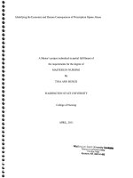
resolution of 1500, and a frequency of 50 0 0. Fig. 1 shows a scheme
of the microfluidic device proposed. Each layer contains a channel
(13 mm length, 70 μm deep and 3mm wide) and a flat membrane
is placed covering the entire channel separating the donor and
acceptor channel from each other. The membrane is impregnated
with 3 μL of organic solvent (TBP) and subsequently closed using
four screws. The device can be reused, opened and closed as many
times as necessary. Each channel has two holes: one for inlet solution and another for outlet solution. Both, the donor phase (containing the analytes) and the acceptor phase are introduced into
the microfluidic device for the microextraction by using a microsyringe pump (Cetoni GmbH, Korbussen, Germany), which operate
at 1 μL min−1 . First, 5 min were waited for SLM stabilization and
later, the acceptor phase was collected during 10 minutes in a micro insert tube and then injected into the HPLC system for analysis.
2. Experimental
2.1. Chemicals and materials
All chemicals were of analytical-reagent grade. Sulfadiazine
(SDI), Sulfamerazine (SMR), Sulfamethazine (SMT) and Sulfamethoxazole (SMZ) were provided from Fluka-Sigma-Aldrich
(Madrid, Spain). Formic acid, sodium hydroxide, chloric acid, 2nitrophenyl octyl ether (NPOE), dihexyl ether (DHE), and 1octanol were purchased from Fluka-Sigma-Aldrich (Madrid, Spain).
Methanol, acetonitrile, nonanol, decanol, undecanol, and tributyl
phosphate (TBP) were supplied from Merck (Darmstadt, Germany).
The stock solutions of the sulfonamides were prepared in methanol
(100 mg L−1 ) and preserved at 4°C in a refrigerator. Working
solutions were daily provided by dilution of the stock solutions
with deionized water (from a Milli-Q Plus water purification system (Millipore, Billerica, MA, USA)). A micro-syringe pump (Cetoni
2
S. Dowlatshah, E. Santigosa, M. Saraji et al.
Journal of Chromatography A 1652 (2021) 462344
Table 1
Extraction efficiencies (RSD %) of the sulfonamides using
different organic solvents as SLM.
2.4. Calculations of extraction efficiency
Extraction efficiency for each analyte was calculated according
to the following equation for each analyte (eq 1):
EE (% ) =
C f,a,outlet
x
Ci,s,inlet
va
x 100
vs
Extraction efficiency % (RSD%, n=4)
(Eq. 1)
Decanol
Undecanol
Nonanol
Octanol
DHE
NPOE
TBP
where C f,a,outlet is the final concentration of the analyte at the outlet of the acceptor channel, Ci,s,inlet is the initial concentration of
the analyte in the sample, and, va and vs , are the acceptor and
sample flow rate, respectively.
SDI
SMR
SMT
SMZ
7 (1)
5 (1)
8 (5)
21 (1)
5 (2)
13 (12)
74 (6)
11 (1)
8 (1)
18 (5)
42 (1)
7 (6)
17 (10)
76 (6)
13
10
14
32
10
21
80
53
43
45
78
46
47
81
(1)
(2)
(5)
(2)
(4)
(9)
(5)
(1)
(1)
(6)
(2)
(4)
(10)
(4)
a Sample: pH 2.5 containing the four compounds each at 1
μg mL−1 , acceptor phase: pH 12, sample and acceptor flow
rate: 1 μL min−1 , extraction time: 10 min.
2.6. Real samples
Urine samples were analyzed using the microchip device to
evaluate its applicability. Both samples were adjusted to pH 4.0
with HCl solution and spiked at three different levels (0.5, 1 and 3
μg mL−1 ). All samples were filtered through Pall NylafloTM nylon
membrane filter 0.45 μm (Pall Corporation, Ann Arbor, Michigan,
USA). Each sample was directly extracted by the microfluidic LPME
device.
3. Results and discussion
3.1. Supported liquid membrane selection
Previous studies carried out on LPME-based microfluidics have
described optimal geometric characteristics for passive diffusion
and good mass transfer [29, 35, 36]. Based on that, an initial device of 13 mm length, 70 μm deep and 3mm wide was designed
for the optimization of the experimental parameters. Sulfonamides
have two dissociation constants related to pKa1 and pKa2 . PKa1 and
pKa2 corresponds to a basic amino group (-NH2 ) and an acid group
(-NH-SO2 -), respectively. The amino group is capable of gaining a
proton while the amide of the acid group is capable of releasing a
proton under specific pH conditions. Within the pH range between
the first and second pKa , the molecule is predominantly neutral,
while at pH above its second pKa value, the molecule is negatively
charged. The pKa1 -pKa2 values are 1.6 - 6.5, 1.58-6.90, 2.07-7.49
and 1.85-5.60 for SDI, SMR, SMT, SMZ respectively [20].
Supported liquid membrane was the first experimental parameter to optimize since it is a critical parameter that is directly related to the nature of the analytes. Solvent selection was based
on the following requirements: water immiscibility, non-volatility,
affinity towards analytes, and compatibility with PMMA plates.
Based on these requirements and previously reported solvents
compatible with sulfonamides and LPME into a chip [20,29,35,36],
2-nitrophenyl octyl ether (NPOE), dihexyl ether, nonanol, decanol,
undecanol, and tributyl phosphate were tested as organic solvent.
Donor phase solution, acceptor phase solution and flow rate were
fixed at pH 2.5 (HCl), pH 12 (NaOH) and 1 μL min−1 , respectively,
for the study. At those conditions, sulfonamides are found in their
neutral form and in their ionized form in the donor and acceptor
phase, respectively. The microfluidic device was cleaned with miliQ
water and a new sheet membrane was used for each different organic solvent. Table 1 shows the highest extraction efficiency for all
analytes when using TBP as SLM. Four replicate experiments were
carried out to test the repeatability and a relative standard deviation (RSDs %) below 7 % was obtained for all analytes, except when
using NPOE (RSD% 9-12). Thus, tributyl phosphate was selected for
further experiments.
3.2. Donor and acceptor solutions optimization
Donor phase pH was studied within the pH range of 2-6 to ensure the analytes to be in their neutral form for passive diffusion,
while the acceptor phase composition was fixed at pH 12 (NaOH)
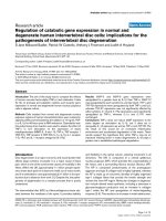
and the flow rate at 1 μL min−1 . As seen in Fig. 2, the highest peak
Fig. 2. Optimization of the donor phase composition. SLM: TBP. Acceptor phase pH
12. Flow rate: 1μL min−1 (acceptor and donor phase).
Fig. 3. Optimization of the acceptor phase composition. SLM: TBP. Donor phase pH
4. Flow rate: 1μL min−1 (acceptor and donor phase).
area was observed at pH 4 for all analytes while no significant difference was observed between the rest of the pHs studied. In the
next step, a range of pH between 9.0-12 was tested to study the
acceptor phase composition while the donor pH was adjusted at 4
for all experiments. Based on Fig. 3, the peak area increased up to
pH 12 and best results were obtained at the mentioned pH for all
sulfonamides. A study of the long-term stability of sulfonamides
was carried out at optimal pH’s, especially at basic pH to ensure
that sulfonamides were not degraded at pH 12. The relative standard deviation based on four replicate experiments was below 6 %.
Therefore, a pH 4 and pH 12 were adjusted throughout the rest of
experiments as donor and acceptor composition, respectively.
3.3. Donor phase flow rate optimization
3
S. Dowlatshah, E. Santigosa, M. Saraji et al.
Journal of Chromatography A 1652 (2021) 462344
Table 2
Calibration parameters using standard solution in water, method detection limit (MLOD), method
quantitation limit (MLOQ) and extraction efficiencies at optimal conditions.
SDI
SMR
SMT
SMX
∗
MLOD (μg mL−1 )
MLOQ (μg mL−1 )
Regresion Equation
R2
EE
0.045
0.057
0.054
0.033
0.15
0.19
0.18
0.11
Y=12519x-987.65
Y= 14358x-1432.7
Y= 9787.5x-126.56
Y= 12836x+340.52
0.9997
0.9996
0.9998
0.9996
101 (3)
96 (5)
98 (2)
98 (3)
∗
% Extraction efficiency (%RSD, n=4) in water
ues over 0.9996 was obtained in all cases. Table 2 shows the calibration parameters of the method: detection limits (LODs, S/N=3),
quantitation limits (LOQs, S/N=10) and extraction efficiencies for
all analytes. LODs between 0.033-0.057 μg mL−1 were obtained
for all sulfonamides. Three concentration levels of the calibration
curve (0.28, 1 and 5 μg mL−1 ) were selected to test the repeatability (n=4) and intraday repeatability (n=4, 15 days), obtaining
a relative standard deviation between 3- 6 % and below 3-5 %
for repeatability and intraday repeatability for all compounds, respectively. Calibration curve was prepared with standard solutions
of the analytes in water. Extraction efficiencies between 96-102 %
were obtained for all analytes. Finally, different microfluidic devices with the same geometry were used to test the reproducibility. Each device was tested replacing the membrane three times
and a relative standard deviation below 6 % was obtained for all
analytes.
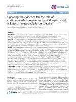
Fig. 4. Donor flow rate optimization. SLM: TBP. Donor phase pH 4. Acceptor phase
pH 12.
There are two flows when working with microfluidics in sample pretreatment: donor and acceptor. Both flows are decisive in
the extraction efficiency of the procedure. However, previous studies have shown that the extraction efficiency would significantly
decrease with greater acceptor flow since the time that this phase
is in contact with the donor phase (containing the analytes) will
decrease [31, 36, 38]. For this reason, the acceptor flow has been
set to 1 μL min−1 avoiding loss of extraction efficiency. In some
cases, depending on the analyte and how fast the mass transfer is,
the extraction efficiency does not decrease drastically at low flows
and its study is interesting since sometimes there is an enrichment phenomenon with acceptable efficiencies [36]. Then, donor
flow rate influence was evaluated within the range of 0.5-20 μL
min−1 using an acceptor flow rate of 1 μL min−1 . As seen in Fig. 4,
highest extraction efficiencies were observed at 0.5 or 1 μL min−1
and no significant difference was observed between both flow rate.
Efficiencies over 96 % was observed for all analytes. The residence
time of the sample decreased when the flow rate increased so,
consequently, a decrease in extraction efficiency was observed at
higher donor flow rate. Therefore, a flow rate of 1 μL min−1 is selected in order to achieve a faster extraction. The efficiencies obtained once the procedure is optimized were between 96-100 %, so
the selected geometry at the beginning has been successful and no
further optimization is needed since the device already has miniaturized characteristics of short (13 mm) and shallow (0.07 mm)
channels.
The carry over effect was tested by analyzing individual extractions with a new membrane and after consecutive extractions,
without observing memory effects.
5. Comparison with other setups
The performance of this chip was compared with previous
methodologies for sulfonamides extraction, in terms of extraction
time, extraction efficiency, relative recovery and sample solution
volume (Table 3). As seen, the authors express the results based
on extraction efficiency (EE %), enrichment factor (EF) and / or relative recovery (RR %). The extraction efficiency is defined as the
percentage of the mole numbers of the analyte extracted into the
acceptor phase respect to the moles number of the analyte originally present in the donor solution and which also depends on the
volume of each phase. On the other hand, the enrichment factor
is defined as the ratio of the analyte concentration in the analytecontaining acceptor to the initial concentration of analytes in the
donor solution and the relative recovery by the percentage of the
amount of analyte recovered in the acceptor solution from spiked
real samples. Different methods for the extraction of sulfonamides
have been reported with high enrichment factors between 121-996
[22], 58-135 [21], 268-664 [19] and 20 0-10 0 0 [20]. Moreover, the
sample volume required is relatively high, with a minimum sample
volume between 40 0 0-80 0 0 μL [21,22] up to 50 0 0 0 μL [20]. The
extraction time of these methods ranges from 20 min [19] to 8 h
[21], a significantly long time. Furthermore, some of them require
more than one sample pretreatment stage prior to analysis [19,21].
Other methods have been reported with shorter extraction times
between 20-30 min [18, 24, 26–28, 40] and extraction efficiencies
between 70-77 % [28], 50-60 % [18], 56-100 % [42] and 12-18 %
[27]. Some of these procedures also require more than one treatment stage prior to analysis, lengthening the total analysis time
[19, 27, 28] and those that only consist of one extraction stage require at least 10 0 0 μL of sample volume. One of the most relevant advantages when using flat membrane microfluidic systems is
that in many cases, the supported liquid membrane is reusable and
therefore allows consecutive extractions to be carried out without the need to change the membrane or add extracting solvent.
As seen in Table 3, the presented microfluidic method provides
the highest extraction efficient for all sulfonamides in real sample
(human urine). In addition, the required sample volume was de-
4. Analytical performance
Microfluidic method based LPME was evaluated for the determination of four sulfonamides by fixing the experimental parameters at optimal conditions as described above. A calibration curve
was constructed using a least-square linear regression analysis at
seven different concentrations from 0.15 to 10 μg mL−1 , 0.19 to
10 μg mL−1 , 0.18 to 10 μg mL−1 and 0.11 to 10 μg mL−1 for SDI,
SMR, SMT and SMX, respectively. A linear relationship with r2 val4
S. Dowlatshah, E. Santigosa, M. Saraji et al.
Journal of Chromatography A 1652 (2021) 462344
Fig. 5. Chromatogram of a (A) spiked human urine at 1 μg mL−1 and (B) blank human urine and (C) blank urine chromatogram of a reused membrane.
Table 3
Comparison of μLPME procedure with other analytical methods for extraction of sulfonamides.
Technique (Analysis)
Real
sample
Extraction
Time (min)
Sample
volume (μl)
Enrichment factor
% Extraction Efficiency/
(∗ R:recovery from spiked
real samples)
Consecutive
extractions
Ref.
HF-LPME-HPLC/UV
SUPRASs-solvent based
LPME- HPLC/UV&
serum
Ion pair Emulsification
LPME-HPLC-UV/Vis
Capsule phase
microextractionHPLC/UV
SD-LPME-HPLC/UV
DLLME-HPLC/UV
SPME-LC/MS
HF-LPME-HPLC/UV
HF- LPME-CE/ED
VA-LPME-HPLC/DAD
IT-SPME-HPLC/UV
IL magnetic
bar-HF-LPME/FLD
HF-LPME-UPLC/FLD
μLPME-HPLC/UV
water
Plasma
360
20
50000
200
200-1000
6.1-6.7
-/∗ (R: 32-100)
70-77 /∗ (R: 85-90)
No
No
[20]
[28]
water
20
10000
268-664
41-97/∗ (R: 104)
No
[19]
milk
30
2000
-
12-18
No
[27]
water
water
meat
water
water
water
milk
water
20
10
40
480
60
30
25
25
1500
5000
15000
4000
8000
4000
>1000
6000
58-135
121-996
-
-/∗ (R: 63-115)
-/∗ (R: 78-117)
4-27
-/∗ (R: 82-103)
-/∗ (R: 75-109)
-/∗ (R: 78-98)
50-60/∗ (R: 11-97)
56-100/∗ (R: 19-94)
No
No
No
No
No
No
No
No
[26]
[24]
[17]
[21]
[22]
[25]
[18]
[42]
water
Urine
60
10
8000
10
14-60
-
-/∗ (R: 56-113)
87-103/(R: 84-100)
No
YES
[23]
This work
HF-LPME: Hollow fiber-liquid phase microextraction, SD-LPME: Single drop liquid phase microextraction, DLLME: Dispersive liquid liquid microextraction, SPME: Solid phase
microextraction, VA-LPME: Voltage-Assisted Liquid-Phase Microextraction, IT-SPME: In-tube solid-phase microextraction, IL magnetic bar-HF-LPME: ionic liquid magneticc
bar based hollow fiber liquid phase microextraction, FLD: Fluorescence detection, ED: electrochemical detection.
Table 4
Extraction efficiencies (average of three determinations ± standard deviation) from 1 μg mL-1 spiked urine
samples.
Samples
Spiked level (μg mL−1 )
SDI
SMR
SMT
SMZ
Urine (non-diluted)
0.5
1
3
97.6 ± 3.9
95.3 ± 2.2
106.1 ± 5.3
86.2 ± 4.0
86.8 ± 3.3
87.0 ± 2.0
88.1 ± 1.6
89.3 ± 2.4
86.8 ± 1.9
91.8 ± 3.2
92.4 ± 1.8
84.8 ± 2.6
creased between 20 and 50 0 0 times compared to existing methods
and it also required shorter extraction times (only 10 min).
samples were spiked at three different concentration levels (0.5,
1 and 3 μg mL−1 ) of SDI, SMR, SMT and SMZ. Experiments were
analyzed in triplicate for each of the concentrations. All samples
were submitted to the microfluidic device using the optimal experimental conditions, and the extract collected was analyzed by
HPLC-DAD. Similar extraction efficiencies were obtained for each
analyte regardless of concentration. Extraction efficiencies between
6. Real samples analysis
The applicability of the microfluidic device proposed was investigated in human urine samples. Urine samples were collected
from a 35 year-old healthy adult female volunteer and undiluted
5
S. Dowlatshah, E. Santigosa, M. Saraji et al.
Journal of Chromatography A 1652 (2021) 462344
95-106 %, 85-88 %, 83-90 % and 83-92 % for SDI, SMR, SMT and
SMZ were obtained, respectively. As seen in Table 4, the relative
standard deviation after triplicate experiments was below 5.5 %
for all analytes and the membrane was stable for more than 20
consecutive extractions using urine samples with no carry over
effect. Fig. 5 shows the corresponding DAD chromatogram from
spiked human urine (A), a blank (B) using two different membranes. Fig. 5C shows a blank chromatogram after washing the
membrane previously used for consecutive extractions.s.
[8] D. Teshima, B. Hino, Y. Itoh, R. Oishi, Simple and simultaneous determination of sulphapyridine and acetylsulphapyridine in human serum by column-switching high-performance liquid chromatography, J. Clin. Pharm. Ther. 27
(2002) 403–408.
[9] J. Tuerk, M. Reinders, D. Dreyer, T.K. Kiffmeyer, K.G. Schmidt, H.M. Kuss, Analysis of antibiotics in urine and wipe samples from environmental and biological
monitoring - Comparison of HPLC with UV-, single MS- and tandem MS-detection, J. Chromatogr. B Anal. Technol. Biomed. Life Sci. 831 (2006) 72–80.
[10] A. Kaufmann, P. Butcher, K. Maden, M. Widmer, Ultra-performance liquid chromatography coupled to time of flight mass spectrometry (UPLC-TOF): A novel
tool for multiresidue screening of veterinary drugs in urine, Anal. Chim. Acta.
586 (2007) 1321.
[11] D.C.G. Bedor, T.M. Gonỗalves, M.L.L. Ferreira, C.E.M. de Sousa, A.L. Menezes,
E.J. Oliveira, D.P. de Santana, Simultaneous determination of sulfamethoxazole
and trimethoprim in biological fluids for high-throughput analysis: Comparison of HPLC with ultraviolet and tandem mass spectrometric detection, J.
Chromatogr. B Anal. Technol. Biomed. Life Sci. 863 (2008) 46–54.
[12] J.K. Johannessen, I. Christiansen, D.R. Schmidt, E. Petersen, S.H. Hansen, Simultaneous determination of pyrimethamine, sulfadiazine and N-acetyl-sulfadiazine in plasma for monitoring infants in treatment of congenital toxoplasmosis, J. Pharm. Biomed. Anal. 36 (2005) 1093–1098.
[13] C.L. Ng, H.K. Lee, S.F.Y. Li, Determination of sulphonamides in pharmaceuticals
by capillary electrophoresis, J. Chromatogr. A. 632 (1993) 165–170.
[14] R.B. Hoff, F. Barreto, T.B.L. Kist, Use of capillary electrophoresis with laser-induced fluorescence detection to screen and liquid chromatography-tandem
mass spectrometry to confirm sulfonamide residues: Validation according to
European Union 2002/657/EC, J. Chromatogr. A. 1216 (2009) 8254–8261.
[15] Q. xun Dang, Z. pei Sun, D.K. Ling, Separation of sulphonamides and determination of the active ingredients in tablets by micellar electrokinetic capillary
chromatography, J. Chromatogr. A. 603 (1992) 259–266.
[16] K.J. Bisceglia, J.T. Yu, M. Coelhan, E.J. Bouwer, A.L. Roberts, Trace determination of pharmaceuticals and other wastewater-derived micropollutants by solid
phase extraction and gas chromatography/mass spectrometry, J. Chromatogr. A.
1217 (2010) 558–564.
[17] K.H. Lu, C.Y. Chen, M.R. Lee, Trace determination of sulfonamides residues
in meat with a combination of solid-phase microextraction and liquid chromatography-mass spectrometry, Talanta 72 (2007) 1082–1087.
[18] Y. Wen, M. Zhang, Q. Zhao, Y.Q. Feng, Monitoring of five sulfonamide antibacterial residues in milk by in-tube solid-phase microextraction coupled
to high-performance liquid chromatography, J. Agric. Food Chem. 53 (2005)
8468–8473.
[19] B. Ebrahimpour, Y. Yamini, M. Rezazadeh, A sensitive emulsification liquid
phase microextraction coupled with on-line phase separation followed by
HPLC for trace determination of sulfonamides in water samples, Environ.
Monit. Assess. 187 (2015) 1–13.
[20] M. Ramos Payán, M.A. Bello López, R. Fernández-Torres, M.V. Navarro,
M.C. Mochón, Hollow fiber-based liquid phase microextraction (HF-LPME) for a
highly sensitive HPLC determination of sulfonamides and their main metabolites, J. Chromatogr. B Anal. Technol. Biomed. Life Sci. 879 (2011) 197–204.
[21] Y. Tao, J.F. Liu, X.L. Hu, H.C. Li, T. Wang, G.Bin Jiang, Hollow fiber supported
ionic liquid membrane microextraction for determination of sulfonamides in
environmental water samples by high-performance liquid chromatography, J.
Chromatogr. A. 1216 (2009) 6259–6266.
[22] F. Tong, Y. Zhang, F. Chen, Y. Li, G. Ma, Y. Chen, K. Liu, J. Dong, J. Ye, Q. Chu, Hollow-fiber liquid-phase microextraction combined with capillary electrophoresis for trace analysis of sulfonamide compounds, J. Chromatogr. B Anal. Technol. Biomed. Life Sci. 942–943 (2013) 134–140.
[23] L. Yang, Y. Shi, J. Li, T. Luan, In situ derivatization and hollow-fiber liquid-phase
microextraction to determine sulfonamides in water using UHPLC with fluorescence detection, J. Sep. Sci. 41 (2018) 1651–1662.
[24] A.V. Herrera-Herrera, J. Hernández-Borges, T.M. Borges-Miquel, M. ángel Rodríguez-Delgado, Dispersive liquid-liquid microextraction combined with ultra-high performance liquid chromatography for the simultaneous determination of 25 sulfonamide and quinolone antibiotics in water samples, J. Pharm.
Biomed. Anal. 75 (2013) 130–137.
[25] M.H. Wang, H.W. Chang, S.P. Wang, Analysis of Sulfonamides by Liquid Chromatography Mass Spectrometry and Capillary Electrophoresis Combing with
Voltage-Assisted Liquid-Phase Microextraction, J. Chinese Chem. Soc. 60 (2013)
1479–1483.
[26] X. Guo, D. Yin, J. Peng, X. Hu, Ionic liquid-based single-drop liquid-phase microextraction combined with high-performance liquid chromatography for the
determination of sulfonamides in environmental water, J. Sep. Sci. 35 (2012)
452–458.
[27] D. Georgiadis, A. Tsalbouris, A. Kabir, K.G. Furton, V. Samanidou, Novel capsule phase microextraction in combination with high performance liquid chromatography with diode array detection for rapid monitoring of sulfonamide
drugs in milk, J. Sep. Sci. 42 (2019) 1440–1450.
[28] P. Bogdanova, A. Pochivalov, C. Vakh, A. Bulatov, Supramolecular solvents formation in aqueous solutions containing primary amine and monoterpenoid
compound: Liquid phase microextraction of sulfonamides, Talanta 216 (2020)
120992.
[29] M. Ramos Payán, Liquid - Phase microextraction and electromembrane extraction in millifluidic devices:A tutorial, Anal. Chim. Acta. 1080 (2019) 12–21.
[30] N.J. Petersen, H. Jensen, S.H. Hansen, S.T. Foss, D. Snakenborg, S.Pedersen Bjergaard, On-chip electro membrane extraction, Microfluid. Nanofluidics. 9 (2010)
881–888.
4. Conclusion
This work presents for the first time an efficient microfluidic
method for the determination of sulfonamides and its successfully
application in urine samples. The presented microfluidic system
significantly improves in terms of sample and reagent volume and
analysis time, offering high extraction efficiencies compared to previous reported methodologies. The method was also successfully
applied in urine sample with extraction efficiencies between 83
and 106 % for all sulfonamides with only a urine sample volume
consumption of 10 μL after 10 minutes extraction time and excellent clean-up. TPB has been demonstrated to be a good organic solvent as extractant which significantly contributes to the stability of
the microfluidic system when the method is applied to consecutive
urine extractions, thus reducing the cost of instrumentation.
Author Statements
Samira Dowlatshah: Formal Analysis, Investigation. Elia
Santigosa: Data curation, Mohammed Saraji: writing original draft,
María Ramos Payán: Methodology, Conceptualization, Supervision,
Writing - review & editing.
Declaration of Competing Interest
The authors declare that they have no known competing financial interests or personal relationships that could have appeared to
influence the work reported in this paper.
Acknowledgements
This work was partially supported by Microliquid S.L. in the
frame of the bilateral collaborative project XploreChip P01158.
Samira Dowlatshah thanks the Ministry of Science, Research and
Technology of the Islamic Republic of Iran and the Research Council of Isfahan University of Technology (IUT) for the scholarship.
References
[1] K. Dost, D.C. Jones, G. Davidson, Determination of sulfonamides by packed column supercritical fluid chromatography with atmospheric pressure chemical
ionisation mass spectrometric detection, Analyst 125 (20 0 0) 1243–1247.
[2] H.C. Wegener, The Consequences for Food Safety of the Use of Fluoroquinolones in Food Animals, N. Engl. J. Med. 340 (1999) 1581–1582.
[3] D. Ferber, Science 288 (20 0 0) 792–794.
[4] P.D. Fey, T.J. Safranek, M.E. Rupp, E.F. Dunne, E. Ribot, P.C. Iwen, P.A. Bradford,
F.J. Angulo, S.H. Hinrichs, Ceftriaxone-Resistant Salmonella Infection Acquired
by a Child from Cattle, N. Engl. J. Med. 342 (20 0 0) 1242–1249.
[5] J. Krungkrai, S.R. Krungkrai, C.T. Supuran, Carbonic anhydrase inhibitors:
Inhibition of Plasmodium falciparum carbonic anhydrase with aromatic/heterocyclic sulfonamides-in vitro and in vivo studies, Bioorganic
Med. Chem. Lett. 18 (2008) 5466–5471.
[6] S. Babic´ , D. Ašperger, D. Mutavdžic´ , A.J.M. Horvat, M. Kaštelan-Macan, Determination of sulfonamides and trimethoprim in spiked water samples by
solid-phase extraction and thin-layer chromatography, J. Planar Chromatogr. Mod. TLC. 18 (2005).
[7] C. Reguera, M.C. Ortiz, A. Herrero, L.A. Sarabia, Optimization of a FIA system
with amperometric detection by means of a desirability function. Determination of sulfadiazine, sulfamethazine and sulfamerazine in milk, Talanta 75
(2008) 274–283.
6
S. Dowlatshah, E. Santigosa, M. Saraji et al.
Journal of Chromatography A 1652 (2021) 462344
[31] B. Li, N.J. Petersen, M. Ramos Payán, S.H. Hansen, S.Pedersen Bjergaard, Design and implementation of an automated liquid-phase microextraction-chip
system coupled on-line with high performance liquid chromatography, Talanta
120 (2014) 224–229.
[32] E. Santigosa Murillo, X. Muñoz Berbel, S. Maspoch, M. Muñoz, M. Ramos Payán,
Impedance model for voltage optimization of parabens extraction in an electromembrane millifluidic device, J. Chromatogr. A. 1625 (2020) 461270.
[33] Y.A. Asl, Y. Yamini, S. Seidi, A novel approach to the consecutive extraction of
drugs with different properties via on chip electromembrane extraction, Analyst 141 (2016) 311–318.
[34] M. Karami, Y. Yamini, Y. Abdossalami Asl, M. Rezazadeh, On-chip pulsed electromembrane extraction as a new concept for analysis of biological fluids in a
small device, J. Chromatogr. A. 1527 (2017) 1–9.
[35] M. Ramos Payán, S. Maspoch, A. Llobera, A simple and fast Double-Flow microfluidic device based liquid-phase microextraction (DF-μLPME) for the determination of parabens in water samples, Talanta 165 (2017) 496–501.
[36] M. Ramos Payán, S. Maspoch, A. Llobera, An effective microfluidic based liquid-phase microextraction device (μLPME) for extraction of non-steroidal anti-inflammatory drugs from biological and environmental samples, Anal. Chim.
Acta. 946 (2016) 56–63.
[37] N.J. Petersen, S.T. Foss, H. Jensen, S.H. Hansen, C. Skonberg, D. Snakenborg,
J.P. Kutter, S. Pedersen-Bjergaard, On-Chip Electro Membrane Extraction with
Online Ultraviolet and Mass Spectrometric Detection, Anal. Chem. 83 (2011)
44–51.
[38] M. Ramos Payán, H. Jensen, N.J. Petersen, S.H. Hansen, S.Pedersen Bjergaard,
Liquid-phase microextraction in a microfluidic-chip – High enrichment and
sample clean-up from small sample volumes based on three-phase extraction,
Anal. Chim. Acta. 735 (2012) 46–53.
[39] Y.A. Asl, Y. Yamini, S. Seidi, M. Rezazadeh, Simultaneous extraction of acidic
and basic drugs via on-chip electromembrane extraction, Anal. Chim. Acta. 937
(2016) 61–68.
[40] M. Ramos Payán, E. Santigosa, R. Fernández Torres, M.Á.Bello López, A New
Microchip Design. A Versatile Combination of Electromembrane Extraction and
Liquid-Phase Microextraction in a Single Chip Device, Anal. Chem. 90 (2018)
10417–10424.
[41] F. Zarghampour, Y. Yamini, M. Baharfar, M. Faraji, Simultaneous extraction of
acidic and basic drugs via on-chip electromembrane extraction using a single–
compartment microfluidic device, Analyst 144 (2019) 1159–1166.
[42] L. Yang, Y. Shi, J. Li, T. Luan, In situ derivatization and hollow-fiber liquid-phase
microextraction to determine sulfonamides in water using UHPLC with fluorescence detection, J. Sep. Sci. 41 (2018) 1651–1662.
7