Báo cáo khoa học: Etoposide upregulates Bax-enhancing tumour necrosis factor-related apoptosis inducing ligand-mediated apoptosis in the human hepatocellular carcinoma cell line QGY-7703 pdf
Bạn đang xem bản rút gọn của tài liệu. Xem và tải ngay bản đầy đủ của tài liệu tại đây (417.3 KB, 11 trang )
Etoposide upregulates Bax-enhancing tumour necrosis factor-related
apoptosis inducing ligand-mediated apoptosis in the human
hepatocellular carcinoma cell line QGY-7703
Lin Miao, Peng Yi, Yi Wang and Mian Wu
Department of Molecular and Cell Biology, Key Laboratory of Structural Biology, School of Life Sciences,
University of Science and Technology of China, Hefei, Anhui, China
Tumour necrosis factor-related apoptosis-inducing ligand
(TRAIL) has attracted much attention because of its ability
to kill tumour cells. In this study, we demonstrated that
treatment of QGY-7703 cells with the combination of
TRAIL and etoposide resulted in synergistic cytotoxic
effects. In dissecting the mechanism underlying this syner-
gistic effect, we found that treatment with etoposide alone
resulted in the upregulation of Bax, while the level of trun-
cated Bid (tBid) was unchanged. In contrast, while treatment
with TRAIL alone significantly increased the level of tBid,
the expression of Bax remained unaffected. The enhanced
apoptosis was accompanied by an increased release of
cytochrome c and second mitochondria-derived activator of
caspase/direct IAP binding protein with low pI (DIABLO)
from mitochondria, leading to the activation of cellular
caspase-8, -9, -3 and -7, as well as poly ADP-ribose polym-
erase. This enhanced release of cytochrome c and second
mitochondria-derived activator of caspase/DIABLO was
inhibited by the general caspase inhibitor N-benzyloxycar-
bonyl-Val-Ala-Asp-fluoromethylketone. The RT–PCR and
Western blotting results demonstrated that the levels of both
mRNA and protein for death receptor-4, death receptor-5
and decoy receptor-2 remained unchanged in response to
etoposide, indicating that the synergistic effect of TRAIL
and etoposide is not a result of increasing the expression for
TRAIL receptors, but rather is associated with amplification
of the mitochondrial signal pathway.
Keywords: p53; Bax; tBid; mitochondrial pathway; death
receptor.
Chemotherapeutic agents are used widely in the treatment
of different types of cancer. Hetapocellular carcinoma, one
of the most common tumours in adults, remains largely
incurable despite intensive multimodality treatment, inclu-
ding surgical eradication, irradiation and chemotherapy.
Besides the difficulties of complete surgical removal,
resistance to chemotherapy and irradiation is a major
hindrance for the successful treatment of liver cancers.
Defects in signalling pathways leading to the activation of
caspases are common in most types of malignancies.
Accumulating data suggest that two major signal pathways
are involved in apoptosis. The first is the mitochondrial
pathway (intrinsic), usually triggered by DNA damage, and
the second is the receptor-mediated pathway (extrinsic),
initiated by death receptor activation. Resistance to
chemotherapeutic agents may be caused by repression of
the mitochondrial pathway. In order to achieve effective
treatment of those drug-resistant tumour cells, new meth-
ods, which could bypass the resistance to chemotherapeutic
drugs through activation of the death receptor-mediated
pathway, need to be developed. Tumour necrosis factor-
related apoptosis-inducing ligand (TRAIL), a type II
membrane protein, is a member of the tumour necrosis
factor death-ligand family [1,2] and it selectively induces
apoptosis in a number of transformed cells in vitro [1–3] and
tumours in vivo, while leaving normal tissues intact [4,5].
Four cognate TRAIL death receptors – death receptor
(DR)4 [6,7], DR5 [7,8], decoy receptor-1 (DcR-1)/TRID
(i.e. TRAIL decoy receptor lacking an intracellular domain)
[9–11] and DcR-2/TRUNDD (i.e. TRAIL decoy receptor
containing a truncated death domain) [12–14] – have been
identified to date. Both DR4 and DR5 contain an intracel-
lular Ôdeath domainÕ that recruits effector molecules, such as
Fas-associated death domain protein (FADD) [15] and
death-associated protein 3 (DAP-3) [16] to activate initiator
caspase-8 and subsequently the effector caspases leading to
apoptosis [6,7]. In contrast, DcR-1/TRID and DcR-2/
TRUNDD contain a truncated or a null intracellular death
Correspondence to M. Wu, Department of Molecular and Cell
Biology, School of Life Sciences, University of Sciences and
Technology of China, Hefei, Anhui, China, 230027.
Fax: + 86 551 360 6264, Tel.: + 86 551 360 6264,
E-mail:
Abbreviations: DcR, decoy receptor; DIABLO, direct IAP binding
protein with low pI; DR, death receptor; FADD, Fas-associated death
domain protein; FITC, fluorescein isothiocyanate; MTT,
3-(4,5-dimethylthiazol-2-yl)-2,5-diphenyltetrazolium bromide; PARP,
poly ADP-ribose polymerase; PI, phosphatidylinositol; Smac, second
mitochondria-derived activator of caspase; tBid, truncated Bid;
TRAIL, tumour necrosis factor-related apoptosis-inducing ligand;
TRID, TRAIL decoy receptor lacking an intracellular domain;
TRUNDD, TRAIL decoy receptor containing a truncated death
domain; zVAD-FMK, N-benzyloxycarbonyl-Val-Ala-Asp-fluoro-
methylketone.
Enzymes: alkaline phosphatase (EC 3.1.3.1).
(Received 13 January 2003, revised 26 March 2003,
accepted 28 April 2003)
Eur. J. Biochem. 270, 2721–2731 (2003) Ó FEBS 2003 doi:10.1046/j.1432-1033.2003.03639.x
domain, respectively, and are unable to transduce the death
signal [6,7].
Mutation in p53 initiates oncogenesis and accounts for
more than 50% of different tumour types [17–19]. p53 acts
as a Ôgenome guardianÕ to scrutinize DNA injury in cell cycle
regulation and cell death control by modulating expression
of a number of target genes in response to DNA-damaging
agents, hypoxia or oncogene activation. A number of
chemotherapeutic drugs, acting as DNA-damaging agents,
enhance the expression of p53. The downstream target
genes of p53 include p21, Bax, DR4, DR5, DcRs and Bcl-2.
Upregulation of p53, resulting in the enhanced expression of
DR4 and/or DR5 in some cancer cell lines, is believed to be
one of the mechanisms by which the susceptibility of the
tumour cells to TRAIL is synergistically increased when
treated with chemotherapeutic agents and TRAIL together
[20,21]. However, some research groups have reported that
the upregulation of DR4 and DR5 is not necessarily p53-
dependent [22,23].
Many chemotherapeutic drugs can promote mitochond-
rial membrane permeabilization and release of caspase-
activating factors, in particular cytochrome c and second
mitochondria-derived activator of caspase (Smac)/DIAB-
LO from mitochondria to the cytosol. Bax is a
pro-apoptotic factor belonging to the Bcl-2 family and
stimulates mitochondria to release cytochrome c and Smac/
DIABLO. Bax, together with its homologue Bak, plays a
vital role in the TRAIL-mediated mitochondrial apoptosis
[24]. The Bid protein, a member of the Bcl-2 family, may
stand at the cross-roads of the mitochondria and the death
receptor, as Bid is cleaved by active caspase-8 to form
truncated Bid (tBid), which, in turn, stimulates the mito-
chondria to release cytochrome c [25].
In this study, we examined the apoptotic effects of the
cytokine TRAIL, in the presence and absence of a
chemotherapeutic agent, on hepatocellular carcinoma
QGY-7703 cells to determine whether co-operative killing
could be achieved and, if so, what possible mechanism
might be underlying this effect. We found that TRAIL plus
etoposide acts synergistically to kill human liver tumour
cells and this synergistic effect involves the upregulation of
Bax and tBid, but not of DR4 or DR5. The interaction
between Bax and tBid results in the amplification of
mitochondrial release of cytochrome c and Smac/DIABLO,
leading to augmented cell death through enhanced activa-
tion of cellular caspases.
Materials and methods
Regents and antibodies
Recombinant human (rh)TRAIL was purchased from
R & D Systems. The general caspase inhibitor, N-benzyl-
oxycarbonyl-Val-Ala-Asp-fluoromethylketone (zVAD-
FMK), was purchased from BIOMOL Research
Laboratories Inc. Most drugs used in this study, including
etoposide, cisplatin, doxorubincin, 5-fluorouracil, metho-
trexate, cytarabine, cyclophophamide and daunorubicin,
were purchased from Sigma. Some other drugs of GCP
(good clinical practice) grade were ordered from a local
pharmaceutical company. Antibodies used in this study
were as follows. Polyclonal antibodies: anti-caspase-7,
anti-actin, anti-DR4, anti-DR5, anti-DcR-2, anti-Bid, anti-
Bax (Santa Cruz Biotech. Inc., Santa Cruz, CA, USA),
anti-caspase-9 (Immunotech), anti-caspase-3/CPP32 (BD
Biosciences) and anti-poly ADP-ribose polymerase (anti-
PARP) (Upstate Biotechnology). Monoclonal antibodies
(mAbs): anti-cytochrome c (R & D Systems), anti-caspase-8
(BD Biosciences) and anti-Smac/DIABLO (Calbiochem).
All the secondary antibodies [anti-goat, anti-mouse, anti-
rabbit (H + L)] were purchased from Promega.
Cell culture
The human hepatocellular carcinoma cell line, QGY-7703,
was kindly provided by D. Lu (Dept of Neurology,
Stanford University, Palo Alto, CA, USA). The cells were
maintained in RPMI-1640 containing 10% heat-inactivated
bovine serum, 1 m
ML
-glutamine, 100 UÆml
)1
penicillin and
100 lgÆml
)1
streptomycin (Life Technologies, Inc. Grand
Island, NY, USA) at 37 °C under an atmosphere of 5%
CO
2
in air.
Cell death assay
The 3-(4,5-dimethylthiazol-2-yl)-2,5-diphenyltetrazolium
bromide (MTT) assay was used to determine tumour cell
viability. Briefly, cells were plated at 1 · 10
4
cells per well in
96-well microtitre plates overnight. Cells were then treated
with 100 lL of fresh medium containing the drug to be
tested, cultured for 20 h, then for a further 4 h with 10 lL
of 5 mgÆmL
)1
MTT. After incubation, the medium was
removed and replaced with 100 lL of dimethylsulfoxide
and the data were analysed by using an EL
X
800 Universal
Microplate Reader (BIO-TEK Instruments, Inc.) at a
wavelength of 570 nm with the reference wavelength set at
630 nm. The effect of drug treatment was expressed as a
percentage of growth inhibition using untreated cells as the
uninhibited control.
Assessment of apoptosis by Annexin V staining
An Annexin V–fluorescein isothiocyanate (FITC) Apoptosis
Detection kit (Pharmingen, San Diego, CA, USA) was used
in this assay. After treatment with the test drugs, cells were
harvested and resuspended in binding buffer [0.01
M
Hepes/
NaOH (pH 7.4), 0.14 m
M
NaCl, 2.5 m
M
CaCl
2
]ata
concentration of 1 · 10
6
cellsÆmL
)1
. One-hundred micro-
litres of this resuspended solution (1 · 10
5
cells) was trans-
ferred to a 5-mL culture tube. After incubation with 5 lLof
Annexin V–FITC and 10 lL of phosphatidylinositol (PI)
(50 lgÆmL
)1
) for 15 min at room temperature in the dark,
the cells were analysed by flow cytometry in a FACSCalibur
using
CELL QUEST
software (Becton Dickinson).
Western blot analysis
Cells were lysed in 50 lL of lysis buffer [50 m
M
Tris/HCl
(pH 7.5), 250 m
M
NaCl, 5 m
M
EDTA, 50 m
M
NaF, 0.5%
Nonidet P-40] supplemented with a protease inhibitor
cocktail (Roche Molecular Biochemicals, Indianapolis,
IN) on ice for 30 min. Fifty micrograms of each sample
was separated by SDS/PAGE (12% or 15% gel) and
transferred to nitrocellulose membrane (Amersham
2722 L. Miao et al. (Eur. J. Biochem. 270) Ó FEBS 2003
Pharmacia Biotech). Filters were blocked with NaCl/Tris
(TBS) containing 5% nonfat milk and 0.1% Tween-20 for
1 h at room temperature and then incubated (for a further
1 h) with primary antibodies. Blots were then probed with
appropriate alkaline phosphatase-conjugated secondary
antibodies and the proteins visualized by using Western
Blue stabilized substrate for alkaline phosphates (Promega).
RT–PCR
Total cellular RNA was extracted from QGY-7703 cells
using the SV total RNA Isolation kit (Promega). The
yield and purity of the RNA sample were determined by
ultraviolet spectrometry using the DU 640 Nucleic Acid and
Protein Analyser (Beckman Coulter). Two micrograms of
total RNA was reverse transcribed using the TaKaRa One
Step RNA PCR kit (TaKaRa Bio Inc., Shiga, Japan)
according to the manufacturer’s instructions. In both RT
and PCR steps, the reaction reagents were prepared as
master mixtures and then aliquotted. PCR primers were
designed to amplify the sequence for the intracellular
domain of the TRAIL/Apo2L receptor. The housekeeping
gene, b-actin, was amplified as an internal control. The
mRNA levels were then normalized to actin mRNA
expression. Equal amounts of RT–PCR products, loaded
onto an agarose gel, were quantified by using the Eagel Eye
Jr Still Video System (Stratagene). Differences in mRNA
levels as a result of treatment with etoposide for different
incubation times are represented as relative units over
basal actin mRNA levels. The sequences of the primers
used in this study are as follows: DR4 forward,
5¢-CGGAATTCGGAGGGGACCCCAAGTGCAT-3¢;
DR4 reverse, 5¢-CGGGATCCTCACTCCAAGGACA
CGGCA-3¢;DR5forward,5¢-CGGAATTCTGCA
AGTCTTTACTGTGGAA-3¢; DR5 reverse, 5¢-CGGA
TCCTTAGGACATGGCAGAG-3¢;DcR2forward,
5¢-CGGAATTCCGCGGAAGAAATTCATTTCT-3¢;
DcR2 reverse, 5¢-CGGGATCCTCACAGGCAGGACG
TAGCAG-3¢; Bax forward, 5¢-GCGAATTCCATGG
ACGGGTCCGGGGAG-3¢; Bax reverse, 5¢-CGCTC
GAGTCAGCCCATCTTCTTCCAG-3¢;Bidforward,
5¢-CGGGATCCCCATGGCGATGGACTGTGAGGT-3¢;
Bid reverse, 5¢-CGGAATTCTCAGTCCATCCCATTTC
TGG-3. The primers for b-actin were kindly provided by
Z. Tian (Department of Immunology, University of Science
and Technology of China, Hefei, Anhui, China).
Cytochrome
c
release assay
Cell were harvested, as described above, and lysed in ice-
cold lysis buffer (20 m
M
Hepes, pH 7.4, 10 m
M
KCl, 5 m
M
EDTA, 2 m
M
MgCl
2
,with2m
M
dithiothreitol and prote-
ase inhibitor cocktail added before use) for 15 min on ice.
Cell lysates were centrifuged at 16 000 g for 2 min and the
supernatants mixed with 2 · Laemmli buffer and resolved
by SDS–PAGE (15% gel). Released cytochrome c and
Smac/DIABLO were subjected to analysis by Western blot.
Statistical analysis
All determinations were made in triplicate, and the results
were expressed as the mean value ± SD. Statistical signi-
ficance was determined by the Student’s t-test. A P-value
of < 0.05 was considered significant. Calculations of
synergistic cytotoxicity were determined by isobolographic
analysis, as described by Berenbaum [26]. The isobolo-
graphic analysis can detect whether the dose-dependent
effects of two compounds in a mixture are more or less
effective than the expected effects based on tests of the
compounds individually. Simply put, the point representing
the dose combination lying on, below, or above the straight
line represents additive, synergistic, or antagonistic effects,
respectively.
Results and discussion
Effect of TRAIL on the liver carcinoma cells QGY-7703
To investigate the apoptotic function of TRAIL containing
the extracellular domain (amino acids 114–281) of the
human liver cancer cell line, QGY-7703 [27], we treated the
cells with increasing concentrations of TRAIL and then
analysed cell viability by using the MTT assay. After
incubation with TRAIL at a concentration of 10 ngÆmL
)1
for 30 h, QGY-7703 cells were found to show morpho-
logical changes, from a spindle-like to a rounded shape, a
characteristic feature of apoptosis (Fig. 1A,c). As controls,
untreated cells (Fig. 1A,a) and cells treated with medium
(Fig. 1A,b) remained healthy and viable. Although TRAIL
can induce the apoptosis of QGY-7703 cells, we found that
the cells were relatively insensitive to TRAIL. In the dose–
response experiment (Fig. 1B), even though the QGY-7703
cells were treated with TRAIL at a high concentration of
100 ngÆmL
)1
for 24 h, only 23% cell death was detected. To
further verify whether the TRAIL-induced cell death
represents apoptosis, we utilized the AnnexinV–FITC
staining method to quantify the apoptotic cell numbers by
flow cytometry, which relies on the property of cells to lose
membrane asymmetry in the early phase of apoptosis. As
shown in Fig. 1C,a, only 1.4% of untreated QGY-7730 cells
were Annexin-V positive and PI negative (early apoptotic
cells). In contrast, approximately 7% of TRAIL
(10 ngÆmL
)1
)-treated QGY-7730 cells were Annexin-V posi-
tive and PI negative (Fig. 1C,c). Approximately 6% of
untreated cells were double-positive for Annexin-V and PI
(Fig. 1C,a). We noted that the percentage of these Annexin-
V- and PI-positive cells remained similar after treatment
with TRAIL (Fig. 1C,c; 7.3%), etoposide (Fig. 1C,b;
6.2%), or both (Fig. 1C,d; 5%), indicating that those
double-positive cells may represent either late-stage apop-
totic cells or necrosis cells, which are independent of
apoptotic induction.
Etoposide, in conjunction with TRAIL, results
in synergistic effects in inducing apoptosis
The resistance of tumours to conventional chemothera-
peutic drugs, and the potential toxicity of conventional
chemotherapeutic drugs to patients, is a major problem in
the treatment of malignant liver tumours. Despite its potent
cytotoxic effect on malignant cells, TRAIL causes little, if
any, damage to normal adult tissues [28]. To examine
whether treatment with the combination of TRAIL and
chemotherapeutic drugs was able to trigger enhanced
Ó FEBS 2003 Augmented apoptosis induced by TRAIL and etoposide (Eur. J. Biochem. 270) 2723
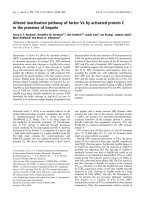
Fig. 1. The effects of tumour necrosis factor-related apoptosis-inducing ligand (TRAIL) on human liver cancer cells QGY-7703. (A) QGY-7703 cells
treated with TRAIL (10 ngÆmL
)1
) for 30 h and the morphological changes associated with apoptosis were photographed under an inverted light
microscope. In contrast to the untreated cells (a) and those treated with medium only (b), viability loss was noted at 30 h in cells treated with TRAIL
(c). (B) Cytotoxicity of TRAIL (1–100 ngÆmL
)1
) on QGY-7703 cells was determined by the MTT assay. (C) Flow cytometry analysis of TRAIL-
induced apoptosis. QGY-7703 cells treated with medium (a), 8 lgÆmL
)1
etoposide (b), 10 ngÆmL
)1
TRAIL (c), or 10 ngÆmL
)1
TRAIL plus
8 lgÆmL
)1
etoposide in the presence (e) or absence (d) of 100 l
M
N-benzyloxycarbonyl-Val-Ala-Asp-fluoromethylketone (zVAD-FMK) for 12 h
were stained with fluorescein isothiocyanate (FITC) conjugated to Annexin V and phosphatidylinositol (PI). Ten-thousand events were analysed.
The percentage of apoptotic cells is indicated in the respective boxes.
2724 L. Miao et al. (Eur. J. Biochem. 270) Ó FEBS 2003
apoptotic induction in the hepatocellular carcinoma cells,
QGY-7703, we treated these cells with eight different agents,
at increasing concentrations, with and without TRAIL,
then analysed apoptosis using the MTT cell death assay and
plotted the results of the cytotoxic effect. The inhibition rate
was calculated as follows:
Inhibition rate ¼½1 Àðabsorbance of drug-treated cells/
absorbance of control cells)]Â100.
Of the eight drugs tested, etoposide was found to be able to
enhance the TRAIL-induced apoptosis in QGY-7703 cells
(Fig. 2A) and was found, by isobolographic analysis, to act
synergistically (Fig. 2B). The remaining seven chemothera-
peutic agents (cisplatin, doxorubincin, 5-fluorouracil,
methotrexate, cytarabine, daunorubicin and cyclophospha-
nide) did not show any synergistic effect when applied
together with TRAIL (data not shown). As shown in
Fig. 2A, only about 25% cell death was detected as a result
of treatment with etoposide alone at a very high concen-
tration (64 lgÆmL
)1
). The QGY-7703 cells were relatively
resistant to etoposide. As shown in Fig. 1C,b, treatment of
QGY-7703 cells with etoposide (8 lgÆmL
)1
) alone for 12 h
caused the induction of early apoptosis in 4.8% of cells,
which is only a slightly increase compared to cells treated
with medium. However, when QGY-7703 cells were treated
with a combination of TRAIL and etoposide, a significant
potentiation of cytotoxicity was achieved. For example,
when combined with TRAIL at a very low concentration of
1ngÆmL
)1
,8lgÆmL
)1
etoposide was sufficient to induce the
same cytotoxicity induced by 64 lgÆmL
)1
etoposide. As
shown in Fig. 1C,d, the percentage of early apoptotic cells
induced by TRAIL (10 ngÆmL
)1
) plus etoposide
(8 lgÆmL
)1
) for 12 h was significantly increased, from 1.4
to 31%. This effect, however, can be prevented by the
general caspase inhibitor, zVAD-FMK (Fig. 1C,e). In the
presence of zVAD-FMK, the percentage of early apoptotic
cells was reduced to 0.59%, indicating complete abrogation
of the synergistic effects resulting from co-treatment with
TRAIL and etoposide. It is also worthy of note that the top
three cell death curves, shown in Fig. 2A become indistin-
guishable when the TRAIL concentration used was
>10 ngÆmL
)1
; this saturation phenomenon could be
explained by the fact that TRAIL-induced apoptosis
involves ligand/receptor (TRAIL/DR4 or TRAIL/DR5)
interaction. Myen et al. and Maccon et al.demonstrated
that cisplatin and etoposide dramatically augment TRAIL-
induced apoptosis in both LNCap and PC3 prostate cancer
cells [29] and malignant breast cells [30]. Taken together, our
results may have provided some clinical significance in the
killing of tumour cells, as combined treatment will help to
achieve more effective therapy with less toxicity by using a
lower dose of chemotherapeutic drugs.
The synergistic effect of augmented apoptosis
is involved in the upregulation of Bax and tBid,
but not of DR4 or DR5
Etoposide is a DNA-damaging agent that affects chromo-
somal DNA [31]. It is well known that the transcription
factor, p53, is essential for the apoptosis caused by DNA
damage [32]. We therefore studied the expression level of
p53 in hepatocellular carcinoma cells, in response to
etoposide or TRAIL. As shown in Fig. 3A, treatment with
etoposide resulted in a time-dependent accumulation of p53,
as expected. Treatment with TRAIL alone did not stimulate
the production of endogenous p53, which is consistent with
the results reported by Ashkenazi & Rieger, who claimed
that TRAIL-induced apoptosis is p53-independent [14,33].
Tumour-suppressor p53 modulates apoptosis through
regulating its target genes involved in apoptosis. We
performed RT–PCR analysis to determine which genes
regulated by p53 could account for the synergistic effect of
etoposide and TRAIL. The QGY-7703 cells were first
treated with etoposide (8 lgÆmL) for the indicated times,
and the subsequent RT–PCR results are shown in Fig. 3B.
The quantitative results from RT–PCR are shown in
Fig. 3C. It is interesting to note that only Bax mRNA
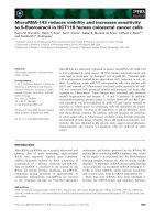
Fig. 2. The synergistic cytotoxicity of tumour necrosis factor-related
apoptosis-inducing ligand (TRAIL) and etoposide on QGY-7703 cells.
(A) The cytotoxicity of TRAIL (1–100 ngÆmL
)1
) and etoposide (8, 16,
32, 64 lgÆmL
)1
), coincubated with QGY-7703 cells for 24 h, was
measured by an MTT assay. All determinations were made in tripli-
cate, and the results are expressed as the mean value ± SD. The data
shown are representative of three independent experiments. Bars, SD.
(B) Synergistic cytotoxicity of TRAIL and etoposide was assessed by
isobolographic analysis.
Ó FEBS 2003 Augmented apoptosis induced by TRAIL and etoposide (Eur. J. Biochem. 270) 2725
was significantly increased at the 12-h time-point and
reached a peak at 18 h, whereas mRNA levels for DR4,
DR5, DcR-2 and Bid remained essentially unchanged at all
time-points tested. We further examined (by Western
blotting) the protein expression of DR4, DR5, DcR-2 and
Bax in QGY-7703 cells treated with and without etoposide.
As shown in Fig. 3D, while no changes in the protein level
for DR4, DR5 or DcR-2 were observed, the expression of
Bax was found to be increased. These Western blot results
are in good accordance with those of RT–PCR (Fig. 3B).
DR4, DR5 and DcR-2 are receptors for TRAIL and their
genes are downstream targets of p53 [34–37]. The upregu-
lation of TRAIL death receptors, especially DR5, by p53
was thought to be the bridge linking chemotherapeutic
agents to the death receptor-elicited apoptotic pathway,
contributing to the synergistic effect of TRAIL and
chemotherapeutic agents [38,39]. In this study, the mRNA
for DR4 and DR5 appeared not to be regulated by p53,
indicating that there exists another mechanism underlying
the synergistic effect. Liu et al. reported that TRAIL and
chemotherapy (such as doxorubicin) can significantly
increase the apoptosis of Mesothelioma cell lines, which is
a highly chemoresistant tumour, but showed no change in
DR5 when treated with chemotherapy [40].
Bax belongs to the Bcl-2 protein family and promotes
apoptosis through increasing the release of cytochrome c
from mitochondria. Recently, Joanna et al. reported that
Bid can be regulated directly by p53 and contributes to
chemosensitivity [41]. Our data demonstrated that Bax, not
Bid, was upregulated upon treatment with etoposide. To
examine whether TRAIL was also able to upregulate Bax,
RT–PCR was performed and the result is shown in Fig. 4A.
No effects on the expression of Bax were observed in QGY-
7703 cells treated with TRAIL. Shi et al. recently reported
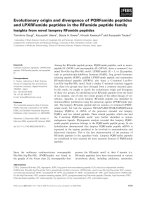
Fig. 3. Etoposide induces the accumulation of p53 and upregulates the mRNA level for Bax, but not for death receptor (DR)4, DR5, decoy receptor-2
(DcR-2) or Bid. (A) Cells were treated with tumour necrosis factor-related apoptosis-inducing ligand (TRAIL) (10 ngÆmL
)1
)oretoposide
(8 lgÆmL
)1
) for 0, 8, 16 and 24 h, and 50 lg of cell lysate was then used to detect the protein level of p53. (B) Total RNA was extracted from QGY-
7703 cells treated with etoposide (8 lgÆmL
)1
) for the indicated time-periods. Two micrograms of total RNA of each sample was used in RT–PCR to
evaluate the level of mRNA for DcR-2, DR4, DR5, Bax and Bid. The housekeeping gene, b-actin, was used as internal control for ensuring that an
equal amount of template was used. PCR was performed under the following conditions: 25 cycles (DR4 and DR5) or 30 cycles (DcR-2, Bax, Bid)
of denaturation at 94 °C for 30 s, annealing at 56 °Cor59 °C (for Bax and Bid) for 30 s, and extension at 72 °C for 90 s. The expected sizes of RT–
PCR amplification fragments are indicated at the right of the panel. The mRNA levels were quantified by densitometry and normalized to basal
b-actin mRNA expression and the results are shown in (C). *, statistically significant result (P < 0.05). (D) Cells were treated with or without
etoposide (8 lgÆmL
)1
) for 16 h and the cell lysates were then used to detect the protein level of DR4, DR5, DcR-2 and Bax. Actin was used as the
loading control.
2726 L. Miao et al. (Eur. J. Biochem. 270) Ó FEBS 2003
that p53 was able to upregulate DR4 and DR5, but not Bax,
in the human lung cancer cells cotreated with TRAIL and
CD437 [38]. Similarly, Kirsten also reported that the DNA-
damaging agent, N-methyl-N¢-nitro-N-nitrosoguanidine
(MNNG), did not affect the expression of Bax [42],
implying that upregulation of Bax upon p53 activation
may be cell-type dependent. Recent studies indicated that
cross-talk might exist between the two pathways by the Bid
protein, a pro-apoptotic protein of the Bcl-2 family. The
BH3 domain of tBid (a cleaved form of Bid) is required to
trigger Bax to release cytochrome c from mitochondria
[43,44]; therefore, tBid is believed to be the linkage protein
between the death receptor pathway and the mitochondrial
pathway. Kim and co-workers have demonstrated that
TRAIL-induced translocation of Bax, subsequent to the
cleavage of Bid, is vital in the TRAIL-induced mitochond-
rial pathway [45]. We therefore examined (by Western
blotting) the protein level of tBid in QGY-7703 cells treated
with TRAIL or etoposide. As shown in Fig. 4B, the level of
tBid was markedly increased with the incubation time when
cells were treated with TRAIL alone, whereas treatment
with etoposide alone did not affect the protein level of tBid.
We also examined the expression of tBid and Bax by
combined treatment of TRAIL and etoposide; these results
are shown in Fig. 4C. The protein levels of both tBid and
Bax increased with the incubation time, and these results are
in agreement with the results shown in Figs 3D and 4B.
Taken together, our results show that the increased level of
active tBid resulting from treatment with TRAIL may link
the death receptor pathway to the mitochondrial pathway
by interaction with upregulated Bax.
Enhanced release of cytochrome
c
and Smac/DIABLO
by combined treatment with TRAIL and etoposide
It is well established that mitochondria play a vital role in
apoptosis and induce cell death by releasing cytochrome c
[46,47] and Smac/DIABLO. Our results also implied that
the mitochondrial pathway is critical for contributing to
the synergistic effect of TRAIL and etoposide in QGY-
7703 cells. To test whether mitochondria are involved in
the co-operative effect of TRAIL and etoposide, we
treated QGY-7703 cells with and without etoposide
(8 lgÆmL
)1
) in the presence or absence of TRAIL
(10 ngÆmL
)1
) for 16 h to determine the release of
cytochrome c and Smac/DIABLO. As expected, little, if
any, cytochrome c and Smac/DIABLO were detected in
the cytoplasm when treated with TRAIL alone (Fig. 5A),
as there was no increase in the expression of Bax.
Treatment with etoposide alone (Fig. 5A) effected some
release of cytochrome c and Smac/DIABLO. Further-
more, both cytochrome c and Smac/DIABLO showed
a marked release from the mitochondria by combined
treatment with TRAIL and etoposide (Fig. 5A). Similarly
to cytochrome c and Smac/DIABLO, the expression of
Bax was also notably increased by cotreatment with
TRAIL and etoposide compared with that of TRAIL or
etoposide alone (Fig. 5A). Smac/DIABLO promotes the
activation of caspases, such as procaspase-9 and caspase-3,
by binding to the inhibitor of apoptosis proteins and thus
disrupts linkage of the caspase–inhibitor of apoptosis
proteins complex during apoptosis [48–50]. Recently,
Deng et al. reported that TRAIL-induced apoptosis
requires Bax-dependent mitochondrial release of Smac/
DIABLO [51]. They have shown that the release of Smac/
DIABLO is required to remove the inhibitory effect of
X-linked inhibitor of apoptosis protein (XIAP) and allow
apoptosis to proceed, and thus mediates the contribution
of the mitochondrial pathway to death receptor-mediated
apoptosis. To our knowledge, ours is the first report to
demonstrate that combined treatment of TRAIL and
etoposide results in an enhanced release of Smac/DIAB-
LO in the hepatocellular carcinoma cell system. zVAD-
FMK is the general caspase inhibitor and used to prevent
caspase-dependent apoptosis. We demonstrated that
zVAD-FMK is able to prevent the early apoptosis induced
by TRAIL plus etoposide (Fig. 1C,e). To investigate
whether the release of cytochrome c and Smac/DIABLO,
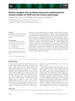
Fig. 4. Activation of Bid in response to tumour necrosis factor-related
apoptosis-inducing ligand (TRAIL), but not to etoposide. (A) Total
RNA was extracted from QGY-7703 cells treated with TRAIL
(10 ngÆmL
)1
) for the indicated time-periods and then RT–PCR was
performed to evaluate the levels of mRNA for Bax. The housekeeping
gene, b-actin, was used as an internal control for ensuring that an equal
amount of template was used. (B) and (C) QGY-7703 cells were
exposed to TRAIL (10 ngÆmL
)1
) alone, etoposide (8 lgÆmL
)1
)alone,
or TRAIL and etoposide together, for the indicated time-periods and
the cell lysates were subject to Western blot analysis for detection of
truncated Bid (tBid) or Bax.
Ó FEBS 2003 Augmented apoptosis induced by TRAIL and etoposide (Eur. J. Biochem. 270) 2727
or the expression of Bax, could also be inhibited by
zVAD-FMK, QGY-7703 cells were exposed to TRAIL
plus etoposide in the presence or absence of zVAD-FMK.
Whole-cell protein lysates or cytosolic protein fractions
were subject to Western blot analysis. As shown in
Fig. 5B, the release of cytochrome c and Smac/DIABLO
were significantly decreased in the presence of zVAD-
FMK. However, the decrease of Bax in the presence of
zVAD-FMK was not as marked as that of cytochrome c
and Smac/DIABLO when compared with Bax expression
in the absence of zVAD-FMK. The exact mechanism
underlying this discrepancy remains unclear. Nevertheless,
our results are similar to those reported by Adrain et al.
[52]. They have proposed a model for explaining how the
caspase inhibitor, zVAD-FMK, could inhibit the release
of cytochrome c and Smac/DIABLO from mitochondria.
They claimed that release of Smac/DIABLO and cyto-
chrome c requires downstream caspase activation. Based
on this model, the results shown in Fig. 5B are plausible,
as zVAD-FMK reduced the release of cytochrome c and
Smac/DIABLO by inhibiting the downstream caspase
activation. Bax is upstream of cytochrome c and Smac/
DIABLO, therefore zVAD-FMK had little effect on its
expression. These results also confirmed that mitochondria
might play a key role in contributing to the synergistic
effect of TRAIL and etoposide in QGY-7703 cells.
Involvement of caspase activation in the enhancement
of TRAIL-induced apoptosis by etoposide
Caspases play key roles in apoptosis triggered by various
pro-apoptotic signals. To identify which caspase is
involved in the process of apoptosis of QGY-7703 cells,
and whether the activation of caspase is enhanced during
the combined treatment with TRAIL and etoposide,
QGY-7703 cells were incubated with TRAIL
Fig. 6. The enhanced cleavage of caspases by combined treatment with
tumour necrosis factor-related apoptosis-inducing ligand (TRAIL) and
etoposide. Total cellular lysates were extracted from QGY-7703 cells
treated with etoposide (8 lgÆmL
)1
), with or without TRAIL
(10 ngÆmL
)1
), for 24 h. Western blot analysis was performed to assess
the processing of caspase-7, -9, -3, -8 and poly ADP-ribose polymerase
(PARP). Human actin was used as the loading control. +, treated;
–, untreated.
Fig. 5. Increased mitochondrial release of cytochrome c and second
mitochondria-derived activator of caspase (Smac)/DIABLO during the
synergistic induction of apoptosis by tumour necrosis factor-related
apoptosis-inducing ligand (TRAIL) and etoposide. (A) Cell lysates were
isolated from QGY-7703 cells treated with etoposide (8 lgÆmL
)1
), with
or without TRAIL (10 ngÆmL
)1
), for 16 h and assessed for Bax
expression (cell lysate) and the contents of released cytochrome c and
Smac/DIABLO (cytosolic fractions) by immunoblot analysis using
respective antibodies. Actin was used as the loading control. (B) QGY-
7703 cells were exposed to the combination of etoposide (8 lgÆmL
)1
)
and TRAIL (10 ngÆmL
)1
), with or without the caspase inhibitor
N-benzyloxycarbonyl-Val-Ala-Asp-fluoromethylketone (zVAD-FMK)
(100 l
M
), for 16 h and both the cell lysate and the cytosolic fractions
were subject to Western blot analysis for detection of the expression of
Bax (cell lysate) and the release of cytochrome c and Smac/DIABLO
(cytosolic fractions). Actin was used as the loading control. +, treated;
–, untreated.
2728 L. Miao et al. (Eur. J. Biochem. 270) Ó FEBS 2003
(10 ngÆmL
)1
) or etoposide (8 lgÆmL
)1
), or the combina-
tion of both, for 24 h (Fig. 6). Etoposide alone and
TRAIL alone slightly activated the initiators caspase-8
and -9, but their activation was much more enhanced
when cells were cotreated with TRAIL plus etoposide.
Similar results were also obtained for effector caspases,
caspase-3 and -7, as seen in Fig. 6. The activation of
caspase-3 and -7 was further confirmed by the accelerated
cleavage of PARP, a direct downstream substrate of
caspase-3 and -7. As shown in Fig. 6, cotreatment with
TRAIL plus etoposide resulted in an enhanced cleavage
of PARP compared to that obtained by treatment with
either TRAIL or etoposide alone. We noted that treat-
ment with etoposide alone also resulted in some cleavage
of procaspase-8; this observation has been documented
previously [53,54], where it was concluded that caspase-8
can be processed by anticancer drugs, independently of
death receptors. In general, activation of the caspase
cascade requires both initiator caspases such as caspase-8
and -9, and effector caspases, such as caspase-3 and -7
[55]. The death ligands and the chemotherapeutic agents
are two distinct classes of signals used to induce apoptosis
and activate the caspase cascade. Caspase-8 is known as
the initiator caspase in the death receptor signal pathway,
while caspase-9 is associated more with the mitochondrial
pathway, which is activated by many chemotherapeutic
drugs [56]. Caspase-3 and -7 are the major effector
caspases and act downstream of caspase-8 and -9. Our
results have demonstrated that cotreatment with TRAIL
and etoposide activated both the caspase-8- and -9-
mediated apoptotic pathway, resulting in augmentation of
the apoptotic death effect. Shi and co-workers have also
reported similar results in human lung cancer cells treated
with TRAIL and CD437 [38]. Taken together, we propose
a hypothetical model to illustrate the possible mechanism
by which TRAIL and etoposide synergistically augment
apoptosis in QGY-7703 cells, and the detailed descriptions
are outlined in Fig. 7.
Acknowledgements
This research was supported by the Key Project Fund (KSCX2-2-01-
004), a special grant (to M. W.) from the Chinese Academy of Sciences,
grants from the National Natural Science Foundation of China
(30121001 and 90208027) and a 973 grant (2002CB713700) from the
Ministry of Science and Technology of China.
References
1. Pitti, R.M., Marsters, S.A., Ruppert, S., Donahue, C.J., Moore,
A. & Ashkenazi, A. (1996) Induction of apoptosis by Apo-2
ligand, a new member of the tumor necrosis factor cytokine
family. J. Biol. Chem. 271, 12687–12690.
2. Wiley, S.R., Schooley, K., Smolak, P.J., Din, W.S., Huang, C P.,
Nicholl,J.K.,Sutherland,G.R.,Smith,T.D.,Rauch,C.,Smith,
C.A. & Goodwin, R.G. (1995) Identification and characterization
of a new member of the TNF family that induces apoptosis.
Immunity 3, 673–682.
3. French, L.E. & Tschopp, J. (1999) The TRAIL to selective tumor
death. Nat. Med. 5, 146–147.
4. Walczak, H., Miller, R.E., Ariail, K., Gliniak, B., Griffith, T.S.,
Kubin, M., Chin, W., Jones, J., Woodward, A., Le, T., Smith, C.,
Smolak, P., Goodwin, R.G., Rauch, C.T., Schuh, J.C. & Lynch,
D.H. (1999) Tumoricidal activity of tumor necrosis factor-related
apoptosis-inducing ligand in vivo. Nat. Med. 5, 157–163.
5. Ashkenazi, A., Pai, R.C., Fong, S., Leung, S., Lawrence, D.A.,
Marsters,S.A.,Blackie,C.,Chang,L.,McMurtrey,A.E.,Hebert,
A., DeForge, L., Koumenis, I.L., Lewis, D., Harris, L., Bussiere,
J., Koeppen, H., Shahrokh, Z. & Schwall, R.H. (1999) Safety and
antitumor activity of recombinant soluble Apo2 ligand. J. Clin.
Invest. 104, 155–162.
Fig. 7. Hypothetical model for illustrating the
proposed apoptosis pathways in QGY-7703
cells cotreated with etoposide and tumour nec-
rosis factor-related apoptosis-inducing ligand
(TRAIL). The double line represents the
cytoplasmic membrane. The broken lines
indicate that etoposide (or p53) has no effect
on the expression of death receptor (DR)4,
DR5, decoy receptor-2 (DcR-2) and Bid. The
mitochondrial pathway is symbolized by
yellow arrows and the death receptor pathway
by blue arrows. The etoposide-activated mit-
ochondrial pathway and the TRAIL-triggered
DR pathway are converged on the mito-
chondria by the interactions of Bax with
truncated Bid (tBid) which, in turn, amplifies
the release of both cytochrome c and second
mitochondria-derived activator of caspase
(Smac)/DIABLO to activate downstream
caspases, resulting in the augmented apoptosis
observed in QGY-7703 hepatocellular carci-
noma cells.
Ó FEBS 2003 Augmented apoptosis induced by TRAIL and etoposide (Eur. J. Biochem. 270) 2729
6. Pan, G., O’Rourke, K., Chinnaiyan, A.M., Gentz, R., Ebner, R.,
Ni, J. & Dixit, V.M. (1997) The receptor for the cytotoxic ligand
TRAIL. Science 276, 111–113.
7. Pan,G.,Ni,J.,Wei,Y F.,Yu,G.,Gentz,R.&Dixit,V.M.(1997)
An antagonist decoy receptor and a death domain-containing
receptor for TRAIL. Science 277, 815–818.
8. Walczak, H., Degli-Esposti, M.A., Johnson, R.S., Smolak, P.J.,
Waugh, J.Y., Boiani, N., Timour, M.S., Gerhart, M.J., Schooley,
K.A.,Smith,C.A.,Goodwin,R.G.&Rauch,C.T.(1997)TRAIL-
R2: a novel apoptosis-mediating receptor for TRAIL. EMBO J.
16, 5386–5397.
9. Degli-Esposti, M.A., Smolak, P.J., Walczak, H., Waugh, J.,
Huang, C P., DuBose, R.F., Goodwin, R.G. & Smith, C.A.
(1997) Cloning and characterization of TRAIL-R3, a novel
member of the emerging TRAIL receptor family. J. Exp. Med.
186, 1165–1170.
10. Sheridan, J.P., Marsters, S.A., Pitti, R.M., Gurney, A., Skubatch,
M., Baldwin, D., Ramakrishnan, L., Gray, C.L., Baker, K.,
Wood, W.I., Goddard, A.D., Godowski, P. & Ashkenazi, A.
(1997) Control of TRAIL-induced apoptosis by a family of sig-
naling and decoy receptors. Science 277, 818–821.
11. Schneider, P., Bodmer, J.L., Thome, M., Hofmann, K., Holler, N.
& Tschopp, J. (1997) Characterization of two receptors for
TRAIL. FEBS Lett. 416, 329–334.
12.Pan,G.,Ni,J.,Yu,G.,Wei,Y.F.&Dixit,V.M.(1998)
TRUNDD, a new member of the TRAIL receptor family that
antagonizes TRAIL signalling. FEBS Lett. 424, 41–45.
13. Marsters, S.A., Sheridan, J.P., Pitti, R.M., Huang, A., Skubatch,
M.,Baldwin,D.,Yuan,J.,Gurney,A.,Goddard,A.D.,
Godowski, P. & Ashkenazi, A. (1997) A novel receptor for
Apo2L/TRAIL contains a truncated death domain. Curr. Biol. 7,
1003–1006.
14. Ashkenazi, A. & Dixit, M.V. (1998) Death receptors: signaling
and modulation. Science 281, 1305–1308.
15. Schneider, P., Thome, M., Burns, K., Bodmer, J.L., Hofmann, K.,
Kataoka, T., Holler, N. & Tschopp, J. (1997) TRAIL receptors 1
(DR4) and 2 (DR5) signal FADD-dependent apoptosis and
activate NF-B. Immunity 7, 831–836.
16. Tadaaki, M. & John, C.R. (2001) A GTP-binding adapter protein
couples TRAIL receptors to apoptosis-inducing proteins. Nature
2, 493–500.
17. Sheikh, M.S. & Albert, J.F. (2000) Role of p53 family members in
apoptosis. J. Cell. Physiol. 182, 171–181.
18. Timothy, F.B. & Wafik, S. (1999) The p53 pathway and apoptosis.
J. Cell. Physiol. 181, 231–239.
19. Alain, G.Z., Karin, R., Jennifer, B., Martin, W. & Martin, H.
(2000) New insights into p53 regulation and gene therapy for
cancer. Biochem. Pharmacol. 60, 1153–1163.
20. Nagane, M., Pan, G., Weddle, J.J., Dixit, V.M., Cavenee, W.K. &
Huang, H.J. (2000) Increased death receptor 5 expression by
chemotherapeutic agents in human gliomas causes synergistic
cytotoxicity with tumor necrosis factor-related apoptosis-inducing
ligand in vitro and in vivo. Cancer Res. 15, 847–853.
21. Gibson, S.B., Oyer, R., Spalding, A.C., Anderson, S.M. & John-
son, G.L. (2000) Increased expression of death receptors 4 and 5
synergizes the apoptosis response to combined treatment with
etoposide and TRAIL. Mol. Cell. Biol. 20, 205–212.
22. Raymond, D.M. & Wafik, S. (2000) p53-independent upregula-
tion of KILLER/DR5 TRAIL receptor expression by glucocorti-
coids and interferon-c. Exp. Cell Res. 262, 154–169.
23. Nimmanapalli, R., Perkins, C.L., Orlando, M., O’Bryan, E.,
Nguyen, D. & Bhalla, K.N. (2001) Pretreatment with paclitaxel
enhances Apo-2 ligand/tumor necrosis factor-related apoptosis-
inducing ligand-induced apoptosis of prostate cancer cells by
inducing death receptors 4 and 5 protein levels. Cancer Res. 61,
759–763.
24. LeBlanc, H., Lawrence, D., Varfolomeev, E., Totpal, K., Morlan,
J., Schow, P., Fong, S., Schwall, R., Sinicropi, D. & Ashkenazi, A.
(2002) Tumor-cell resistance to death receptor-induced apoptosis
through mutational inactivation of the proapoptotic Bcl-2
homolog Bax. Nat. Med. 8, 274–281.
25. Luo, X., Budihardjo, I., Zuo, H., Slaughter, C. & Want, X. (1998)
Bid, a Bcl-2 interacting protein, mediates cytochrome c release
from mitochondria in response to activation of cell surface death
receptors. Cell 94, 481–490.
26. Berenbaum, M.C. (1978) A method for testing for synergy with
any number of agents. J. Infect. Dis. 137, 122–130.
27. Samuel, C., Cheng, S., Dan, L. & Yong, X. (2001) Taxol induced
Bcl-2 protein phosphorylation in human hepatocellular carcinoma
QGY-7703 cell line. Cell Biol. Int. 25, 261–265.
28. Jo, M., Kim, T.H., Seol, D.W., Esplen, J.E., Dorko, K., Billiar,
T.R. & Strom, S.C. (2000) Apoptosis induced in normal human
hepatocytes by tumor necrosis factor-related apoptosis-inducing
ligand. Nat. Med. 6, 564–567.
29. Munshi, A., McDonnell, T.J. & Meyn, R.E. (2002) Chemother-
apeutic agents enhance TRAIL-induced apoptosis in prostate
cancer cells. Cancer Chemother. Pharmacol. 50, 46–52.
30. Maccon, M.K., Ettenberg, S.A., Nau, M.M., Russell, E.K. &
Lipkowitz, S. (1999) Chemotherapy augments TRAIL-induced
apoptosis in breast cell lines. Cancer Res. 59, 734–741.
31. Scott, H.K. & William, C.E. (2000) Induction of apoptosis by
cancer chemotherapy. Exp. Cell Res. 256, 42–49.
32. Karpinich, N.O., Tafani, M., Rothman, R.J., Russo, M.A. &
Farber, J.L. (2002) The course of etoposide-induced apoptosis
from damage to DNA and p53 activation to mitochondrial release
of cytochrome c. J. Biol. Chem. 277, 16547–16552.
33. Rieger,J.,Naumann,U.,Glaser,T.,Ashkenazi,A.&Weller,M.
(1998) Apo2 ligand: a novel lethal weapon against malignant
glioma? FEBS Lett. 427, 124–128.
34. Miyashita, T. & Reed, J.C. (1995) Tumor suppressor p53 is a
direct transcriptional activator of the human bax gene. Cell 80,
293–299.
35. Wu, G.S., Burns, T.F., McDonald, E.R., Jiang, W., Meng, R.,
Krantz, I.D., Kao, G., Gan, D.D., Zhou, J.Y., Muschel, R.,
Hamilton, S.R., Spinner, N.B., Markowitz, S., Wu, G. &
El-Deiry, W.S. (1997) KILLER/DR5 is a DNA damage-
inducible p53-regulated death receptor gene. Nat. Genet. 17,
141–143.
36. Baoxiang, G., Ping, Y., Gary, L.C. & Sun, S.Y. (2001) Evidence
that the death receptor DR4 is a DNA damage-inducible,
p53-regulated gene. J. Cell. Physiol. 188, 98–105.
37. Raymond, D.M., McDonald, E.R., Sheikh, M.S., Albert, J.F. &
Wafik, S. (2000) The TRAIL decoy receptor TRUNDD (DcR2,
TRAIL-R4) is induced by adenovirus-p53 overexpression and can
delay TRAIL-, p53-, and KILLER/DR5-dependent colon cancer
apoptosis. Mol. Ther. 1, 130–144.
38. Shi, Y.S., Yue, P., Hong, W.K. & Lotan, R. (2000) Augmentation
of tumor necrosis factor-related apoptosis-inducing ligand
(TRAIL)-induced apoptosis by the synthetic retinoid 6-[3-(1-
Adamantyl)-4-hydroxyphenyl]-2-naphthalene carboxylic acid
(CD437) through up-regulation of TRAIL receptors in human
lung cancer cells. Cancer Res. 60, 7149–7155.
39. Sheikh, M.S., Burns, T.F., Huang, Y., Wu, G.S., Amundson, S.,
Brooks, K.S., Fornace, A.J. Jr, & el-Deiry, W.S. (1998) p53-
dependent and -independent regulation of the death receptor
KILLER/DR5 gene expression in response to genotoxic stress and
tumornecrosisfactoralpha.Cancer Res. 58, 1593–1598.
40. Liu, W., Bodle, E., Chen, J.Y., Gao, M., Rosen, G.D. &
Broaddus, V.C. (2001) Tumor necrosis factor-related apoptosis-
inducing ligand and chemotherapy cooperate to induce apoptosis
in mesothelioma cell lines. Am.J.Respir.CellMol.Biol.25,
111–118.
2730 L. Miao et al. (Eur. J. Biochem. 270) Ó FEBS 2003
41. Sax,J.K.,Fei,P.,Murphy,M.E.,Bernhard,E.,Korsmeyer,S.J.&
Walfik, S.E D. (2002) BID regulation by p53 contributes to
chemosensitivity. Nat. Cell Biol. 4, 842–849.
42. Kirsten Ochs and Bernd, K. (2000) Apoptosis induced by DNA
damage: O
6–
Methylguanine is Bcl-2 and caspase-9/3
regulated and Fas/caspase-8 independent. Cancer Res. 60,
5815–5824.
43. Korsmeyer,S.J.,Wei,M.C.,Saito,M.,Weiler,S.,Oh,K.J.&
Schlesinger, P.H. (2000) Pro-apoptotic cascade activates BID,
which oligomerizes BAK or BAX into pores that result in the
release of cytochrome c. Cell Death Differ. 7, 1166–1173.
44. Li, H., Zhu, H., Xu, C.J. & Yuan, J. (1998) Cleavage of BID by
caspase8 mediated the mitochondria in response to activation of
cell surface death receptors. Cell 94, 491–450.
45. Kim,J.Y.,Kim,Y.H.,Chang,I.,Kim,S.,Pak,Y.K.,Oh,B.H.,
Yagita, H., Jung, Y.K., Oh, Y.J. & Lee, M.S. (2002) Resistance of
mitochondrial DNA-deficient cells to TRAIL: role of Bax in
TRAIL-induced apoptosis. Oncogene 21, 3139–3148.
46. Green, D.R. & Reed, J.C. (1998) Mitochondria and apoptosis.
Science 281, 1309–1312.
47. Ruth,M.K.,Ella,B W.,Douglas,R.G.&Donald,D.N.(1997)
The release of cytochrome c from mitochondria: a primary site for
Bcl-2 regulation of apoptosis. Science 275, 1132–1136.
48. Li, P., Nijhawan, D., Budihardjo, I., Srinivasula, S.M., Ahmad,
M.,Alnemri,E.S.&Wang,X.(1997)Cytochromec and dATP-
dependent formation of Apaf-1/caspase-9 complex initiates an
apoptotic protease cascade. Cell 91, 479–489.
49. Dn,C.,Fang,M.,Li,Y.&Wang,X.(2000)Smac,amitochon-
drial protein that promotes cytochrome c-dependent caspase
activation by eliminating IAP inhibition. Cell 102, 33–42.
50. Ekert, P.G., Silke, J., Hawkins, C.J., Verhagen, A.M. & Vaux,
D.L. (2001) DIABLO promotes apoptosis by removing MIHA/
XIAP from processed caspase9. J. Cell Biol. 152, 483–490.
51. Yibin, D., Yahong, L. & Xiang, W.W. (2002) TRAIL-induced
apoptosis requires Bax-dependent mitochondrial release of Smac/
DIABLO. Genes Dev. 16, 33–45.
52. Adrain, C., Creagh, E.M. & Martin, S.J. (2001) Apoptosis-asso-
ciated release of Smac/DIABLO from mitochondria requires
active caspases and is blocked by Bcl-2. EMBO J. 20, 6627–6636.
53. Engel, I.H., Strpczynska, A., Stroh, C., Lauber, K., Berg, C.,
Schwenzer, R., Wajant, H., Janicke, R.U., Porter, A.G., Belka, C.,
Gregor, M., Schulze, O.K. & Wesselborg, S. (2000) Caspase-8/
FLICE functions as an executioner caspase in anticancer drug-
induced apoptosis. Oncogene 19, 4563–4573.
54. Wesselborg, S., Engel, I.H., Rossmann, E., Los, M. & Schulze,
O.K. (1999) Anticancer drugs induce caspase-8/FLICE activation
and apoptosis in the absence of CD95 receptor/ligand interation.
Blood 93, 3053–3063.
55. Nancy, A., Thornberry, N.A. & Yuri, L. (1998) Caspases: enemies
within. Science 281, 1312–1316.
56. Sun, X.M., MacFarlane, M., Zhuang, J., Wolf, B.B., Green, D.R.
& Cohen, G.M. (1999) Distinct caspase cascades are initiated in
receptor-mediated and chemical-induced apoptosis. J. Biol. Chem.
274, 5053–5060.
Ó FEBS 2003 Augmented apoptosis induced by TRAIL and etoposide (Eur. J. Biochem. 270) 2731