Báo cáo khoa học: p53-induced inhibition of protein synthesis is independent of apoptosis pdf
Bạn đang xem bản rút gọn của tài liệu. Xem và tải ngay bản đầy đủ của tài liệu tại đây (340.48 KB, 11 trang )
p53-induced inhibition of protein synthesis is independent
of apoptosis
Constantina Constantinou
1
, Martin Bushell
1
*, Ian W. Jeffrey
1
, Vivienne Tilleray
1
, Matthew West
1
,
Victoria Frost
2
†, Jack Hensold
3
and Michael J. Clemens
1
1
Translational Control Group, Department of Basic Medical Sciences, St George’s Hospital Medical School, Cranmer Terrace,
London;
2
Biochemistry Group, School of Biological Sciences, University of Sussex, Falmer, Brighton, UK;
3
Department of
Hematology and Oncology, Case Western Reserve University and the Veterans Administration, Cleveland, Ohio, USA
Activation of a temperature-sensitive form of p53 in murine
erythroleukaemia cells results in a rapid impairment of
protein synthesis that precedes inhibition of cell proliferation
and loss of cell viability by several hours. The inhibition of
translation is associated with specific cleavages of polypep-
tide chain initiation factors eIF4GI and eIF4B, a pheno-
menon previously observed in cells induced to undergo
apoptosis in response to other stimuli. Although caspase
activity is enhanced in the cells in which p53 is activated, both
the effects on translation and the cleavages of the initiation
factors are completely resistant to inhibition of caspase
activity. Moreover, exposure of the cells to a combination of
the caspase inhibitor z-VAD.FMK and the survival factor
erythropoietin prevents p53-induced cell death but does not
reverse the inhibition of protein synthesis. We conclude that
the p53-regulated cleavages of eIF4GI and eIF4B, as well as
the overall inhibition of protein synthesis, are caspase-inde-
pendent events that can be dissociated from the induction of
apoptosis per se.
Keywords: caspases; erythroleukaemia; p53; protein synthe-
sis; temperature-sensitive mutants.
The tumour suppressor protein p53 is a key regulator of
both cell cycle progression and cell death by apoptosis [1–5].
Inactivating mutations of p53 have been found with high
frequency in a broad spectrum of tumours and the
inactivation of p53 is central to the transforming function
of several viral oncoproteins [6–8]. Primarily, p53 functions
as a transcription factor controlling expression of genes that
affect cell proliferation, induce DNA repair or regulate cell
survival [9–12]. Expression of p53 in p53-negative cell lines
induces a cell cycle block and in many cases results in cell
death by apoptosis [1,13]. p53 has also been demonstrated
to control the activity of RNA polymerases I and III,
suggesting that p53 regulates the synthesis of ribosomes and
tRNAs [14]. Furthermore, the tumour suppressor protein
has been found in association with ribosomes [15,16] and
has been shown to have an effect on the translation of
specific mRNAs, such as those encoding cdk4, fibroblast
growth factor (FGF) 2 and p53 itself [14,17–21].
Recently, we reported that p53 down-regulates overall
translation at the level of polypeptide chain initiation [22]. In
those studies we utilized a murine erythroleukaemia (MEL)
cell line expressing a temperature-sensitive p53 mutant
(Val135) [23] and showed that activation of p53 by placing
the cells at 32 °C caused a rapid decrease in the overall rate
of protein synthesis. However it has not been established
whether this translational inhibition is an early part of the
programme of induced cell death or whether it is associated
with the block to cell cycle progression mediated by the
activation of p53. There are strong precedents for the
former as several studies have shown that the induction of
apoptosis by other agents is accompanied by a substantial
down-regulation of translation and the caspase-mediated
cleavage of certain polypeptide chain initiation factors
[24–29].
In the work described here we have employed the same
MEL cell system to address some of these issues. Careful
comparisons of the kinetics of translational down-regula-
tion vs. the inhibition of cell cycle progression and
induction of apoptosis show that the effect of p53
activation on protein synthesis is an early event that
precedes both overt inhibition of cell proliferation and the
loss of cell viability. We show that although caspase
Correspondence to M. J. Clemens, Department of Basic Medical
Sciences, St George’s Hospital Medical School, Cranmer Terrace,
London SW17 0RE, UK.
Fax: + 44 (0)20 87252992, Tel.: + 44 (0)20 8725 5762,
E-mail:
Abbreviations: 4E-BP1, eIF4E binding protein 1; Ac-DEVD-AMC,
acetyl-Asp-Glu-Val-Asp7-amino-4-methylcoumarin; Ac-IETD-
AMC, acetyl-Ile-Glu-Thr-Asp7-amino-4-methylcoumarin;
Ac-LEHD-AMC, acetyl-Leu-Glu-His-Asp7-amino-4-methylcou-
marin; eIF, eukaryotic initiation factor; Epo, erythropoietin; MEL,
murine erythroleukaemia; mTOR, mammalian target of rapamycin;
PARP, poly(ADP-ribose) polymerase; RFU, relative fluorescence
units; TNF-a, tumour necrosis factor a; TRAIL, tumour necrosis
factor-related apoptosis-inducing ligand; z-VAD.FMK,
benzyloxycarbonyl-Val-Ala-Asp-fluoromethylketone.
*Present address: Department of Biochemistry, University of
Leicester, University Road, Leicester, LE1 7RH, UK.
Present address: School of Biological Sciences, University of
Manchester, 2.205 Stopford Building, Oxford Road,
Manchester M13 9PT, UK.
(Received 11 March 2003, revised 21 May 2003,
accepted 27 May 2003)
Eur. J. Biochem. 270, 3122–3132 (2003) Ó FEBS 2003 doi:10.1046/j.1432-1033.2003.03687.x
activities increase within a few hours of activating p53, and
specific proteolytic cleavages of some polypeptide chain
initiation factors are observed, the factor cleavages do
not depend on caspase activity. Clonogenicity assays have
established that the cells do not become irreversibly
committed to apoptosis until several hours after the initial
inhibition of translation. Moreover, conditions that block
apoptosis do not prevent the p53-induced translational
down-regulation. Our results are consistent with a mech-
anism whereby the p53-mediated inhibition of protein
synthesis in MEL cells is at least partially mediated by
initiation factor cleavages. However caspase activity is not
required for these cleavages and the down-regulation of
translation can be dissociated from the p53-induced
apoptotic programme.
Materials and methods
Cell culture conditions
The Val135 and Pro190 MEL cell lines were obtained from
S. Benchimol [30] and were grown in stationary suspension
culture in DMEM medium supplemented with glutamine
(300 mgÆL
)1
) and 10% (v/v) fetal bovine serum in a 5%
CO
2
atmosphere at 38 °C. Under these conditions the cells
had a doubling time of approximately 12 h. Cultures were
maintained at densities between 2 and 8 · 10
5
cells per
milliliter. Continued expression of p53 was assured by
weekly selection of the cells in G418 (200 lgÆmL
)1
). For
activation of p53 in the Val135 cells the cultures were
transferred to 32 °C for the times indicated. The control
Pro190 cells were treated similarly. Where indicated, the
cells were treated with erythropoietin (Epo) and caspase
inhibitors at the concentrations described in Table 2 and
legends to Figs 4–6.
Analysis of cell proliferation, the cell cycle and clonal
growth potential
Cells were counted in triplicate using a haemocytometer and
cell viability was determined by trypan blue exclusion. For
cell cycle analysis cells were examined by flow cytometry as
described in [31]. Cells that had been grown at 38 °Cor
incubated at 32 °C for various periods of time (10
7
cells per
sample) were centrifuged at 1000 g for 5 min and washed
three times in 5 mL NaCl/P
i
. The pellets were resuspended
in approximately 500 lLofNaCl/P
i
, 5 mL of cold ethanol
were added and the cells were fixed at 4 °Covernight.
The fixed cells were washed in NaCl/P
i
and stained with
propidium iodide (500 lgÆmL
)1
). After treatment with
boiled RNAse A the cells were analyzed on a FACS flow
cytometer (Beckton Dickinson).
To determine the ability of the cells to proliferate clonally
after exposure to the p53 permissive temperature, Pro190
and Val135 cells were diluted 1 · 10
5
such that the final
concentration was three cells per milliliter. Aliquots of
100 lL were added to the wells of 96-well microtiter plates
(giving an average of one cell for every three wells). The
plates were incubated for various times up to 72 h at 32 °C
andthenreturnedto38°C. The wells were observed
microscopically after a total of 10 days and were scored for
the number of clones that had proliferated.
Measurement of protein synthesis rates
Overall rates of protein synthesis were measured by pulse-
labeling intact cells for up to 1 h with 10–15 lCiÆmL
)1
of
[
35
S]methionine (in the presence of the normal level of
methionine in the cell culture medium). The cells were
centrifuged briefly at 1000 g, washed once in cold NaCl/P
i
,
dissolved in 0.3
M
NaOH and precipitated with 10%
trichloroacetic acid in the presence of 0.5 mg bovine serum
albumin carrier protein. Precipitates were harvested on GF/
C filters under suction and washed with 5% trichloroacetic
acid and industrial methylated spirit. The acid-insoluble
radioactivity was determined by scintillation counting.
Preparation of cell extracts and analysis
by immunoblotting
Cytoplasmic extracts were prepared for immunoblotting by
washing the cells in NaCl/P
i
and lyzing them in a buffer
containing a cocktail of protease and protein phosphatase
inhibitors [24]. The extracts were analyzed by SDS gel
electrophoresis using equal amounts of protein in each lane
of the gel (3–10 lg protein per sample). After transfer of the
proteins to poly(vinylidene difluoride) membranes the blots
were blocked and incubated with the appropriate primary
antibodies against polypeptide chain initiation factors
eIF4B and eIF4GI. The blots were developed with alkaline
phosphatase-linked secondary antibodies using nitroblue
tetrazolium as the substrate [24], or with horseradish
peroxidase-linked secondary antibodies followed by
enhanced chemiluminescence. As a positive control for the
effects of z.VAD-FMK, the same initiation factors were
also examined in extracts from Jurkat cells treated with
an agonistic anti-Fas (CD95) antibody, as described
previously [32,33].
Measurements of apoptosis
The progress of apoptosis in the MEL cells was assessed
by measuring the activities of caspases-3, -8 and -9 in cell
extracts. At appropriate times after incubation at 32 °C, in
the absence or presence of z.VAD-FMK, aliquots of 10
7
cells were washed with NaCl/P
i
, resuspended in 1 mL cell
lysis buffer (10 m
M
Hepes, pH 7.3, 2 m
M
EDTA, 0.1%
NP-40, 5 m
M
dithiothreitol, 1 m
M
phenylmethanesulfonyl
fluoride, 10 lgÆmL
)1
pepstatin A, 20 lgÆmL
)1
leupeptin,
10 lgÆmL
)1
aprotinin) and incubated on ice for 10–15 min.
After centrifugation of the extracts at 10 000 g for 1 min at
4 °C the supernatants were frozen at )80 °C. Caspase
activities using fluorogenic substrates were determined in a
Packard Fusion microplate reader. Twenty microliters of
each cell extract was incubated with 200 lL of reaction
buffer [100 m
M
Hepes, pH 7.3, 20% (v/v) glycerol, 0.5 m
M
EDTA, 5 m
M
dithiothreitol] and 2 lL of substrate for
caspases-3, -8 or -9 (Ac-DEVD-AMC, Ac-IETD-AMC or
Ac-LEHD-AMC, respectively) (Biosource International),
each at 5 m
M
. Reactions were incubated at 37 °Cfor1h
and the product was quantified by fluorescence using an
excitation wavelength of 380 nm and an emission wave-
length of 460 nm. Protein concentrations were determined
and caspase activities expressed in relative fluorescence
units (RFU) per microgram of protein. Apoptosis was
Ó FEBS 2003 Regulation of protein synthesis by p53 (Eur. J. Biochem. 270) 3123
also assessed by the cleavage of the caspase substrates
poly(ADP-ribose) polymerase (PARP) and p27
KIP1
,using
immunoblotting procedures as described elsewhere
[24,34,35].
Results
Inhibition of protein synthesis and cell proliferation
following activation of p53
Activation of the temperature-sensitive Val135 p53 mutant
in MEL cells (or of the equivalent Val138 mutant in human
cells) can be achieved by reducing the incubation tempera-
ture from 38 to 32 °C and results in inhibition of cell
proliferation and subsequent induction of apoptosis [36–39].
Figure 1A shows the kinetics of cell growth at the two
temperatures of the Val135 cells, containing the tempera-
ture-sensitive p53, in comparison with that of Pro190 cells
which express a mutant form of p53 that is inactive at either
temperature. Both cell lines grew at approximately equal
rates at 38 °C, with a doubling time of about 12 h. At 32 °C
the growth rates were slower but again approximately the
same for the first 24 h, during which both cell lines
completed at least one traverse of the cell cycle. After this
time, whereas the Pro190 cells continued to proliferate, the
Val135 cells showed no further increase in number and
indeed exhibited a decline over the ensuing 24–48 h. Cell
cycle analysis of the Val135 cells (Fig. 1B) indicates that
after about 24 h at 32 °C there was a substantial decrease in
the fraction of cells in G2/M relative to G1, consistent with a
cell cycle block in the G1 phase. At this time very few cells
showed a sub-G1 DNA content, in contrast to the situation
at later times (Fig. 1B), suggesting that overt apoptosis does
not begin until after 24 h. Pro190 cells showed neither any
significant shift in cell cycle distribution nor any evidence of
apoptosis, even after 72 h at 32 °C (data not shown).
Consistent with the cell cycle analysis, the viability of the
Val135 cells remained high up to 20 h at 32 °C but declined
substantially thereafter, as judged by trypan blue exclusion
assays (Fig. 2A).
In contrast to the delayed effects of p53 activation on cell
proliferation and viability, the shift to the lower temperature
resulted in an early inhibition of the overall rate of protein
synthesis in the Val135 cells, relative to that in the Pro190
cells (Fig. 2A). Thus comparison of the kinetics of inhibition
with the rate of decline in cell viability and the appearance of
apoptotic cells shows that the p53-mediated decrease in
translational activity preceded cell death by several hours.
We also investigated whether the translational inhibition,
although preceding overt apoptosis, may nevertheless act
as a signal to commit cells to death. To test this, we
measured the ability of Val135 cells to recover when
replaced at 38 °C after various lengths of exposure to the
p53-permissive temperature. Figure 2B shows that the
majority of cells retained the ability to survive and recover
after incubation at 32 °C for up to 16 h (as judged by their
potential for subsequent clonal growth at 38 °C). This was
in spite of the fact that the overall rate of protein synthesis
progressively declined by up to 50% over this time period.
However, after 20 h or more at 32 °C the ability of the
cells to recover declined sharply, coinciding with the onset
of cell death indicated by the failure to exclude trypan blue.
Taken together with the data in Fig. 1 these results suggest
that the effect of p53 on protein synthesis cannot merely be
a consequence of either the cessation of cell proliferation or
the loss of cell viability as it precedes both these events in the
temperature-sensitive MEL cells incubated at the permissive
temperature. Moreover, translational inhibition per se for
up to 16 h is not sufficient to induce cell death. Neverthe-
less, as we have not yet identified a means of preventing the
down-regulation of protein synthesis, we cannot exclude
a requirement for longer periods of inhibition for the
p53-mediated induction of subsequent apoptosis.
Fig. 1. Inhibition of cell proliferation and changes in cell cycle distri-
bution following activation of p53. (A) Exponentially growing Val135
and Pro190 MEL cells were diluted to 1.3 · 10
5
cellsÆmL
)1
and incu-
bated at 38 °Cor32°C for the times indicated. Total cell numbers
were determined in quadruplicate in a haemocytometer. The values
shown are means ± SEM. (B) Exponentially growing Val135 MEL
cells were maintained at 38 °C or transferred to 32 °Cfor24hor48h.
The cells were fixed with ethanol and then stained with propidium
iodide as described in Materials and methods. The distribution of the
cells in the cell cycle was determined by FACS analysis of DNA
content. The peaks corresponding to cells with a sub-G1, G1 or G2/M
DNA content are indicated.
3124 C. Constantinou et al. (Eur. J. Biochem. 270) Ó FEBS 2003
Initiation factor cleavages following activation of p53
Previously several changes to the protein synthetic machin-
ery have been observed to occur during the early stages of
apoptosis in a range of cell types. These include the specific,
caspase-dependent cleavage of polypeptide chain initiation
factors such as eIF4GI, eIF4B and 4E-BP1 [24–29]. We
have therefore examined extracts from Val135 and Pro190
MEL cells for the integrity of eIF4GI and eIF4B following
the shift to 32 °C. Figure 3 shows that, although the effect
was variable from one experiment to another, both eIF4GI
and eIF4B underwent partial cleavage within 6–8 h
following the activation of p53 in the Val135 cells, giving
rise to discrete fragments. These changes were not seen in
the Pro190 cells after the shift to 32 °C. The fragments that
were generated correspond in size to the eIF4GI cleavage
products N-FAG + M-FAG and M-FAG alone [27]
(Fig. 3A,B) and to the eIF4B cleavage product DeIF4B
[28,29] (Fig. 3C). These products have previously been
observed in both human and mouse cells induced to
undergo apoptosis in response to treatment with cyclo-
heximide, anti-Fas (CD95) antibody, TNF-a,TRAIL,
staurosporine or etoposide [24–29,39]. Partial cleavage was
also seen in the case of the eIF4E binding protein 4E-BP1 in
the Val135 cells (data not shown).
Fig. 2. Activation of p53 results in rapid impairment of protein synthesis
that precedes the loss of cell viability and irreversible commitment to cell
death. (A) Exponentially growing Val135 and Pro190 cells were
counted, transferred from 38 °Cto32°C and incubated for the times
shown. Rates of protein synthesis per 10
5
cells were then measured by
pulse-labeling the cells with [
35
S]methionine (10 lCiÆmL
)1
)for15min
(two incubations, each in triplicate), as described in Materials and
methods. Methionine incorporation in the Val135 cells is shown as a
percentage of that in the Pro190 cells at the same temperature (s). The
cell viabilities were determined by trypan blue exclusion and are
plotted on the same time scale (d,m). (B) Val135 and Pro190 cells were
extensively diluted and placed in multiwell plates such that an average
of only one cell was present in every three wells of the plates. After
incubation for various times at 32 °C, the cells were shifted back to
38 °C and allowed to proliferate. The wells were observed micro-
scopically 10 days later and scored for the numbers of colonies formed.
The data are the means ± the ranges of duplicate determinations.
Dark-shaded bars, Pro190 cells; light-shaded bars, Val135 cells.
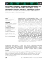
Fig. 3. Activation of p53 causes cleavage of initiation factors eIF4GI
and eIF4B. (A) Characterization of cleavage products of eIF4GI:
Pro190 and Val135 cells were incubated at 32 °C for 15 h and extracts
prepared and analyzed by immunoblotting for initiation factor eIF4-
GI. The positions of migration of the intact factor ( 200 kDa), an
intermediate cleavage product comprising the N-terminal and middle
fragments of eIF4GI (N-FAG plus M-FAG) ( 150 kDa) and the
middle fragment of eIF4GI (M-FAG) ( 76 kDa) [27] are indicated.
(B) Time-course of cleavage of eIF4GI following activation of p53 at
32 °C. Pro190 and Val135 cells were incubated at 32 °Cforthetimes
indicated and extracts were analyzed for the disappearance of intact
eIF4GI and the appearance of M-FAG as in (A). (C) Time-course of
cleavage of eIF4B following activation of p53 at 32 °C. Pro190 and
Val135 cells were incubated at 32 °C for the times indicated and
extracts were analyzed for the presence of eIF4B (80 kDa) and its
cleavage product DeIF4B (60 kDa) as described previously [28,29]. No
cleavage of eIF4B was observed when either cell line was maintained at
38 °C(seeFig.5C).
Ó FEBS 2003 Regulation of protein synthesis by p53 (Eur. J. Biochem. 270) 3125
Effects of p53 on protein synthesis and initiation factor
cleavages are independent of caspases
To determine whether caspases are activated under the same
conditions that result in the initiation factor cleavages we
have assayed caspases-3, -8 and -9 in extracts from the two
cell lines. As shown in Table 1, within 6 h at 32 °Cthe
activities of all three caspases increased significantly in the
Val135 cells but not in the Pro190 cells, reaching a
maximum at about 20 h. In addition, we determined the
extent of cleavage of two well characterized caspase
substrates, PARP and the cyclin-dependent protein kinase
inhibitor p27
KIP1
(Fig. 4A,B). Consistent with the activa-
tion of caspase-3, processing of PARP to give rise to the
characteristic p89 cleavage product was observed (Fig. 4A).
A proportion of p27
KIP1
was also cleaved to produce a
discrete fragment, Dp27 (Fig. 4B). Both the p53-induced
increase in caspase-3 activity and the cleavages of PARP
and p27
KIP1
were inhibited by the broad specificity caspase
inhibitor z-VAD.FMK (Table 2, Fig. 4A,B).
The above results show that, although cell viability does
not decline until about 20 h (Fig. 2), p53 activation does
cause increased caspase activity within 6 h. We therefore
investigated whether caspase activity is responsible for the
early changes in protein synthesis and the initiation factor
cleavages that occur following the activation of p53.
Figure 5(A,B) shows that the p53-induced inhibition of
protein synthesis was completely resistant to treatment of
the cells with z-VAD.FMK, both at early and late times
after the temperature shift. Moreover, neither the cleavage
of eIF4GI nor that of eIF4B was prevented by the caspase
inhibitor, even when these cleavages involved only a
relatively small fraction of the respective proteins (Fig. 5C).
This was the case even though the z-VAD.FMK was able to
block completely the activity of caspase-3 (Table 2), as well
as that of caspases-8 and -9 (data not shown), and prevented
the cleavages of PARP and p27
KIP1
induced by p53
activation (Fig. 4A,B). Moreover, the same z-VAD.FMK
preparation inhibited the extensive cleavages of eIF4GI and
eIF4B that occur in another apoptotic system, viz. Jurkat
cells treated with an agonistic anti-Fas antibody [32,33]
(Fig. 5C). In some experiments where more extensive
cleavage of eIF4GI occurred z-VAD-FMK had a very
slight protective effect but a substantial level of M-FAG was
still generated in the presence of the caspase inhibitor
Table 1. Activation of p53 rapidly enhances caspase activity. Pro190 and Val135 cells were incubated at 32 °C for the times indicated. Cell extracts
were prepared and the activities of caspases-3, -8 and -9 were assayed as described in Materials and methods. The data are expressed as RFU per lg
of protein and show the means ± the ranges of duplicate determinations.
Cell line
Caspase activity (RFUÆlg
)1
protein)
Caspase-8 Caspase-9 Caspase-3
Pro190 Val135 Pro190 Val135 Pro190 Val135
Hours at 32 °C
0 73.6 ± 6.7 63.9 ± 5.7 42.8 ± 5.5 53.6 ± 5.2 82.0 ± 3.6 127.7 ± 2.0
2 80.6 ± 1.9 89.5 ± 8.3 39.6 ± 4.7 62.5 ± 10.1 67.4 ± 0.1 168.3 ± 17.5
6 79.4 ± 2.9 117.6 ± 13.6 43.6 ± 2.0 94.3 ± 1.2 67.5 ± 0.1 413.8 ± 17.5
20 62.8 ± 2.1 230.3 ± 69.1 34.0 ± 0.7 213.0 ± 5.0 56.1 ± 3.1 966.6 ± 78.9
30 74.8 ± 8.4 243.9 ± 22.6 43.0 ± 8.5 163.3 ± 3.0 55.1 ± 1.4 577.5 ± 13.5
Fig. 4. Activation of p53 in MEL cells causes caspase-dependent
cleavages of PARP and p27
KIP1
. (A) Pro190 and Val135 cells were
incubated at 32 °C in the presence and absence of z-VAD.FMK for
15 h. Extracts were prepared and immunoblotted for (A) the apoptotic
cleavage product of PARP (89 kDa) and (B) p27
KIP1
and its caspase
cleavage product Dp27 as described previously [33–35].
Table 2. p53-induced caspase activity is sensitive to inhibition by
z.VAD-FMK. Val135 cells were incubated at 38 °Corat32°Cfor6h
in the presence and absence of the caspase inhibitor z.VAD-FMK
(10 l
M
and 50 l
M
). Extracts were prepared and assayed for caspase-3
activity by cleavage of the substrate Ac-DEVD.AMC as described in
Materials and methods. The data are expressed as RFU per micro-
gram of protein and are the means ± the ranges of duplicate deter-
minations.
Condition
Caspase-3 activity
(RFUÆlg protein
)1
)
38 °C 68.6 ± 4.8
32 °C 290.0 ± 5.1
32 °C plus z.VAD-FMK (10 l
M
) 50.7 ± 1.6
38 °C plus z.VAD-FMK (50 l
M
) 17.5 ± 1.6
3126 C. Constantinou et al. (Eur. J. Biochem. 270) Ó FEBS 2003
(Fig. 5D). These data therefore suggest that p53 regulates
protein synthesis by mechanism(s) that do not require
caspase activity and are consistent with the conclusion that
the translational inhibition occurs independently of the
induction of apoptosis.
It is likely that other proteases are involved in causing the
initiation factor cleavages and these may be responsible for
regulating translation following p53 activation [40,41].
Incubation of the Val135 cells with a range of protease
inhibitors, viz. the chymotrypsin inhibitor TPCK, the
calpain inhibitors N-acetyl-Leu-Leu-Nle-CHO (ALLN),
calpain inhibitor IV (z-LLY.FMK) and calpeptin (z-Leu-
Nle-CHO), and the cathepsin B inhibitor z-FA.FMK,
prevented neither the p53-induced cleavage of eIF4GI nor
the inhibition of protein synthesis at 32 °C (data not
shown). Further investigations utilizing a wider range of
protease inhibitors will therefore be necessary to identify the
enzyme(s) involved.
Although the inhibition of protein synthesis by p53
activation can be dissociated temporally from the progress
of apoptosis, we wanted to determine whether the preven-
tion of cell death had an effect on the down-regulation of
translation. To address this question we took advantage
of the observation that treatment of cells either with
z-VAD.FMK or with cytokines that function as survival
factors inhibits p53-induced apoptosis [13,30,42–46]. In our
hands, although z-VAD.FMK and the erythroid cell-
specific survival factor erythropoietin (Epo) each had a
marked antiapoptotic effect, both were required together to
prevent completely the loss of viability of Val135 cells at
32 °C (Fig. 6A). In spite of this dramatic protective effect,
however, neither z-VAD.FMK nor Epo, alone or in
combination, showed any ability to rescue protein synthesis
from p53-induced inhibition (Fig. 6B). This again suggests
that the down-regulation of translation by p53 does not
require the activity of caspases or other apoptotic mediators.
It also indicates that the inhibition does not involve other
pathways that are inactivated in the presence of Epo.
Discussion
The tumour suppressor protein p53 is activated in cells by
a number of stresses, including UV irradiation, chemically
induced DNA damage and hypoxia [47–50]. Activation of
p53 results in a variety of cellular responses, notably
inhibition of cell cycle progression and stimulation of DNA
repair [51]. If p53 activity is sustained it can also lead to cell
death by apoptosis [52]. Many of these effects require
nuclear translocation of p53 and subsequent transcriptional
activation of a large number of target genes [10,53].
However there is also evidence for direct cytoplasmic effects
of activated p53, including association of the protein with
Fig. 5. p53-induced translational inhibition and initiation factor cleavages do not require caspase activity. (A and B) Protein synthesis measurements.
Pro190 and Val135 cells were incubated at 32 °Cfor(A)4hor6hor(B)15hinthepresenceandabsenceofz-VAD.FMK(50l
M
) and protein
synthesis was determined as described in Materials and methods. (C) Pro190 and Val135 cells were incubated at 32 °Cfor15hinthepresenceand
absence of z-VAD.FMK and cell extracts were immunoblotted for eIF4GI or eIF4B and their cleavage products. As a positive control for the
efficacy of the z-VAD.FMK, Jurkat cells were incubated for 2 h with or without an agonistic anti-Fas antibody [32], in the presence or absence of
the same preparation of the inhibitor, and extracts were blotted for the same initiation factors. (D) Val135 cells were incubated at 38 °Cor32 °Cfor
6 h in the presence or absence of z-VAD.FMK as indicated. Cell extracts were immunoblotted for eIF4GI and its cleavage products.
Ó FEBS 2003 Regulation of protein synthesis by p53 (Eur. J. Biochem. 270) 3127
mitochondria and ribosomes [15,16,54], and several studies
have shown that pro-apoptotic effects of p53 do not
necessarily require transcriptional transactivation activity of
the protein [55–57].
Well documented reports have revealed a role for p53 in
the control of translation of individual mRNA species such
as those encoding cdk4, FGF-2 and even p53 itself [14,17–
21]. However the mechanisms responsible have not been
elucidated. Our data show that p53 can also control the rate
of global protein synthesis. Although the inhibition of
translation precedes the impairment of cell cycle progression
such an effect may ultimately contribute to the growth
inhibitory effects of the activated tumour suppressor
protein. Detailed analysis of the kinetics of the inhibition
of translation has shown that this effect begins within 2–4 h
of activating wild-type p53 [22] (manuscript in preparation).
Thus it is unlikely that the regulation of protein synthesis is
simply a consequence of either the impairment of cell cycle
progression or the induction of apoptosis, both of which are
associated with inhibition of translation in other systems
[58–60]. However the question of whether common signal-
ing pathways are involved in the control of translation and
in the effects of p53 on the cell cycle and/or apoptosis will
require the use of further mutants of p53 defective in
inducing one or other of the latter effects. It is possible that
the effects of p53 on protein synthesis are a result of new
gene transcription events, although the early response time
would tend to mitigate against this. Unfortunately we have
been unable to use transcription inhibitors to investigate this
possibility directly because such agents alone affect p53
function [61].
We have reported elsewhere that the extent of phos-
phorylation of the inhibitor of polypeptide chain initiation
factor eIF4E, 4E-BP1, is reduced following activation of
p53 in the Val135 cells and that this results in sequestra-
tion of eIF4E away from the eIF4F initiation complex
[22]. No changes in the phosphorylation state of other key
protein synthesis initiation factors such as eIF2a or eIF4E
could be observed. The possibility that the dephosphory-
lation of 4E-BP1 results in the inhibition of translation of
specific mRNAs, including those known to be regulated at
the translational level by p53, remains to be tested. The
changes in 4E-BP1 function, in combination with the
partial cleavages of eIF4GI and eIF4B reported here,
may be sufficient to bring about the overall inhibition of
protein synthesis by p53. However the present data do
not address this issue directly. In many experiments a
significant proportion of both eIF4GI and eIF4B
remained intact following p53 activation; nevertheless it
is possible that the cleavage products that accumulate
could exert an inhibitory (dominant negative) effect on the
activity of the remaining full-length protein. A further
consequence of the cleavage of eIF4GI could be the
stimulation of cap-independent translation. Fragments
that are generated in apoptotic cells from both eIF4GI
itself [62] and the related protein DAP5 [63,64] have been
shown to enhance the utilization for translation of
mRNAs with internal ribosome entry sites.
At first sight the specific cleavages of initiation factors
eIF4GI and eIF4B, both of which are known substrates for
proteolysis in apoptosing cells, would seem to be in accord
with the established pro-apoptotic effects of p53. These
factors have been shown previously to be cleaved in cells
undergoing apoptosis in response to treatment with anti-Fas
antibody [28,32,33], cycloheximide [24,28], staurosporine or
tumour necrosis factor a [26]. However, as shown in Figs 2
and 3, the initial down-regulation of translation, as well as
the cleavages of eIF4GI and eIF4B, occurs at a time when
there is little loss of cell viability and during a period when
the p53-induced inhibition of cell growth is reversible [65].
This indicates that neither the translational inhibition nor
the initiation factor modifications are simply consequences
of apoptosis. Moreover these events are clearly not sufficient
to commit the cells to death, although of course later
changes that affect translation may be. Consistent with these
conclusions, progression into apoptosis is not required for
translational inhibition by p53 as essentially complete
protection of the Val135 cells against death at 32 °Cby
the combination of z.VAD-FMK and Epo did not rescue
protein synthesis (Fig. 6).
Both eIF4GI and eIF4B can be cleaved by caspase-3
in vivo and in vitro [27,29]. As caspase-3 is activated in
the Val135 cells following the temperature shift, and the
cleavage products of the two initiation factors appear to be
Fig. 6. The p53-induced inhibition of protein synthesis can be dissociated
from apoptosis. (A) Pro190 and Val135 cells were incubated at 38 °Cor
32 °C for 48 h in the absence or presence of Epo (10 unitsÆmL
)1
)and/
or z.VAD-FMK (10 l
M
)asindicated.Attheendofthisperiodcell
viability was determined by trypan blue exclusion. (B) Cells were
incubated as in (A). After 24 h, protein synthesis was monitored by the
incorporation of [
35
S]methionine (10 lCiÆmL
)1
) into acid-insoluble
material during the last 1 h of incubation. The data are expressed as a
percentage of the incorporation in Pro190 cells incubated at 38 °Cin
the absence of Epo and z.VAD-FMK.
3128 C. Constantinou et al. (Eur. J. Biochem. 270) Ó FEBS 2003
very similar, if not identical, to those seen as a result of
caspase-dependent degradation in other systems, we were
surprised to find that the appearance of M-FAG and
DeIF4B was not inhibited by the broad specificity caspase
inhibitor z.VAD-FMK. This was not due to a failure of the
latter to act on MEL cells as the compound inhibited the
activation of caspase-3 and completely blocked the cleavage
of the caspase substrates PARP and p27
KIP1
in Val135 cells
shiftedto32°C.Moreoverthesamez.VAD-FMKprepar-
ation was effective in inhibiting the cleavage of eIF4GI and
eIF4B in Jurkat cells treated with anti-Fas. Along with an
inability to prevent the factor cleavages in the Val135 cells
z.VAD-FMK was also unable to prevent the overall
inhibition of protein synthesis.
Although we cannot rule out the possibility that eIF4GI
and eIF4B are cleaved by caspase(s) that are at least
partially active even in the presence of z.VAD-FMK [66] it
is possible that other proteases that are activated directly or
indirectly by p53 are responsible [40,41]. Such alternative
pathways may also operate in other systems. Whereas p53 is
required for radiation-induced neuronal cell death, caspase
activity is not required for this process [67]. Several studies
have established the phenomenon of caspase-independent
cell death. Moreover noncaspase proteases are involved in
some forms of apoptosis mediated by p53 and other
pathways, and specific protein cleavages occur in some cases
[68–72]. Morley and Pain [73] reported that eIF4GI and
eIF4GII can be cleaved by a z.VAD-FMK-resistant mech-
anism in cells undergoing apoptosis in response to treatment
with the immunosuppressant drugs FTY720 and cyclo-
sporin A. If noncaspase mediated proteolytic events are
responsible for the cleavage of eIF4GI and eIF4B the
enzyme(s) involved must presumably act on sites that are
identical or very close to those targeted by the caspases
[27,28]. These sites may lie in relatively accessible or
unstructured regions of the proteins. In spite of using a
wide range of protease inhibitors we have not yet identified
the protease(s) responsible for the initiation factor cleavages
following p53 activation.
The effects of p53 on the translational machinery are
very similar to those seen following treatment of cells with
DNA damaging agents such as etoposide, mitomycin-C or
cisplatin [32,39,74]. Common features include the caspase-
independent nature of the inhibition of overall translation,
the lack of effect on eIF2a phosphorylation and, in contrast,
the marked dephosphorylation of 4E-BP1 [22]. These
observations suggest that the effects of DNA-damaging
agents on translation could be mediated, at least in part, by
p53. The p53-regulated effects we observe are also similar to
those seen following inhibition of proteasome activity [75].
Proteasome inhibition not only induces p53-dependent
apoptosis [76–78] but also causes dephosphorylation of
4E-BP1 and the cleavage of initiation factors, effects which
are partially caspase-independent (S. Morley, personal
communication). Cyclosporin A, which can induce eIF4G
cleavage [73], also inhibits proteasome activity [79,80].
Moreover, inhibition of proteasome-mediated proteolysis
induces p53 expression and caspase-independent apoptosis
[78,81].
Another potential mechanism of action of p53 may
involve signaling by ceramide as a second messenger.
Ceramide causes caspase-independent apoptosis and also
induces p53 in at least one system [82,83]. Irradiation-
induced DNA damage activates ceramide production [84],
and p53 is required for the induction of ceramide by some
cell stresses [85]. Whether the tumour suppressor protein
is required for ceramide-induced growth inhibition and
apoptosis remains controversial however, [85–87]. If p53
functions upstream of ceramide then the latter may indeed
contribute to the down-regulation of translation observed in
this study. However ceramide has been reported to activate
the eIF2a-specific protein kinase PKR and thereby inhibit
translation [88], whereas p53 activation has no effect on
eIF2a phosphorylation [22]. Few other studies of the effects
of ceramide on protein synthesis or initiation factor
modifications have been reported and the possibility of
regulation by this second messenger in cells expressing active
p53 remains to be evaluated.
In summary, we have reported the novel observation that
activation of p53 results in the caspase-independent down-
regulation of translation, together with the cleavages of at
least two polypeptide chain initiation factors that are critical
for protein synthesis. Moreover, we have shown that these
events are not simply the consequences of p53-induced
apoptosis and indeed occur independently of this process.
Further details of the mechanisms involved await future
study.
Acknowledgements
This research was supported by grants to M. J. Clemens from the
Wellcome Trust (056778), the Leukaemia Research Fund and Glaxo-
Wellcome and by grants to J. Hensold from the Office of Research and
Development, Medical Research Service, Department of Veterans’
Affairs and the NIH (DK43414). J. Hensold was also funded during a
period of sabbatical leave by an award from Burroughs-Wellcome.
C. Constantinou is supported by a PhD studentship from the Cancer
Prevention Research Trust, with additional funding from the AG
Leventis Foundation and an Overseas Research Scholarship from
Universities UK. M. Bushell is supported by a Fellowship from The
Wellcome Trust (063233).
References
1. Johnson, P., Gray, D., Mowat, M. & Benchimol, S. (1991)
Expression of wild-type p53 is not compatible with continued
growth of p53-negative tumor cells. Mol. Cell Biol. 11, 1–11.
2. Burns, T.F. & El-Deiry, W.S. (1999) The p53 pathway and
apoptosis. J. Cell. Physiol. 181, 231–239.
3. Sionov, R.V. & Haupt, Y. (1999) The cellular response to p53: the
decision between life and death. Oncogene 18, 6145–6157.
4. Appella, E. (2001) Modulation of p53 function in cellular regu-
lation. Eur. J. Biochem. 268, 2763–2763.
5. Vousden, K.H. (2000) p53: death star. Cell 103, 691–694.
6. Farrell,P.J.,Allan,G.J.,Shanahan,F.,Vousden,K.H.&Crook,
T. (1991) p53 Is frequently mutated in Burkitt’s lymphoma cell
lines. EMBO J. 10, 2879–2887.
7. Bates, S. & Vousden, K.H. (1999) Mechanisms of p53-mediated
apoptosis. Cell. Mol. Life Sci. 55, 28–37.
8. Wang, X.W. (1999) Role of p53 and apoptosis in carcinogenesis.
Anticancer Res. 19, 4759–4771.
9. Owen-Schaub, L.B., Zhang, W., Cusack, J.C., Angelo, L.S.,
Santee, S.M., Fujiwara, T., Roth, J.A., Deisseroth, A.B., Zhang,
W.W., Kruzel, E. & Radinsky, R. (1995) Wild-type human p53
and a temperature-sensitive mutant induce Fas/APO-1 expression.
Mol. Cell Biol. 15, 3032–3040.
Ó FEBS 2003 Regulation of protein synthesis by p53 (Eur. J. Biochem. 270) 3129
10. Miyashita, T. & Reed, J.C. (1995) Tumor suppressor p53 is a
direct transcriptional activator of the human bax gene. Cell 80,
293–299.
11. Kato, M.V., Sato, H., Anzai, H., Nagayoshi, M. & Ikawa, Y.
(1997) Up-regulation of cell cycle-associated genes by
p53 in apoptosis of an erythroleukemic cell line. Leukemia 11,
389–392.
12. Tokino, T. & Nakamura, Y. (2000) The role of p53-target genes in
human cancer. Crit. Rev. Oncol. Hematol. 33, 1–6.
13. Lin, Y.P. & Benchimol, S. (1995) Cytokines inhibit p53-mediated
apoptosis but not p53-mediated G
1
arrest. Mol. Cell Biol. 15,
6045–6054.
14. Ewen, M.E. & Miller, S.J. (1996) p53 and translational control.
Biochim. Biophys. Acta Rev. Cancer 1242, 181–184.
15. Fontoura, B.M.A., Atienza, C.A., Sorokina, E.A., Morimoto, T.
& Carroll, R.B. (1997) Cytoplasmic p53 polypeptide is associated
with ribosomes. Mol. Cell Biol. 17, 3146–3154.
16. Marechal, V., Elenbaas, B., Piette, J., Nicolas, J.C. & Levine, A.J.
(1994) The ribosomal L5 protein is associated with mdm-2 and
mdm-2-p53 complexes. Mol. Cell Biol. 14, 7414–7420.
17. Ewen, M.E., Oliver, C.J., Sluss, H.K., Miller, S.J. & Peeper, D.S.
(1995) p53-dependent repression of CDK4 translation in TGF-b-
induced G
1
cell-cycle arrest. Genes Dev. 9, 204–217.
18. Miller, S.J., Suthiphongchai, T., Zambetti, G.P. & Ewen, M.E.
(2000) p53 binds selectively to the 5¢ untranslated region of cdk4,
an RNA element necessary and sufficient for transforming growth
factor b- and p53-mediated translational inhibition of Cdk4. Mol.
Cell Biol. 20, 8420–8431.
19. Galy, B., Cre
´
ancier, L., Zanibellato, C., Prats, A.C. & Prats, H.
(2001) Tumour suppressor p53 inhibits human fibroblast growth
factor 2 expression by a post-transcriptional mechanism. Onco-
gene 20, 1669–1677.
20. Galy, B., Cre
´
ancier, L., Prado-Lourenco, L., Prats, A C. & Prats,
H. (2001) p53 directs conformational change and translation
initiation blockade of human fibroblast growth factor 2 mRNA.
Oncogene 20, 4613–4620.
21. Mosner, J., Mummenbrauer, T., Bauer, C., Sczakiel, G., Grosse,
F. & Deppert, W. (1995) Negative feedback regulation of wild-
type p53 biosynthesis. EMBO J. 14, 4442–4449.
22. Horton, L.E., Bushell, M., Barth-Baus, D., Tilleray, V.J.,
Clemens, M.J. & Hensold, J. (2002) p53 activation results in rapid
dephosphorylation of the eIF4E-binding protein 4E-BP1, inhibi-
tion of ribosomal protein S6 kinase and inhibition of translation
initiation. Oncogene 21, 5325–5334.
23. Michalovitz, D., Halevy, O. & Oren, M. (1990) Conditional
inhibition of transformation and of cell proliferation by a tem-
perature-sensitive mutant of p53. Cell 62, 671–680.
24. Clemens, M.J., Bushell, M. & Morley, S.J. (1998) Degradation of
eukaryotic polypeptide chain initiation factor (eIF) 4G in response
to induction of apoptosis in human lymphoma cell lines. Oncogene
17, 2921–2931.
25. Marissen, W.E. & Lloyd, R.E. (1998) Eukaryotic translation
initiation factor 4G is targeted for proteolytic cleavage by caspase
3 during inhibition of translation in apoptotic cells. Mol. Cell Biol.
18, 7565–7574.
26. Bushell, M., McKendrick, L., Janicke, R.U., Clemens, M.J. &
Morley, S.J. (1999) Caspase-3 is necessary and sufficient for clea-
vage of protein synthesis eukaryotic initiation factor 4G during
apoptosis. FEBS Lett. 451, 332–336.
27. Bushell, M., Poncet, D., Marissen, W.E., Flotow, H., Lloyd, R.E.,
Clemens, M.J. & Morley, S.J. (2000) Cleavage of polypeptide
chain initiation factor eIF4GI during apoptosis: characterisation
of an internal fragment generated by caspase-3-mediated cleavage.
Cell Death Differ. 7, 628–636.
28.Bushell,M.,Wood,W.,Clemens,M.J.&Morley,S.J.(2000)
Changes in integrity and association of eukaryotic protein
synthesis initiation factors during apoptosis. Eur. J. Biochem. 267,
1083–1091.
29. Bushell, M., Wood, W., Carpenter, G., Pain, V.M., Morley, S.J. &
Clemens, M.J. (2001) Disruption of the interaction of mammalian
protein synthesis eukaryotic initiation factor 4B with the poly (A)-
binding protein by caspase- and viral protease-mediated cleavages.
J. Biol. Chem. 276, 23922–23928.
30. Johnson, P., Chung, S. & Benchimol, S. (1993) Growth suppres-
sion of Friend virus-transformed erythroleukemia cells by p53
protein is accompanied by hemoglobin production and is sensitive
to erythropoietin. Mol. Cell Biol. 13, 1456–1463.
31. Spector, D.L., Goldman, R.D. & Leinwand, L.A. (1998) Cells:
a Laboratory Manual. Cold Spring Harbor Laboratory Press,
New York.
32. Morley, S.J., McKendrick, L. & Bushell, M. (1998) Cleavage of
translation initiation factor 4G (eIF4G) during anti-Fas IgM-
induced apoptosis does not require signalling through the p38
mitogen-activated protein (MAP) kinase. FEBS Lett. 438, 41–48.
33. Morley, S.J., Jeffrey, I., Bushell, M., Pain, V.M. & Clemens, M.J.
(2000) Differential requirements for caspase-8 activity in the
mechanism of phosphorylation of elF2a, cleavage of eIF4GI and
signaling events associated with the inhibition of protein synthesis
in apoptotic Jurkat T cells. FEBS Lett. 477, 229–236.
34. Frost, V. & Sinclair, A.J. (2000) p27
KIP1
is down-regulated by two
different mechanisms in human lymphoid cells undergoing apop-
tosis. Oncogene 19, 3115–3120.
35. Frost, V., Al-Mehairi, S. & Sinclair, A.J. (2001) Exploitation of a
non-apoptotic caspase to regulate the abundance of the cdkI
p27
KIP1
in transformed lymphoid cells. Oncogene 20, 2737–2748.
36. Martinez, J., Georgoff, I. & Levine, A.J. (1991) Cellular localiza-
tion and cell cycle regulation by a temperature-sensitive p53 pro-
tein. Genes Dev. 5, 151–159.
37. Yamato, K., Yamamoto, M., Hirano, Y. & Tsuchida, N. (1995) A
human temperature-sensitive p53 mutant p53Val-138: modulation
of the cell cycle, viability and expression of p53-responsive genes.
Oncogene 11,1–6.
38. Kato, M.V. (1999) The mechanisms of death of an erythroleuke-
mic cell line by p53: involvement of the microtubule and mito-
chondria. Leuk. Lymphoma 33, 181–186.
39. Jeffrey, I.W., Bushell, M., Tilleray, V.J., Morley, S. & Clemens,
M.J. (2002) Inhibition of protein synthesis in apoptosis: Differ-
ential requirements by the tumour necrosis factor a family and a
DNA damaging agent for caspases and the double-stranded
RNA-dependent protein kinase. Cancer Res. 62, 2272–2280.
40. Lotem, J. & Sachs, L. (1996) Differential suppression by protease
inhibitors and cytokines of apoptosis induced by wild-type p53
and cytotoxic agents. Proc.NatlAcad.Sci.USA93, 12507–12512.
41. Yuan, X.M., Li, W., Dalen, H., Lotem, J., Kama, R., Sachs, L. &
Brunk, U.T. (2002) Lysosomal destabilization in p53-induced
apoptosis. Proc. Natl Acad. Sci. USA 99, 6286–6291.
42. Abrahamson, J.L., Lee, J.M. & Bernstein, A. (1995) Regulation of
p53-mediated apoptosis and cell cycle arrest by Steel factor. Mol.
Cell Biol. 15, 6953–6960.
43. Li, P.F., Dietz, R. & von Harsdorf, R. (1999) p53 regulates
mitochondrial membrane potential through reactive oxygen spe-
cies and induces cytochrome c-independent apoptosis blocked by
Bcl-2. EMBO J. 18, 6027–6036.
44. Gao, C.F., Ren, S., Zhang, L., Nakajima, T., Ichinose, S., Hara,
T., Koike, K. & Tsuchida, N. (2001) Caspase-dependent cytosolic
release of cytochrome c and membrane translocation of Bax in
p53-induced apoptosis. Exp. Cell Res. 265, 145–151.
45. Yamaguchi, A., Tamatani, M., Matsuzaki, H., Namikawa, K.,
Kiyama, H., Vitek, M.P., Mitsuda, N. & Tohyama, M. (2001) Akt
activation protects hippocampal neurons from apoptosis by
inhibiting transcriptional activity of p53. J. Biol. Chem. 276,
5256–5264.
3130 C. Constantinou et al. (Eur. J. Biochem. 270) Ó FEBS 2003
46. Di Bacco, A.M.A. & Cotter, T.G. (2002) p53 expression in K562
cells is associated with caspase-mediated cleavage of c-ABL and
BCR-ABL protein kinases. Br. J. Haematol. 117, 588–597.
47. Lutzker, S.G., Mathew, R. & Taller, D.R. (2001) A p53 dose–
response relationship for sensitivity to DNA damage in isogenic
teratocarcinoma cells. Oncogene 20, 2982–2986.
48. Lakin, N.D. & Jackson, S.P. (1999) Regulation of p53 in response
to DNA damage. Oncogene 18, 7644–7655.
49. Oren, M. (1999) Regulation of the p53 tumor suppressor protein.
J. Biol. Chem. 274, 36031–36034.
50. Xie,S.,Wu,H.,Wang,Q.,Cogswell,J.P.,Husain,I.,Conn,C.W.,
Stambrook, P., Jhanwar-Uniyal, M. & Dai, W. (2001) Plk3 links
DNA damage to cell cycle arrest and apoptosis at least in part via
the p53 pathway. J. Biol. Chem. 276, 43305–43312.
51. Ryan, K.M., Phillips, A.C. & Vousden, K.H. (2001) Regulation
and function of the p53 tumor suppressor protein. Curr. Opin. Cell
Biol. 13, 332–337.
52. Lowe, S.W., Schmitt, E.M., Smith, S.W., Osborne, B.A. & Jacks,
T. (1993) p53 is required for radiation-induced apoptosis in mouse
thymocytes. Nature 362, 847–849.
53. Rodriguez-Lopez, A.M., Xenaki, D., Eden, T.O., Hickman, J.A.
& Chresta, C.M. (2001) MDM2-mediated nuclear exclusion of
p53 attenuates etoposide-induced apoptosis in neuroblastoma
cells. Mol. Pharmacol. 59, 135–143.
54. Marchenko, N.D., Zaika, A. & Moll, U.M. (2000) Death signal-
induced localization of p53 protein to mitochondria – a potential
role in apoptotic signaling. J. Biol. Chem. 275, 16202–16212.
55. Caelles, C., Helmberg, A. & Karin, M. (1994) p53-dependent
apoptosis in the absence of transcriptional activation of p53-target
genes. Nature 370, 220–223.
56. Gao, C. & Tsuchida, N. (1999) Activation of caspases in p53-
induced transactivation-independent apoptosis. Jpn. J. Cancer
Res. 90, 180–187.
57. Ding, H.F., Lin, Y.L., McGill, G., Juo, P., Zhu, H., Blenis, J.,
Yuan, J.Y. & Fisher, D.E. (2000) Essential role for caspase-8 in
transcription-independent apoptosis triggered by p53. J. Biol.
Chem. 275, 38905–38911.
58. Pyronnet, S., Dostie, J. & Sonenberg, N. (2001) Suppression of
cap-dependent translation in mitosis. Genes Dev. 15, 2083–2093.
59. Pyronnet, S. & Sonenberg, N. (2001) Cell-cycle-dependent trans-
lational control. Curr. Opin. Genet. Dev. 11, 13–18.
60. Clemens, M.J., Bushell, M., Jeffrey, I.W., Pain, V.M. & Morley,
S.J. (2000) Translation initiation factor modifications and the
regulation of protein synthesis in apoptotic cells. Cell Death Differ.
7, 603–615.
61. David-Pfeuty, T., Nouvian-Dooghe, Y., Sirri, V., Roussel, P. &
Hernandez-Verdun, D. (2001) Common and reversible
regulation of wild-type p53 function and of ribosomal bio-
genesis by protein kinases in human cells. Oncogene 20, 5951–
5963.
62. Nevins, T.A., Harder, Z.M., Korneluk, R.G. & Holcik, M.
(2003) Distinct regulation of internal ribosome entry site-mediated
translation following cellular stress is mediated by apoptotic
fragments of eIF4G translation initiation factor family members
eIF4GI and p97/DAP5/NAT1. J. Biol. Chem. 278, 3572–3579.
63. Henis-Korenblit, S., Strumpf, N.L., Goldstaub, D. & Kimchi, A.
(2000) A novel form of DAP5 protein accumulates in apoptotic
cells as a result of caspase cleavage and internal ribosome entry
site-mediated translation. Mol. Cell Biol. 20, 496–506.
64. Henis-Korenblit, S., Shani, G., Sines, T., Marash, L., Shohat, G.
& Kimchi, A. (2002) The caspase-cleaved DAP5 protein supports
internal ribosome entry site-mediated translation of death pro-
teins. Proc. Natl Acad. Sci. USA 99, 5400–5405.
65. Geske, F.J., Lieberman, R., Strange, R. & Gerschenson, L.E.
(2001) Early stages of p53-induced apoptosis are reversible. Cell
Death Differ. 8, 182–191.
66. MacFarlane, M., Cohen, G.M. & Dickens, M. (2000) JNK (c-Jun
N-terminal kinase) and p38 activation in receptor-mediated and
chemically-induced apoptosis of T-cells: differential requirements
for caspase activation. Biochem. J. 348, 93–101.
67. Johnson, M.D., Xiang, H., London, S., Kinoshita, Y., Knudson,
M.,Mayberg,M.,Korsmeyer,S.J.&Morrison,R.S.(1998)Evi-
dence for involvement of Bax and p53, but not caspases, in
radiation-induced cell death of cultured postnatal hippocampal
neurons. J. Neurosci. Res. 54, 721–733.
68. Denecker, G., Vercammen, D., Declercq, W. & Vandenabeele, P.
(2001) Apoptotic and necrotic cell death induced by death domain
receptors. Cell. Mol. Life Sci. 58, 356–370.
69. Sperandio, S., De Belle, I. & Bredesen, D.E. (2000) An alternative,
nonapoptotic form of programmed cell death. Proc. Natl Acad.
Sci. USA 97, 14376–14381.
70. Belmokhtar, C.A., Hillion, J. & Se
´
gal-Bendirdjian, E. (2001)
Staurosporine induces apoptosis through both caspase-
dependent and caspase-independent mechanisms. Oncogene 20,
3354–3362.
71. Foghsgaard, L., Wissing, D., Mauch, D., Lademann, U., Bast-
holm, L., Boes, M., Elling, F., Leist, M. & Ja
¨
a
¨
ttela
¨
, M. (2001)
Cathepsin B acts as a dominant execution protease in tumor
cell apoptosis induced by tumor necrosis factor. J. Cell Biol. 153,
999–1009.
72. Jin, S., Kalkum, M., Overholtzer, M., Stoffel, A., Chait, B.T. &
Levine, A.J. (2003) CIAP1 and the serine protease HTRA2 are
involved in a novel p53-dependent apoptosis pathway in mam-
mals. Genes Dev. 17, 359–367.
73. Morley, S.J. & Pain, V.M. (2001) Proteasome inhibitors and
immunosuppressive drugs promote the cleavage of eIF4GI and
eIF4GII by caspase-8-independent mechanisms in Jurkat T cell
lines. FEBS Lett. 503, 206–212.
74. Tee, A.R. & Proud, C.G. (2000) DNA-damaging agents cause
inactivation of translational regulators linked to mTOR signalling.
Oncogene 19, 3021–3031.
75. Wojcik, C. (1999) Proteasomes in apoptosis: villains or guardians?
Cell. Mol. Life Sci. 56, 908–917.
76. Chen,F.,Chang,D.,Goh,M.,Klibanov,S.A.&Ljungman,M.
(2000) Role of p53 in cell cycle regulation and apoptosis following
exposure to proteasome inhibitors. Cell Growth Differ. 11,
239–246.
77. MacLaren, A.P., Chapman, R.S., Wyllie, A.H. & Watson, C.J.
(2001) p53-dependent apoptosis induced by proteasome inhibition
in mammary epithelial cells. Cell Death Differ. 8, 210–218.
78. Kurland, J.F. & Meyn, R.E. (2001) Protease inhibitors restore
radiation-induced apoptosis to Bcl-2-expressing lymphoma cells.
Int. J. Cancer 96, 327–333.
79. Meyer, S., Kohler, N.G. & Joly, A. (1997) Cyclosporine A is an
uncompetitive inhibitor of proteasome activity and prevents
NF-kappaB activation. FEBS Lett. 413, 354–358.
80. Marienfeld, R., Neumann, M., Chuvpilo, S., Escher, C., Kneitz,
B.,Avots,A.,Schimpl,A.&Serfling,E.(1997)CyclosporinA
interferes with the inducible degradation of NF-kappa B
inhibitors, but not with the processing of p105/NF-kappa B1 in T
cells. Eur. J. Immunol. 27, 1601–1609.
81. Monney, L., Otter, I., Olivier, R., Ozer, H.L., Haas, A.L., Omura,
S. & Borner, C. (1998) Defects in the ubiquitin pathway induce
caspase-independent apoptosis blocked by Bcl-2. J. Biol. Chem.
273, 6121–6131.
82. Belaud-Rotureau, M A., Lacombe, F., Durrieu, F., Vial, J P.,
Lacoste, L., Bernard, P. & Belloc, F. (1999) Ceramide-induced
apoptosis occurs independently of caspases and is decreased by
leupeptin. Cell Death Differ. 6, 788–795.
83. Lopez-Marure, R., Ventura, J.L., Sanchez, L., Montano, L.F. &
Zentella, A. (2000) Ceramide mimics tumour necrosis factor-alpha
in the induction of cell cycle arrest in endothelial cells. Induction of
Ó FEBS 2003 Regulation of protein synthesis by p53 (Eur. J. Biochem. 270) 3131
the tumour suppressor p53 with decrease in retinoblastoma/pro-
tein levels. Eur. J. Biochem. 267, 4325–4333.
84. Radford, I.R. (1999) Initiation of ionizing radiation-induced
apoptosis: DNA damage-mediated or does ceramide have a role?
Int. J. Radiat. Biol. 75, 521–528.
85. Dbaibo, G.S., Pushkareva, M.Y., Rachid, R.A., Alter, N., Smyth,
M.J., Obeid, L.M. & Hannun, Y.A. (1998) p53-dependent cer-
amide response to genotoxic stress. J. Clin. Invest. 102, 329–339.
86. Oh, W.J., Kim, W.H., Kang, K.H., Kim, T.Y., Kim, M.Y. &
Choi, K.H. (1998) Induction of p21 during ceramide-mediated
apoptosis in human hepatocarcinoma cells. Cancer Lett. 129,
215–222.
87. Pruschy, M., Resch, H., Shi, Y.Q., Aalame, N., Glanzmann, C. &
Bodis, S. (1999) Ceramide triggers p53-dependent apoptosis in
genetically defined fibrosarcoma tumour cells. Br.J.Cancer80,
693–698.
88. Ruvolo, P.P., Gao, F.Q., Blalock, W.L., Deng, X.M. & May,
W.S. (2001) Ceramide regulates protein synthesis by a novel
mechanism involving the cellular PKR activator RAX. J. Biol.
Chem. 276, 11754–11758.
3132 C. Constantinou et al. (Eur. J. Biochem. 270) Ó FEBS 2003