Báo cáo khoa học: The )148 to )124 region of c-jun interacts with a positive regulatory factor in rat liver and enhances transcription Dipali Sharma*, Sujata Ohri and Aparna Dixit ppt
Bạn đang xem bản rút gọn của tài liệu. Xem và tải ngay bản đầy đủ của tài liệu tại đây (243.21 KB, 9 trang )
The )148 to )124 region of c-
jun
interacts with a positive
regulatory factor in rat liver and enhances transcription
Dipali Sharma*, Sujata Ohri and Aparna Dixit
Gene Regulation Laboratory, Center for Biotechnology, Jawaharlal Nehru University, New Delhi-110067, India
The c-jun gene encodes the protein Jun, a component of the
essential transcription factor, AP1. Jun/AP-1 occupies a
central position in signal transduction pathways as it is
responsible for the induction of a number of genes in
response to growth promoters. However, the exact mecha-
nisms leading to an enhanced expression of the c-jun gene
itself during proliferation, differentiation, cell growth and
development are not fully understood. Cell culture studies
have given some insight in the mechanisms involved in the
up-regulation of c-jun expression by UV irradiation and
phorbol esters. However, it is well known that transformed
cells do not accurately reflect the biology of a normal cell. We
now report the identification of a positive regulatory factor
from normal rat liver that activates transcription from the
c-jun promoter by binding to the )148 to )124 region of
c-jun. Preincubation of fractionated rat liver nuclear extract
with an oligonucleotide encompassing this region of the gene
significantly reduced transcription from cloned c-jun pro-
moter. In vitro transfection studies using green fluorescent
protein as a reporter gene under the control of the c-jun
promoter with ()148 to +53) and without ()123 to +53)
this region further confirmed its role in transcription. A
DNA-binding protein factor, interacting with this region of
c-jun was identified from rat liver by using electrophoretic
mobility shift assays. This factor binds to its recognition
sequence only in the phosphorylated form and exhibits high
affinity and specificity. UV cross-linking studies, South-
Western analysis and affinity purification collectively indi-
cated the factor to be 40 kDa and to bind to its recognition
sequence as a dimer.
Keywords:c-jun; DNA–protein interaction; in vitro tran-
scription; rat liver positive regulatory factor; transcriptional
regulation.
Elucidation of the molecular mechanisms regulating eu-
karyotic gene expression is essential for an understanding of
the complex processes that occur during normal cellular
development, differentiation and oncogenic transformation.
Proto-oncogene c-jun encodes a protein Jun, a major
component of transcription factor AP-1 [1–3]. Jun/AP-1
plays a role in the flow of information from cell surface
receptors to the nucleus [4,5]. Jun has been reported to be
involved in different aspects of cell growth, differentiation
and development [6–8]. Expression of the c-jun gene is
induced as an early response by serum active phorbol esters,
ionizing radiation and tumour necrosis factor-alpha [9–11].
An increase in the expression of c-jun precedes DNA
synthesis in proliferating cells. Jun/AP-1 is responsible for
the induction of a number of genes in response to phorbol
ester and tumour promoters and thus holds a central place
in the signal transduction pathway. However, the exact
mechanism(s) regulating c-jun expression during cell prolif-
eration, differentiation, growth and development are not
clearly understood except for its autoregulation by AP-1.
AP-1 is known to autoregulate c-jun expression by binding
to the AP-1 site present within the c-jun promoter [4,5].
Further, AP-1 transcription factors of different composition
have been reported to control c-jun transcription in resting
or stimulated cells [12].
c-jun expression and activity are partly regulated by Jun
N-terminal kinases (JNKs) and mitogen activated protein
kinases. JNKs phosphorylate the N terminus of the trans-
acting domain of Jun, thereby increasing its transactiva-
tion potency [13–16]. Inhibition of the stress-dependent
signal cascade (JNK/SAPK pathway) by culture confluency
inhibits c-jun N-terminal phosphorylation in response to
platelet-derived growth factor, epidermal growth factor or
UV irradiation [14]. Hence, Jun/AP-1 activity is regulated
at two different levels. Immediately after stimulation with
12-O-tetradecanoylphorbol 13-acetate (TPA), a post-trans-
lational event leads to an increased activity of pre-existing
Jun/AP-1 molecules. The second step involves increased
synthesis of Jun mediated by the interaction of activated
Jun/AP-1 with the jun promoter, resulting in transcrip-
tional activation [4,5]. The positive autoregulation of c-jun
can therefore function as a major genetic switch respon-
sible for the conversion of transient early events in signal
transduction into long lasting effects on cellular gene
expression.
Correspondence to A. Dixit, Gene Regulation Laboratory,
Centre for Biotechnology, Jawaharlal Nehru University,
New Delhi 110067, India.
Fax: +91 11 6198234, Tel.: +91 11 6102164,
E-mail: ;
Abbreviations: RNE-d, rat liver nuclear extract-fraction D; EMSA,
electrophoretic mobility shift assay; TPA, 12-O-tetradecanoyl
phrobol 13-acetate.
*Present address: The Johns Hopkins Oncology Center,
The Johns Hopkins University School of Medicine,
Baltimore, Maryland 21231, USA.
(Received 10 September 2002, revised 4 November 2002,
accepted 6 November 2002)
Eur. J. Biochem. 270, 181–189 (2003) Ó FEBS 2003 doi:10.1046/j.1432-1033.2003.03369.x
Regulation of c-jun is likely to involve many more cis-
acting elements and a number of factors differentially
interacting with these elements under different physiological
conditions and may vary between cell types. All of the studies
to understand c-jun transcriptional regulation have been
conducted in cultured cells which do not mimic in vivo
conditions. The present investigation was therefore under-
taken to develop an understanding of regulation of c-jun
expression in quiescent rat liver. We have identified a positive
regulatory factor from normal rat liver that binds to the
region )148 to )124 of c-jun and stimulates transcription.
Materials and methods
Reagents and animals
All chemicals were of reagent grade and were from Sigma
Chemical Co. unless stated otherwise. Healthy female
inbred rats of Wistar strain weighing 150–170 g were
procured from the Animal Facility, Jawaharlal Nehru
University, New Delhi, India. Animals were fed water and
standard rat chow ad libitum.
Plasmid DNA isolation
Escherichia coli cells, HB101 transformed with plasmid
)1100/+170 jun-CAT were grown in liquid culture and
plasmid DNA was isolated by the alkaline lysis method [17].
Plasmid )1100/+170 jun-CAT consists of the indicated
region of the c-jun gene upstream of the promoterless CAT
gene [4].
Fractionation of nuclear extract
Animals were killed by cervical dislocation, livers were
removed immediately, washed in chilled saline and pro-
cessed further for the preparation of nuclear extract as
described [18,19]. The fraction designated RNE-d contain-
ing maximum RNA polymerase II activity and essential
transcription factors was used in in vitro transcription assay
and electrophoretic mobility shift assay (EMSA).
In vitro
run-off transcription assay
In vitro transcription reactions were carried out using
conditions described earlier [19,20]. The transcription reac-
tion was carried out using 12 lgÆmL
)1
EcoRI linearized
plasmid )1100/+170 jun-CAT and 1.6 mgÆmL
)1
nuclear
protein (RNE-d) at 30 °C for 30 min. Transcripts extracted
with phenol/chloroform/isoamylalcohol (25 : 24 : 1) were
precipitated with ethanol and separated on a 6% acryl-
amide, 8
M
urea gel in 1 · Tris/borate/EDTA buffer [17].
The transcripts were visualized by autoradiography. EcoRI
linearized plasmid )1100/+170 jun-CAT should yield a
370-nucleotides long run-off transcript.
Transient transfection and reporter gene assay
Promoter constructs. Green fluorescent protein (GFP)
does not require any exogenous substrate and cofactors
for its fluorescence and its expression can be used to
monitor gene expression [21]. Also, GFP is a highly stable
protein and fluorescence from GFP can be used as a
quantitative measure of GFP content per cell [22].
Therefore, to assay jun promoter activity, two promoter
constructs ) p123jun-eGFP and p148jun-eGFP ) were
made by cloning PCR amplified )123 to +53 region
and )148 to +53 region of c-jun, respectively. For both
the amplifications, AseIandEcoRI restriction sites were
included in the forward and reverse primers, respectively.
PCR amplified fragments digested with AseIandEcoRI
were cloned into AseI–EcoRI digested plasmid pEGFP-N1
(GenBank Accession # U55762, Invitrogen), thus placing
the GFP coding region under the control of the )123 to
+53 and )148 to +53 regions of c-jun in p123jun-eGFP
and p148jun-eGFP, respectively. Recombinant clones were
confirmed for insertion of the promoter regions of c-jun by
sequencing.
Cells and cell culture. Chinese hamster ovary (CHO) cells
were maintained in Eagle’s modified essential medium
(Biological Industries, Israel) supplemented with 10% heat-
inactivated foetal bovine serum, 100 UÆmL
)1
penicillin and
100 lgÆmL
)1
streptomycin at 37 °C in a humidified atmos-
phere containing 5% CO
2
.
Transfection assay. CHO cells were plated at a density of
2 · 10
5
cells per well (35 mm diameter) in 2 mL Eagle’s
modified essential medium containing foetal bovine serum,
penicillin and streptomycin in six-well tissue culture plates
(Falcon, Becton Dickinson) to achieve 50–80% confluency
in 24 h. The cells were transfected with 2.5 lgeither
p123jun-eGFP or p148jun-eGFP DNA and 5 lL Lipofec-
tin reagent (Gibco-BRL) according to the manufacturer’s
protocol. One lg pSV-bgal (Promega) was included as a
control plasmid to monitor transfection efficiency. Twenty-
four h after transfection, the DNA-containing medium was
replaced with 2 mL normal growth medium and incubated
at 37 °Cina5%CO
2
incubator for an additional 48 h.
Medium was again removed and the cells were rinsed with
NaCl/P
i
followed by an incubation in 500 lL lysis buffer
(100 m
M
Tris/HCl pH 7.4, 0.15
M
NaCl, 1.5 m
M
magne-
sium acetate, 0.5% NP-40) at 37 °C for 5 min. The lysates
were assayed for both GFP and b-galactosidase activity.
GFP activity was assessed by measuring the fluorescence at
480 nm (excitation maximum) and 507 nm (emission
maximum) in a Varian fluorescence spectrofluorometer
(Varian Ltd, Germany). The b-galactosidase activity was
measured using O-nitrophenol b-
D
-galactoside in phosphate
buffer as per the manufacurer’s protocol. The results are
reported as the ratio of the observed fluorescence to
b-galactosidase activity in the respective sample to account
for differences in transfection efficiency.
EMSA
EMSA using fraction RNE-d and a-
32
P-labelled oligonu-
cleotide encompassing the )148 to )124 region of c-jun
(designated Jun)25) was performed essentially as described
by Garg et al. [23]. Two complementary synthetic oligonu-
cleotides [(a) 5¢-CTAGGGTGGAGTCTCCATGGT
GAC-3¢ ()148 to )124 of c-jun)and(b)5¢-GTCACCATG
GAGACTCCA-3¢ (designed in such a way as to leave a
seven base 5¢ overhang upon annealing with oligonucleotide
182 D. Sharma et al. (Eur. J. Biochem. 270) Ó FEBS 2003
ÔaÕ)] were obtained from Rama Biotechnologies (Hyderabad,
India). Annealed oligonucleotide (Jun)25) was labelled by
end filling using Klenow fragment and [a-
32
P]dCTP and
purified on 15% polyacrylamide gel prior to its use in
EMSA [23]. Various concentrations of RNE-d (prein-
cubated with 500 ng fragmented calf thymus DNA for
20 min) were incubated with 1 ng (0.06 pmol) labelled
Jun)25 ( 10
4
c.p.m.), in a reaction mixture containing
1 · binding buffer (10 m
M
Tris/HCl pH 7.5, 50 m
M
NaCl,
2.5 m
M
MgCl
2
,1m
M
dithiothreitol, 1 m
M
EDTA, 0.1%
Triton-X-100, 5% glycerol) in a final reaction volume
of 40 lLat30°C for 30 min (unless otherwise stated). The
complex was immediately loaded on a pre-electrophoresed
6% nondenaturing polyacrylamide gel and electrophoresed
in 1 · Tris/glycine buffer (0.192
M
glycine, 25 m
M
Tris/
HClpH8.3)at11VÆcm
)1
for 3 h. The products were
analysed by autoradiography. For competition experi-
ments, unlabeled Jun)25 oligonucleotide or nonspecific
DNA (pBR322 and fragmented calf thymus DNA) were
added to the reaction mixture prior to the addition of
labelled Jun)25.
Alkaline phosphatase treatment
Fraction RNE-d (100 lg nuclear protein) was treated with
2–20 U calf intestine alkaline phosphatase (Boehringer
Manheim, Germany) for 30 min at 37 °C [24] in the
presence of 1 · binding buffer. RNE-d treated with
heat-inactivated phosphatase was used as a control. Phos-
phatase-treated nuclear extracts were assayed for their
DNA-binding capacity in standard EMSA.
UV crosslinking of DNA–protein adduct
The EMSA reaction was carried out using 1 ng labelled
Jun)25 and 100 lg nuclear protein as described earlier.
After 15 min, the reaction mixture was placed on ice and
UV irradiated (254 nm) for 15 min [25]. Following irradi-
ation, the mixture was separated by SDS/PAGE (15%
acrylamide) and analysed by autoradiography.
South-Western blot analysis
South-Western analysis of RNE-d with labelled probe
(tetramer of Jun)25) was performed essentially as described
by Philippe [26]. Fraction RNE-d of rat liver nuclear extract
was separated by SDS/PAGE on a 12% acrylamide gel and
transferred electrophoretically to a nitrocellulose mem-
brane. All of the following steps were performed at 4 °C.
The membrane strip containing the sample was cut and
incubated in denaturing solution (6
M
guanidine/HCl in 1 ·
binding buffer) for 10 min. To this, an equal volume of 1 ·
binding buffer was sequentially added to dilute guanidine/
HCl in the denaturing buffer to 3
M
,1.5
M
,0.75
M
,0.38
M
and 0.185
M
with a 5-min incubation after each addition.
The membrane was then blocked for 1 h in blocking buffer
(5% BSA in 1 · binding buffer) and washed four times
with 1 · binding buffer for 10 min each. Finally, 1 ·
binding buffer consisting of labelled tetramer of Jun)25
(10
6
c.p.m.ÆmL
)1
), fragmented calf thymus DNA
(10 lgÆmL
)1
) and 0.25% BSA was added and allowed to
incubate overnight. The strip was washed with three
changes of 1 · binding buffer over a period of 30 min and
autoradiographed.
Affinity purification of the factor(s) interacting
with the )148 to )124 region of c-
jun
This was carried out essentially as described by Kadonaga
and Tjian [27]. First, 220 lg annealed oligonucleotides
encompassing the )148 to )124 region of c-jun were 5¢end
labelled using polynucleotide kinase and [c-
32
P]ATP. The
radiolabelled oligonucleotides were ligated and analysed for
the presence of oligomers ranging from 3 · to 75 · of
Jun)25 on nondenaturing PAGE. The concatemers were
coupled to commercially available CNBr-activated seph-
arose CL-4B resin in the presence of 10 m
M
potassium
phosphate pH 8.0. The oligonucleotide-affinity resin thus
prepared was collected on a sintered glass funnel, washed
with 200 mL H
2
O and 100 mL 1
M
ethanolamine/HCl
pH 8.0. The oligonucleotide-affinity resin was finally sus-
pended in 14 mL 1
M
ethanolamine/HCl. All procedures
were carried out at 4 °C. DNA-affinity resin was poured in
a syringe column plugged with glass wool and equilibrated
with 1 · binding buffer excluding Triton-X-100. The salt
concentration of the protein sample (RNE-d) was adjusted
to 0.1
M
NaCl. Fraction RNE-d (10 mg) was then incuba-
ted for 10 min on ice with fragmented calf thymus DNA at
100 ngÆlg
)1
protein to block nonspecific binding followed
by incubation with the resin in a 15-mL tube with end-over-
end mixing for 30 min at 4 °C. Resin incubated with RNE-d
and fragmented calf thymus DNA was packed in a 3-mL
syringe column followed by washing with binding buffer
and was eluted with binding buffer containing increasing
concentrations of NaCl at a flow rate of 15 mLÆh
)1
.The
fractions collected were frozen rapidly in liquid nitrogen and
stored at )70 °C. Aliquots from the various fractions were
analysed by EMSA. The fractions were also analysed by
SDS/PAGE and silver staining [28].
Results and discussion
Role of the )148 to )124 region of c-
jun
in transcription
Angel et al. [4] have reported that binding of AP-1 to its
consensus sequence within the c-jun promoter positively
autoregulates c-jun expression. It was also reported that sites
further upstream of the AP-1 site may be involved in the
transcriptional regulation of c-jun [29]. In order to investi-
gate the functional properties of upstream regions of c-jun,
several oligonucleotides encompassing various upstream
regions were synthesized and analysed for their role in
transcription, if any. Fractionated nuclear extract prepared
from normal rat liver could accurately transcribe EcoRI-
linearized plasmid )1100/+170 jun-CAT (Fig. 1A). Prein-
cubation of RNE-d with the )148 to )124 region of c-jun
resulted in a significant decrease in intensity of the
transcripts obtained (Fig. 1B, lanes 5–7) while no decrease
in the transcription was obtained when RNE-d was
preincubated with equimolar concentrations of pBR322
(lanes 2–4). These results suggest that this region specifically
binds to some positive regulatory factors present in
normal rat liver and preincubation with this oligonucleotide
Ó FEBS 2003 Regulation of c-jun expression in rat liver (Eur. J. Biochem. 270) 183
titrates out these factors thus resulting in a decreased
transcription.
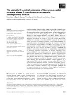
To establish the direct role of this region in c-jun
transcription, CHO cells were transfected with p123jun-
eGFP and p148jun-eGFP plasmids containing GFP as a
reporter gene as shown in Fig. 2A. It is clear from Fig. 2B
that the presence of the )148 to )124 region significantly
increased GFP expression when compared to the control
promoter present in pjun123-eGFP, substantiating the
positive role of this region in c-jun transcription in normal
rat liver.
The )148 to )124 region of c-
jun
binds to factors
present in fractionated rat liver nuclear extract
As preincubation of nuclear extract with the oligonucleotide
()148 to )124) had resulted in a decrease in transcription,
suggesting its interaction with positive factors present
therein, binding reactions were carried out using different
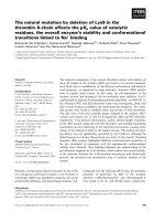
amount of RNE-d. As shown in Fig. 3A, optimum complex
formation was obtained with 100, 150 and 200 lg nuclear
protein in RNE-d (lanes 2–4) while at higher concentrations
of RNE-d (250 and 300 lg, lanes 5 and 6), a decrease in the
complex formation was observed. The factor(s) involved in
the complex formation are designated as RLjunRP [rat liver
jun regulatory protein(s)]. Binding of factors, present in
normal liver, with this region of c-jun intrigued us as earlier
studies [30,31] had shown that the )139 to )129 region of
c-jun is recognized by NF-jun or NF-jun-like transcription
factors present in cellular extracts from TPA-induced
leukaemic cells. This activity was reported to be absent
from nonproliferating diploid cells.
Sequence-specific binding of RLjunRP
Specificity of the complex formation between the factors
and the )148 to )124 region of c-jun was examined
(Fig. 3B) by preincubating 100 lg of the fraction RNE-d
with a 100-fold excess of unlabelled nonspecific DNA
[fragmented calf thymus DNA (lane 7), pBR322 (lane 8)
and unlabelled oligonucleotide (5–20 ng, lanes 3–6)] prior to
the addition of labelled oligonucleotide Jun)25 (1 ng). As is
evident, the complex formation was completely abolished
when RNE-d was preincubated with unlabelled Jun)25
whereas no effect on the complex formation was observed
when a 100- to 200-fold excess of nonspecific DNA was
used for competition, indicating the specificity of complex
formation. The complex formation did not take place in the
Fig. 2. Effect of )148 to )124regiononc-jun promoter activity. (A)
Schematic diagram of plasmids p123jun-eGFP and p148jun-eGFP
used in reporter gene assay. Plasmid p123jun-eGFP consists of the
)123 to +53 region of c-jun cloned upstream of the GFP coding
region and p148jun-eGFP consists of the )148 to +53 region of c-jun
cloned upstream of the GFP coding region. (B) Transfection assay and
GFP expression under the control of the c-jun promoter. CHO cells
(2 · 10
5
cellsÆmL
)1
, in triplicate) were transfected with 2.5 lg p123jun-
eGFP or p148jun-eGFP along with 1 lgofpSV-bgal plasmid. Cells
transfected with 2.5 lgpEGFP-N1and1lgpSV-bgal served as a
positive control. Relative fluorescence shown here represent
mean + SEM of three independent transfections performed in tripli-
cate for the respective plasmids.
Fig. 1. (A) In vitro transcription of EcoRI-linearized )1100/+
+
170 jun-
CAT plasmid with fractionated rat liver nuclear extract (RNE-d) and (B)
effectofthe)148 to )124 region of c-jun on in vitro transcription of
linearized )1100/+
+
170 jun-CAT plasmid. (A) Linearized template
(12 lgÆmL
)1
) was transcribed with rat liver fraction RNE-d (0.4 and
0.8 lgÆmL
)1
, lanes 1 and 2, respectively). The arrow points to the
370-nucleotide-long run-off transcript and M indicates end-labelled
molecularmassmarkers/X174 DNA digested with HaeIII. (B)
In vitro transcription reactions were carried out using 10 lgÆmL
)1
EcoRI linearized plasmid )1100/+170 jun-CAT as template and
1.6 mgÆmL
)1
RNE-d (lane 1). Lanes 5–7 represent the transcripts
obtained from in vitro transcription reactions carried out with fract-
ionated nuclear extract preincubated with 10, 20 and 40 ng oligonu-
cleotide Jun)25, encomapassing the )148 to )124 region of c-jun for
20 min prior to the addition of template. Lanes 2–4 represent tran-
scription reaction carried out with RNE-d preincubated with equi-
molar concentrations of pBR322 to the amount of oligonucleotide
used in lanes 5–7, respectively.
184 D. Sharma et al. (Eur. J. Biochem. 270) Ó FEBS 2003
presence of 7.5% formamide further confirming the speci-
ficity of protein–oligo interaction (lane 2) as formamide is
known to dissociate the protein factors from the recognition
sequence.
The presence of high affinity of RLjunRP for its cognate
sequence was established by performing binding reactions in
the absence of fragmented calf thymus DNA (Fig. 3B, lane
2) which is used to titrate out nonspecific DNA binding
protein. RLJunRP present in crude nuclear extract could
bind even in the absence of nonspecific DNA showing that
it has a high binding affinity enabling it to compete with the
nonspecific DNA-binding proteins present in the extract.
Regulatory proteins are known to bind to their specific
recognition sites with higher affinity than unrelated DNA
sequence [32].
Specific DNA-binding proteins can bind nonspecifically
to nontarget DNA, albeit with low affinity. Therefore, if
excessive nonspecific DNA is added, it will compete for the
specific factor of interest and the level of the specific
complex will decrease. Binding reactions were performed
using 100 lg RNE-d and 1 ng labelled )148 to )124 region
of c-jun in the presence of much higher excess of fragmented
calf thymus DNA to inspect the specificity of the interac-
tions between RLjunRP and the )148 to )124 region of
c-jun. When RNE-d was incubated with labelled Jun)25
oligonucleotide in the presence of a 1000-, 10 000-, 20 000-
and 40 000-fold excess of nonspecific fragmented calf
thymus DNA (Fig. 3C; lanes 1–4), specific DNA–protein
adducts were observed confirming the remarkable specificity
of RLjunRP.
The optimum concentration of monovalent cations was
determined by carrying out EMSA using 100 lg nuclear
proteins and 1 ng labelled )148 to )124 region of c-jun in
the presence of different concentrations of NaCl. Complex
formation was observed over a range of concentration of
monovalent cations, i.e. 25–250 m
M
(Fig. 4A, lanes 1–5). At
500 m
M
(lane6),therewasadecreaseinthecomplex
formation. The fact that RLjunRP retained its binding
activity even in the presence of 0.5
M
NaCl indicated that
the factor has a higher than usual affinity to the recognition
sequence. Most of the DNA-binding proteins exhibit
binding activity with a rather limited range of monovalent
cations with optimal binding at either low or high salt
concentrations. The RNA polymerase II transcription
factor TFIIB (which is considered to be unusual in terms
of high salt resistance) can be stripped off its cognate DNA
sequence by high salt concentrations [33]. It was observed
that TFIIB could bind to its specific sequence only at low
salt concentration, following which it can withstand increa-
ses in NaCl concentration. However, TFIIB cannot bind at
high salt concentration. RLjunRP, in contrast, can actually
bind to its recognition sequence at a relatively higher salt
concentration. The fact that the complex formation between
RLjunRP and the )148 to )124 region of c-jun was not
highly affected by the fluctuation in NaCl concentration
indicates that the protein–DNA association is probably
through interactions that are nonionic.
The involvement of divalent cations that are required for
certain protein–cognate sequence interaction was investi-
gated by carrying out EMSA in the presence of EDTA
(Fig. 4B). Inclusion of 100 m
M
EDTA in the binding
reaction resulted in a slight decrease in complex formation
(lane 3) and no complex was observed in the presence of
150 m
M
EDTA (lane 4). It is likely that in the presence of 50
or 100 m
M
EDTA (Fig. 4B, lanes 2 and 3, respectively),
most of the divalent cations are chelated but there might still
be small amounts of free divalent cations (unchelated),
which are sufficient for complex formation. When the
EDTA concentration is raised to 150 m
M
(Fig. 4B, lane 4),
all of these ions are chelated and no complex formation is
observed. These data suggest that very small amounts of
divalent cations are necessary for the formation of complex
between RLjunRP and the )148 to )124 region of c-jun,
and so the optimum amount of MgCl
2
required for complex
formation was then titrated (Fig. 4C). Complex formation
couldbeseeninthepresenceof1m
M
MgCl
2
(lane 1).
Binding was found to be maximal in the presence of 2.5 m
M
MgCl
2
(lane 2).
Studies on the effect of temperature (Fig. 4D) on complex
formation revealed that the factors present in RNE-d
formed the complex even at temperature as low as 0 °C
Fig. 3. Specificity of complex formation between )148 to )124 region of
c-jun and factors present in RNE-d. (A) Titration of optimum concen-
tration of nuclear extract for binding. EMSA reactions were carried out
in the presence of 1 ng )148 to )124 region of c-jun and various con-
centrations of nuclear proteins as indicated. (B) jun-RP forms specific
complex with the )148 to )124 region of c-jun. Lane 1 represents the
interaction of factor(s) present in fraction RNE-d with 1 ng )148 to
)124 region of c-jun. EMSA reactions were carried out using 100 lgof
RNE-d preincubated with a 100-fold excess of unlabelled nonspecific
DNA [fragmented calf thymus DNA (lane 7), pBR322 (lane 8)], and in
the presence of various concentrations of unlabeled Jun)25 oligo-
nucleotide encompassing the )148 to )124 region of c-jun (lanes 3–6)
prior to the addition of labelled Jun)25. Lane 2 depicts the binding
reaction carried out in the presence of 7.5% of formamide. (C)
RLjunRP can form complexes even in the presence of a 40 000-fold
excess of fragmented calf thymus DNA. The binding reactions were
carried out with 1 ng labelled )148 to )124 region of c-jun and 100 lg
fractionated nuclear extracts in the presence of 1 lg(lane1),10lg
(lane 2), 20 lg(lane3)and40 lg (lane 4)fragmented calf thymus DNA.
Ó FEBS 2003 Regulation of c-jun expression in rat liver (Eur. J. Biochem. 270) 185
(lane 1). No significant change in complex formation was
observed untill 30 °C (lanes 2–6). However, very little
complex formation occurred when EMSA was carried out
at 45 °C (lane 7) and no complex was formed at 55 °C
onwards. Unlike TATA binding protein that becomes
totally inactivated within 15 min of heat treatment at 47 °C
[34], junRP retains its DNA-binding activity, although at a
relatively low level, even when the binding reaction was
carried out at 45 °C for 30 min.
Phosphorylation of RLjunRP is imperative for its
DNA-binding activity
Inducible phosphorylation or dephosphorylation of tran-
scription factors is an important mechanism of signal
dependent gene regulation in eukaryotic cells [35,36]. It is
generally assumed that protein phosphorylation stabilizes
different conformational states of the regulated and
regulatory molecule to enhance or inhibit biological
activity [36–40]. To check whether RLjunRP interacts
with the )148 to )124 region of c-jun in the phospho-
rylated or dephosphorylated form, nuclear extract from
normal liver was treated with various concentrations of
calf intestinal alkaline phosphatase prior to its addition to
the EMSA reaction (Fig. 4E). A decrease in complex
formation was observed with increasing concentrations of
alkaline phosphatase from 4 U upwards and the treat-
ment of RNE-d with 20 U of enzyme completely
abolished DNA binding (lane 2) suggesting that
RLjunRP interacts with the cis-element only in phos-
phorylated form. The inhibitory effect of phosphorylation
on DNA binding is depicted by a number of trans-acting
factors whereas phosphorylation is necessary for DNA
binding in very few cases [35], making RLjunRP unique
in this respect. It is possible that phosphorylation of
RLjunRP is imperative to maintain its DNA-binding
domain in an active conformation.
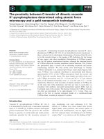
RLjunRP is an 40 kDa protein that forms an 80-kDa
protein–DNA adduct
To assess approximate molecular mass of the factors
interacting with the )148 to )124 region of c-jun, RLjunRP
complexed with this region was UV irradiated (254 nm) for
15 min. After separation by SDS/PAGE on a 15%
acrylamide gel, the complex was visualized by autoradio-
graphy (Fig. 5A). The molecular mass of the cross-linked
junRP was 80 kDa as evident from lane 1. The protein–
DNA complex shows a retarded electrophoretic mobility as
compared with the free DNA fragment. The parameter for
the degree of retardation of a linear DNA fragment bound
in a complex with its specific factors reflects the molecular
mass of the bound protein(s), as the molecular mass of
DNA is negligible [41–43] in terms of the charge : mass
ratio required to alter the mobility of the complex. South-
Western analysis of rat liver nuclear extract using the )148
to )124 region as a probe, revealed a hybridized band of
40 kDa (Fig. 5B). These data suggest that RLjunRP
binds to its recognition sequence as a dimer.
Affinity purified RLjunRP is a protein of 40 kDa
To confirm that RLjunRP is indeed a protein of 40 kDa,
it was affinity purified from rat liver nuclear extract (Fig. 6).
Major peak fractions eluted between 0.1
M
and 0.2
M
NaCl
(Fig. 6A) contained nonspecific DNA binding proteins, as
these fractions did not show any complex formation in
EMSA (Fig. 6B). The factor(s) interacting with the )148 to
Fig. 4. Binding characteristics of RLjunRP. (A) Titration of optimum
monovalent cation concentration. Binding reactions between junRP
and the )148 to )124 region of c-jun were carried out in the presence of
25, 50, 75, 100, 250 and 500 m
M
NaCl (lanes 1–6, respectively) using
100 lg nuclear extract and 1 ng labelled )148 to )124 region of c-jun.
(B) Divalent cations are absolutely essential for the binding activity of
junRP(s). EMSA were carried out with 100 lgfractionatednuclear
extract RNE-d and 1 ng labelled )148 to )124 region of c-jun in the
presence of 25, 50, 100 and 150 m
M
EDTA (lanes 1–4, respectively). (C)
Determination of optimum divalent cation concentration for complex
formation. EMSA were carried out using 1 ng labelled )148 to )124
region of c-jun and 100 lg fractionated nuclear extract from normal rat
liver in the presence of 1 m
M
(lane 1), 2.5 m
M
(lane 2), 5 m
M
(lane 3),
10 m
M
(lane 4), 15 m
M
(lane 5) and 20 m
M
(lane 6) MgCl
2
and analysed
by nondenaturing PAGE on 6%acrylamide gels.(D) Complex between
Jun)25 and RLjunRP forms over a wide temperature range. The
binding reactions between 100 lg fraction RNE-d from normal liver
and 1 ng labelled )148 to )124 region of c-jun were carried out at
temperatures ranging from 0 to 65 °C (lanes 1–9). (E) Phosphorylation
of RLjunRP is necessary for its DNA-binding activity. One-hundred
micrograms fractionated nuclear extract from normal rat liver was
treated with different concentrations of calf intestine alkaline phos-
phatase (shown at the top) prior to its addition to EMSA. Lane 1 shows
the complex formed between RNE-d treated with heat inactivated
alkaline phosphatase (10 U) and labelled Jun)25.
186 D. Sharma et al. (Eur. J. Biochem. 270) Ó FEBS 2003
)124 region of c-jun eluted in the 2.0
M
NaCl fraction
(fractions 38–42) as evident from the formation of retarded
complex with labelled )148 to )124 region of c-jun.Allof
the proteins that do not interact with the )148 to )124
region of c-jun, may nonspecifically bind to the affinity
matrix and be eluted at a lower salt concentration. SDS/
PAGE of different peaks obtained from affinity chroma-
tography showed a band of 40 kDa (Fig. 6C, lane P).
Presence of a purified factor of 40 kDa is consistent with
our South-Western data. These data further confirm that
RLjunRP is indeed a protein of 40 kDa and binds to its
recognition sequence as a dimer. Dimerization of several
transcription factors has been found to be necessary for
their interaction with recognition sequence [44,45]. It is
likely that dephosphorylation (which results in complete
loss of complex formation) results in the dissociation of the
dimers and the monomers are not able to bind to the )148
to )124 region of c-jun.
This study thus provides an insight into the molecular
mechanisms regulating the c-jun expression in quiescent
cells. The data indicate that the )148 to )124 region of c-jun
is a functional motif present upstream of the gene promoter
region, interacting with positive regulatory trans-acting
factors present in rat liver. Although previous studies have
reported the presence of an inducible factor, NF-jun, in
human myeloid leukaemia cells that protected the )139 to
)129 region of c-jun [30], NF-jun binding activity was found
to be absent from nonproliferating diploid cells and
appeared to be restricted to dividing cells [30,31] as growth
arrested human embryonic lung fibroblasts, granulocytes
and resting human T cells did not express NFjun constitu-
tively. Further in Hela cells, it has been shown that NF-jun
is already bound to its recognition sequence (before
transcriptional activation of c-jun by TPA and UV irradi-
ation). Thus, NF-jun behaves differently in different cell
types, being translocated from the cytosol to the nucleus
upon induction by an external stimulus in human myeloid
leukaemia cells but found already bound to c-jun gene in
uninduced Hela cells.
Thus, RLjunRP differs from the factor NF-jun reported
by Brach et al. [30] (that interacts with the )139 to )132
Fig. 6. Affinity Purification of factors interacting with the )148 to )124
region of c-jun. (A) Spectrophotometric elution Profile: RNE-d was
subjected to sequence-specific affinity column chromatography and all
fractions obtained were analysed spectrophotometrically. Absorbance
at 280 nm was measured and plotted. (B) Assessment of complex
formation ability of eluted fractions from DNA affinity column.
Presence of RLjunRP in different fractions obtained by affinity chro-
matography was checked using EMSA with labelled )148 to )124
oligonucleotide fragment of c-jun. L represents EMSA reaction with
the loaded fraction and the numbers on top represent the fraction
numbers. The numbers at the bottom represent the salt concentration
in the respective fraction. (C) SDS/PAGE of RLjunRP-positive frac-
tion. The fractionated nuclear extract, RNE-d fraction (L), flow-
throughfraction(F)andpeakfractionnumber38(P)showingDNA
binding ability in EMSA, were subjected to SDS/PAGE and silver
stained. M represents the mid-range molecular mass markers.
Fig. 5. UV cross-linking and South-Western blot analysis. (A) Deter-
mination of the molecular mass of complex between junRP and the
)148 to )124 region of c-jun by UV cross-linking. Complex between
RLjunRP (lane 1) with its cognate sequence was formed under
standard conditions using 100 lgRNE-dand1 ng)48 to )124 region
of c-jun followed by UV irradiation (254 nm) for 15 min. DNA–pro-
tein complex was separated from free DNA by SDS/PAGE. Autora-
diography revealed the presence of complex (shown by arrowhead).
Numbers represent protein molecular mass markers. (B) South-West-
ern blot analysis of fraction RNE-d with Jun)25. Fifty and 75 lg
nuclear extract fraction RNE-d were fractionated by SDS/PAGE
(lanes 1 and 2), transferred onto a nitrocellulose sheet and probed with
radiolabelled tetramer of Jun)25 oligonucleotide. The molecular mass
of the markers is shown on the left.
Ó FEBS 2003 Regulation of c-jun expression in rat liver (Eur. J. Biochem. 270) 187
region) with respect to it being present in resting liver cells
whereas NF-jun is found to be restricted to rapidly dividing
cells such as myeloid leukaemia cells and is not detectable in
nonproliferating diploid lung fibroblasts, blood monocytes,
granulocytes or resting T cells. Thus, in vivo occupancy of
the )148 to )124 region in the c-jun promoter with
RLjunRP cannot generally be associated with the prolifer-
ative state of the cells. Further, NF-jun forms DNA–protein
adducts of 55 and 125 kDa as established by UV cross-
linking studies suggesting that it can bind to the sequence
both as a monomer and dimer [20]. Unlike NF-jun,
RLjunRP shows only a single complex at 80 kDa in
UV cross-linking studies whereas the purified protein is only
40 kDa, suggesting that it binds only as a dimer. Absence
of an 40 kDa DNA-protein adduct in UV cross-linking
studies indicates that RLjunRP is not able to bind as a
monomer.
Thus, we have clearly demonstrated a direct involvement
of the )148 to )124 region of c-jun in its transcription and
its interaction with positive regulatory factor (RLjunRP) in
normal rat liver. The positive regulatory factor interacting
with this region was purified to homogeneity and the cDNA
cloning of the gene encoding this factor is in progress to help
in understanding its structural and functional aspects.
Acknowledgements
P. Angel, Institute for Genetik, Kernforschungszentrum Karlruhe,
GmBH Postfach 3640 D-76021, Karlsruhe, Germany is gratefully
acknowledged for providing the )1100/+170 jun-CAT plasmid. This
work was supported by a research grant (#37(834)/94-EMR-II) from
the Council of Scientific and Industrial Research (CSIR), India to A.D.
CSIR, India is duly acknowledged for the Senior Research Fellowships
to D.S. and S.O. The technical assistance of S. Singh is sincerely
appreciated. The animal work included in this paper had the approval
of Institutional Animal Ethics Committee, JNU (IAEC-JNU Project
Code no. 27/1999).
References
1. Curran, T. & Franza, B.R., Jr (1988) Fos and Jun: the AP-1
connection. Cell 55, 395–397.
2. Hirai, S., Bourachot, B. & Yaniv, M. (1990) Both Jun and Fos
contribute to transcription activation by the heterodimer. Onco-
gene 5, 39–46.
3. Vogt, P.K. & Bos, T.J. (1990) Jun: oncogene and transcription
factor. Adv. Cancer Res. 55, 1–35.
4. Angel, P., Allegretto, E.A., Okino, S.T., Hattori, K., Boyle, W.J.,
Hunter, T. & Karin, M. (1988) Oncogene jun encodes a sequence-
specific trans-activator similar to AP-1. Nature 332, 166–170.
5. Angel, P., Hattori, K., Smeal, T. & Karin, M. (1988) The jun
proto-oncogene is positively autoregulated by its product, Jun/
AP-1. Cell 55, 875–885.
6. Angel, P. & Karin, M. (1991) The role of Jun, Fos and the AP-1
complex in cell proliferation and transformation. Biochem. Bio-
phys. Acta 1072, 129–157.
7. Goswami, S.K., Shafiq, S. & Siddiqui, M.A. (2001) Modulation of
MLC-2v gene expression by AP-1: complex regulatory role of Jun
in cardiac myocytes. Mol. Cell. Biochem. 217, 13–20.
8. Yuen, M.F., Wu, P.C., Lai, V.C., Lau, J.Y. & Lai, C.L. (2001)
Expression of c-myc, c-fos and c-jun in hepatocellular carcinoma.
Cancer Res. 91, 106–112.
9. Bohmann,D.,Bos,T.J.,Admon,A.,Nishimura,T.,Vogt,P.K.&
Tjian, R. (1987) Human protooncogene c-jun encodes a DNA
binding protein with structural and functional properties of tran-
scription factor AP-1. Science 238, 1386–1392.
10. Karin, M., Liu, Z. & Zandi, E. (1997) AP-1 function and reg-
ulation. Curr. Opin. Cell Biol. 9, 240–246.
11. Xia, Y., Buja, L.M. & McMillin, J.B. (1998) Activation of the
cytochrome c gene by electrical stimulation in neonatal rat cardiac
myocytes. Role of NRF-1 and c-jun.J.Biol. Chem. 273, 12593–12598.
12. Steinmuller, L., Cibelli, G., Moll, J.R., Vinson, C. & Thiel, G.
(2001) Regulation and composition of activator protein 1 (AP-1)
transcription factors controlling collagenase and c-jun promoter
activities. Biochem. J. 360, 599–607.
13. Coso, O.A., Chiariello, M., Kalinec, G., Kyriakis, J.M., Wood-
gett, J. & Gutkind, J.S. (1995) Transforming G protein-coupled
receptors potently activate JNK (SAPK). J. Biol. Chem. 270,
5620–5624.
14. Lallemand, D., Ham, J., Garbay, S., Bakiri, L., Traincard, F.,
Jeannequin, O., Pfarr, C.M. & Yaniv, M. (1998) Stress-activated
protein kinases are negatively regulated by cell density. EMBO J.
17, 5615–5626.
15. Oguro, T., Hayashi, M., Nakajo, S., Numazawa, S. & Yoshida, T.
(1998) The expression of heme oxygenase-1 gene responded to
oxidative stress produced by phorone, a glutathione depletor, in
the rat liver; the relevance to activation of c-jun n-terminal kinase.
J. Pharmacol. Exp. Ther. 287, 773–778.
16. Wisdom, R., Johnson, R.S. & Moore, C. (1999) c-jun regulates cell
cycle progression and apoptosis by distinct mechanisms. EMBO J.
8, 188–197.
17. Sambrook, J., Fritsch, E.F. & Maniatis, T. (1989) Molecular
Cloning: a Laboratory Manual, 2nd edn. Cold Spring Harbor
Laboratory Press, Cold Spring Harbor, NY.
18. Dixit, A., Garg, L.C., Chao, W. & Jacob, S.T. (1987) An enhancer
element in the far upstream spacer region of rat ribosomal RNA
gene. J. Biol. Chem. 262, 11616–11622.
19. Sharma, D., Choudhary, S.K. & Dixit, A. (1998) In vitro tran-
scription of c-jun gene using fractionated nuclear extract from
regenerating rat liver. Biochem. Mol. Biol. Int. 44, 1175–1185.
20. Dixit, A., Garg, L.C. & Jacob, S.T. (1989) A cis-acting sequence
within the rat ribosomal DNA enhancer region can modulate
RNA polymerase II-directed transcription of the metallothionein I
gene in vitro. DNA 8, 311–320.
21. Chalfie, M., Tu, Y., Euskirchen, G., Ward, W.W. & Prasher, D.C.
(1994) Green fluorescent protein as a marker for gene expression.
Science 263, 802–805.
22. Subramanian, S. & Sriene, F. (1996) Quantitative analysis of
transient gene expression in mammalian cells using the green
fluorescent protein. J. Biotechnol. 49, 137–151.
23. Garg, L.C., Dixit, A. & Jacob, S.T. (1989) A 37-base pair element
in the far upstream spacer region can enhance transcription of rat
rDNA in vitro and can bind to the core promoter-binding factor
(s). J. Biol. Chem. 264, 220–224.
24. Parthun, M.R. & Jaehning, J.A. (1992) A transcriptionally active
form of Gal4 is phosphorylated and associated with Gal80. Mol.
Cell. Biol. 12, 4981–4987.
25. Ho, D.T., Sauve, D.M. & Roberge, M. (1994) Detection and
isolation of DNA-binding proteins using single pulse ultraviolet
laser crosslinking. Anal. Biochem. 218, 248–254.
26. Philippe, J. (1994) The Southwestern Assay. In Methods in
Moleuclar Biology, Protocols for Gene Analysis 31 (Harwood, A.J.,
ed.), pp. 349–361. Humana Press Inc., Totowa, NJ.
27. Kadonaga, J.T. & Tjian, R. (1986) Affinity purification of
sequence-specific DNA binding protein. Proc. Natl Acad. Sci.
USA 83, 5889–5893.
28. Dunn, M.J. & Crisp, S.J. (1994) Detection of proteins in poly-
acrylamide gels using an ultrasensitive silver staining technique. In
Basic Protein and PeptideProtocols, Methods in Molecular Biology 32
(Walker, J.M., ed.), pp. 113–118. Humana Press Inc., Totowa, NJ.
188 D. Sharma et al. (Eur. J. Biochem. 270) Ó FEBS 2003
29. Devary, Y., Gottlieb, R.A., Smeal, T. & Karin, M. (1992) The
mammalian ultraviolet response is triggered by activation of Src
tyrosine kinases. Cell 72, 1081–1091.
30. Brach, M.A., Herrmann, F., Yamada, H., Bauerele, P.A. & Kufe,
D.W. (1992) Identification of NF-jun, a novel inducible tran-
scription factor that regulates c-jun gene transcription. EMBO J.
11, 1479–1486.
31. Rozek, D. & Pfeifer, G.P. (1993) In vivo protein–DNA interac-
tions at the c-jun promoter: Preformed complexes mediate the UV
response. Mol. Cell. Biol. 13, 5490–5499.
32. Strauss, F. & Varshavsky, A. (1984) A protein binds to a satellite
DNA repeat at three specific sites that would be brought into
mutual proximity by DNA folding in the nucleosome. Cell 37,
889–901.
33. Kassavetis,G.A.,Braun,B.R.,Nguyen,L.H.&Geiduschek,E.P.
(1990) S. cerevisiae TFIIIB is the transcription initiation factor
proper of RNA-polymerase III, while TFIIIA and TFIIIC are
assembly factors. Cell 60, 235–245.
34. Nakajima, N., Horikoshi, M. & Roeder, R.G. (1988) Factors
involved in specific transcription by mammalian RNA polymerase
II: purification, genetic specificity and TATA box–promoter
interactions of TFIID. Mol. Cell. Biol. 8, 4028–4040.
35. Hunter, T. & Karin, M. (1992) The regulation of transcription by
phosphorylation. Cell 70, 375–387.
36. Papavassiliou, A.G., Bohmann, K. & Bohmann, D. (1992)
Determining the effect of inducible protein phosphorylation on the
DNA-binding activity of transcription factors. Anal. Biochem.
203, 302–309.
37. Sprang, S.R., Acharya, K.R., Goldsmith, E.J., Stuart, D.I.,
Varvill, K., Fletterick, R.J., Madsen, N.B. & Johnson, L.N. (1988)
Structural changes in glycogen phosphorylase induced by phos-
phorylation. Nature 336, 215–221.
38. Yamamoto, K.K., Gonzalez, G.A., Biggs, W.H. & Montminy,
M.R. (1988) Phosphorylation-induced binding transcriptional
efficacy of nuclear factor CREB. Nature 334, 494–498.
39. Luscher, B., Christenson, E., Litchifield, D.W., Krebs, E.G. &
Eisenman, R.N. (1990) Myb DNA binding inhibited by phos-
phorylation at a site deleted during oncogenic activation. Nature
344, 517–521.
40. Copenhaver, G.P., Putnam, C.D., Denton, M.L. & Pikaard, C.S.
(1994) The RNA polymerase I transcription factor UBF is a
sequence-tolerant HMG-box protein that can recognize structured
nucleic acids. Nucleic Acids Res. 22, 2651–2657.
41. Chodosh, L.A., Carthew, R.W. & Sharp, P.A. (1986) A single
polypeptide possesses the binding and transcription activities of
the adenovirus major late transcription factor. Mol. Cell. Biol. 6,
4723–4733.
42. Feavers,I.M.,McEwan,I.J.,Liang,H.&Jost,J.P.(1989)An
estradiol-dependent protein from chicken liver binds single-
stranded DNA and RNA. J. Biol. Chem. 264, 9114–9117.
43. Hughes,M.J.&Jost,J.P.(1989)Theubiquitousnuclearprotein,
NHPI, binds with high affinity to different sequences of the
chicken vitellogenin II gene. Nucleic Acid Res. 17, 8511–8520.
44. Sengchanthalangsy, L.L., Datta, S., Huang, D.B., Anderson, E.,
Braswell, E.H. & Ghosh, G. (1999) Characterization of the dimer
interface of transcription factor NfkappaB p50 homodimer.
J. Mol. Biol. 289, 1029–1040.
45. Shen, Q. & Subauste, J.S. (2000) Dimerization interfaces of v-erbA
homodimers and heterodimers with retinoid X receptor alpha.
J. Biol. Chem. 275, 41018–41027.
Ó FEBS 2003 Regulation of c-jun expression in rat liver (Eur. J. Biochem. 270) 189