Báo cáo khoa học: Fluorescence and FTIR study of pressure-induced structural modifications of horse liver alcohol dehydrogenase (HLADH) potx
Bạn đang xem bản rút gọn của tài liệu. Xem và tải ngay bản đầy đủ của tài liệu tại đây (426.54 KB, 10 trang )
Fluorescence and FTIR study of pressure-induced structural
modifications of horse liver alcohol dehydrogenase (HLADH)
Marie Trovaslet
1
, Sandrine Dallet-Choisy
1
, Filip Meersman
2
, Karel Heremans
2
, Claude Balny
3
and Marie Dominique Legoy
1
1
Laboratoire de Ge
´
nie Prote
´
ique et Cellulaire, Universite
´
de La Rochelle, France;
2
Department of Chemistry,
Katholieke Universiteit Leuven, Belgium;
3
INSERM U 128, Montpellier Cedex 5, France
The process of pressure-induced modification of horse
liver alcohol dehydrogenase (HLADH) was followed by
measuring in situ catalytic activity (up to 250 MPa),
intrinsic fluorescence (0.1–600 MPa) and modifications of
FTIR spectra (up to 1000 MPa). The tryptophan fluor-
escence measurements and the kinetic data indicated that
the pressure-induced denaturation of HLADH was a
process involving several transitions and that the observed
transient states have characteristic properties of molten
globules. Low pressure (< 100 MPa) induced no
important modification in the catalytic efficiency of the
enzyme and slight conformational changes, characterized
by a small decrease in the centre of spectral mass of the
enzyme’s intrinsic fluorescence: a native-like state was
assumed. Higher pressures (100–400 MPa) induced a
strong decrease of HLADH catalytic efficiency and fur-
ther conformational changes. At 400 MPa, a dimeric
molten globule-like state was proposed. Further increase
of pressure (400–600 MPa) seemed to induce the dissoci-
ation of the dimer leading to a transition from the first
dimeric molten globule state to a second monomeric
molten globule. The existence of two independent struc-
tural domains in HLADH was assumed to explain this
transition: these domains were supposed to have different
stabilities against high pressure-induced denaturation.
FTIR spectroscopy was used to follow the changes in
HLADH secondary structures. This technique confirmed
that the intermediate states have a low degree of unfold-
ing and that no completely denatured form seemed to be
reached, even up to 1000 MPa.
Keywords: alcohol dehydrogenase; FTIR spectroscopy; high
hydrostatic pressure; molten globule; tryptophan fluores-
cence.
Several papers have reported on the effects of high pressure
on protein structures [1–6] or on enzyme catalytic activities
[7–12]. These works have shown that high hydrostatic
pressure can modify the structure or the function of
enzymes by altering intra- or intermolecular interactions
involved in protein stability [13,14]. However, relatively few
studies have been performed to correlate conformational
modifications of an enzyme to changes in its catalytic
activity; although it is generally recognized that conform-
ational integrity is important for preserving the activity of
an enzyme [15–17].
Horse liver alcohol dehydrogenase (HLADH) is a metal
protein containing two zinc ions. This enzyme is mesostable
and dimeric, consisting of two identical subunits.
The reasons for investigating HLADH in the present
work are several. This enzyme is believed to be represen-
tative of a group of proteins: dehydrogenases. Its kinetics
and its three-dimensional structure are well known at
ambient pressure (0.1 MPa). Its catalytic behaviour under
pressure has already been studied [7,8]: Morild has shown
that HLADH presented a complicated behaviour at high
pressure (which was believed to be due to the pressure-
induced modifications of the substrate inhibition pheno-
menon occurring at ethanol concentrations > 10 m
M
).
Moreover, the conformational changes of this enzyme have
already been monitored as a function of pressure (up to
300 MPa) by means of the intrinsic tryptophan fluores-
cence, phosphorescence emission and binding of ANS
fluorophore [18,19]. These studies have revealed unequivo-
cal perturbations of HLADH structure in the region of the
chromophores. Analysis of these structural modifications
seemed to lead to the conclusion that, at about 300 MPa,
the pressure induced a dimeric molten globule-like state
rather than HLADH subunit dissociation.
In the present work, pressure effects on HLADH
structure also have been studied by intrinsic fluorescence.
In fact, the intrinsic fluorescence of a protein is due mainly
to the tryptophan residues [20,21] with some exceptions
where the emission could be dominated by tyrosine [22]. The
wavelength at maximum fluorescence, thus the centre of
the spectral mass (CSM), depends on the polarity of the
environment around these residues; for example, the CSM
decreases as the polarity of the environment increases [4].
Based on this characteristic, the environment of the
tryptophans can be monitored upon pressure-induced
Correspondence to S. Dallet-Choisy, Laboratoire de Ge
´
nie
Prote
´
ique et Cellulaire, Universite
´
de La Rochelle,
Avenue Michel Cre
´
peau, La Rochelle, France.
Fax: +33 5 46 45 82 47, Tel.: +33 5 46 45 82 77,
E-mail:
Abbreviations: HLADH, horse liver alcohol dehydrogenase;
CSM, center of spectral mass; FTIR, Fourier transform infrared;
MG,moltenglobule;DAC,diamondanvilcell.
Enzyme: horse liver alcohol dehydrogenase (EC 1.1.1.1).
(Received 20 June 2002, revised 30 September 2002,
accepted 18 November 2002)
Eur. J. Biochem. 270, 119–128 (2003) Ó FEBS 2003 doi:10.1046/j.1432-1033.2003.03370.x
protein denaturation [23]. Moreover, tryptophan residues
have specific positions in the HLADH molecule. The
enzyme has two tryptophans per subunit: tryptophan 15 is
always exposed to the solvent whereas tryptophan 314 lies
right at the subunit interface [24] and upon dissociation will
become exposed to the solvent. So any modifications of the
enzyme’s intrinsic fluorescence would be a proof of
pressure-induced unfolding, denaturation and/or dissoci-
ation of the dimeric enzyme. However, because of technical
limitations, the pressure in the fluorescence experiments did
not exceed 600–700 MPa. In order to follow possible
changes at higher pressures, Fourier Transform Infrared
Spectroscopy (FTIR) was used. With this technique, in
combination with the diamond anvil cell (DAC), it was
possible to determine the evolution of secondary structures
of a protein, up to pressures of 1000 MPa [25,26]. More-
over, kinetic modifications of HLADH have been followed
under pressure (up to 250 MPa), in a reactor described
previously [27]. Experimental conditions were as those used
for structural studies (30 °C, pH 8). This allowed us to
correlate changes in HLADH catalytic activity to the
pressure-induced conformational changes of this enzyme.
Materials and methods
Reagents
HLADH (alcohol:NAD
+
oxidoreductase), NAD
+
, NADH,
Tris and Mes were from Sigma. Ethanol was from Fluka.
All chemicals were of analytical grade.
Enzyme assays
The enzyme reactions were carried out in 50 m
M
Tris/HCl
pH 8 at 30 °C. Assays of HLADH activity for ethanol
oxidation were followed by NADH absorption increase at
340 nm, directly under pressure, in a reactor described
previously [27]. The initial reaction velocity was expressed
as lmoles NADHÆmin
)1
Æmg protein
)1
using the molar
absorption coefficient of NADH calculated at all pres-
sures.
All reactant solutions were prepared prior to use.
Assays were performed for all substrates (coenzyme and
ethanol) by keeping the concentration of one substrate
constant (always saturating but never inhibitory for ethanol
concentration) whilst varying the concentration of the other,
as shown in Table 1. The enzyme concentration was
0.625 lgÆmL
)1
.
The apparent Michaelis constant (K
m
) and the maximum
velocity (V
m
) were determined using
STATISTICA
Ò software
from the least-squares fit at all given pressures.
Fluorescence spectroscopy
Experimental procedures. Intrinsic fluorescence measure-
ments were carried out on an AB2 fluorospectrophotometer
(US SLM Co.), modified in the Montpellier laboratory to
measure fluorescence in the pressure range 0.1–700 MPa,
through a thermostated cell [4]. The maximum pressure
applied was 600 MPa and the temperature was constant at
30 °C.
The enzyme concentration was 0.5 mgÆmL
)1
,1mgÆmL
)1
and 5 mgÆmL
)1
(in 50 m
M
Mes buffer pH 8), depending on
the experiments. Samples were placed in a 0.5 mL quartz
cuvette (5 mm path length) and were allowed to reach
equilibrium for 5 min before data collection. In order to
investigate selectively tryptophan fluorescence, the excita-
tion wavelength was 295 nm (4 nm slit, 1 nm step size). The
emission spectra were recorded between 300 nm and
370 nm (8 nm slit, 1 nm step size). Each spectrum was the
result of three accumulations.
Fluorescence intensities were first corrected by subtract-
ing the fluorescence spectra of the buffer at each pressure
and then they were corrected for volume contraction under
high pressure. The CSM, defined below (Eqn 1), was used to
quantify the spectra [21,28,29].
CSM ¼
X
vi  Fi=
X
Fi ð1Þ
where mi is the wavenumber and Fi the fluorescence intensity
at mi.
Determination of thermodynamic parameters. Thermody-
namic parameters DG
0
and DV of the pressure-induced
spectral transitions were determined by analysing the CSM.
A two state-transition (n for native and d for denatured)
could be assumed. Generally, if CSM was assumed to take
different values in the two states (CSMn and CSMd) and an
increase of pressure to cause a transition from one state to
the other, then an equilibrium constant K(p) can be defined
at a given pressure (Eqn 2):
KðpÞ¼
CSMd À CMSp½
CSMp À CSMn½
ð2Þ
where CSMp is the intermediate value of the CSM at a given
pressure (P).
Table 1. Substrate concentrations used for activity measurements under pressure.
Pressure (MPa)
< 100 > 100 > 200
Varying concentration of coenzyme
Ethanol concentration (m
M
) 20 200
NAD
+
concentration (m
M
) Varying from 0 to 3
Varying concentration of alcohol
Ethanol concentration (m
M
) Varying from 0 to 700
NAD
+
concentration (m
M
) 0.62 12
120 M. Trovaslet et al. (Eur. J. Biochem. 270) Ó FEBS 2003
The pressure dependence of K(p)canberelatedtoDG
0
(at atmospheric pressure) and DV (3) [30]:
ÀRT ln½KðpÞ ¼ DG
0
þ PDV ð3Þ
Combining Eqns (2) and (3):
CSM ¼ CSMd þ
CSMn À CSMd
1 þ e
ÀDG
0
þPDV
RT
ÀÁ
ð4Þ
Eqn (4) was used to determine HLADH thermodynamic
parameters upon pressure-induced structural modifications.
The pressure of half denaturation (P
1/2
)isgivenbyEqn(5):
P
1=2
¼ÀDG
0
=DV ð5Þ
FTIR spectroscopy
The pressure-induced structural modifications of HLADH
was also investigated by FTIR, in the Leuven laboratory, by
using the DAC (Diacell Products, Leicester, UK). The
gasket between the diamonds had an original thickness of
50 lm. The DAC was connected to a thermostat and the
temperature inside the cell was measured by means of a
thermocouple localized near the diamonds [5,25]. This
allowed measurements at stable temperature of 30 °C.
Barium sulfate was used as an internal pressure standard
[31]. The infrared spectra were obtained with a Bruker
IFS66 FTIR spectrometer equipped with a liquid nitrogen
cooled broad-band mercury–cadmiun–telluride detector.
The sample compartment was continuously purged with
dry air. A total of 250 interferograms were coadded after
registration at a resolution of 2 cm
–1
. Spectral noises
originating from water vapour were removed through the
subtraction of the water vapour spectrum using the
BRUKER software. Spectra shown were smoothed 9
points after vapour correction. The second-derivative spec-
tra containing the amide I¢ band of HLADH were obtained
through application of the 9-data-point Savitzky–Golay
function available from Grams Research software.
HLADH concentration was 50 mgÆmL
)1
. The lyophi-
lized protein was dissolved in 10 m
M
deuterated Tris/DCl
pD 8. Due to its high concentration, the protein itself could
buffer the solution [5]. In this case, the pH value of the
buffer was the same as the pH obtained by measuring that
of the protein solution. After solubilization, the sample was
stored overnight at 25 °C to ensure that all the solvent
accessible protons are exchanged for deuterons.
Results and discussion
Pressure-induced kinetic modifications
In our preliminary report on HLADH reaction under
pressure [32], kinetic parameters and thermodynamic acti-
vation volumes of HLADH oxidation of ethanol with the
coenzyme NAD
+
as oxidizing agent were determined.
Based on this previous study, kinetic parameters of this
reaction (V
m
and K
m
) were now determined always using
noninhibitory ethanol concentrations. Fig. 1 shows the
pressure dependence of these kinetic parameters and of
HLADH catalytic efficiency (inset), respectively, at
saturating NAD
+
concentration and varying ethanol
concentration (Fig. 1A), and at saturating (but not inhibi-
tory) ethanol concentration and varying NAD
+
concentra-
tion (Fig. 1B).
Whatever the varying substrate concentration, up to
200 MPa, the V
m
values increased with an increase in
pressure: at 200 MPa, V
m
was about 10 times higher than at
0.1 MPa. In the same range of pressure, HLADH affinities
for both substrates (NAD
+
and ethanol) seemed to be
strongly decreased. The K
m
values for NAD
+
and ethanol,
at atmospheric pressure and at 225 MPa, were 18.92 ±
1.97 l
M
and 561.82 ± 68.93 l
M
,0.56±0.03m
M
and
24.51 ± 3.17 m
M
, respectively (Fig. 1A and B). Conse-
quently, these variations could influence HLADH catalytic
efficiency (k
cat
/K
m
). Up to 75–100 MPa (depending on the
varying substrate concentration), k
cat
/K
m
values were
almostthesameasat0.1MPa:thepressuredidnotseem
to influence HLADH catalytic efficiency. Above 100 MPa,
further elevation of pressure gave a lower catalytic effi-
ciency. At 225 MPa, the k
cat
/K
m
value was reduced by
% 70% compared with the value obtained at 0.1 MPa. As
shown in Fig. 1A and B (inset), the plot of k
cat
/K
m
vs.
pressure seemed to be ÔbiphasicÕ. This behaviour could be
related to a multistep process of pressure-induced unfolding,
Fig. 1. Pressure-induced modifications in HLADH kinetics parameters
(V
m
and K
m
), during the oxidation of ethanol with the coenzyme NAD
+
as
oxidizing agent (30 °C, Tris/HCl pH 8). (A) At saturating NAD
+
concentration and varying ethanol concentration. Inset: k
cat
/K
m
vs.
pressure. (B) At saturating (but not inhibitory) ethanol concentration
and varying NAD
+
concentration. Inset: k
cat
/K
m
vs. pressure.
Ó FEBS 2003 HLADH under pressure (Eur. J. Biochem. 270) 121
denaturation and/or dissociation of HLADH molecule. So,
a better knowledge of the pressure-induced structural
modifications of the dimeric enzyme seemed to be essential
to understand this kinetic behaviour.
Fluorescence spectroscopy
To monitor the effects of high pressure on HLADH tertiary
and quaternary structures (pressure-induced denaturation
or dissociation of the dimeric enzyme), fluorescence spec-
troscopy under pressure was used.
The intrinsic fluorescence spectra of HLADH excited at
295 nm, under different pressures from 0.1 to 600 MPa, are
shown in Fig. 2. Fluorescence intensity of aromatic residues
seems to vary in somewhat unpredictable manner, but the
wavelength of the emitted light seems to be a better
indication and can be used to follow the environment of the
fluorophores [20]. At atmospheric pressure, the enzyme
displayed a typical fluorescence emission spectrum with a
maximum at 332 nm. This fluorescence emission maximum
is characteristic of tryptophans placed in a relatively
hydrophobic environment, buried in protein [20]. The
three-dimensional structure of HLADH is well character-
ized [24]. The dimeric enzyme has two tryptophans per
subunit. Tryptophan 15 is always exposed to the solvent
whereas tryptophan 314 is buried at the subunit interface
within a large b-sheet that extends from one subunit to the
other (Fig. 3). Tryptophan 314 is thus buried in an
extremely hydrophobic environment in contrast with tryp-
tophan 15 [24]. So, any change in the fluorescence charac-
teristics of HLADH with pressure could be attributed
exclusively to alterations in the environment of tryptophan
314.
When pressure was raised to 600 MPa, the fluorescence
intensity decreased by 60% and the maximum emission
wavelength had a red shift of 8 nm (Fig. 2), indicating the
pressure induced unfolding of HLADH molecule. In fact, in
high pressure studies of proteins, red shifts in tryptophan
fluorescence have invariably been attributed to the hydra-
tion of fluorophores as a result of either subunit dissociation
and/or penetration of water molecules to interior sites of the
protein globule [21].
To monitor tryptophan hydration during the high
pressure treatment, the CSM, which reflects global changes
of the population of fluorophores, was calculated for each
spectrum and then was plotted against pressure in Fig. 4.
Between 0.1 and 600 MPa, a gradual decrease in the CSM
occurred with increasing pressure. In this range of pressure,
the transition curve did not follow a simple two-state
transition. The pressure-induced modifications of HLADH
seemed to occur through a multi-step process, of at least
three transitions and four different states, although the final
state did not seem to be completely reached. The first
Fig. 2. Fluorescence spectra of tryptophan in HLADH at 30 °Cand
pH 8, under different pressures. The spectra from top to bottom cor-
respond to the pressures: 0.1, 100, 400 and 600 MPa. The concentra-
tion of HLADH was 1 mgÆmL
)1
in 50 m
M
Mes buffer, excitation
wavelength: 295 nm.
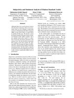
Fig. 3. Strands diagram of HLADH, based on X-ray crystallographic
data of Eklund et al. [33] (6ADH) obtained from the Swiss Prot data-
base. The two different domains of one subunit are coloured orange
(coenzyme-binding domain) and yellow (catalytic domain); trypto-
phans 15 and 314 are shown in red and NAD
+
isshowninwhite.
Fig. 4. Pressure dependence of the CSM of HLADH intrinsic fluores-
cence. d, Compression; s, decompression. Conditions: 30 °C, pH 8,
concentration of HLADH, 1 mgÆmL
)1
in 50 m
M
Mes buffer, excita-
tion wavelength: 295 nm. Dotted lines show the half of the second
transition.
122 M. Trovaslet et al. (Eur. J. Biochem. 270) Ó FEBS 2003
transition was over at about 100 MPa, the second one
occurred between 100 and 400 MPa and the last one began
at higher pressures (% 500 MPa).
Relatively low pressure (< 100 MPa) seemed to induce
small conformational changes of HLADH, because the
amplitude of the change in the CSM was moderate. Since
pressure promotes structural rearrangement of the protein/
solvent interactions, the application of low pressure may
provide pathways for water to penetrate into the protein
and probably between subunits favouring the protein
hydration. It could explain the decrease of CSM for this
range of pressure and hence the increase of polarity of the
environment of Trp314. Moreover, pressure could also
induce a reduction of size of internal cavities, voids that
result from imperfect packing of amino acids and a change
in the length of chemical bonds [34]. The protein could reach
a new conformation state that could be assumed to a native-
like state (N¢) and could correspond to the first intermediate
state of pressure-induced structural modification. These
conformational changes do not alter the molecular folding
which is confirmed by the weak effect of pressure on the
catalytic efficiency of HLADH, as shown in the previous
section (Fig. 1A and B inset): more precisely, pressure
favours the catalytic step whereas the substrate binding
steps are influenced by pressure only slightly. At atmo-
spheric pressure and noninhibitory primary alcohol con-
centration, the dissociation rate of the enzyme–NADH
complex is the rate-limiting step [35,36]. Hence, the positive
effect of pressure on HLADH catalysis could be due to a
new conformation of the enzyme which enhances the
dissociation rate of NADH from the enzyme [32]. Under
pressure, hydration and decrease of volume have antagon-
istic effects on the flexibility of proteins. In the range of
75–100 MPa, these antagonisms have probably induced an
optimal conformation of HLADH as far as the kinetics are
concerned. As the environment of Trp314 is only slightly
more polar in this pressure range, we deduce that the
hydrophobic core of HLADH is not much affected. As a
consequence, the dissociation of the dimeric form of
HLADH cannot take place.
Higher pressures, between 100 and 400 MPa, induced
greater changes in the intrinsic fluorescence of HLADH. In
this pressure range, the enzyme also progressively lost its
catalytic efficiency. Then, the increase of catalytic activity
(V
m
) was strongly counteracted by the decrease of HLADH
affinities for its substrates (NAD
+
and alcohol) (Fig. 1A
and B). Therefore, conformational changes observed prob-
ably corresponded to alterations of the dimeric enzyme
active sites, where both NAD
+
and ethanol were bound.
These suggestions could be in good agreement with Tsou’s
conclusions [37,38]. Based on studies of inactivation/dena-
turation of several enzymes (including alcohol dehydroge-
nase from baker’s yeast), Tsou has proved that enzyme
active sites are formed by relatively weak molecular
interactions and may be conformationally more flexible
than the whole molecule. In the HLADH molecule, the
position of the active site is well known: the enzyme subunits
are divided into two different domains (the coenzyme
binding domain and the catalytic domain). These domains
are separated by a crevice that contains a wide and deep
pocket which is the binding site for the substrate and the
nicotinamide moiety of the coenzyme [24]. This specific
position of the active site could explain why it was more
sensitive to pressure than the molecule as a whole.
Because of the sigmoidal shape of the transition observed
between 100 and 400 MPa, a two-state transition was
assumed and pseudo-thermodynamic parameters were
calculated. The apparent molecular standard volume
change (DV
app
) and the apparent free energy ðDG
app
0
Þ of
HLADH upon pressure-induced structural modifications
(between 100 and 400 MPa), were )41.5 ± 4.6 mLÆmol
)1
and +10.7 ± 1.2 kJÆmol
)1
, respectively. The pressure of
half-denaturation was 260 MPa.
The value of the apparent molecular standard volume
change (DV
app
¼ )41.5 ± 4.6 mLÆmol
)1
)wasnotvery
important and did not suggest HLADH dissociation. In
fact, molecular standard volume changes upon oligomeric
protein dissociation usually vary between )100 and
)500 mLÆmol
)1
, depending on proteins and experimental
conditions [39,40]. So, our result seemed to confirm the
conclusions of Cioni and Strambini [18]. Based on a
HLADH phosphorescence study, they have shown that the
dimeric molecule is not dissociated up to 300 MPa, even if
the tryptophan 314 becomes more exposed to the solvent. It
is generally admitted that oligomeric proteins dissociate
under moderate pressures [41] with numerous exceptions
[42]. For example, butyrylcholinesterase studies have shown
that pressure-induced modifications of this tetrameric
enzyme is not a dissociation (up to 350 MPa), but a
multi-step process of denaturation and that the observed
transient pressure-denatured state has characteristics of the
molten globule state [43]. Furthermore, work on RNase A
[6] or on the 7-kDa protein P2 from Sulfolobus solfataricus
[44] has shown a major role of hydrophobic residues and
interactions among aromatics in barostability. So, the large
hydrophobic interaction area present between the HLADH
subunits could explain why it was difficult to separate the
enzyme subunits without denaturation [24].
The modifications of the enzyme’s tertiary structure, the
loss of catalytic activity and the capacity to bind ANS [18]
are characteristic properties of a molten globule-like state
[43]. Hence the existence of a pressure-induced dimeric
molten globule like state (MG1) could be assumed at
400 MPa.
The higher pressures applied (> 400 MPa) showed an
additional transition in the CSM variation vs. pressure,
although the final state did not seem to be completely
reached. Two different questions were then addressed: did
this new transition correspond to a conversion from a
dimeric state to a monomeric state? Did it correspond to
conversion from a molten globule to a second molten
globule like state or to a completely denatured state?
While there was no direct evidence that the final state
corresponds to a second molten globule-like state, the
incomplete fluorescence red shift strongly supported this
conclusion. In fact, as water-exposed indole side chains
fluoresce with a kmax between 350 and 355 nm [20] and for
HLADH kmax reached only at 340 nm at 600 MPa, the
HLADH fluorophores seemed to be partly shielded from
the solvent. This incomplete fluorescence red shift could be
characteristic of a dimeric intermediate state at 600 MPa.
However, it did not exclude monomer formation because
structural rearrangements in the monomer may partly shield
tryptophan 314 from the solvent.
Ó FEBS 2003 HLADH under pressure (Eur. J. Biochem. 270) 123
In order to characterize better the new state obtained at
600 MPa, reversibility of pressure-induced modifications of
HLADH structure was followed. The fluorescence spectra
and the CSM values were followed upon enzyme decom-
pression. When the pressure was released from 600 MPa
to atmospheric pressure, the fluorescence spectrum of
HLADH did not return to the original (data not shown)
and the CSM did not exactly come back to the original
value although the sample was stored more than 30 min at
0.1 MPa after decompression (the recovery of the signal was
% 80% of the value at 0.1 MPa) (Fig. 4). So, under these
conditions, the pressure-induced modifications of HLADH
structure seemed to be partly reversible, supporting that at
600 MPa, no completely denatured state was reached: a
new intermediate state seemed to be obtained. Interestingly,
the recovery of the CSM showed a strong hysteresis when
the pressure was gradually released: the CSM at a pressure
in the decompression direction was much lower than at the
same pressure in the compression direction. This Ôconform-
ational driftÕ could be explained by assuming that the last
transition represented the dissociation of HLADH subunits,
the modifications of these subunits’ structure and that the
subunit reassociation started before complete refolding of
individual subunits was completed [45]. These suggestions
could explain why the pressure-induced modifications of
HLADH were only partly reversible: a possible aggregation
of the (partly) unfolded monomer could occur. It is known
that the renaturation of proteins in native and aggregated
form is different. So, all our results suggested that a second
intermediate state was observed at 600 MPa: it could be a
monomeric molten globule-like state.
Thus, the native like state (N¢)seemedtobefurther
unfolded into a first molten globule (MG1) and a second
molten globule (MG2) as pressure increased. Different
models could be proposed to explain this process: (a) each of
the transitions observed in the CSM changes with pressure
concerned the whole molecule. Then, the whole HLADH
molecule was melted into a first molten globule, which was
melted into a second molten globule state. In such cases,
both intermediate states still have partially secondary
structure but their compactness is different [46]; (b) the
protein molecule was assumed to be composed of at least
two different domains each of which unfolded independ-
ently. Then, MG1 corresponded to the melting of one
domain into a molten globule-like state, whereas the second
domain preserved its native conformation, and MG2
corresponded to the melting of the second domain into a
molten globule-like state.
As shown in Fig. 3, each subunit of HLADH is divided
into two separate domains: the coenzyme binding domain
and the catalytic domain. These two domains are unequal in
size and in amount of secondary structure [24]. It is possible
that they underwent conformational changes nonsimulta-
neously, showing that they are independent and different in
stability against high pressure-induced structural modifica-
tions. The coenzyme-binding domain could be less pressure
sensitive than the catalytic domain because of its amount of
secondary structure. In the catalytic domain, a large number
of residues – 32% – have no regular secondary structure,
whereas in the coenzyme-binding domain only % 10% of
the residues have no regular secondary structure, and no
continuous stretch of irregular structure is longer than four
residues [24]. According to the second model proposed, at
moderate pressures alterations of the catalytic domain could
explain the decrease of HLADH affinity for its substrates
and thus the modifications of the enzyme catalytic efficiency
without dissociation of the dimeric enzyme; the dissociation
happened at higher pressures, when modifications of the
coenzyme-binding domain start. A similar behaviour has
already been observed with creatine kinase. Zhou et al.[16]
have shown that creatine kinase inactivation, at low
pressure, may precede the enzyme dissociation and the
unfolding of the hydrophobic core, occurring at higher
pressure. They have postulated that the multi-state transi-
tions induced both by pressure and guanidine denaturation
were in direct relationship with the existence of hydrogen
bonds which maintain the dimeric structure of the enzyme.
At last, in order to determine whether the observed effects
of pressure were due to HLADH dissociation or not, the
same experiment was performed at different enzyme
concentrations. Usually dissociation of oligomeric enzymes
exhibits a strong concentration dependence as expected
from the law of mass action [28,29,47]. Fig. 5 shows that the
half-transition pressure values of the two intermediate states
(N¢fiMG1 and MG1 fi MG2)remainthesamewhat-
ever the enzyme concentration. The concentration inde-
pendence of pressure effects suggests that unfolding (and
inactivation) of the enzyme is not a result of the dissociation
of the dimer. It seems that our results are in agreement with
Ruan and Weber [29] who have shown that the degree of
dissociation of an oligomeric protein could be insensitive to
protein concentration. However our results do not com-
pletely exclude the formation of a monomer. Recent
investigations of the reversible subunit dissociation of
several oligomers (ranging from dimer to viral particles)
by hydrostatic pressure has revealed significant deviations
from the law of mass action and hence an anomalous or
complete lack of protein concentration dependence [47].
HLADH could fit to the previous case as the dissociation of
the dimers seems to follow different rates and therefore the
dimer population is heterogeneous in terms of thermody-
namic stability. In the case of the dimeric triosephosphate
Fig. 5. Pressure variation of the CSM of HLADH intrinsic fluores-
cence, at 30 °C and pH 8. The concentration of HLADH was
0.5 mgÆmL
)1
(d)and5mgmL
)1
(n)in50 m
M
Mes buffer. Excitation
wavelength: 295 nm.
124 M. Trovaslet et al. (Eur. J. Biochem. 270) Ó FEBS 2003
isomerase from rabbit muscle it was shown that the
activation free-energy barriers for dimer dissociation are
quite high corresponding to a characteristic time of
dissociation of 15 h or longer [47]. Perhaps, it should be
considered that the HLADH dimer dissociation is a slow
process and that the reaction could be controlled by kinetic
rather than thermodynamic factors [47,48].
FTIR studies of pressure-induced structural
modifications of HLADH
Mozhaev et al. [49] have suggested that the data obtained
by fluorescence spectroscopy could be complementary to
those obtained by other techniques such as FTIR. In fact, in
the few instances when these techniques have been applied
to the same protein (trypsin, gliadin, staphylococcal nucle-
ase, Trp apo-repressor [2,50–52] for example), almost
identical denaturation pressures have often been found,
except the case of Trp apo-repressor of Escherichia coli for
which the different behaviour could be explained by the use
of different solvent conditions. But, in contrast with the
tryptophan fluorescence experiments, where local pressure
effects were observed, FTIR spectroscopy was used to look
at the secondary structures of the whole molecule. In fact,
the secondary structures of the protein may be determined
from the analysis of the amide I¢ bandshape of the infrared
spectrum. The amide I¢ band of proteins occurs due to the
in-plane C ¼ O stretching vibrations which are weakly
coupled with the C–N stretching and in-plane N–H bending
vibrations. It is located in the frequency band of 1600–
1700Æcm
)1
and usually consists of many overlapping com-
ponent bands that represent different structural elements
such as a-helices, b-sheets, turns, nonordered or irregular
structures [53,54]. Several different approaches can be used
to determine these structures: the spectrum can be compared
with a database of the amide I¢ bands of several proteins
with known secondary structures, curve fitting after Fourier
self-deconvolution can be observed or second derivative of
the spectrum can be calculated. For studies of pressure-
induced structural changes of proteins, the first approach
cannot be easily used because of the need of a data set of
reference proteins as a function of pressure. In pressure-
induced structural modifications of HLADH, the second
derivative of the absorption spectrum was used. Then, a
qualitative analysis can be carried out in order to identify
the secondary structures present in the protein and to detect
theirs pressure-induced modifications. Furthermore, the use
of the DAC made it possible to extend the pressure range up
to 1000 MPa and thus allowed us to follow possible changes
in the protein conformation beyond the reach of fluores-
cence conditions.
Fig. 6 shows the absorbance spectra of HLADH in D
2
O
in the frequency region 1350–1750Æcm
)1
for pressures of
0.1–1000 MPa. The amide I¢ bands is centred at 1640Æcm
)1
and the amide II¢ band at % 1455Æcm
)1
. The band at
% 1557Æcm
)1
is attributed to the amide II mode which is a
mixed vibration involving N–H in plane bending and the
CN stretching [55]. This band shifts to % 1455Æcm
)1
as a
result of deuteration of labile protons on the amide groups.
On this graph we observe a simultaneous decrease of the
band at 1557Æcm
)1
and an increase of the band at 1450Æcm
)1
with pressure increase up to 800 MPa. This phenomenon
could be attributed to H/D exchange during the increase of
pressure for hydrogens buried in the core of the protein.
Nevertheless this H/D exchange is not total which is in good
concordance with the partial unfolding of the protein.
Fig. 7 shows the original spectra of amide I¢ band (1600–
1700Æcm
)1
) and Fig. 8 shows the second derivative of
nondeconvoluted spectra for the same range. At atmo-
spheric pressure, the amide I¢ band consists of several
frequency regions, summarized in Table 2, showing that
different secondary structures could be observed in the
HLADH molecule. Up to relative low pressure (90–
100 MPa), when the native-like state (N¢) was assumed,
no change in the infrared spectrum of HLADH seemed to
take place. At higher pressures (between 100 and 600 MPa),
when molten globule like states MG1 and MG2 were
assumed, only a few changes in the secondary structures of
HLADH were observed. These changes were rather small,
as can be seen in Fig. 8. The band at 1610Æcm
)1
,which
corresponds to the amino acid side chains, exhibits the main
change suggesting that pressure induced modifications of
HLADH tertiary structure. The bands at 1658Æcm
)1
,
1666Æcm
)1
and 1672Æcm
)1
have been assigned to a-struc-
tures, b-turns and b-sheets [53,54]. Pressurization of
HLADH seemed to cause a small modification of the
amount of these structures. These trends continued as the
Fig. 6. Original spectra of the amide I¢ and II¢ areas of HLADH
(50 mgÆmL
-1
) in Tris/DCl 10 m
M
at varying pressures 0.1–1000 MPa
(pH 8, 30 °C).
Fig. 7. Original spectra of the amide I¢ areas of HLADH (50 mgÆmL
-1
)
in 10 m
M
Tris/DCl at varying pressures 0.1–1000 MPa (pH 8, 30 °C).
Ó FEBS 2003 HLADH under pressure (Eur. J. Biochem. 270) 125
pressure was increased up to 1000 MPa; but no further
modifications were observed up to the maximum pressures.
These results were consistent with the molten globule like
states observed at 400 MPa (MG1) and 600 MPa (MG2) in
fluorescence experiments. In fact, one of the characteristic
properties of the molten globules is the partially secondary
structures present in these intermediate states [30,43].
Fig. 9 gives a more quantitative representation of the
pressure-induced spectral changes of HLADH, by plotting
the bandwidth at half height of the amide I¢ band of the
enzyme vs. pressure (up to 1000 MPa). It is of interest to
note that no cooperative change in the spectrum was
observed in that pressure range. Moreover, the shape of the
amide I¢ band of the protein (Fig. 7) contained distinct
features up to 1000 MPa, which suggested that HLADH
was not completely unfolded at this pressure. In fact,
whatever the applied pressure – up to 1000 MPa – no
plateau was obtained (Fig. 9), showing that HLADH was
not transformed into a stable pressure denatured state.
All of these FTIR results were in agreement with the
fluorescence data. They allowed us to confirm the existence
of at least two molten globule like states upon pressure-
induced unfolding of HLADH, while no completely dena-
turedstatewasobtainedupto1000MPa.
Fig. 9 also led to the conclusion that the pressure-induced
modifications of HLADH were irreversible, which was in
contrast with the fluorescence results. However, several
authors have shown that the high concentrations of proteins
used in FTIR studies often lead to irreversible inactivation
or denaturation [5,49]. In fact, in FTIR studies, the
intermolecular interactions (protein–protein interactions)
became more pronounced and this could explain the
findings on the irreversibility [56].
Conclusion
It is well known that the activity of an enzyme is strongly
dependent on its conformational integrity [57,58]. So, the
aim of this study was to characterize the pressure-induced
conformational changes of the dimeric HLADH and to
correlate them with the modifications of the enzyme’s
catalytic activity. To this end, the pressure-induced modi-
fications of the enzyme’s activity were studied (up to
250 MPa) as well as the pressure effects on protein
fluorescence (up to 600 MPa) and the changes in HLADH
secondary structures (studied by FTIR technique up to
1000 MPa). All of these studies indicated that the pressure-
induced modifications of HLADH represents a multi-step
process leading to several different intermediate states of
denaturation. A native like state (N¢) and two different
molten globules (at 400 and 600 MPa) were assumed, while
no completely denatured state seemed to be reached up to
1000 MPa (Figs 4 and 9). Although it is widely accepted
that oligomeric enzymes are dissociated under moderate
pressures [28,29,59,60], our fluorescence and FTIR studies
have confirmed that pressure (up to 400 MPa) did not
induce HLADH dissociation: our structural studies sup-
ported the existence of a dimeric molten globule at
400 MPa. Moreover, at 600 MPa, the intermediate seemed
to be monomeric because of the hysteresis observed when
the pressure was released from 600 to 0.1 MPa. Further
experiments (like electrophoresis under high hydrostatic
pressure) could be necessary to confirm that the last
Table 2. Amide I¢ frequencies (cm
-1
) characteristic of the amide bond in
various conformations [51,52].
Conformation Band position (cm
)1
)
Amino acid side chains % 1610
Unordered structures % 1643
% 1656–1660
a-Helices % 1648–1655
b-Turns % 1662–1667
b-sheets % 1628–1638
% 1672–1678
% 1690–1693
Fig. 9. Pressure effects on the bandwidth at half height of the amide I¢
band of HLADH (in Tris/DCl 10 m
M
pH 8), at 30 °C, upon compres-
sion (d)anddecompression(s).
Fig. 8. Second derivative IR spectra of the amide I¢ area of HLADH
(50 mgÆmL
-1
)in10m
M
Tris/DCl at various pressures (pH 8, 30 °C).
126 M. Trovaslet et al. (Eur. J. Biochem. 270) Ó FEBS 2003
transition observed in the fluorescence experiments (Fig. 4)
really corresponded to conversion from a dimeric state to a
monomeric state. At last, in order to test the contribution of
the pressure insensitive interactions involved in HLADH
subunit contacts, to the pressure insensitivity of the
molecule, specific hydrophobic residues (present at the
subunit interface) could be replaced with nonhydrophobic
amino acids. Then, high pressure studies of HLADH
mutants could be considered to observe their pressure-
induced unfolding, denaturation and/or dissociation. This
could allow us to understand the molecular basis of the
pressure stability of this enzyme and perhaps of the other
proteins of the alcohol dehydrogenase family.
Acknowledgements
We thank N. Bec and R. Lange for stimulating discussions and fruitful
advice.
References
1. Silva, J.L., Foguel, D. & Royer, C.A. (2001) Pressure provides new
insights into protein folding, dynamics and structure. Trends
Biochem. Sci. 26, 612–618.
2. Balny, C., Masson, P. & Heremans, K., eds. (2002) Frontiers
in High Pressure Biochemistry and Biophysics. Elsevier,
Amsterdam.
3. Ikeuchi, Y., Suzuki, A., Oota, T., Hagiwara, K., Tatsumi, R., Ito,
T. & Balny, C. (2002) Fluorescence study of the high pressure-
induced denaturation of skeletal muscle actin. Eur. J. Biochem.
269, 364–371.
4. Ruan, K., Lange, R., Bec, N. & Balny, C. (1997) A stable partly
denatured state of trypsin induced by high hydrostatic pressure.
Biochem. Biophys. Res. Comm. 239, 150–154.
5. Ruan, K., Lange, R., Meersman, F., Heremans, K. & Balny, C.
(1999) Fluorescence and FTIR study of the pressure-induced
denaturation of bovine pancreas trypsin. Eur. J. Biochem. 265,
79–85.
6. Torrent, J., Rubens, P., Ribo, M., Heremans, K. & Vilanova, M.
(2001) Pressure versus temperature unfolding of ribonuclease A:
An FTIR spectroscopic characterization of 10 variants at the
carboxy-terminal site. Protein Sci. 10, 725–734.
7. Morild, E. (1977) Pressure neutralization of substrate inhibition in
the alcohol dehydrogenase reaction. J. Phys. Chem. 81, 1162–
1166.
8. Morild, E. (1977) Pressure variation of enzymatic reaction rates:
yeast and liver alcohol dehydrogenase. Biophys. Chem. 6, 351–362.
9. Hamon, V., Dallet, S. & Legoy, M.D. (1996) The pressure-
dependence of two b-glucosidases with respect to their thermo-
stability. Biochim. Biophys. Acta 1294, 195–203.
10. Ciardiello, M.A., Schmitt, B., di Prisco, G. & Herve
´
, G. (1999)
Influence of hydrostatic pressure on 1-glutamate dehydrogenase
from the Antarctic fish Chaenocephalus aceratus. Marine Biol. 134,
631–636.
11. Northrop, D.B. & Cho, Y K. (2000) Effects of high pressure on
solvent isotope effects of yeast alcohol dehydrogenase. Biophys. J.
79, 1621–1628.
12. Northrop, D.B. (2002) Effects of high pressure on enzymatic
activity. Biochim. Biophys. Acta 1595, 71–79.
13. Deville-Bonne, D. & Else, A.J. (1991) Reversible high hydrostatic
pressure inactivation of phosphofructokinase from Escherichia
coli. Eur. J. Biochem. 200, 747–750.
14. Masson, P., Froment, M T., Fort, S., Ribes, F., Bec, N., Balny, C.
& Schopfer, L.M. (2002) Butyrylcholinesterase-catalyzed hydro-
lysis of N-methylindoxyl acetate: analysis of volume changes upon
reaction and hysteresis behavior. Biochim. Biophys. Acta 1597,
229–243.
15. Kornblatt, M.J., Lange, R. & Balny, C. (1998) Can monomers
of yeast enolase have enzymatic activity? Eur. J. Biochem. 251,
775–780.
16. Zhou, J M., Zhu, L. & Balny, C. (2000) Inactivation of creatine
kinase by high pressure may precede dimer dissociation. Eur. J.
Biochem. 267, 1247–1253.
17. Roitel, O., Bec, N., Lange, R., Balny, C. & Branlant, G. (2001)
Pressure denaturation of phosphorylating glyceraldehyde-
3-phosphate dehydrogenase from Bacillus stearothermophilus.
Biochem. Biophys. Res. Comm. 283, 347–350.
18. Cioni, P. & Strambini, G.B. (1996) Pressure effects on the structure
of oligomeric proteins prior to subunit dissociation. J. Mol. Biol.
263, 789–799.
19. Cioni, P. & Strambini, G.B. (2002) Tryptophan phosphorescence
and pressure effects on protein structure. Biochim. Biophys. Acta
1595, 116–130.
20. Schmid, F.X. (1989) Spectral methods of characterizing protein
conformation and conformational changes. In Protein Structure: a
Practical Approach (Creighton, T. E., ed.), pp. 251–285. IRL
Press, Oxford.
21. Ruan, K. & Balny, C. (2002) High pressure static fluorescence to
study macromolecular structure-function. Biochim. Biophys. Acta
1595, 94–102.
22. Ruan, K., Li, J., Liang, R. & Xu, C., YuY., Lange, R. &
Balny, C. (2002) A rare protein fluorescence behavior where the
emission is dominated by tyrosine: case of the 33-kDa protein
from spinach photosystem II. Biochem. Biophys. Res. Comm. 293,
593–597.
23. Ruan, K., Tian, S., Lange, R. & Balny, C. (2000) Pressure effects
on tryptophan and its derivatives. Biochem. Biophys. Res. Comm.
269, 681–686.
24. Eklund, H., Nordstro
¨
m, B., Zeppezauer, E., So
¨
derlund, G.,
Ohlsson, I., Boiwe, T., So
¨
derberg, B.O., Tapia, O. & Bra
¨
nde
´
n, C.I.
(1976) Three-dimensional structure of horse liver alcohol dehy-
drogenase at 2.4 A
˚
resolution. J. Mol. Biol. 102, 27–59.
25. Smeller, L. & Heremans, K. (1997) In situ observation of the
pressure-induced changes in proteins with infrared spectroscopy in
the diamond anvil cell. In High Pressure Research in the Bios-
ciences and Biotechnology (Heremans, K., ed.), Leuven University
Press, Leuven. 131–134.
26. Dzwolak, W., Kato, M. & Taniguchi, Y. (2002) Fourier transform
infrared spectroscopy in high-pressure studies on proteins.
Biochim. Biophys. Acta 1595, 131–144.
27. Dallet, S. & Legoy, M.D. (1996) Hydrostatic pressure induces
conformational and catalytic changes on two alcohol dehy-
drogenases but not oligomeric dissociation. Biochim. Biophys.
Acta 1294, 15–24.
28. Silva, J.L., Miles, E.W. & Weber, G. (1986) Pressure dissociation
and conformational drift of the dimer of tryptophan synthase.
Biochemistry 25, 5780–5786.
29. Ruan, K. & Weber, G. (1989) Hysteresis and conformational drift
of pressure dissociated glyceraldehyde phosphate dehydrogenase.
Biochemistry 28, 2144–2153.
30. Dumoulin, M., Ueno, H., Hayashi, R. & Balny, C. (1999) Con-
tribution of the carbohydrate moiety to conformational stability
of the carboxypeptidase Y. High pressure study. Eur. J. Biochem.
262, 475–483.
31. Wong, P.T.T. & Moffat, D.J. (1989) A new internal pressure
calibrant for high-pressure infrared spectroscopy of aqueous
systems. Appl. Spectrosc. 43, 1279–1281.
32. Trovaslet, M., Dallet, S. & Legoy, M.D. (2000) Effects of
hydrostatic pressure and temperature on catalytic activity of horse
liver alcohol dehydrogenase (HLADH). High Pressure Res. 19,
303–309.
Ó FEBS 2003 HLADH under pressure (Eur. J. Biochem. 270) 127
33. Eklund, H., Samma, J.P., Wallen, L., Branden, C.I., Akeson, A. &
Jones, T.A. (1981) Structure of a triclinic ternary complex of horse
liver alcohol dehydrogenase at 2.9 A
˚
resolution. J. Mol. Biol. 146,
561–587.
34. Heremans, K. & Smeller, L. (1988) Protein structure and dynamics
at high pressure. Bioch. Biophys. Acta 1386, 353–370.
35. Dalziel, K. & Dickinson, F.M. (1966) The kinetics and mechanism
of liver alcohol dehydrogenase with primary and secondary alco-
hols as substrates. Biochem. J. 100, 34–46.
36. Bra
¨
nde
´
n, C.I., Jo
¨
rnvall, H., Eklund, H. & Furugen, B. (1975)
Alcohol dehydrogenases. In The Enzymes,Vol.XI(BoyerP.D,
eds), 3rd edn, pp. 103–190. Academic Press, New York.
37. Tsou, C.L. (1986) Location of the active sites of some enzymes in
limited and flexible molecular regions. Trends Biochem. Sci. 11,
427–429.
38. Tsou, C.L. (1993) Conformational flexibility of enzyme active
sites. Science 262, 380–381.
39. Erijman, L., Lorimer, G.H. & Weber, G. (1993) Reversible
dissociation and conformational stability of dimeric ribulose
bisphosphate carboxylase. Biochemistry 32, 5187–5195.
40. Schade, B.C., Rudolph, R., Ludemann, H.D. & Jaenicke, R.
(1980) Reversible high-pressure dissociation of lactic dehy-
drogenase from pig muscle. Biochemistry 19, 1121–1126.
41. Mozhaev, V.V., Heremans, K., Frank, J., Masson, P. & Balny, C.
(1994) Exploiting the effects of high hydrostatic pressure in bio-
technological applications. Trends Biotechnol. 12, 493–501.
42. Balny, C. (2002) High pressure and protein oligomeric dissoci-
ation. High Press Res. 22, 737–741.
43. Cle
´
ry, C., Renault, F. & Masson, P. (1995) Pressure-
induced molten globule state of cholinesterase. FEBS Lett. 370,
212–214.
44. Fusi, P., Goossens, K., Consonni, R., Grisa, M., Puricelli, P.,
Vecchio,G.,Vanoni,M.,Zetta,L.,Heremans,K.&Tortora,P.
(1997) Extreme heat- and pressure-resistant 7-kDa protein P2
from the archaeon Sulfolobus solfataricus is dramatically destabi-
lized by a single-point amino acid substitution. Proteins 29, 381–
390.
45. Weber, G. (1986) Phenomenological description of the associ-
ation of protein subunits subjected to conformational drift. Effects
of dilution and of hydrostatic pressure. Biochemistry 25, 3626–
3631.
46. Levitt,M.,Gerstein,M.,Huang,E.,Subbiah,S.&Tsai,J.(1997)
Protein folding: the endgame. Annu. Rev. Biochem. 66, 549–579.
47. Ferreira, S.T. & De Felice, F.G. (2001) Protein dynamics, folding
and misfolding: from basic physical chemistry to human con-
formational diseases. FEBS Lett. 498, 129–134.
48. Cioni, P. & Strambini, G.B. (1997) Pressure-induced dissoci-
ation of yeast glyceraldehyde-3-phosphate dehydrogenase:
heterogeneous kinetics and perturbations of subunit structure.
Biochemistry 36, 8586–8593.
49. Mozhaev, V.V., Heremans, K., Frank, J., Masson, P. & Balny, C.
(1996) High pressure effects on protein structure and function.
Proteins: Struct., Funct. Genet. 24, 81–91.
50. Lullien-Pellerin, V., Popineau, Y., Meersman, F., Morel, M.H.,
Heremans, K., Lange, R. & Balny, C. (2001) Reversible
changes of the wheat gamma46 gliadin conformation submitted
to high pressures and temperatures. Eur. J. Biochem. 268,
5705–5712.
51. Royer, C.A., Hinck, A.P., Loh, S.N., Prehoda, K.E., Peng, X.,
Jonas, J. & Markley, J.L. (1993) Effects of amino acid substitu-
tions on the pressure denaturation of staphylococcal nuclease as
monitored by fluorescence and nuclear magnetic resonance spec-
troscopy. Biochemistry 32, 5222–5232.
52. Desai,G.,Panick,G.,Zein,M.,Winter,R.&Royer,C.A.(1999)
Pressure-jump studies of the folding/unfolding of trp repressor.
J. Mol. Biol. 288, 461–475.
53. Jackson, M. & Mantsch, H.H. (1995) The use and misuse of FTIR
spectroscopy in the determination of protein structure. Crit. Rev.
Biochem. Mol. 30, 95–120.
54. Arrondo, J.L.R., Muga, A., Castresana, J. & Goni, F.M. (1993)
Quantitative studies of the structure of proteins in solution by
Fourier-transform infrared spectroscopy. Prog. Biophys. Mol.
Biol. 59, 23–56.
55. Wong, P.T.T. &.Heremans, K. (1988) Pressure effects on protein
secondary structure and hydrogen deuterium exchange in chy-
motrypsinogen: a fourier transform infrared spectroscopic study.
Biochim. Biophys. Acta 956, 1–9.
56. Goossens, K., Smeller, L., Heremans, K. & Frank, J. (1992)
Pressure effect on lipoxygenase: a FTIR study with the diamond
anvil cell. High Pressure and Biotechnology. Colloque INSERM
(Balny, C., Hayashi, R., Heremans, K. & Masson, P., eds), pp.
171–173. John Libbey Eurotext Ltd, Paris.
57. Ruan, Q., Ruan, K., Balny, C., Glaser, M. & Mantulin, W.W.
(2001) Protein folding pathways of adenylate kinase from E. coli:
hydrostatic pressure and stopped-flow studies. Biochemistry 40,
14706–14714.
58. Chatani, E., Nonomura, K., Hayashi, R., Balny, C. & Lange, R.
(2002) Comparison of heat- and pressure-induced unfolding of
ribonuclease A: the critical role of Phe-46 which appears to belong
to a new hydrophobic chain-folding initiation site. Biochemistry
41, 4567–4574.
59. King, L. & Weber, G. (1986) Conformational drift of dissociated
lactate dehydrogenase. Biochemistry 25, 3632–3637.
60. Ruan, K. & Weber, G. (1993) Physical heterogeneity of muscle
glycogen phosphorylase revealed by hydrostatic pressure dis-
sociation. Biochemistry 32, 6295–6301.
128 M. Trovaslet et al. (Eur. J. Biochem. 270) Ó FEBS 2003