Báo cáo khoa học: A hydrophobic segment within the C-terminal domain is essential for both client-binding and dimer formation of the HSP90-family molecular chaperone pptx
Bạn đang xem bản rút gọn của tài liệu. Xem và tải ngay bản đầy đủ của tài liệu tại đây (340.76 KB, 9 trang )
A hydrophobic segment within the C-terminal domain is essential
for both client-binding and dimer formation of the HSP90-family
molecular chaperone
Shin-ichi Yamada
1,2
, Toshio Ono
2
, Akio Mizuno
1
and Takayuki K. Nemoto
2
1
Division of Oral and Maxillofacial Surgery and
2
Division of Oral Molecular Biology, Department of Developmental
and Reconstructive Medicine, Course of Medical and Dental Sciences, Nagasaki University Graduate
School of Biomedical Sciences, Japan
The a isoform of human 90-kDa heat shock protein
(HSP90a) is composed of three domains: the N-terminal
(residues 1–400); middle (residues 401–615) and C-terminal
(residues 621–732). The middle domain is simultaneously
associated with the N- and C-terminal domains, and the
interaction with the latter mediates the dimeric configuration
of HSP90. Besides one in the N-terminal domain, an addi-
tional client-binding site exists in the C-terminal domain of
HSP90. The aim of the present study is to elucidate the
regions within the C-terminal domain responsible for the
bindings to the middle domain and to a client protein, and to
define the relationship between the two functions. A bac-
terial two-hybrid system revealed that residues 650–697 of
HSP90a were essential for the binding to the middle domain.
An almost identical region (residues 657–720) was required
for the suppression of heat-induced aggregation of citrate
synthase, a model client protein. Replacement of either
Leu665-Leu666 or Leu671-Leu672 to Ser-Ser within the
hydrophobic segment (residues 662–678) of the C-terminal
domain caused the loss of bindings to both the middle
domain and the client protein. The interaction between the
middle and C-terminal domains was also found in human
94-kDa glucose-regulated protein. Moreover, Escherichia
coli HtpG, a bacterial HSP90 homologue, formed hetero-
dimeric complexes with HSP90a and the 94-kDa glucose-
regulated protein through their middle-C-terminal domains.
Taken together, it is concluded that the identical region
including the hydrophobic segment of the C-terminal
domain is essential for both the client binding and dimer
formation of the HSP90-family molecular chaperone and
that the dimeric configuration appears to be similar in the
HSP90-family proteins.
Keywords: GRP94; HtpG; molecular chaperone; dimer;
client binding.
The 90-kDa heat shock protein (HSP90) is a ubiquitously
distributed molecular chaperone and is an essential protein in
eukaryotic cells [1]. Most, if not all, compartments of
mammalian cells contain specific members of HSP90. For
instance, two HSP90 isoforms, HSP90a [2] and HSP90b [3],
are present in the cytosol; the 94-kDa glucose-regulated
protein (GRP94/gp96) is expressed in the lumen of endo-
plasmic reticulum [4]; and TRAP1/hsp75 is expressed in
mitochondria [5]. Also, HtpG exists in prokaryotic cells [6],
although its expressionis not essential for the organisms [7,8].
HSP90 is either transiently or stably associated with
specific client proteins that are unstable unless chaperoned
with HSP90. Various regions of HSP90 have been proposed
to be involved in the interactions with such target proteins.
For instance, a highly charged region of chick HSP90 (amino
acids 221–290) is essential for the binding to estrogen and
mineralocorticoid receptors [9]; this region is also involved in
the binding to the a subunit of casein kinase CK2 [10].
However, the corresponding highly charged region and
C-terminal 35 residues that are specific to mammalian
HSP90 can be deleted from yeast HSP82 [11]. Serial deletion
experiments on HSP90b demonstrated that amino acids 327–
340, which are distinct but proximal to the charged region,
are essential for chaperoning of serine/threonine kinase Akt/
PKB [12]. Two separate regions were proposed to be
involved in the binding to the progesterone receptor [13].
At present, it is ambiguous whether this discrepancy is caused
by the variation in the binding sites of HSP90 for the
respective substrates or if the respective regions are respon-
sible for certain aspects of the chaperoning mechanism.
Another approach by use of model client proteins has
been employed to clarify the client-binding sites of HSP90.
By use of citrate synthase (CS) and insulin, it was observed
that mammalian HSP90 possesses two distinct client-
binding sites [14,15]: one of them is located in the
N-terminal domain and its activity is modulated by ATP
Correspondence to T. K. Nemoto, Division of Oral Molecular
Biology, Nagasaki University School of Dentistry,
1-7-1, Sakamoto, Nagasaki 852-8588, Japan.
Fax: + 81 95 849 7642, Tel.: + 81 95 849 7640,
E-mail:
Abbreviations: HSP90, the 90-kDa heat shock protein; HSP90a and
HSP90b,thea and b isoforms of HSP90, respectively; HtpG,
an E. coli homologue of mammalian HSP90; GRP94, the 94-kDa
glucose-regulated protein; GST, glutathione S-transferase;
GST-HSP90a and H
6
HSP90a,HSP90a tagged with GST and a
histidine hexamer (MRGSH
6
GS), respectively, at the N-terminus;
CS, citrate synthase.
(Received 19 August 2002, revised 12 November 2002,
accepted 20 November 2002)
Eur. J. Biochem. 270, 146–154 (2003) Ó FEBS 2003 doi:10.1046/j.1432-1033.2003.03375.x
and geldanamycin, a specific inhibitor of HSP90 molecular
chaperone; and the other is in the C-terminal domain.
Minami et al. [16] and Tanaka et al. [17] confirmed the
existence of these respective client-binding sites in the
N- and C-terminal domains. Similarly, the C-terminal
fragment (residues 494–782 as a mature form) of human
GRP94 protects the catalytic subunit of protein kinase CK2
against thermal aggregation [18]. In contrast, we found a
single client-binding site in Escherichia coli HtpG, which
was localized solely in the N-terminal domain (residues
1–336) of the 624-amino acid protein [17].
All members of the HSP90 family proteins so far studied
exist as dimers [19–22]. HSP90b analyzed by PAGE under
nondenaturing conditions predominantly existed as a
monomer [19]; it has been reported to exist as a dimers or
as oligomers in rat liver cytosol, but tends to dissociate into
monomers under the electrophoretic conditions [23]. Dis-
ruption of the dimeric structure of HSP90 is lethal in yeast
[24], although some of the monomeric mutants of HSP90
are able to confer viability and interact with the estrogen
receptor [25].
The C-terminal 49 amino acids are essential for the dimer
formation of HSP90 [24] and 191 amino acids are sufficient
for the function [20]. We previously proposed on human
HSP90a [20] and E. coli HtpG [26] that they form a dimer in
an antiparallel fashion through a pair of the interactions
between the middle domain and the C-terminal domain.
Similarly, the C-terminal 326 amino acids of barley GRP94
[22] and 200 amino acids of canine GRP94 [27] are sufficient
for the dimer formation. However, Wearsch and Nicchitta
[27] proposed a distinct mechanism of dimer formation, on
which the hydrophobic segment localized in the C-terminal
domain interacts with each other.
In the present study, we investigated two issues with
respect to the C-terminal domain of HSP90. One was the
identification of the minimal essential region required for
the interaction with the middle domain, which mediates the
dimerization of HSP90, and the other, the identification of
the minimal region of the C-terminal domain for the client
binding. Bearing in mind the fact that the 35-amino-acid
residues corresponding to the C-terminus of HSP90 are
deleted in HtpG, we postulated that the regions within the
C-terminal domain responsible for dimerization, i.e. an
interaction with the middle domain, and client binding,
could be separated into the N- and C-terminal parts,
respectively. However, the present study demonstrates that
the two regions are unable to be separated and that the two
functions are closely related to each other. We also
reinvestigated the mode of dimer formation in the HSP90-
family proteins.
Experimental procedures
Materials
Expression vector pQE9 and plasmid pREP4 were pur-
chased from Qiagen Inc. (Chatsworth, CA, USA) and
expression vector pGEX4T-1, glutathione-Sepharose and
low-molecular-mass markers, from Amersham Pharmacia
Biotech (Uppsala, Sweden). Talon metal affinity resin was
obtained from Clontech Laboratories Inc. (Palo Alto, CA,
USA). Porcine heart CS was purchased from Roche
Molecular Biochemicals (Mannheim, Germany). All other
reagents were of analytical grade.
Plasmid construction
DNA fragments carrying truncated forms of human
HSP90a amplified by PCR and cut with BamHI and SalI,
were inserted into a BamHI/SalIsiteofpQE9(desig-
nated pH
6
HSP90a). Construction of truncated forms of
pH
6
HSP90a, i.e. pH
6
HSP90a542–732, 542–728, 542–720,
542–697 and 542–687 was described previously [28]. Trun-
cated forms of HSP90a were also expressed as glutathione
S-transferase (GST)-fusion proteins. The DNA fragments
encoding HSP90a657–732, 676–732 and 697–732 were
amplified by PCR and inserted into a BamHI/SalIsiteof
pGEX-4T-1. Construction of the plasmid encoding amino
acids 1–43/604–732 was described previously [28].
We also expressed the middle and C-terminal domains of
human GRP94 as GST-fusion proteins. Although the
domain structures and the domain boundaries of human
GRP94 have not been determined, we tentatively defined
the boundary between the N-terminal and middle domains
to be Arg427-Glu428 and that between the middle and
C-terminal domains to be Lys650-Asp651 by comparison
with the amino acid sequence of human GRP94 [29] with
those of human HSP90a [28] and E. coli HtpG [26]. Amino
acid numbers refer to those of the mature form. Accord-
ingly, the initial Met in the prepeptide corresponds to )21
and the mature form corresponds to Asp1–Leu782. The
DNA fragments encoding the middle domain (Glu428–
Lys650) and the C-terminal domain (Asp651–Leu782) of
human GRP94 [29] were amplified by PCR and then
inserted into a BamHI/SalI site of pGEX4T-1 (designated
pGST-GRP94-M and pGST-GRP94-C, respectively).
Y1090 cells transformed with these plasmids were selected
on Luria broth agar containing 50 lgÆmL
)1
of ampicillin.
Constructed plasmids were verified by DNA sequencing.
Expression and purification of recombinant proteins
A histidine hexamer-tagged form of recombinant proteins
was expressed and purified by use of Talon affinity resin
according to the manufacturer’s protocol, except that
10 m
M
imidazole was added to the lysis/washing buffer.
Bound proteins were eluted with 0.1
M
imidazole (pH 8.0)
containing 10% (v/v) glycerol. GST-fusion proteins were
expressed and purified by binding to glutathione-Sepharose
as described previously [30].
Bacterial two-hybrid system
Bacterial strain BTH101 [F
–
, cya-99, araD139, gal15,
galK16, rpsL1 (Strl
r
), hsdR2, mcrA1, mcrB1] and plasmids
pKT25
kan
and pUT18C
amp
were provided by D. Ladant
(Pasteur Institute, Paris, France) and L. Selig (Hybrigenics,
S.A.,Paris,France).Animprovedversionofthebacterial
two-hybrid system [31] was employed to evaluate domain–
domain interactions of HSP90, GRP94 and HtpG. This
method is based on the interaction-mediated reconstitution
of an adenylate cyclase activity in the enzyme-deficient
E. coli strain, BTH101. Because human HSP90a was
proteolysed at Lys615-Ala616 and Arg620-Ala621 by
Ó FEBS 2003 Two roles of the C-terminal domain of HSP90 (Eur. J. Biochem. 270) 147
trypsin [20,28], the border between the middle and
C-terminal domains in the present study was set to
Lys618-Leu619. The PCR fragment carrying the middle-
C-terminal domains (residues 401–732) of HSP90a was
inserted in a PstI/BamHI site of pUT18C
amp
. The DNA
fragments carrying the C-terminal domain (residues 619–
732) of HSP90a or its truncated forms amplified by PCR
were inserted into a PstI/BamHI site of pKT25
kan
.The
DNA fragments encoding tentative middle domain and
C-terminal one of human GRP94 were amplified by PCR
andtheninsertedinanXbaI/BamHI site of both
pUT18C
amp
and pKT25
kan
. The DNA fragment encoding
the middle-C-terminal domains of GRP94 was amplified by
PCR and then inserted in an XbaI/BamHI site of
pUT18C
amp
. The construction of pKT25
kan
-HtpG 337–
624/the middle-C-terminal domains was described previ-
ously [17].
An E. coli strain BTH101 was cotransformed with
pUT18C
amp
- and pKT25
kan
-derived plasmids. The extent
of reconstitution of the catalytic domains of Bordetella
pertussis adenylate cyclase through the fused portions was
quantified as b-galactosidase activity, which was measured
after the bacteria had been cultured overnight at 30 °Cin
Luria broth medium containing 50 lgÆmL
)1
of ampicillin
and 25 lgÆmL
)1
kanamycin in the presence of 0.5 m
M
isopropyl thio-b-
D
-galactoside [31].
Suppression assay for heat-induced aggregation of CS
Heat-induced aggregation of CS and its suppression in the
presence of recombinant proteins were measured as des-
cribed previously [32]. In brief, CS (8 lg) in the presence or
absence of 6–48 lg of recombinant proteins in 0.4 mL of
40 m
M
Hepes, pH 7.4, was transferred at 45 °C. The
absorbance at 360 nm was measured at 80 min, when the
turbidity reached a plateau.
SDS/PAGE
PAGE was performed at a polyacrylamide concentration of
12.5% in the presence of 0.1% SDS. Separated proteins
were stained with Coomassie Brilliant Blue. Low-molecular-
mass markers (Amersham Pharmacia BioTech) were used
as references.
Protein concentration
Protein concentrations were determined by the bicincho-
ninic acid method (Pierce, Rockford, IL, USA).
Results
Minimal region of the C-terminal domain sufficient
for dimerization with the middle domain
The C-terminus of the C-terminal domain (Leu619–
Asp732) of human HSP90a was serially truncated, and
the binding activity to the middle domain (Glu401–Lys618)
was quantified by using the bacterial two-hybrid system
(Fig. 1). As reported previously on human HSP90a [17],
because the C-terminal domain could not associate with the
middle domain, but associated with the middle-C-terminal
domains, we used the middle-C-terminal domains as a
binding partner of the C-terminal domain in the two-hybrid
system. As a result, H
6
HSP90a619–728, 619–720 and 619–
707 bound to the partner. Even H
6
HSP90a619–697 pos-
sessed 72.5% of the maximal binding. However, truncation
by additional 10 amino acids resulted in loss most of the
binding. Thus, the C-terminal 35 amino acids of HSP90a
were dispensable without significant loss of the dimer-
forming activity, whereas further 10-amino acid truncation
disrupted the function.
In turn, the N-terminal side of the C-terminal domain
was truncated. Residues 629–732 as well as 619–732 (the
C-terminal domain) had the binding activity (Fig. 1). It
should be noted that the full-length form of HSP90a was
cleaved with chymotrypsin at Tyr627-Met628 and Met628-
Ala629 bonds [20]. Thus, the Ala616-Met628 segment may
not be essential for the function of the C-terminal domain.
Hence, it was reasonable that residues 629–732 still retained
the binding activity. The binding activity of residues 650–
732, i.e. a further deletion of the N-terminus up to Lys649,
was 48.5% of the positive control (Fig. 1). Thus, residues
650–732 were essential for the binding, although its
N-terminal proximal site (residues 629–649) may also be
involved in the association.
Minimal region sufficient for the binding to a model
client protein
Next, we measured the client-binding activity of the
C-terminal site. We started from H
6
HSP90a542–732, an
N-terminally histidine hexamer-tagged form, for the
C-terminal truncation experiment (Fig. 2A). The truncated
proteins were purified to near homogeneity (Fig. 2B, lanes
1–3 and lane 5) with an exception of GST-HSP90a 542–697
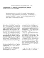
Fig. 1. Minimal region of the C-terminal domain that is required for the
dimerization. The truncated forms of the C-terminal domain (residues
619–732) of human HSP90a were expressed in combination with the
middle-C-terminal domains (resides 401–732). Residues 662–678 con-
stitute the hydrophobic segment (see Fig. 3A). Residues 698–732
correspondtothedeletedregioninE. coli HtpG. The extent of the
association was estimated by the b-galactosidase activity. The value of
the combination of intact C-terminal domain (residues 619–732) with
the middle-C-terminal domains was set to 100%. Values are means
± SD of three samples.
148 S i. Yamada et al. (Eur. J. Biochem. 270) Ó FEBS 2003
(lane 4). Aggregation of CS induced at 45 °Cwas
suppressed in the presence of H
6
HSP90a542–732 in a
dose-dependent manner. Its C-terminal truncation forms,
i.e. H
6
HSP90a542–728 suppressed the CS aggregation
(Fig.2C).H
6
HSP90a542–720 still suppressed the aggrega-
tion, but the efficiency appeared to be lower than those of
H
6
HSP90a542–732 and H
6
HSP90a542–728. A further
truncated form, HSP90a542–687, showed no suppression.
We could not test whether or not GST-HSP90a542–697
would suppress the aggregation of CS, because the prepar-
ation contained doublet bands (Fig. 2B, lane 4) and self-
aggregated at 45 °C even in the absence of CS (data not
shown).
We attempted to express even smaller fragments of
N-terminal truncation than those of the C-terminal trunca-
tions. However, recombinant proteins were not quantita-
tively recovered in the expression system presumably due to
the instability of exogenous proteins with small molecular
masses in E. coli. Accordingly, the N-terminal-truncated
forms were expressed as GST-fusion proteins with a
relatively large moiety (Fig. 2A,B, lanes 6–9). As shown
in Fig. 2D, GST-HSP90a1–43/604–732 and GST-HSP90
a657–732 suppressed the aggregation of the client protein.
However, GST-HSP90a697–732 and GST-HSPa676–732,
as well as GST, did not affect the process. Taken together,
the data indicate that residues 657–720 are indispensable for
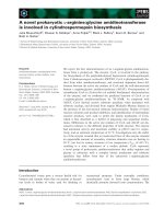
Fig. 2. Suppression of the heat-induced aggregation of CS by the C-terminal regions of HSP90a. (A) HSP90a542–732 and its C-terminally truncated
forms were expressed with an N-terminal histidine hexamer tag. HSP90a657–732, 676–732 and 697–732 were expressed as GST-fusion proteins. A
dotted line indicates the boundary between the middle and C-terminal domains. (B) Purified proteins (1 lg) were electrophoresed on SDS/PAGE
gels. Lane numbers are identical to those in Fig. 2A. M, low-molecular-mass markers. (C and D) The increase in the turbidity, representing the
aggregation of CS, was measured after incubation with various concentrations of recombinant proteins at 45 °C for 80 min. Values are expressed as
percents of the absorbance of CS in the absence of additional proteins (100%). (C) C- and (D) N-terminal truncation series. BSA, bovine serum
albumin. Experiments (C and D) were repeated three times and essentially identical results were obtained. The data of one typical experiment are
represented.
Ó FEBS 2003 Two roles of the C-terminal domain of HSP90 (Eur. J. Biochem. 270) 149
the client-binding function. The activities of GST-fusion
proteins were consistently higher than those of the histidine-
tagged forms (compare Fig. 2C,D), which may be related to
the dimeric nature of GST-fusion proteins as reported
previously [33]. The dimeric form may more efficiently bind
to a client protein like a clamp, as proposed for the
mechanism of the action of the N-terminal domain of
HSP90 [34–36].
Effect of amino acid replacements within
the hydrophobic segment
The above findings revealed an overlap or even identity
between the region required for the dimer formation
(residues 650–697) and that for the binding to a client
protein (residues 657–720). Notably, a hydrophobic segment
(residues 662–678) is located in the region (Fig. 3A). It is well
known that high ionic strength does not induce the
dissociation of an HSP90 dimer. Thereby, it is reasonable
to postulate that the hydrophobic segment is involved in
dimeric interaction, and presumably in client binding as well.
In fact, Wearsch and Nicchitta [27] previously proposed that
45 amino acids carrying this hydrophobic segment were
sufficient for the dimerization of GRP94. Hence, on the
C-terminal domain of HSP90a, we substituted Leu665-
Leu666 or Leu671-Leu672 located in this segment to Ser-Ser
(Fig. 3A). As shown in Table 1, the C-terminal domain with
either of these mutations completely lost its activity to bind
to the middle-C-terminal domains.
HSP90a657–732 with substitutions as represented in
Fig. 3A was also expressed as GST-fusion proteins
(Fig. 3B), and the suppression on CS aggregation at an
elevated temperature was tested. The substitutions caused
the loss of or a dose-dependent reduction in the suppression
activity (Fig. 3C).
Reinvestigation of the mode of dimer formation of
GRP94
Because the C-terminal 326 residues of barley GRP94 [22]
and 200 residues of canine GRP94 [27] are sufficient for the
dimer formation, it is reasonable to postulate that the mode
of the dimer formation is common among the HSP90-
family proteins. However, it was reported that the 45 amino
acids carrying the hydrophobic segment (see Fig. 3A) could
self-dimerize when expressed as a fusion protein with a
Table 1. Effect of amino acid substitutions in the hydrophobic segment
in the C-terminal domain of HSP90a. The bacterial two-hybrid system
was used to evaluate the binding activity. The binding activity of the
C-terminal domain (100%) or its mutated forms toward the middle-
C-terminal domains was quantified as the b-galactosidase activity of
the bacterial two-hybrid system. Activities are given as mean ± SD
(n ¼ 4).
pKT25
kan
- pUT18C
amp
- Activity (%)
Vector Vector 6.5 ± 0.6
HSP90a-C HSP90a-MC 100.0 ± 0.8
HSP90a-C L665S/L666S HSP90a-MC 9.5 ± 0.4
HSP90a-C L671S/L672S HSP90a-MC 8.6 ± 2.4
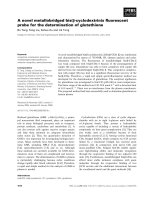
Fig. 3. Effects of amino acid substitutions in
the hydrophobic segment. (A) The amino acid
sequences around the hydrophobic segment of
4 HSP90-family proteins are compared.
Arrowheads indicate Leu-Leu replaced to
Ser-Ser at amino acids 665 and 666 or at 671
and 672. Asterisks indicate identical amino
acids. A bar represents the hydrophobic
region (amino acids Leu662-Leu678 of human
HSP90a). (B) SDS/PAGE of GST-
HSP90a657–732 (lane 1), GST-HSP90a657–
732 L665S/L666S (lane 2), GST-HSP90a657–
732 L671S/L672S (lane 3) and GST (lane 4).
M, low-molecular-mass markers. (C) The
increase in the turbidity of CS (8 lg) with
increasing amounts of recombinant proteins
was measured as described in ÔExperimental
ProceduresÕ. Experiments were repeated three
times and identical results were obtained. The
data of one typical experiment are represented.
150 S i. Yamada et al. (Eur. J. Biochem. 270) Ó FEBS 2003
maltose-binding protein [27]. This configuration of a
GRP94 dimer is apparently distinct from our dimer model
on HSP90, in which the middle domain is associated with
the C-terminal domain in an antiparallel fashion [20,37]. It
has also been reported that purified HSP90, GRP94 and
HtpG self-oligomerize at elevated temperatures and that
this phenomenon is closely related to the client-binding
function of the proteins [32,38]. Taken together, we assumed
that formation of the complex of the region carrying the
hydrophobic segment of GRP94 is mediated via its client-
binding activity. To settle this issue, we reinvestigated the
domain–domain interaction of human GRP94 by use of the
bacterial two-hybrid system.
Table 2 shows that the dimerization was mediated via the
interaction between the middle domain and the C-terminal
one. Hence, we conclude that the C-terminal domain, which
contains the hydrophobic region, does not associate with
each other.
Above findings let us further examine the possibility that
hybrid dimers could be formed among three HSP90-family
proteins. In a control experiment, the two-hybrid experi-
ment demonstrated homodimer formation of the middle-C-
terminal domains of HSP90a (Table 3). The two-hybrid
experiment using the middle-C-terminal domains showed
heterodimer formation between HSP90a and HtpG and
between GRP94 and HtpG. On the other hand, a complex
was not formed between HSP90a and GRP94 as reported
previously [22].
We finally investigated whether the client-binding site of
GRP94 is localized in either the middle domain or the
C-terminal one. GRP94-M and GRP94-C were expressed
as GST-fusion proteins. They were purified to near homo-
geneity, although the preparation of GST-GRP94-M con-
tained some amounts of 29-kDa GST species (Fig. 4A, lane
1). Figure 4B clearly demonstrated that GST-GRP94-C
suppressed the aggregation of CS at 45 °C, but that GST-
GRP94-M did not.
Discussion
Several biochemical properties and the roles have been
characterized on the C-terminal domain of HSP90. At first,
the C-terminal pentapeptide of HSP90 was recognized by
the tetra-tricopeptide repeat (TPR)-domain containing
cochaperone Hop, which connects HSP90 with the
HSP70-family proteins [39]. Secondly, residues 702–716
adjacent to the C-terminus form one of the two most
immunogenic regions [28], which strongly suggests that this
region is exposed at the outer surface of an HSP90 dimer.
Thirdly, the C-terminal 49 amino acids are essential for the
dimer formation [24]. Fourthly, the C-terminal domain of
HSP90 contains a client-binding site with characteristics
distinct from those of the site located at the N-terminal
domain [14–17]. This C-terminal client-binding site also
exists in GRP94 [40], but not in HtpG [17]. However, the
respective studies dealt with one of these properties, and
Fig. 4. Suppression of the heat-induced
aggregation of CS by the C-terminal domain of
GRP94. (A) One microgram of GST-GRP94-
M (lane 1), GST-GRP94-C (lane 2) and GST
(lane 3) were electrophoresed on SDS/PAGE.
M, low-molecular-mass markers. (B) The
increase in the turbidity of CS (8 lg) with
increasing amounts of GST-GRP94-M and
GST-GRP94-C was measured. Experiments
were repeated twice and identical results were
obtained. The data of one typical experiment
are represented.
Table 3. Hybrid dimer formation in the C-terminal regions of 3 HSP90-
family proteins. The bacterial two-hybrid system was used to evaluate
the binding activity. The value of the combination of pKT25
kan
-
HSP90a-MC and pUT18C
amp
-HSP90a-MC was set to 100%. Activ-
ities are given as mean ± SD (n ¼ 4).
pKT25
kan
- pUT18C
amp
- Activity (%)
1
Vector Vector 10.1 ± 0.2
HSP90a-MC HSP90a-MC 100.0 ± 1.3
GRP94-MC HSP90a-MC 13.9 ± 6.9
GRP94-MC HtpG-MC 81.2 ± 40.7
HSP90a-MC HtpG-MC 87.7 + 38.6
Table 2. Interaction between the middle and C-terminal domains of
GRP94. The bacterial two-hybrid system was used to evaluate the
binding activity. The value of the combination of the middle and the
C-terminal domains was set to 100%. Activities are given as mean ±
SD (n ¼ 3).
pKT25
kan
- pUT18C
amp
- Activity (%)
vector vector 2.1 ± 0.4
GRP94-M GRP94-C 100.0 ± 0.7
GRP94-M GRP94-M 1.8 ± 0.5
GRP94-C GRP94-C 2.5 ± 0.2
Ó FEBS 2003 Two roles of the C-terminal domain of HSP90 (Eur. J. Biochem. 270) 151
therefore, it is still ambiguous whether the regions, especially
the region responsible for dimer formation and that for
client binding, exist at distinct sites of the C-terminal region,
or they are closely related to each other.
In our approach we initially focused on the C-terminal 35
amino acids of HSP90, of which the equivalent region is
deleted in HtpG. Our hypothesis that the C-terminal 35
amino acids were not essential for the dimerization was
verified by the data shown in Fig. 1. On the other hand, the
second assumption that the 35 amino acids were involved in
the client binding was not true, but the central part of the
C-terminal domain, residues 657–720, was shown to be
essential. Therefore, the two regions that were sufficient for
both functions overlapped or were indistinguishable from
each other. Their close relationship was ascertained by
amino acid substitutions in the hydrophobic segment
(Fig. 3 and Table 1).
The present study demonstrated that, in HSP90a, double
mutations of Leu to Ser at positions 665 and 666 or 671 and
672 in the hydrophobic segment diminished or completely
destroyed the client-binding and dimer-forming activities
simultaneously. The amino acid sequence of the hydropho-
bic segment of HSP90a was relatively conserved with those
of human GRP94 and E. coli HtpG (Fig. 3A). However,
the difference was evident in the hydropathy plot of the
C-terminal domain according to Kyte and Doolittle [41].
As shown in Fig. 5, the corresponding region of HtpG is
less hydrophobic, which may explain the lack of the binding
of the C-terminal domain of HtpG to a client protein [17].
We critically reviewed the previous study that demon-
strated dimer formation of the hydrophobic segment of
GRP94 [27]. The maltose-binding protein-fused GRP94
segment migrated with a wide range of apparent molecular
masses on a size-exclusion chromatography column, indi-
cating the formation of oligomers larger than a dimer. The
present study on GRP94 demonstrated a direct interaction
between the middle domain and the C-terminal one, and
that neither the C-terminal domain nor the middle domain
homo-dimerized. Accordingly, we propose that the dimeri-
zation of the HSP90-family protein is generally achieved
through a pair of heteromeric interactions between the
middle and C-terminal domains. Self-oligomer formation of
the hydrophobic segment of GRP94 [27] may reflect its
potent client-binding capacity located in the C-terminal
domain.
The perfect dimer configuration of the HSP90-family
protein seems to be accomplished through a pair of
the intermolecular interactions between the middle and
C-terminal domains as proposed previously [20], even if a
single interaction between the middle and C-terminal
domains might be sufficient to maintain the complex under
the experimental conditions. Bearing in mind the finding
that the hybrid formation of the N-terminal and middle
domains between human HSP90a and E. coli HtpG [17],
the conformational similarity of the HSP90-family proteins
can be expanded to all domains of the protein.
Bacterial two-hybrid experiments demonstrated the
interaction between the middle and C-terminal domains of
GRP94 (Table 2) as well as those of HtpG [17]. In contrast,
the combination failed to form a complex in HSP90a [17],
but the combination of the middle-C-terminal domains
either with the middle domain or the C-terminal domain is
required for the interaction (Fig. 1 and [17]). Presumably,
the fine mode of the dimeric structure may be not identical
among all members of the HSP90-family protein. Addi-
tionally, it should be noted that this phenomenon made it
difficult to reconstitute the complex between the middle and
C-terminal domains with purified samples in vitro,because
HSP90a-MC formed a stable dimer; neither the middle
domain nor the C-terminal domain added afterwards was
replaced (data not shown). Accordingly, an attempt to
reconstitute such a complex of HSP90a in vitro was not
successful (data not shown).
The importance of the C-terminal region for the HSP90
molecular chaperone has been indicated by Sullivan and
Toft [13]: two separate regions of chicken HSP90b (amino
acids 381–441 and 601–677) are particularly important for
the binding of the progesterone receptor. Hartson et al.
[42] also proposed that a specific region near residue 600
determines the mode by which HSP90 interacts with
substrates. Moreover, Glu651-Ile698 of human HSP90a,
which carries the hydrophobic segment, is required for
activation of basic helix-loop-helix-helix (bHLH) proteins,
such as MyoD and E12 [43]. The findings in the present
study on the client binding are consistent with these
reports.
Human GRP94 and mouse HSP90 were identified as
tumor-specific antigens expressed on the surface of various
tumor cells [44,45]. Recently, the C-terminal site of GRP94
bound to a vesicular stomatitis virus capsid-derived peptide
was attributed to a charged region, Lys602-Asp-Lys-Ala-
Leu-Lys-Asp-Lys609, by a photoaffinity labeling experi-
ment [40]. This region is located in the middle domain
(Glu428-Lys650), not in the C-terminal domain (Asp651-
Leu782), in contrast to the results in the present study. At
present, it remains unknown why this discrepancy occurred,
but the dimer topology of the family proteins may provide
a hint. That is, the middle domain associates with the
Fig. 5. Hydropathy plot of the C-terminal domain of the 3 HSP90-
family proteins. The hydropathy of the C-terminal domain of human
HSP90a (light line), human GRP94 (dotted line) and E. coli HtpG
(bold line) were plotted according to the methods of Kyte and Doo-
little [41]. The amino acid numbers are represented as those of human
HSP90a.
152 S i. Yamada et al. (Eur. J. Biochem. 270) Ó FEBS 2003
C-terminal domain in a GRP94 dimer, and accordingly, the
charged region of the middle domain may be adjacent to the
hydrophobic segment of domain C in the tertiary structure
in a dimer. Therefore, it should be possible to confirm
whether the Lys602-Lys609 charged region is truly indis-
pensable for client binding or simply present adjacent to the
client-binding site with the result of being affinity-labeled
with the client peptide. This issue is now under investigation
in our laboratory.
Acknowledgements
We greatly appreciate Drs D. Ladant (Pasteur Institute, Paris, France)
and L. Selig (Hybrigenics S.A., Paris, France) for generous providing
with the bacterial two-hybrid system. We also thank Mr T.
Kobayakawa (Nagasaki University, Nagasaki, Japan) for the technical
assistance. This work was supported by Grants-in-Aid for Scientific
Research from the Ministry of Education, Culture, Sports, Science and
Technology of Japan and from Japan Society for the Promotion of
Science (to T. K. N.).
References
1. Borkovich, K.A., Farrelly, F.W., Finkelstein, D.B., Taulien, J. &
Lindquist, S. (1989) Hsp82 is an essential protein that is required
in higher concentrations for growth of cells at higher temperatures.
Mol. Cell. Biol. 9, 3919–3930.
2. Hickey,E.,Brandon,S.E.,Smale,G.,Lloyd,D.&Weber,L.A.
(1989) Sequence and regulation of a gene encoding a human
89-kDa heat-shock protein. Mol. Cell. Biol. 9, 2615–2626.
3. Rebbe, N.F., Ware, J., Bertina, R.M., Modrich, P. & Sttafford,
D.W. (1987) Nucleotide sequence of a cDNA for a member of the
human 90-kDa heat-shock protein family. Gene 53, 235–245.
4. Pelham, H.R.B. (1986) Speculations on the functions of the major
heat shock and glucose-regulated proteins. Cell 46, 959–961.
5. Felts, S.J., Owen, B.A.L., Nguyen, P M., Trepe, J., Donner, D.T.
& Toft, D.O. (2000) The hsp90-related protein TRAP1 is a
mitochondrial protein with distinct functional properties. J. Biol.
Chem. 275, 3305–3312.
6. Bardwell, J.C.A. & Craig, E.A. (1987) Eukaryotic M
r
83,000 heat
shock protein has a homologue in Esherichia coli. Proc.NatlAcad.
Sci. USA 84, 5177–5181.
7. Bardwell, J.C. & Craig, E.A. (1988) Ancient heat shock gene is
dispensable. J. Bacteriol. 170, 2977–2983.
8. Tanaka,N.&Nakamoto,H.(1999)HtpGisessentialforthe
thermal management in cyanobacteria. FEBS Lett. 458, 117–123.
9. Binart, N., Lombes, M. & Baulieu, E E. (1995) Distinct functions
of the 90 kDa heat shock protein (hsp90) in oestrogen and
mineralocorticoid receptor activity: effects of hsp90 deletion
mutants. Biochem. J. 311, 797–804.
10. Miyata,Y.&Yahara,I.(1995)Interactionbetweencaseinkinase
II and the 90-kDa stress protein. Biochemistry 34, 8123–8129.
11. Louvion, J.F., Warth, R. & Picard, D. (1996) Two eukaryotic-
specific regions of Hsp82 are dispensable for its viability and signal
transduction functions in yeast. Proc.NatlAcad.Sci.USA93,
13937–13942.
12. Sato. S., Fujita, N. & Tsurui, T. (2000) Modulation of Akt kinase
activity by binding to Hsp90. Proc. Natl Acad. Sci. USA 97,
10832–10837.
13. Sullivan, W.P. & Toft, D.O. (1993) Mutational analysis of hsp90
binding to the progesterone receptor. J. Biol. Chem. 268, 20373–
20379.
14. Young, J.C., Schneider, C. & Hartl, F.U. (1997) In vitro evidence
that hsp90 contains two independent chaperone sites. FEBS Lett.
418, 139–143.
15. Scheibel, T., Weikl, T. & Buchner, J. (1998) Two chaperone sites in
Hsp90 differing in substrate specificity and ATP dependence. Proc.
NatlAcad.Sci.USA95, 1495–1499.
16. Minami,M.,Nakamura,M.,Yasuhumi,E.&Minami,Y.(2001)
Both the N- and C-terminal chaperone sites of Hsp90 participate
in protein folding. Eur. J. Biochem. 268, 2520–2524.
17. Tanaka, E., Nemoto, T.K. & Ono, T. (2001) Liberation of the
intramoleular interaction as the mechanism of heat-induced acti-
vation of HSP90 molecular chaperone. Eur. J. Biochem. 268,
5270–5277.
18. Roher, N., Miro, F., Boldyreff, B., Llorens, F., Plana, M.,
Issinger, O G. & Itarte, E. (2001) The C-terminal domain of
human grp94 protects the catalytic subunit of protein kinase CK2
(CK2a) against thermal aggregation: role of disulfide bonds. Eur.
J. Biochem. 268, 429–436.
19. Minami, Y., Kawasaki, H., Miyata, Y., Suzuki, K. & Yahara, I.
(1991) Analysis of native forms and their isoform compositions of
the mouse 90-kDa heat-shock protein, HSP90. J. Biol. Chem. 266,
10099–10103.
20. Nemoto, T., Ohara-Nemoto, Y., Ota, M., Takagi, T. &
Yokoyama, K. (1995) Mechanism of dimer formation of the
90-kDa heat-shock protein. Eur. J. Biochem. 233, 1–8.
21. Spence, J. & Georgopoulos, C. (1989) Purification and properties
of the Escherichia coli heat shock protein, HtpG. J. Biol. Chem.
264, 4398–4403.
22. Nemoto, T., Matsusaka, T., Ota, M., Takagi, T., Collinge, D.B. &
Walther-Larsen, H. (1996) Dimerization characteristics of the
94-kDa glucose-regulated protein. J. Biochem. 120, 249–256.
23. Nemoto, T. & Sato, N. (1998) Oligomeric forms of the 90-kDa
heat shock protein. Biochem. J. 330, 989–995.
24. Minami, Y., Kimura, Y., Kawasaki, H., Suzuki, K. & Yahara, I.
(1994) The carboxy-terminal region of mammalian HSP90 is
required for its dimerization and function in vivo. Mol. Cell. Biol.
14, 1459–1464.
25. Meng, X., Devin, J., Sullivan, W.P., Toft, D., Bauleu, E E. &
Catelli, M G. (1996) Mutational analysis of HSP90a dimerization
and subcellular localization: dimer disruption does not impede
Ôin vivoÕ interaction with estrogen receptor. J. Cell Sci. 109,
1677–1687.
26. Nemoto, T.K., Ono, T., Kobayakawa, T., Tanaka, E., Baba, T.T.,
Tanaka, K., Takagi, T. & Gotoh, T. (2001) Domain–domain
interactions of HtpG, an Esherichia coli homologue of eukaryotic
HSP90 molecular chaperone Eur. J. Biochem. 268, 5258–5269.
27. Wearsch, P.A. & Nicchitta, C.V. (1996) Endoplasmic reticulum
chaperone GRP94 subunit assembly is regulated through a defined
oligomerization domain. Biochemistry 35, 16760–16769.
28. Nemoto, T., Sato, N., Iwanari, H., Yamashita, H. & Takagi, T.
(1997) Domain structures and immunogenic regions of the 90-kDa
heat-shock protein (HSP90): probing with a library of anti-HSP90
monoclonal antibodies and limited proteolysis. J. Biol. Chem. 272,
26179–26187.
29. Maki, R.G., Old, L.J. & Srivastava, P.K. (1990) Human homo-
logue of murine tumor rejection antigen gp96: 5¢-regulatory and
coding regions and relationship to stress-induced proteins. Proc.
NatlAcad.Sci.USA87, 5658–5662.
30. Nemoto, T., Ohara-Nemoto, Y., Shimazaki, S. & Ota, M. (1994)
Dimerization characteristics of the DNA- and steroid-binding
domains of the androgen receptor. J. Steroid Biochem. Mol. Biol.
50, 225–233.
31. Karimova, G., Ullmann, A. & Ladant, D. (2001) Protein–protein
interactions between Bacillus stearothernophilus tyrosyl-tRNA-
synthase subdomains revealed by a bacterial two-hybrid system.
J. Mol Microbiol. Biotechnol. 3, 73–82.
32. Nemoto, T.K., Ono, T. & Tanaka, K. (2001) Substrate-binding
characteristics of proteins in the 90 kDa heat shock protein family.
Biochem. J. 354, 663–670.
Ó FEBS 2003 Two roles of the C-terminal domain of HSP90 (Eur. J. Biochem. 270) 153
33. Chadli, A., Bouhouche, I., Sullivan, W., Stensgard, B., McMahon,
N., Catelli, M.G. & Toft, D.O. (2000) Dimreization and
N-terminal proximity underlie the function of the molecular cha-
perone heat shock protein 90. Proc. Natl Acad. Sci. USA 97,
12524–12529.
34. Prodromou, C., Roe, S.M., O’Brien, R., Ladbury, J.E., Piper,
P.W. & Pearl, L.H. (1997) Identification and structural char-
acterization of the ATP/ADP-binding site in the Hsp90 molecular
chaperone. Cell 90, 65–75.
35. Prodromou, C., Roe, S.M., Piper, P.W. & Pearl, L.H. (1997) A
molecular clamp in the crystal structure of the N-terminal domain
of the yeast Hsp90 chaperone. Nat. Struct. Biol. 4, 477–482.
36. Prodromou, C., Panaretou, B., Chohan, S., Siligardi, G., O’Brien,
R., Ladbury, J.E., Roe, S.M., Piper, P.W. & Pearl, L.H. (2000)
The ATPase cycle of Hsp90 drives a molecular ÔclampÕ via tran-
sient dimerization of the N-terminal domains. EMBO J. 16, 4383–
4392.
37. Maruya, M., Sameshima, M., Nemoto, T. & Yahara, I. (1999)
Monomer arrangement in HSP90 dimer as determined by dec-
oration with N- and C-terminal specific antibodies. J. Mol. Biol.
285, 903–907.
38. Yonehara,M.,Minami,Y.,Kawata,Y.,Nagai,J.&Yahara,I.
(1996) Heat-induced chaperone activity of HSP90. J. Biol. Chem.
271, 2641–2645.
39. Scheufler, C., Brinker, A., Bourenkov, G., Pegoraro, S.,
Moroder,L.,Bartunik,H.,Hartl,F.U.&Moarefi,I.(2000)
Structure of TPR domain-peptide complexes: critical elements in
the assembly of the Hsp70-Hsp90 multichaperone machine. Cell
101, 199–210.
40. Linderoth, N.A., Popowicz, A. & Sastry, S. (2000) Identifica-
tion of the peptide-binding site in the heat shock chaperone/
tumor rejection antigen gp96 (Grp94). J. Biol. Chem. 275, 5472–
5477.
41. Kyte, J. & Doolittle, R.F. (1982) A simple method for displaying
the hydrophobic character of a protein. J. Mol. Biol. 157, 105–132.
42. Hartson, S.D., Thulasiraman, V., Huang, W., Whitesell, L. &
Matts, R.L. (1999) Molybdate inhibits Hsp90, induces structural
changes in its C-terminal domain, and alters its interactions with
substrates. Biochemistry 38, 3837–3849.
43. Shue, G. & Kohtz, D.S. (1994) Structural and functional aspects
of basic helix-loop-helix protein folding by heat-shock protein 90.
J. Biol. Chem. 269, 2707–2711.
44. Srivastava, P.K., DeLeo, A.B. & Old, L.J. (1986) Tumor rejection
antigens of chemically induced tumors of inbred mice. Proc. Natl
Acad. Sci. USA 83, 3407–3411.
45. Ullrich, S.J., Robinson, E.A., Lav, L.W., Willingham, M. &
Appella, E. (1986) A mouse tumor-specific transplantation antigen
is a heat shock protein. Proc. Natl Acad. Sci. USA 83, 3121–3125.
154 S i. Yamada et al. (Eur. J. Biochem. 270) Ó FEBS 2003