Báo cáo khoa học: Upstream and intronic regulatory sequences interact in the activation of the glutamine synthetase promoter pot
Bạn đang xem bản rút gọn của tài liệu. Xem và tải ngay bản đầy đủ của tài liệu tại đây (304.67 KB, 7 trang )
Upstream and intronic regulatory sequences interact in the
activation of the glutamine synthetase promoter
Rocio M. Garcia de Veas Lovillo, Jan M. Ruijter, Wil T. Labruye
`
re, Theodorus B. M. Hakvoort
and Wouter H. Lamers
AMC Liver Center, Academic Medical Center, University of Amsterdam, Amsterdam, the Netherlands
Glutamine synthetase (GS) is expressed at high levels in
subsets of cells in some tissues and at low levels in all cells of
other tissues, suggesting that the GS gene is surrounded by
multiple regulatory elements. We searched for such elements
in the 2.5-kb upstream region and in the 2.6-kb first intron of
the GS gene, using FTO-2B hepatoma and C2/7 muscle cells
as representatives of both cell types and transient transfec-
tion assays as our tools. In addition to the entire upstream
region and entire intron, an upstream enhancer module at
)2.5 kb, and 5¢, middle and 3¢ modules of the first intron
were tested. The main effects of the respective modules and
their combinatorial interactions were quantified using the
analysis of variance (
ANOVA
) technique. The upstream
enhancer was strongly stimulatory, the middle intron mod-
ule strongly inhibitory, and the 3¢-intron module weakly
stimulatory in both hepatoma and muscle cells. The
5¢-intron module was strongly stimulatory in muscle cells
only. The major new finding was that in both cell types, the
upstream enhancer and 5¢-intron module needed to be pre-
sent simultaneously to fully realize their transactivational
potencies. This interaction was responsible for a pronounced
inhibitory effect of the 5¢-intron module in the absence of the
upstream enhancer in hepatoma cells, and for a strong
synergistic effect of these two modules, when present sim-
ultaneously in muscle cells. The main difference between
hepatoma and muscle cells therefore appeared to reside in
tissue-specific differences in activity of the respective regu-
latory elements due to interactions rather than in the exist-
ence of tissue-specific regulatory elements.
Keywords: enhancer; glutamine synthetase; hepatoma;
muscle; transient transfection.
Glutamine synthetase (GS; EC 6.3.1.2), the enzyme that
catalyses the ATP-dependent conversion of glutamate and
ammonia into glutamine, is expressed in a tissue-specific and
developmentally controlled manner. GS functions to
remove ammonia or glutamate, or to produce glutamine.
Cells that function primarily to remove glutamate or
ammonia, contain very high GS levels (30–160 l
M
),
whereas cells that synthesize glutamine contain much lower
levels (1–8 l
M
) [1]. Another highly characteristic and
functionally important feature of GS is its topographic
distribution: in organs in which GS is present at relatively
high concentrations, it is usually expressed in a subset of
cells only, whereas in organs in which it is present at low
concentrations, it is expressed in the majority of cells.
Examples of the first group of organs are the pericentral
hepatocytes in the liver, the astrocytes in nervous tissue, the
epithelial cells of the caput epididymis, and the gastric
antrum. Examples of the second group are adipocytes and
muscle cells (for a review, see [1]).
Because of these interorgan differences in distribution
and cellular concentration of GS, and because only a single
functional copy of the GS gene is present per haploid
genome in rodents [2–4], it is to be anticipated that the
regulation of GS expression is complex [1]. Studies aimed
towards delineating the transcriptional regulation of GS
expression in rodents have thus far revealed upstream
enhancer elements at )6.0 kb and )2.5 kb, and intron
enhancer elements at +0.35 kb and +1.6 kb, by transient
or stable transfections to cultured cells [5–8]. The sequence
of the far-upstream mouse GS enhancer that is active in
adipocytes [9] is 80% similar to that of the far-upstream rat
GS enhancer and, like the rat far-upstream enhancer, also
maps at )6.0 kb in the Celera Discovery System mouse
genome database. The upstream enhancer at )2.5 kb
confers pericentral localization to reporter gene expression
in the liver of transgenic mice [10]. The significance of the
far-upstream and intron elements for the in vivo expression
pattern of GS remains to be assessed.
In the liver, genes are expressed in a porto-central
gradient. Studies with transgenic animals have shown that
many of these gradients in gene expression, including that of
GS [10], are determined at the transcriptional level. Porto-
central gradients in gene expression have been distinguished
into dynamic and stable gradients [11]. The dynamic type of
zonation is characterized by adaptive changes in expression
in response to changes in the metabolic or hormonal state,
whereas the stable type of zonation, of which GS is an
example [12,13], is characterized by the virtual absence of
such adaptive changes. A relatively simple model to explain
Correspondence to W. H. Lamers, AMC Liver Center, Academic
Medical Center, University of Amsterdam, Meibergdreef 69-71,
1105 BK, Amsterdam, the Netherlands.
Fax: + 31 20 5669190, Tel.: + 31 20 5665948,
E-mail:
Abbreviations: GS, glutamine synthetase; TK, thymidine kinase.
Enzymes: Glutamine synthetase (EC 6.3.1.2).
(Received 22 August 2002, revised 10 October 2002,
accepted 17 October 2002)
Eur. J. Biochem. 270, 206–212 (2003) Ó FEBS 2003 doi:10.1046/j.1432-1033.2003.03424.x
such a stable expression pattern is to assume a Ôdouble-lockÕ
regulatory mechanism, meaning that GS expression depends
on the synergistic interaction of two or more factors [14].
The observation that very high levels of GS are present in
subsets of cells in some organs, and moderate-to-low levels
in all cells of other organs, in combination with the
hypothetical Ôdouble-lockÕ mechanism to account for the
stable expression of GS in pericentral hepatocytes suggested
that multiple regulatory modules would control GS expres-
sion and that at least some of these modules would be
interdependent with respect to their regulatory activity. To
test this hypothesis, we examined the combinatorial effects
of modular deletions in the distal upstream and first intron
regions of the rat GS gene on reporter gene expression in
hepatoma and muscle cells. We now report that interactions
of upstream and intronic regulatory elements do indeed
determine the degree of activation of the GS promoter and
that these interactions differ quantitatively between cells
from hepatic and muscular origin.
Materials and methods
Sequence of the first intron of GS
The nucleotide sequences of the upstream region and of the
first 1938 nucleotides of the first intron of the rat GS gene
were reported [5,6]. This intron sequence ends at the EcoRI
restriction site (Fig. 1). The remaining 877 nucleotides of the
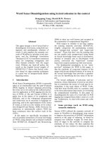
Fig. 1. Schematic representation of GS sequences used in constructs A–Q and their reporter gene activities. At the top, a restriction map of the
genomic GS region analysed in these experiments, is shown. The exons are shown as boxes, with the arrows indicating the start sites of transcription
(left) and translation (right), and ÔpAÕ both polyadenylation sites of the gene. The upstream boundary ()2520), the transcription start site (0) and the
downstream boundary (+2774) of the genomic DNA segment that was analysed, are indicated. The sequences present in the respective constructs
are shown in the left portion of the figure as solid black lines. The upper line shows the linkage of the 5.3-kb genomic GS segment with the luciferase
reporter gene at the NcoI site (translation start site) in the second exon and with the bovine growth hormone transcription termination and
polyadenylation signal (bGH). The first and second exon up to the translation start site are represented as black boxes. The right portion of the
figure shows luciferase activity of the respective constructs in FTO-2B hepatoma cells (light grey) and C2/7 muscle cells (dark grey). Panels I and II
show activities of the respective upstream and intron elements, respectively, when present in conjunction with the minimal promoter and minimized
first intron. Panels III and IV show activities of combinations of upstream enhancer and the entire upstream region, respectively, in conjunction
with the minimal promoter, the minimized first intron and the respective intron elements. Luciferase activity (± SEM) is expressed relative to
construct A containing only the GS promoter and minimized first intron, which was set at 100. Restriction sites: H, HindIII; E, EcoRV; P, PstI; Bg,
BglII; S, SmaI; B, BamHI; EI, EcoRI; N, NcoI.
Ó FEBS 2003 Regulatory elements of the glutamine synthetase gene (Eur. J. Biochem. 270) 207
first intron of GS were sequenced in both orientations. The
sequence data has been deposited with the EMBL nucleo-
tide sequence data bank and is available under accession
number AF170107.
Construction of plasmids
Rat GS genomic DNA sequences were cloned into the
vector pSPluc+ (Promega). The starting construct (Fig. 1,
construct Q) was made by inserting the genomic GS
segment from )2520 bp (relative to the transcription start
site) to +2774 bp (corresponding to position +132 in the
GS cDNA, that is, the translation start site in the second
exon) between the HindIII and NcoI sites in the polylinker
upstream of the luciferase cDNA. The 305 bp bovine
growth-hormone polyadenylation sequence (the XbaI–
PvuII fragment from pcDNA3; Invitrogen) was inserted
between the XbaIandEcoRV sites in the polylinker
downstream of the luciferase cDNA. The other constructs
were generated by modular deletions of construct Q. The
upstream modules were: the entire region downstream of
HindIII ()2520), the region downstream of EcoRV
()2148), the region downstream of PstI()965), or the
upstream enhancer element ()2520 to )2148 [5]). The first
intron was subdivided into three modules: a 5¢ SmaI–
BamHI fragment (+153 to +856), a middle BamHI–
SmaI fragment (+856 to +1791) and a 3¢ SmaI–BamHI
fragment (+1791 to +2712). The extended GS promoter
(BglII ()368) to the transcription-start site [5], the first
exon, a minimized first intron, containing its 5¢-most
portion (+119 to +153) and 3¢-most portion (+2712 to
+2760), and the second exon up to the translation start
site (+132) were present in all constructs. The construct
carrying only these elements (construct A) was used as
reference construct.
For the transfection assays, the respective constructs were
purified in CsCl gradients or on Nucleobond columns
(Machery-Nagel, Du
¨
ren, Germany).
Cell culture
FTO-2B rat hepatoma cells [15] were cultured in DMEM/
F-12 medium (Gibco), supplemented with 10% (w/v)
foetal bovine serum (Gibco). C2/7 cells (generously
provided by M. Buckingham, Institut Pasteur, Paris,
France) are a subclone of the C2 cell line that was
originally derived from Soleus muscle of adult C3H mice
[16]. These cells were cultured in DMEM (Gibco),
supplemented with 10% (w/v) foetal bovine serum. All
cells were cultured at 37 °C in humidified air containing
5% CO
2
. Cell lines were tested monthly for contamination
with mycoplasms.
DNA transfection
Exponentially growing FTO-2B cells were transfected by
electroporation [17] and C2/7 cells were transfected at the
myoblast stage by the calcium-phosphate method [18]. In
both cases 20 lg supercoiled plasmid was used. Cotrans-
fection with 5 lg of the vector pRSVcat [19] enabled
correction for differences in transfection efficiency. After
electroporation, the cell suspension was divided into two
equal parts, one being grown in culture medium and the
other in culture medium supplemented with 100 n
M
dexamethasone. Sixteen h after transfection, the cells were
washed with NaCl/P
i
and given fresh medium. In the case
of the C2/7 cells, the concentration of foetal bovine serum
was reduced to 1% to induce the formation of myotubes.
Harvest of the cells was carried out 48 h after transfection
in the case of FTO-2B cells, and 72 h in the case of C2/7
cells, that is, when all cells were fused into myotubes.
Cells were lysed in 100 m
M
KH
2
PO
4
/K
2
HPO
4
pH 7.6,
0.1% (v/v) Triton X-100 buffer and tested for chloram-
phenicol acetyltransferase activity [20], luciferase activity
[21] and protein concentration (bicinchoninic acid reagent;
Pierce).
Splicing of modified first intron
Constructs carrying the different modules of the GS first
intron were tested for proper splicing of the mRNA.
RT-PCR was carried out with primers in the first exon (+1
to +18) and in the luciferase-coding region (+78 to +60 in
the luciferase cDNA). All constructs generated correctly
spliced mRNAs (data not shown).
Correction for experimental variation and statistics
The transactivation potential of the tested DNA con-
structs was expressed as the ratio between their luciferase
activity (light unitsÆmg protein
)1
) and the chloramphenicol
acetyltransferase activity (unitsÆmg protein
)1
)ofthe
cotransfected pRSVcat construct. The data were collected
from 33 experiments with FTO-2B cells and 13 experi-
ments with C2/7 cells. In each experiment, different
combinations of constructs were tested. The number of
transfections per construct was 8–20. Interexperimental
variation in reporter gene activity was removed using
log-transformed values and the
GENERAL LINEAR MODEL/
ANOVA
without interaction (
SPSS
version 10.0.7; SPSS
Inc.).
The activity of a specific construct (X,Y) containing
upstream module (X) and intron module (Y) can be
modelled to consist of the sum of a basal activity (produced
by the promoter and minimized intron), the effects of the
respective upstream (X) and intron (Y) modules, and the
interaction (X,Y) between these modules:
activity
construct ðX;YÞ
¼ activity
construct A
þ effect
upstream module ðXÞ
þ effect
intron module ðYÞ
þ effect
interaction ðX;YÞ
In this model, the value of the main effects of the upstream
and intron modules, and that of their interactions can be
calculated with an approach based on the analysis of
variance (
ANOVA
) technique. To normalize the data, the
activity of reference construct A was set to 100 arbitrary
units (AU) in these calculations. The activity of the
respective modules and their interactions, including 95%
confidence intervals, was expressed relative to construct A.
Whenever a difference is mentioned in the text, it is
significant at the 5% level.
208 R. M. Garcia de Veas Lovillo et al. (Eur. J. Biochem. 270) Ó FEBS 2003
Results
We based the design of our analysis of the regulatory
properties of sequences in the upstream region and within
the first intron of the rat GS gene on the assumption that
two or more interdependent regulatory elements were
responsible for transactivation of the GS promoter [14].
For this reason, the study was designed to reveal which
DNA sequences do interact with respect to transactivation
of this promoter. We also wished to avoid that changes in
the position of the regulatory sequences might affect the
regulatory behaviour of the DNA modules. For that reason,
the experimental constructs were made by modular ortho-
topic additions to construct A (Fig. 1).
To delineate regulatory elements upstream of the GS
structural gene, the upstream sequence present in construct
A was extended to )965 nucleotides (construct B), to )2148
nucleotides (construct C), or to )2520 nucleotides (construct
M) (Fig. 1, panel I). None of these upstream modules
significantly enhanced the activity of the GS promoter in
either FTO-2B hepatoma cells or C2/7 myotubes. Previous
experiments had shown that the upstream region was able
to transactivate the heterologous thymidine kinase (TK)
promoter, and that this activity was localized between
)2520 and )2148 bp [5]. When placed directly in front of
the GS promoter, this distal upstream enhancer element
(construct H, Fig. 1, panel III) caused a small but significant
increase in reporter gene expression (1.4-fold in FTO-2B
and 1.8-fold in C2/7 cells).
The transactivational capacity of the first intron of the GS
gene was tested as such (construct G), as a 703-bp 5¢ intron
module (construct D), a 935-bp middle intron module
(construct E), a 921-bp 3¢ intron module (construct F), or
after deleting all intron sequences except 35 nucleotides at the
5¢ end and 48 nucleotides at the 3¢ end (construct A, the
reference construct) (Fig. 1, panel II). In FTO-2B cells, the
entire intron (construct G) decreased reporter gene activity
significantly to 40% of that of construct A. The inhibitory
activity was found to reside in the 5¢ and middle intron
fragments (constructs D and E). In muscle cells, the entire
intron depressed reporter gene activity to 50% of that of
reference construct A. When the intron fragments were
tested individually, the 5¢ intron fragment (construct D) was
without effect on the promoter, whereas the middle fragment
slightly decreased reporter gene activity (to 70%) and the 3¢
fragment (construct F) stimulated promoter activity 1.9-fold.
Interactions between upstream and intron regulatory
modules
When different combinations of upstream and intron
sequences were tested for transactivation of the GS promo-
ter, the highest activities were observed for constructs
containing the upstream enhancer (constructs H-L), whereas
the lowest activities were consistently associated with the
presence of the middle intron module (constructs E, J and O)
(Fig. 1, panels II–IV). The effect of partner choice appeared
to matter most for constructs containing the 5¢-intron
module (constructs D, I and N). These findings demonstra-
ted that the degree of transactivation of the GS promoter
depended to a substantial degree on interactions between the
upstream and intron regulatory sequences. We therefore
used an approach that is based upon the
ANOVA
technique to
quantify the main (that is, ÔintrinsicÕ) effects of upstream and
intron modules, and to segregate these effects from those due
to interaction between the respective elements. In this
approach, the activity of construct A was set at 100 AU.
FTO-2B hepatoma cells
The computation of the main effects revealed that the
presence of the upstream enhancer increased promoter
activity with 87 AU in hepatoma cells, whereas this number
was slightly lower (67 AU) for the entire upstream region
(Fig. 2, upper panel). Both effects were statistically signifi-
cant. The 5¢- and 3¢-intron modules both increased promo-
ter activity with 23 AU. The middle intron module
decreased promoter activity with 84 AU. Although the
effect of the entire intron on promoter activity was not
significant (14 AU), it neutralized the negative effect of its
middle fragment. The actual activity of the respective
constructs often resulted from less than additive effects
between the upstream and intron modules. Such negative
interactions were observed for constructs containing the
upstream enhancer, but lacking the 5¢-intron fragment
(constructs H, J and K), and vice versa (constructs D, G).
Furthermore, the effects of the upstream region and the
entire intron were not additive (construct Q). The other
combinations did not show significant interactions, meaning
that the main activities of their components accounted for
the observed effect. These findings demonstrate that the
simultaneous presence of the upstream enhancer and the
5¢-intron module is necessary for full transactivation of
theGSpromoterinhepatomacells.
C2/7 muscle cells
The upstream enhancer significantly increased promoter
activity in muscle cells with 121 AU, whereas this number
was not significant (13 AU) for the entire upstream region
(Fig. 2, lower panel). The intron modules all had significant
effects on the promoter: the 5¢- and 3¢-intron modules
increased promoter activity with 127 AU and 47 AU,
respectively, whereas the middle intron module decreased
promoter activity with 58 AU. The entire intron did not
affect promoter activity significantly. The inhibitory effect of
the middle intron module therefore outweighed the strongly
stimulatory effects of the 5¢- and 3¢-intron modules. The
interaction between the upstream enhancer and the 5¢-intron
fragment produced a more than additive transactivational
effect on the promoter. Similar to liver cells, the stimulatory
effect of either element was largely lost if the other element
was absent. Furthermore, and again similar to liver cells, the
effects of the upstream region and the entire intron were not
additive. These findings show that the upstream enhancer
and the 5¢-intron modules are mutually dependent for full
activityoftheGSpromoterinbothliverandmusclecells.
Comparison of FTO-2B with C2/7 cells
The comparison of both cell lines revealed that the main
activities of the upstream enhancer and 5¢-intron modules, as
well as their interaction, were higher in C2/7 cells than in
FTO-2B cells. Both cell types resembled each other in that
Ó FEBS 2003 Regulatory elements of the glutamine synthetase gene (Eur. J. Biochem. 270) 209
the upstream enhancer and 5¢-intron module had to be
simultaneously present for the highest level of reporter gene
expression, whereas significant negative interactions were
observed if either element was absent. Apparently as a result
of the latter effect the upstream enhancer increased the
inhibitory effect of the middle intron module, but did not
support the stimulatory effect of the 3¢-intron module in both
cell types. In C2/7, but not in FTO-2B cells, the upstream
enhancer and the 5¢-intron modules lost their stimulatory
activity in the context of the upstream region and the entire
intron, respectively. As the presence of both the entire
upstream region and the entire intron negatively affected
promoter activity in both cell types, the 5.3-kb region
encompassing the entire upstream region and first intron,
was threefold less active in muscle than in hepatoma cells.
The differences between hepatoma and muscle cells therefore
can be explained by tissue-specific differences in activity
of the respective regulatory elements due to interactions
rather than in the use of distinct, tissue-specific regulatory
elements.
Glucocorticoid sensitivity of the regulatory sequences
All modules were tested for sensitivity to glucocorticoid
treatment. Only constructs containing the middle intron
module showed a threefold induction of reporter gene
activity when tested in C2/7 cells (data not shown). In
hepatoma cells, no effects of the hormone were observed.
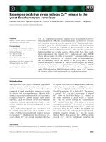
Fig. 2. Main activity and interactions of GS upstream and intron modules in FTO-2B hepatoma and C2/7 muscle cells. Main activities and interactions
(±95% confidence interval) were calculated from the activities of constructs A and D-Q, using the
ANOVA
technique. Main activities were expressed
as increase in activity upon addition to reference construct A, which was set at 100 arbitrary units. Interactions were expressed as the difference
between the observed activity of a construct and the main activities of its components. Main activities and interactions marked in bold differ
significantly (P < 0.05) from construct A and from 0. The activity of a construct (e.g. I: FTO-2B: 220; C2/7: 430) can be calculated from the figure
as the sum of the basal activity (¼ 100), the main activity of the upstream element (UE; FTO-2B: 87; C2/7: 121), the intron element (5¢;FTO-2B:23;
C2/7: 127) and their interaction (FTO-2B: 10; C2/7: 82).
210 R. M. Garcia de Veas Lovillo et al. (Eur. J. Biochem. 270) Ó FEBS 2003
Discussion
We have studied the capacity of the upstream region and
first intron of the GS gene to transactivate its promoter,
as well as the effect of interactions between these regions
on GS promoter activity. Furthermore, we aimed to
determine if any of the regulatory elements in these
regions behaved differently in cells which can express high
levels of GS (hepatocytes) and in cells which do express
low levels of the gene (muscle). The application of the
ANOVA
technique allowed us to segregate the main
(intrinsic) activities of the respective elements from the
effects of their interactions. Using this approach, the
upstream enhancer was identified as a strongly stimulatory
element in both hepatoma and muscle cells, whereas the
5¢-intron module was strongly stimulatory in muscle cells
only. In both cell types, however, the upstream enhancer
and 5¢-intron module depended on each other for effective
transactivation of the GS promoter. Other intriguing
findings were that the upstream enhancer lost most of
its activity when present in the context of the entire
upstream region in muscle cells, but not in hepatoma cells.
Furthermore, the inhibitory effect of the middle intron
module appeared to be constitutive in hepatoma cells, but
dependent on glucocorticoids in muscle cells. Apparently,
both positive and negative elements, and extensive inter-
actions between them, regulate GS promoter activity.
In addition to transcriptional control, translatability of
the GS mRNA and stability of the GS protein appear to
be important post-transcriptional levels of control [1,22].
In fact, we did show that it is necessary to consider these
post-transcriptional control levels when analysing the
expression of GS in transgenic animals [23,24]. However,
as both the reporter gene and the transcription termin-
ation and polyadenylation sequences that were used are
not normally expressed in either liver or muscle, post-
transcriptional control is an unlikely level of control to
explain the observed differences between hepatoma and
muscle cells.
The similarity of the activity of constructs A, B and C
argues against the presence of an inhibitory sequence within
the upstream region. Nevertheless, the activity of the
upstream enhancer in muscle cells and, to a lesser extent, in
hepatoma cells, is clearly mitigated in the context of the entire
upstream region, i.e. by the interposition of 1780 bp. We
interpret the increase in activity of the upstream enhancer
when positioned in close proximity to the promoter as the
consequence of a distance effect (see [25,26]): apparently, the
upstream enhancer has difficulty contacting the promoter
when the 1780 bp intervene. We have previously observed
such a distance effect for the carbamoylphosphate syn-
thetase enhancer in conjunction with the TK promoter, but
not with the carbamoylphosphate synthetase promoter
alone [26].
The 5¢ and middle intron modules of the first intron of GS
correspond tp two DNaseI-hypersensitive sites [5]. Three
studies [6–8] have analysed the enhancer activity of these
modules in conjunction with the heterologous TK promoter
[27]. The 5¢ intron module behaves as a conditional enhancer
element when positioned downstream of the promoter (this
study), but as a constitutive stimulatory element when tested
upstream of the TK promoter [6]. In this configuration, the
activity of the 5¢ intron element resided between positions
+153 and +627 [6]. In contrast, the strong and consistently
inhibitory effect of the middle intron module on GS
promoter activity does not appear to be context sensitive,
as it was also observed when tested upstream of the TK
promoter [8]. This inhibitory activity resided in a 325-bp
fragment (position +1466 to +1791) and was, similar to our
finding, relieved by glucocorticoids [8]. A putative GRE
(glucocorticoid-responsive element) was identified at posi-
tion +1656 to +1670. The middle intron regulatory element
inthemouseGSgene[7]wastestedinstabletransfection
assays in a differentiating adipocyte cell line. Its core activity
was found to be limited to a 310-bp fragment, the sequence of
which corresponds with that of position +1450 to +1752 in
the rat GS intron. This sequence was found to contain C/
EBP and HNF3 consensus-binding sites at position +1580
to +1592. The middle intron regulatory module may
therefore qualify as a glucocorticoid-responsive unit (see
[28]). The presence of an inhibitory GRU (glucocorticoid-
responsive unit) in the GS gene and a strongly stimulatory
one in the carbamoylphosphate synthetase gene [28] may
explain the frequently reciprocal behaviour of both genes
with respect to expression [13].
Co-operative interactions in the binding of transcription
factors to arrays of response elements within an enhancer
module appear to be the rule rather than the exception. The
explanation for these co-operative effects is that the binding
of a factor to an element within such an array entails an
increase in the affinity of adjacent elements for their
corresponding transcription factors. Due to the presence
of protein–protein interactions within an enhancer–promo-
ter complex, the transcription factor-binding sites do not
have to be adjacent [29,30]. However, co-operative interac-
tions between distant enhancer modules as now reported for
GS are described infrequently. Reported examples include
the synergistic interaction between a far-upstream and an
upstream enhancer [31], between an upstream and an intron
enhancer [32,33], and between an intron and a downstream
enhancer [34]. Interestingly, the GS gene itself may present
yet another example of an interaction between distant
regulatory modules, as both the )6.0 kb far-upstream
enhancer and the middle intron element enhance reporter
gene activity in stably transfected adipocytes [7,9]. Notwith-
standing this association, it remains to be shown that these
two elements do indeed interact. Whether the cooperative
interactions between distant enhancers obey the same
rules as observed for elements within a single enhancer
and for enhancer–promoter interactions, remains to be
established.
In transgenic animals, the spatio-temporal expression
pattern of a reporter gene that is driven by the GS upstream
region, revealed several discrepancies between the expression
of the endogenous GS gene and the reporter gene [23,24].
This finding suggested that one or more regulatory elements
that were not present in this transgene, were responsible for
the expression pattern of endogenous GS. Furthermore, our
modelling of gene expression patterns in the liver had
predicted that an interaction between at least two regulatory
elements was necessary to generate the remarkably stable
expression gradient of GS [14]. The present study has
identified the upstream enhancer and the 5¢-intron module as
two such interacting regulatory elements.
Ó FEBS 2003 Regulatory elements of the glutamine synthetase gene (Eur. J. Biochem. 270) 211
References
1. Lie-Venema, H., Hakvoort, T.B.M., van Hemert, F.J., Moorman,
A.F.M. & Lamers, W.H. (1998) Regulation of the spatiotemporal
pattern of expression of the glutamine synthetase gene. Prog. Nucl.
Acid Res. 61, 243–308.
2. Kuo, C.F. & Darnell, J.E. Jr (1989) Mouse glutamine synthetase is
encoded by a single gene that can be expressed in a localized
fashion. J.Mol.Biol.208, 45–56.
3. Magnuson, S.R. & Young, A.P. (1988) Murine glutamine syn-
thetase: cloning, developmental regulation and glucocorticoid
inducibility. Dev. Biol. 130, 536–542.
4. van de Zande, L.P.W.G., Labruye
`
re, W.T., Arnberg, A.C., Wil-
son, R.H., van den Bogaert, A.J.W., Das, A.T., van Oorschot,
D.A.J., Frijters, C., Charles, R., Moorman, A.F.M. & Lamers,
W.H. (1990) Isolation and characterization of the rat glutamine
synthetase- encoding gene. Gene 87, 225–232.
5. Fahrner, J., Labruye
`
re, W.T., Gaunitz, C., Moorman, A.F.M.,
Gebhardt, R. & Lamers, W.H. (1993) Identification and func-
tional characterization of regulatory elements of the glutamine
synthetase gene from rat liver. Eur. J. Biochem. 213, 1067–1073.
6. Gaunitz, F., Gaunitz, C., Papke, M. & Gebhardt, R. (1997) Cis-
regulatory sequences from the first intron of the rat glutamine
synthetase gene are involved in hepatocyte specific expression of
the enzyme. Biol. Chem. 378, 11–18.
7. Hadden, T.J., Ryou, C. & Miller, R.E. (1998) Elements in the
distal 5¢-flanking sequence and the first intron function cooper-
atively to regulate glutamine synthetase transcription during adi-
pocyte differentiation. Nucleic Acids Res. 25, 3930–3936.
8. Chandrasekhar, S., Souba, W.W. & Abcouwer, S.F. (1999)
Identification of glucocorticoid-responsive elements that control
transcription of rat glutamine synthetase. Am.J.Physiol.276,
L319–L331.
9. Hadden, T.J., Ryou, C., Zhu, L. & Miller, R.E. (2002) CAAT/
Enhancer-binding protein activates an enhancer in the glutamine
synthetase distal 5¢-flanking sequence. Arch. Biochem. Biophys.
397, 258–261.
10. Lie-Venema, H., Labruye
`
re, W.T., van Roon, M.A., de Boer,
P.A.J., Moorman, A.F.M., Berns, A.J.M. & Lamers, W.H. (1995)
The spatio-temporal control of the expression of glutamine syn-
thetase in the liver is mediated by its 5¢-enhancer. J. Biol. Chem.
270, 28251–28256.
11. Jungermann, K. (1995) Zonation of metabolism and gene
expression in liver. Histochemistry 103, 81–91.
12. Gebhardt, R. & Mecke, D. (1983) Heterogeneous distribution of
glutamine synthetase among rat liver parenchymal cells in situ and
in primary cultures. EMBO J. 2, 567–570.
13. de Groot, C.J., ten Voorde, C.H.J., van Andel, R.E., te Kortschot,
A., Gaasbeek Janzen, J.W., Wilson, R.H., Moorman, A.F.M.,
Charles, R. & Lamers, W.H. (1987) Reciprocal regulation of
glutamine synthetase and carbamoylphosphate synthetase levels in
rat liver. Biochim. Biophys. Acta 908, 231–240.
14. Christoffels, V.M., Sassi, H., Ruijter, J.M., Moorman, A.F.M.,
Grange,T.&Lamers,W.H.(1999)Amechanisticmodelforthe
development and maintenance of porto-central gradients in gene
expression in the liver. Hepatology 29, 1180–1192.
15. Killary, A.M. & Fournier, R.E.K. (1984) A genetic analysis of
extinction: trans-dominant loci regulate expression of liver-specific
traits in hepatoma hybrid cells. Cell 38, 523–534.
16. Catala, F., Wanner, R., Barton, P., Cohen, A., Wright, W. &
Buckingham, M. (1995) A skeletal muscle-specific enhancer
regulatedbyfactorsbindingtoEandCArGboxesispresentinthe
promoter of the mouse myosin light-chain 1A gene. Mol. Cell Biol.
15, 4585–4596.
17. van den Hoff, M.J.B., Christoffels, V.M., Labruye
`
re, W.T.,
Moorman, A.F.M. & Lamers, W.H. (1995) Electrotransfection
with ÔintracellularÕ buffer. In Animal Cell Electroporation and
Electrofusion Protocols (Nickoloff, J.A., ed.), pp. 185–197.
Humana Press Inc., Totowa, NJ, USA.
18. Kelly,R.,Alonso,S.,Tajbaksh,S.,Cossu,G.&Buckingham,M.
(1995) Myosin light chain 3F regulatory sequences confer
regionalised cardiac and skeletal muscle expression in transgenic
mice. J.CellBiol.129, 383–396.
19. Gorman, C.M., Merlino, G.T., Willingham, M.C., Pastan, I. &
Howard, B.H. (1982) The Rous sarcoma virus long terminal
repeat is a strong promoter when introduced into a variety of
eukaryotic cells by DNA-mediated transfection. Proc.NatlAcad.
Sci. USA 79, 6777–6781.
20. Seed, B. & Sheen, J.Y. (1988) A simple phase-extraction assay for
chloramphenicol acetyltransferase activity. Gene 67, 271–277.
21. Brasier, A.R., Tate, J.E. & Habener, J.F. (1989) Optimized use of
the firefly luciferase assay as a reporter gene in mammalian cell
lines. Biotechniques 7, 1116–1122.
22. Haupt, W., Gaunitz, F. & Gebhardt, R. (2000) Post-transcrip-
tional inhibition of glutamine synthetase induction in rat liver
epithelial cells exerted by conditioned medium from rat hepato-
cytes. Life Sci. 67, 3191–3198.
23. Lie-Venema, H., de Boer, P.A.J., Moorman, A.F.M. & Lamers,
W.H. (1997) Role of the 5¢ enhancer of the glutamine synthetase
gene in its organ-specific expression. Biochem. J. 323, 611–619.
24. Lie-Venema, H., de Boer, P.A.J., Moorman, A.F.M. & Lamers,
W.H. (1997) Organ-specific activity of the 5¢ regulatory region of
the glutamine synthetase gene in developing mice. Eur. J. Biochem.
248, 644–659.
25. Boulet, A.M., Erwin, C.R. & Rutter, W.J. (1986) Cell-specific
enhancers in the rat exocrine pancras. Proc. Natl Acad. Sci. USA
83, 3599–3603.
26. Christoffels, V.M., van den Hoff, M.J.B., Moorman, A.F.M. &
Lamers, W.H. (1995) The far-upstream enhancer of the carba-
moylphosphate synthetase I gene is responsible for the tissue
specificity and hormone inducibility of its expression. J. Biol.
Chem. 270, 24932–24940.
27. Nordeen, S.K. (1988) Luciferase reporter gene vectors for analysis
of promoters and enhancers. Biotechniques 6, 454–457.
28. Christoffels, V.M., Grange, T., Kaestner, K.H., Cole, T.J., Dar-
lington, G.J., Croniger, C.M. & Lamers, W.H. (1998) Glucocorti-
coid receptor, C/EBP, HNF3 and protein kinase A, coordinately
activate the glucocorticoid-response unit of the carbamoylphos-
phate synthetase-I gene. Mol. Cell. Biol. 18, 6305–6315.
29. Carey, M. (1998) The enhanceosome and transcriptional synergy.
Cell 92,5–8.
30. Bulger, M. & Groudine, M. (1999) Looping versus linking: toward
a model for longdistance gene activation. Genes Dev 13, 2465–2477.
31. Grange,T.,Roux,J.,Rigaud,G.&Pictet,R.(1989)Tworemote
glucocorticoid responsive units interact cooperatively to promote
glucocorticoid induction of rat tyrosine aminotransferase gene
expression. Nucleic Acids Res. 17, 8695–8709.
32. Maekawa, T., Imamoto, F., Merlino, G.T., Pastan, I. & Ishii, S.
(1989) Cooperative function of two separate enhancers of the
human epidermal growth factor receptor proto-oncogene. J. Biol.
Chem. 264, 5488–5494.
33. Schlaeger, T.M., Bartunkova, S., Lawitts, J.A., Teichmann, G.,
Risau, W., Deutsch, U. & Sato, T.N. (1997) Uniform vascular-
endothelial-cell-specific gene expression in both embryonic and
adult transgenic mice. Proc. Natl Acad. Sci. USA 94, 3058–3063.
34. Ong, J., Stevens, S., Roeder, R. & Eckhardt, L.A. (1998) 3¢ IgH
Enhancer elements shift synergistic interactions during B cell
development. J. Immunol. 160, 4896–4903.
212 R. M. Garcia de Veas Lovillo et al. (Eur. J. Biochem. 270) Ó FEBS 2003