Báo cáo khoa học: Proteome analysis at the level of subcellular structures Mathias Dreger pot
Bạn đang xem bản rút gọn của tài liệu. Xem và tải ngay bản đầy đủ của tài liệu tại đây (282.73 KB, 11 trang )
MINIREVIEW
Proteome analysis at the level of subcellular structures
Mathias Dreger
Institute for Chemistry/Biochemistry, Free University Berlin, Germany
The targeting of proteins to particular subcellular sites is an
important principle of the functional organization of cells at
the molecular level. In turn, knowledge about the subcellular
localization of a protein is a characteristic that may provide a
hint as to the function of the protein. The combination of
classic biochemical fractionation techniques for the enrich-
ment of particular subcellular structures with the large-scale
identification of proteins by mass spectrometry and bio-
informatics provides a powerful strategy that interfaces cell
biology and proteomics, and thus is termed Ôsubcellular
proteomicsÕ. In addition to its exceptional power for the
identification of previously unknown gene products, the
analysis of proteins at the subcellular level is the basis for
monitoring important aspects of dynamic changes in the
proteome such as protein transloction. This review sum-
marizes data from recent subcellular proteomics studies with
an emphasis on the type of data that can retrieved from such
studies depending on the design of the analytical strategy.
Keywords: subcellular proteomics; mass spectrometry;
organelle; synapse; nucleus; membrane protein; functional
genomics.
Introduction
With the increasing degree of complexity, organisms acquire
a broader repertoire of options to meet enviromental
challenges. This increased complexity of organisms is
realized at two levels: firstly, not all cells of the organism
serve the same purpose; the organism contains several
different subsets of cells with distinct properties, for example
neurons, germ cells, or epithelial cells. Secondly, within a
given cell, functions such as storage of genetic material,
degradation of proteins, or the provision of energy-rich
metabolites to fuel cellular reactions are compartmentalized.
Different subcellular compartments contain different and
compartment-specific subsets of gene products in order to
provide suitable biochemical environments, in which they
exert their particular function. The identification of subsets
of proteins at the subcellular level is therefore an initial step
towards the understanding of cellular function.
There are subsets of proteins that are associated with
subcellular structures only in certain physiological states,
but localized elsewhere in the cell in other states (for
examples, see [1,2]). Among the possible mechanisms that
underlie such conditional association, there is protein
translocation between different compartments, cycling of
proteins between the cell surface and intracellular pools or
shuttling between nucleoplasm and cytoplasm. In many
cases, initial states of developing diseases are likely to be
characterized by translocation events that precede altera-
tions in gene expression.
For comparative studies, in order to elucidate the
molecular basis of biological processes, the analysis of
dynamic changes of the subcellular distribution of gene
products is necessary. In order to be able to monitor these
changes, the classic proteome analysis approach must be
modified. Performing proteomics at a subcellular level is an
appropriate strategy for this kind of analysis as it is suited to
the way in which cells are organized.
Deficits of the classic proteome analysis approach
What is termed here the Ôclassic approachÕ in proteomics
is characterized by a one-step sample preparation from a
crude homogenate followed by two-dimensional electro-
phoretical protein separation in order to display the whole
body of expressed proteins within the studied system under
the given physiological conditions. This approach bears the
advantage of a very fast and easily reproducible sample
preparation. It theoretically provides a complete overview
over all proteins in the sample based on protein spot
patterns. These patterns may be compared between two
samples obtained from the investigated system under
different physiological conditions. There were three basic
assumptions on which the expectations of the approach
were grounded: (a) the separation system is capable of
representing all proteins of the sample, (b) all proteins may
not only be visualized, but also identified (including their
post-translational modifications), and (c) biological proces-
ses manifest as changes in gene expression and/or identifi-
able post-translational modifications that affect the
migration behaviour of the protein on the 2D gel. Despite
the exceptional analytical power of this approach, system-
atic limitations of the approach at the present state of the
technology became apparent. There are certain classes of
proteins, such as integral membrane proteins, that are not
Correspondence to M. Dreger, Institute for Chemistry/Biochemistry,
Free University Berlin, Thielallee 63, 14195 Berlin, Germany.
Tel.: + 49 30 83852232,
E-mail:
Abbreviations: NPC, nuclear pore complex; NE, nuclear envelope;
IGC, interchromatin granule cluster; ICAT, isotope-coded
affinity tag; PSD, postsynaptic density; LC, liquid chromatography.
(Received 12 September 2002, accepted 12 December 2002)
Eur. J. Biochem. 270, 589–599 (2003) Ó FEBS 2003 doi:10.1046/j.1432-1033.2003.03426.x
represented proportional to their abundance. Furthermore,
the analysis of post-translational modifications like protein
phosphorylation requires a complex repertoire of analytical
tools [4]. There are also limitations with respect to the
dynamic range of proteins that can be displayed on a gel [5].
This problem increases with sample complexity. The classic
approach may fail in the discovery of gene products that are
major proteins of particular subcellular compartments, but
are minor proteins of the whole crude homogenate. Even if
sequential extractions of crude homogenate samples are
performed to visualize more proteins [6], the approach still
remains blind towards the cellular architecture and thus also
towards protein translocation events, and therefore inevit-
ably will miss significant alterations in the proteome.
Characteristics of subcellular proteomics strategies
Fractionation techniques to isolate distinct subcellular
compartments have been among the standard strategies
established in biochemistry-oriented laboratories for dec-
ades. The efficiency of the subcellular fractionation was
assessed based on the determination of marker enzyme
activities, and a major analytical goal was the identification
of single new proteins specifically localized to the subcellular
structure. However, due to the limited power of protein
identification techniques in traditional protein chemistry,
the systematic characterization of the protein subsets
specific to subcellular compartments was time-comsuming,
of limited sensitivity, or even impossible.
This changed with the introduction of peptide mass
spectrometry along with the availability of comprehensive
protein and DNA databases that made easy and quick
protein identification feasible. The analytical tools that are
available nowadays allow the identification of many
proteins in a single experiment. This enables systematic
studies that are designed to describe the proteome of the
whole subcellular entity. In spite of the large overlap with
traditional approaches with respect to the subcellular
fractionation protocols, this change of the scope of the
protein analytical studies at the subcellular level now
justifies the introduction of the term Ôsubcellular proteo-
micsÕ.
The scheme in Fig. 1 summarizes several characteristics
of the subcellular proteomics approach. As a feature unique
to this experimental approach, subcellular proteomics
allows the mapping of the components of particular
subcellular structures at the level of the endogenous
proteins. In addition, the identification and subcellular
assignment of previously unknown gene products at the
level of the endogenous protein is feasible. However, with
respect to the completeness or ÔcoverageÕ of the proteome,
there will be limitations due to the differential abundance of
proteins similar to the situation in classic proteome analysis
experiments. Due to the presence of gene products derived
from other subcellular structures than the one investigated,
the subcellular assignment of newly discovered gene
products requires validation by independent techniques
such as immunocytochemistry (see below).
In contrast to the classic proteome analysis approach, no
unifying experimental procedure applies to the analysis at
the subcellular level. In most cases, the preparation of
subcellular structures is optimized for single structures
prepared from distinct sources. Apart from the subcellular
structure to be isolated, the rest of the preparation is usually
regarded as waste. A standardized preparation protocol,
working with every experimental system, does not exist. The
preparation conditions may only refer to a particular cell
line and may not work in a different one. To give an
example: under conditions in which neuroblastoma neuro
2a cells are lysed to prepare intact nuclei devoid of other
organelles [7], pheochromoytoma PC12 cells remain largely
intact. Under conditions suited for the isolation of nuclei
from this cell line [8], neuro 2a nuclei would already be
severely damaged.
Problems of this kind have to be kept in mind when
studies on the same subcellular structures prepared from
Fig. 1. Subcellular proteomics as a functional
genomics strategy. The comprehensive identi-
fication of the proteins present in the prepar-
ation may reveal true previously unknown
components of the structure investigated at
the level of the endogenous gene products, but
will also yield a certain amount of false-posi-
tives, depending on the degree of impurities
derived from other subcellular structures pre-
sent in the preparation. Classic cell biological
methods as well as sequence analyses by bio-
informatics tools are required to validate the
findings.
590 M. Dreger (Eur. J. Biochem. 270) Ó FEBS 2003
different sources are to be compared. This problem also
highlights the need for independent validation methods in
subcellular proteomics studies. This may be achieved, e.g. by
assessing the subcellular localization of selected gene
products by indirect immunofluorescence.
For studies on dynamic changes of the proteome at
subcellular level, there is a strong need for the optimization
of preparation protocols, as several subcellular structures
have to be monitored in parallel.
The scope of this minireview is to present data obtained
from exemplary studies that can be described as Ôsubcellular
proteomicsÕ.
Not all recent studies dealing with the identification of
proteins of subcellular structures can be mentioned, nor can
there be a reasonable effort to review all the classic papers
that describe subcellular fractionation protocols, as there
are hundreds, if not more. A number of studies that address
the proteomes of subcellular compartments are listed in a
recent review by Jung and Hochstrasser [9].
Instead of pointing out unifying strategies, this
minireview covers exemplary studies which, depending
on the approach, contain different kinds of information
exceeding the mere identification of proteins. These
comprise of studies on different part of the nucleus of
eukaryotic cells to demonstrate how proteome analysis
can be used to elucidate the functional architecture of
cell nuclei. These also comprise of studies on vesicle-like
organelles, including structures that up to now lack one
particular marker protein but are distinguished from
other structures based on the description of there entire
proteome. Many proteomic studies deal with tissue
samples. A number of proteomic studies have targeted
synaptic structures of the CNS. As their study is central
to the understanding of the molecular basis of the
function of the nervous system, studies on synaptic
structures like the postsynaptic density will be covered
in this minireview.
Finally, exemplary studies will be mentioned in which
subcellular fractionation was performed to compare cell
proteomes in different physiological states to point out
specific problems and potentials when studied at the
subcellular level.
The gain in information yielded by subcellular
proteomics studies, in which protein chemical methods
are combined with established cell biological methods
such as indirect immunofluorescence or immunoelec-
tron microscopy, will be pointed out in this mini-
review.
Except for the nuclear pore complex (NPC), which can
be prepared based on subcellular fractionation without
affinity purification, the issue of analysis of multiprotein
complexes will be discussed in an accompanying minireview
[9a].
Proteome analysis of subnuclear structures:
the functional architecture of a complex
organelle
The functional architecture of the nucleus of eukaryotic
cells is one of the central topics in current cell biology. A
simplified schematic representation of a cell nucleus is
shown in Fig. 2. Instead of representing a nonstructured
container for the chromatin, nuclei contain functionally
distinct substructures like the nucleolus, the nuclear
speckles, coiled bodies and some more (for a review
see [10]), many of which were discovered based on
electron microscopy and the distribution of single specific
marker proteins. The nuclear architecture is thought to
be related to the epigenetic control of gene expression.
Some of the structures seem to be dynamic, and the
overall nuclear structure appears distorted in transformed
cells [12]. The nuclear envelope not only represents a
barrier which separates the genetic information from the
cytosol, but also may take part in the regulation of
chromatin structure through binary or ternary contacts
between proteins of the inner nuclear membrane, of the
nuclear lamina, and DNA [13]. Furthermore, the nuclear
envelope contains the NPCs, multiprotein complexes that
enable the cell to exchange molecules between nucleus
and cytoplasm [14].
Both subnuclear structures and nuclear multiprotein
complexes have been subject to proteomic analysis. The
analysis of the mammalian spliceosome ([15], see also
accompanying minireview by Bauer and Ku
¨
ster) repre-
sented an exemplary study for the whole field of
subcellular proteomics as it demonstrates the analytical
power of the approach, especially the efficiency of protein
identification by mass spectrometry in an organism whose
entire genome is sequenced. A similarly exemplary study
was the analysis the spindle pole complex of yeast [16],
which was isolated by subcellular fractionation. Here, the
power of the combination of mass spectrometric identi-
fication of numerous novel gene products followed by
immunoelectron microscopic subcellular localization of
tagged versions of these gene products was demonstrated.
Similar as in the case of the nuclear pore complex
analysis published later by Rout et al.[17],astructural
model of the yeast spindle pole could be derived from the
data.
Various subnuclear structures and complexes have been
analysed in a number of recent studies which are reviewed
in the following sections (Table 1).
Fig. 2. Schematic representation of a mammalian cell nucleus. Different
subnuclear structures, some of which have been investigated by sub-
cellular proteomics studies, are indicated.
Ó FEBS 2003 Subcellular structure level proteome analysis (Eur. J. Biochem. 270) 591
Nuclear pore complex (NPC)
In a comprehensive study Rout et al. [17] identified
probably all core components of the yeast NPC. A
preparation highly enriched in yeast nuclear core complexes
was separated by three different liquid separation systems as
the first separation dimension and SDS/PAGE as the
second dimension. Proteins were identified both using mass
spectrometric peptide mass fingerprints as well as fragment
ion spectra containing partial sequence information of
selected peptides. In total, 174 different proteins were
identified. A total of 34 gene products that at that time
corresponded to uncharacterized open reading frames were
expressed and localized by indirect immunofluorescence. In
total, 40 gene products were assigned to be associated with
the NPC. Others represented proteins that were either
assumed to be contaminants derived from other structures,
or protein with unknown relation to the NPC. The
localization of 27 tagged nucleoporins within the NPC
structure was determined by immunoelectron microscopy.
Aided by literature data, a detailed structural model for the
yeast NPC was proposed.
Apart from the gene products assessed in more detail,
Rout et al. interpreted the significance of the identification
of the other proteins in three ways: firstly, there are proteins
that according to the literature are known NPC interactors,
e.g. transport factors with a role in nucleocytoplasmic
transport. Second, there are mere contaminants like
subunits of the mitochondrial ATP synthase. Third, there
are proteins that likely will turn out to be new transient
nucleoporin interactors, but this issue cannot be addressed
on the basis of the reported proteome analysis alone.
This interpretation highlights important features of
informations retrieved by a subcellular proteomics
approach: Firstly, there are findings on known proteins
that confirm literature data. Secondly, there are findings on
known proteins that are not covered by the literature, but
that are additionally validated in the respective study by
classic cell biological tools. Thirdly, there remains a body of
information of unknown or speculative significance. This is
likely to contain new significant information on the
subcellular structure investigated, but also likely contains
artifacts. Therefore no decision can be taken based on the
proteomic data alone.
Nuclear envelope (NE)
The nuclear envelope comprises an outer and inner nuclear
membrane (ONM and INM, respectively), the pore mem-
brane, the NPCes and the nuclear lamina [13]. These
subcompartments differ with respect to their protein
components, but there is no method by which the nuclear
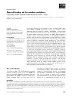
membranes can be separated from each other. Dreger et al.
[18] therefore used a strategy of alternative extraction of
the raw nuclear envelope preparation from mouse
neuroblastoma neuro 2a cells, to prepare different nuclear
envelope substructures characterized by the presence or
absence of substructure-specific marker proteins (Fig. 3A).
The protein subsets present in these fractions were identified
separately. The methods applied for separation and
Table 1. Selected subnuclear proteomics studies.
Subnuclear
structure
New
proteins
Total
proteins
Preparative
approach
Separation and
identification
technique
Additional
techniques Major outcome
Nuclear pore
complex (NPC)
(yeast) [17]
34 174 NPC preparation by
subcellular fractionation.
Alternative LC
SDS/PAGE as
second dimension,
peptide mass
fingerprints. CID.
Protein tagging,
immunoelectron
microscopy.
Structural model
of NPC.
Spindle pole (yeast)
[16]
11 23 Subcellular fractionation. SDS/PAGE,
peptide mass
fingerprints.
Protein tagging,
immunoelectron
microscopy.
Structural model
of spindle pole.
Interchromatin
granule clusters (IGC)
(mouse liver) [20]
3 36 Subcellular fractionation,
WB: enrichment of
markers.
2DE for enrichment
monitoring, direct
protein digestion,
LC/MS.
Immunofluorescence
with transiently
expressed proteins.
New IGC proteins.
Nuclear envelope (NE)
(neuro 2a cells,
mouse-derived) [18]
19 147 Subcellular fractionation,
alternative extraction of
the NE preparation.
BAC-SDS/PAGE
peptide mass
fingerprints.
Post-source decay.
Immunofluorescence
with transiently
expressed proteins.
Assignment of
novel proteins
within NE; two
new INM proteins.
Nucleolus (HeLa cells,
human-derived) [21,22]
84 271 Nucleolus preparation by
subcellular fractionation.
2D, several 1D
systems
Immunofluorescence
with transiently
expressed proteins.
Many new nucleolar
proteins; discovery
of new compartment
ÔparaspecklesÕ.
592 M. Dreger (Eur. J. Biochem. 270) Ó FEBS 2003
identification of the proteins were the two-dimensional
protein separation by the 16-BAC-/SDS/PAGE system [19],
followed by standard methods of mass spectrometric
protein identification based on peptide mass fingerprinting
and post source decay fragmentation of selected peptides.
Within each fraction, identified known proteins were
grouped according to literature data on their subcellular
localization (Fig. 3B) and according to features of their
primary structures as determined by bioinformatic analysis
tools. The distribution of identified proteins over the
different fractions analyzed allowed a tentative assignment
of nuclear envelope proteins to NE substructures without a
physical preparation of the substructure. The subcellular
localization of novel identified gene products in this study
could be predicted accordingly. LUMA and murine
KIAA0810 were the only previously unknown gene pro-
ducts that behaved like integral membrane proteins (chao-
trope-resistance), nuclear lamina-interacting proteins
(Triton X-100-resistance), and contained putative trans-
membrane regions within their primary structures. These
proteins were thus predicted to reside within the inner
nuclear membrane as integral membrane proteins. This was
independently confirmed by heterologous expression of
tagged versions of the proteins in transiently transfected
cells followed by indirect immunofluorescence using confo-
cal laserscanning microscopy. However, the accuracy of a
Fig. 3. Isolation and characterization of nuclear envelope subfractions. (A) Distribution of the marker proteins calnexin (outer nuclear membrane/
endoplasmic reticulum membrane), lamina-associated polypeptide 2b (LAP 2b, inner nuclear membrane), and lamin B1 (nuclear lamina)
throughout the different nuclear envelope subfractions. Calnexin is absent from the TX-100-resistant fraction; lamin B1 is almost absent from the
chaotrope-resistant fraction. (B) Distribution of NE proteins in the different fractions. Selected proteins detected in the TX-100-resistant NE
fraction (Tx) and in the chaotrope-resistant fraction (U/C) grouped according to their subcellular localization. INM, inner nuclear membrane; ER/
ONM, endoplasmic reticulum/outer nuclear membrane; L/M, nuclear lamina and attached protein scaffold; NPC, nuclear pore complex; CS,
cytoskeleton; Mito, mitochondria. Note the differences in the distribution of ER/ONM, L/M and NPC proteins.
Ó FEBS 2003 Subcellular structure level proteome analysis (Eur. J. Biochem. 270) 593
prediction made based on the proteomic data is consider-
ably reduced by the presence of contaminants derived from
other subcellular structures, as well as by the Ôresolution of
the studyÕ which is determined by the availability of different
subfractionation procedures. The use of independent meth-
odstovalidatetheresultsisalwaysrequired.
Interchromatin granule clusters
Interchromatin granule clusters (IGCs) are microscopically
defined subnuclear structures associated with enhanced
transcriptional activity [10]. Mintz et al. [20] addressed these
structures in a proteome analysis approach using either a
particular subfractionation procedure or immunoaffinity
isolation of presumed IGC-related protein complexes with a
known IGC protein as the bait. 2D gel electrophoresis was
used for visualization of proteins enriched in the IGC
preparation as compared to other subnuclear fractions.
Thus, the display of the protein pattern was used to monitor
proteins that were coenriched and were candidates for
colocalization within the same subnuclear structure. Using
Western blot analysis subsequent to 1D-separation of the
proteins, the enrichment of known IGC residents was
monitored. Protein identification was performed by an
LC-MS strategy subsequent to direct proteolytic digest of
the preparation and 36 different gene products were
identified. Among these, three previously unknown IGC-
associated protein were identified. The subcellular localiza-
tion was validated by indirect immunofluorescence of
transiently transfected cells.
Nucleolus
Numerous different separation and analysis methods have
been used in the recent study by Andersen et al.[21]to
explore the proteome of the nucleolus, a subnuclear
structure which is known to be the site of synthesis of the
ribosomal RNA and assembly of ribosomal subunits.
Andersen et al. prepared highly purified nucleoli from
human HeLa cells. Proteins were separated and analysed
according to two major strategies: first, classic 2D gel
electrophoresis was conducted, spots were picked and the
respective proteins identified by peptide mass fingerprinting
of the tryptic digests. Second, different 1D SDS/PAGE
methods using different gradients of acrylamide concentra-
tion and different buffer systems were used to separate the
proteins. This was followed by gel slicing, tryptic digestion
and nano-LC/MS analysis. Here proteins could be covered
that escaped analysis on classic 2D gel electrophoresis, e.g.
because of their basic pI values. The use of different
separation systems yielded partially nonoverlapping sets of
identified proteins. The efficiency of this analytic approach
is demonstrated by the very high number of 271 identified
proteins in the preparation of which only a very low
percentage had to be assigned to contaminants. More than
30% of the identified gene products were previously
unknown or uncharacterized, 82 of them were termed
Ônovel nucleolar proteinÕ. The subcellular localization of
several of them was assessed by indirect immunofluores-
cence of cells transiently transfected with DNA encoding
tagged versions of these gene products. Two of the newly
discovered gene products, as assessed by indirect immuno-
fluorescence and immunoelectron microscopy using anti-
bodies that recognize the endogenous proteins, defined a
new subnuclear structure, termed Ôparaspeckle compart-
mentÕ [22]. This finding represents an example for the
identification of novel subcellular structures driven initially
by a proteomic approach.
Proteomic analysis of small organelles
and vesicles
Golgi apparatus
A number of proteomic studies have been conducted on
other cellular organelles such as the Golgi apparatus and
peroxisomes. The work on the Golgi apparatus is mentioned
here as it has been subject to several proteomic studies
designed to create a Golgi complex protein map [24,25]. The
particular problem of the preparation of the Golgi appar-
atus, as compared to the relatively straightforward prepar-
ation of nuclei, is that the procedure comprises a series of
density centrifugation steps as the physical properties of the
material differ minimaly from those of, e.g. microsomal
material [26]. Further fractionation of the Golgi preparation
was performed by triton X-114 phase partioning, with the
triton-soluble fraction in the focus of the analysis. Both Bell
et al. [23] and Taylor et al. [24] succeeded in the identifica-
tion of new gene products of which one, termed either
GPP34 [23] or GMx33 [25], was unamibigiously localized to
the Golgi apparatus as a peripheral membrane protein using
immunoelectron microscopy. In addition, Wu et al.[27]
reported upregulation of a number of Golgi proteins in
Golgi preparations from rat mammary gland cells in the
state of maximal secretion at lactation as compared to that
in a state of basal secretion. This upregulation was observed
at the protein level by comparison of protein patterns
displayed by classic 2D gel electrophoretic separation of
proteins from the Golgi preparation.
Mitochondria
A number of studies have been performed using 2D gel
electrophoresis and mass spectrometric protein identifica-
tion to create two-dimensional protein maps for mitochon-
dria (for a review see [28]). However, in a number of studies
concerning the mitochondrial proteome strategies were used
that address additional aspects of the proteome. As early as
1991, Scha
¨
gger and Jagow used a native gel system for the
separation of intact protein complexes in the first dimension
and SDS/PAGE under denaturing conditions as the second
dimension to display the components of the complexes
[29]. A similar approach with three separation dimensions
using Blue native electrophoresis as the first dimension in
preparative electrophoresis followed by two-dimensional
separation of the eluted fractions of the preparative gel was
reported by Werhahn and Braun [30]. Using sucrose density
centrifugation as a first dimension, Hanson et al.[31]aimed
to create a Ôthree dimensional protein mapÕ of the mito-
chondrial proteome. Both methods are either restricted by
limited resolution or limited use for very complex samples.
However, they share the basic idea that the information
594 M. Dreger (Eur. J. Biochem. 270) Ó FEBS 2003
content of proteomic screens could be extended by
addressing protein interactions in one of the separation
dimensions. This differs from the analysis of multiprotein
complexes subsequent to affinity purification.
Vesicles charcterized on the basis of comprehensive
proteome analysis
There are a number of studies on vesicular structures that
are characterized not by containing specifically localized
proteins, but are characterized by a particular protein
population as determined by proteomic approaches. One
example is the analysis of phagosomes [32], organelles that
occur upon phagocytic internalization of foreign material
by macrophages. In this analysis, in addition to the
description of the phagosome proteome, the maturation
of the organelle was monitored by comparative analysis of
phagosomes in different stages. The authors demonstrated
that the phagosomes acquire cathepsins, key catabolic
enzymes of mature phagosomes, in a sequential manner
during pahgosome maturation.
There has also been a proteomics approach to charac-
terize exosomes, secreted organelles that, among potential
other functions, may play a role in the immune response
[33]. A special feature of this analysis was that the exosomes
were separated from other vesicular organelles by means of
free-flow electrophoresis, and that the whole population of
identified proteins served to distinguish exosomes from
apoptotic vesicles.
There have been a number of other proteome analysis
studies to characterize vesicular organelles based on their
entire proteome. One example is the proteomic character-
ization of prespore secreted vesicles of Dictyostelium discoi-
dum [34,35].
A common theme of these studies is the requirement of a
comprehensive proteome analysis in order to acquire an
image of the organelle investigated. This highlights the
unique potential of subcellular proteomics as compared to
other, more traditional approaches, where the analysis was
designed to identify single specifically localized proteins.
Subcellular proteomics at the tissue level:
tackling the synapse
Many current proteome analysis projects are aimed at
the comparative analysis of tissue samples, e.g. prepared
from CNS structures. Tissue samples are more complex
than samples from cultured cells as any tissue contains
many different cell types and contains structural material
like connective tissue that may not be the target of the
analysis.
Samples derived from synaptic structures have been
targeted by proteomic analysis in various studies. Walikonis
et al. [36] analysed proteins present in the classic post-
synaptic density (PSD) preparation from rat brain. This
preparation starts from the isolation of synaptosomes,
vesicles that form spontaneously upon homogenization of
nervous tissue and that contain pre- and postsynaptic
structures. The final PSD preparation contains postsynaptic
neurotransmitter receptors as well as their anchoring
proteins together with the underlying cytoskeleton and
docked signalling molecules. The enrichment of the PSD is
achieved by different density centrifugation steps subse-
quent to the lysis of the synaptosomes, and by detergent
extraction of membrane proteins not bound to the PSD. A
total of 24 different proteins were identified by mass
spectrometry subsequent to 1D gel electrophoretic separ-
ation of the PSD fraction. However, at least one presynap-
tically localized protein as well as a few mitochondrial
contaminants were identified in addition to known key
postsynaptic proteins. A similar analysis was preformed by
Satoh et al. [37] who separated their PSD fraction in two
dimensions and detected difference spots depending on
synaptic activity. In total, 47 different proteins were
identified. However, subunits of ionotropic glutamate
receptors, which are key PSD proteins, were not detected,
in contrast to the aforementioned study and in line with the
assumed underrepresentation of integral membrane pro-
teins on 2D gels.
Phillips et al. [38] reported the preparation of specific
presynaptic structures and the preparative separation of the
presynaptic membrane from the postsynaptic membrane.
Only a few selected proteins have been identified in this
study, many of which can be assigned to the presynaptic side
of the synapse. It will be interesting to observe what the
outcome of a detailed proteome analysis of this fraction will
be.
Special aspects of comparative studies
at the subcellular level
In addition to the description of the proteome of a
subcellular entity, the analysis of dynamic proteome chan-
ges at a subcellular level promises to yield significant insight
into biological mechanisms. In this section I would like to
point out analytical aspects and potentials specific to the
analysis at the subcellular level.
Microsomal fractions are comprised of membrane vesi-
cles that spontaneously form during cell homogenization.
They do not represent distinct cellular organelles; they are
of heterogenous origin and may contain, e.g. material from
the endoplasmic reticulum and other cytosolic organelles.
However, they are a source for membrane proteins that can
be easily and quickly prepared. In a comparative study, Han
et al. [39] used microsomes from HL60 cells, a human acute
myeloid leukemia cell line that is cultured in suspension, but
that upon certain stimuli (e.g. phorbol ester) differentiates
into an adherent form, to detect alterations in the micro-
somal fraction upon cell differentiation by the application of
the isotope-coded affinity tag (ICAT) technique. In this
technique, the proteins of the control sample and the test
sample are alkylated by the cysteine-specific biotinylated
ICAT reagent in its nondeuterated or in an eightfold
deuterated form, respectively [40]. Subsequent to alkylation,
the proteins from both samples are pooled and proteo-
lytically cleaved. Peptides that carry the cysteine-specific
modification can be isolated from the whole peptide mixture
by application of the mixture directly or subsequent to
further prefractionation to an affinity column loaded with
monomeric avidin. Bound peptides are then eluted from the
column and analysed by LC-MS. Peptides with the same
amino-acid sequence derived from the two samples will
Ó FEBS 2003 Subcellular structure level proteome analysis (Eur. J. Biochem. 270) 595
differ in mass due to the mass difference between the
nondeuterated and the deuterated ICAT reagent. As these
ICAT pair peptides behave chemically the same during
chromatography and mass spectrometric analysis, the ratio
of their intensities in the mass spectra is a semiquantitative
measure for the abundance of the proteins they are derived
from.
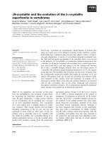
Microsomes from control cells or differentiated cells were
isolated, the proteins were labelled by the ICAT reagents,
proteolytically cleaved and analysed by liquid chromato-
graphy/MS (Fig. 4A). The analysis of ICAT pairs yielded
semiquantitative information on more than 400 microsomal
proteins, of which several displayed a differential abundance
in the control as compared with the phorbol ester-stimula-
ted sample. This study highlighted some important aspects
concerning the interpretation of data obtained from com-
parative proteomics at the subcellular level. Firstly, virtually
all classes of proteins were represented, including regulatory
proteins like protein kinases and multispanning integral
membrane proteins (Fig. 4B), which are thought to be
596 M. Dreger (Eur. J. Biochem. 270) Ó FEBS 2003
underrepresented on classic 2D gels [3] (see [41] for
contrasting data). Secondly, the question arises, which ratio
of abundance of a particular protein is considered as a true
quantitative difference. Many of the identified proteins
differ by a ratio of around two, which is not considered a
significant difference by the authors. Thirdly, as one
particular subcellular fraction has been analysed, Han et al.
point out several mechanisms that can account for the
increased or decreased abundance of particular proteins in
the preparation dependent on the status of cellular differ-
entiation. There may be upregulation due to increased
protein synthesis, but there may also be signal-induced
translocation of proteins towards cellular membranes,
which accounts for the occurrence of these proteins in the
microsomal fraction. Decreased abundance of proteins may
be due to reduced protein synthesis, but also due to signal-
induced protein degradation or signal-induced detachment
of proteins from the microsomal membranes.
If the biochemical mechanism of the alterations in the
subcellular proteome is to be addressed, it is necessary to
monitor several different subcellular fractions in parallel.
An example for such a study is given by Gerner et al.[42]in
their study of Fas-induced apoptosis in Jurkat T-lympho-
cytes. The authors monitored in parallel the nucleoplasmic
and the cytosolic fraction of the cells. Their data suggested
signal-induced entrance of the protein TCP-1a into the
nucleus as well as translocation of nuclear annexin IV from
the nucleus to the cytosol, as deduced from the comparative
analysis of the protein pattern of the respective fractions
obtained by classic two-dimensional gel electrophoresis.
Concluding remarks
With the option to identify large numbers of proteins rather
than single proteins specifically localized to particular
structures, the combination of subcellular fractionation
and protein identification, in other terms Ôsubcellular
proteomicsÕ, can be used as a multifunctional tool in cell
biology. The first line of information (and the best-
established approach) is the discovery of novel gene
products and their assignment to subcellular structures. A
second line of information is the characterization of
subcellular structures based on their entire protein popula-
tion in addition to known physical and biochemical
properties of these structures. As it starts from subcellular
fractions and is based on the identification of endogenous
proteins in functional contexts, this approach is comple-
mentary to recent molecular biology-based studies to
systematically probe the subcellular localization of large
numbers of gene products. As examples for such molecular
biology-based approaches, see [43] for the systematic
assessment of the subcellular localization of gene products
based on the heterologous overexpression of GFP fusion
proteins derived from cDNA libraries, and [44] for the
systematic assessment of the subcellular localization of yeast
gene products based on overexpression of tagged gene
products. In order to detect the subnuclear localization of
gene products at the endogenous expression level, a gene
trap approach with the introduction of a reporter tag into
endogenous genes in embryonic stem cells has been used
[45]. Each method has its potentials and drawbacks, so it
will be interesting to compare data on the same subcellular
structure obtained by different approaches.
A strategy to acquire a third line of information derived
from subcellular proteomics studies is still in the beginning:
the study of dynamic changes at the subcellular level, e.g.
upon protein translocation and altered protein–protein
interactions. Major requirements are the simultaneous
preparation and analysis of different subcellular structures
and the development of strategies for the simultaneous
display of many different protein interactions at an appro-
priate resolution.
With an increasing number of subcellular proteomic
studies, most of them directed to the discovery of novel
gene products, the need arises for storage of data in
organelle databases. In typical studies, more than one
hundred different proteins are identified. As the functional
investigation of novel gene products is much more difficult
and time-consuming than protein identification, only a few
will be subject to further research by the research group
that identified the gene product. To prevent loss of
information on the other detected gene products, this
information should be collected in a publicly accessible
database. One such example is the Nuclear Protein
Database at which contains
information on nuclear proteins from many different
studies.
In summary, subcellular proteomics may be more than
separating proteins on gels and identifying them by mass
spectrometry. Depending on the design of the study,
functional insight into cellular processes may be obtained.
Fig. 4. Comparative subcellular proteome analysis of microsomal
membranes using the ICAT method. (A) The ICAT strategy for quan-
titating differential protein expression. Two protein mixtures repre-
senting two different cell states have been treated with the isotopically
light and heavy ICAT reagents, respectively; an ICAT reagent is cov-
alently attached to each cysteinyl residue in every protein. The protein
mixtures are combined, proteolyzed to peptides, and ICAT-labeled
peptides are isolated utilizing the biotin tag. These peptides are separ-
ated by microcapillary high performance liquid chromatography. A
pair of ICAT-labeled peptides are chemically identical and are easily
visualized because they essentially co-elute and there is an eight dalton
mass difference measured in a scanning mass spectrometer (four m/z
units difference for a doubly charged ion). The ratios of the original
amounts of proteins from the two cell states are strictly maintained in
the peptide fragments. The relative quantification is determined by the
ratio of the peptide pairs. Every other scan is devoted to fragmenting
and then recording sequence information about an eluting peptide
(tandem mass spectrum). The protein is identified by computer
searching for the recorded sequence information against large protein
databases. In theory, every peptide pair in the mixture is, in turn,
measured and fragmented resulting in the relative quantitation and
identification of mixture proteins in a single analysis. (B) Categories of
proteins identified from HL-60 cell microsomal fraction. The 491
proteins identified and quantified in this study were classified by broad
functional criteria. The numbers in parentheses indicate the percentage
fraction of identified proteins represented by each category. Some
proteins are represented in more than one category.
Ó FEBS 2003 Subcellular structure level proteome analysis (Eur. J. Biochem. 270) 597
Acknowledgements
I would like to thank Dr Chris Weise and Stephanie Williams for
critically reading this manuscript. I would also like to thank Dr Ruedi
Aebersold for providing graphic material for Fig. 4. This work was
supported by the German ministry for research and education and by
the Deutsche Forschungsgemeinschaft.
References
1. Malinow, R. & Malenka, R.C. (2002) AMPA receptor trafficking
and synaptic plasticity. Annu. Rev. Neurosci. 25, 103–126.
2. Bryant, N.J., Govers, R. & James, D.E. (2002) Regulated
transport of the glucose transporter GLUT4. Nat. Rev. Mol.
Cell Biol. 3, 267–277.
3. Santoni, V., Molloy, M. & Rabilloud, T. (2000) Membrane
proteins and proteomics: un amour impossible? Electrophoresis 21,
1054–1070.
4. Mann, M., Ong, S.E., Gronborg, M., Steen, H., Jensen, O.N. &
Pandey, A. (2002) Analysis of protein phosphorylation using
mass spectrometry: deciphering the phosphoproteome. Trends
Biotechnol. 20, 261–268.
5. Gygi, S.P., Corthals, G.L., Zhang, Y., Rochon, Y. & Aebersold,
R. (2000) Evaluation of two-dimensional gel electrophoresis-based
proteome analysis technology. Proc. Natl Acad. Sci. USA 97,
9390–9395.
6. Molloy, M.P., Herbert, B.R., Walsh, B.J., Tyler, M.I., Traini, M.,
Sanchez,J.C.,Hochstrasser,D.F.,Williams,K.L.&Gooley,A.A.
(1998) Extraction of membrane proteins by differential
solubilization for separation using two-dimensional gel electro-
phoresis. Electrophoresis 19, 837–844.
7. Emig, S., Schmalz, D., Shakibaei, M. & Buchner, K. (1995) The
nuclear pore complex protein p62 is one of several sialic acid-
containing proteins of the nuclear envelope. J. Biol. Chem. 270,
13787–13793.
8. Tang,T.,Hirata,Y.,Whalin,M.&Guroff,G.(1996)Nerve
growth factor-stimulated nuclear S6 kinase in PC12 cells.
J. Neurochem. 66, 1198–1206.
9. Jung, E., Heller, M., Sanchez, J.C. & Hochstrasser, D.F. (2000)
Proteomics meets cell biology: the establishment of subcellular
proteomes. Electrophoresis 21, 3369–3377.
9a. Bauer, A. & Kuster, B. (2003) Affinity purification-mass
spectrometry. Powerful tools for the characterization of protein
complexes. Eur. J. Biochem. 270, 570–578.
10. Lamond, A.I. & Earnshaw, W.C. (1998) Structure and function in
the nucleus. Science 280, 547–553.
11. Misteli, T. (2001) Protein dynamics: implications for nuclear
architecture and gene expression. Science 291, 843–847.
12. Nickerson, J.A. (1998) Nuclear dreams: the malignant alteration
of nuclear architecture. J. Cell Biochem. 70, 172–180.
13. Wilson, K.L. (2000) The nuclear envelope, muscular dystrophy
and gene expression. Trends Cell Biol. 10, 125–129.
14. Mattaj, I.W. & Englmeier, L. (1998) Nucleocytoplasmic transport:
the soluble phase. Annu. Rev. Biochem. 67, 265–306.
15. Neubauer, G., King, A., Rappsilber, J., Calvio, C., Watson,
M.,Ajuh,P.,Sleeman,J.,Lamond,A.&Mann,M.(1998)
Mass spectrometry and EST-database searching allows
characterization of the multi-protein spliceosome complex. Nat.
Genet. 20, 46–50.
16. Wigge, P.A., Jensen, O.N., Holmes, S., Soues, S., Mann, M. &
Kilmartin, J.V. (1998) Analysis of the Saccharomyces spindle pole
by matrix-assisted laser desorption/ionization (MALDI) mass
spectrometry. J. Cell Biol. 141, 967–977.
17. Rout, M.P., Aitchison, J.D., Suprapto, A., Hjertaas, K., Zhao, Y.
& Chait, B.T. (2000) The yeast nuclear pore complex:
composition, architecture, and transport mechanism. J. Cell
Biol. 148, 635–651.
18. Dreger, M., Bengtsson, L., Schoneberg, T., Otto, H. & Hucho, F.
(2001) Nuclear envelope proteomics: novel integral membrane
proteins of the inner nuclear membrane. Proc. Natl Acad. Sci.
USA 98, 11943–11948.
19. Hartinger, J., Stenius, K., Hogemann, D. & Jahn, R. (1996)
16-BAC/SDS-PAGE: a two-dimensional gel electrophoresis
system suitable for the separation of integral membrane
proteins. Anal. Biochem. 240, 126–133.
20.Mintz,P.J.,Patterson,S.D.,Neuwald,A.F.,Spahr,C.S.&
Spector, D.L. (1999) Purification and biochemical characterization
of interchromatin granule clusters. EMBO J. 18, 4308–4320.
21. Andersen,J.S.,Lyon,C.E.,Fox,A.H.,Leung,A.K.,Lam,Y.W.,
Steen, H., Mann, M. & Lamond, A.I. (2002) Directed proteomic
analysis of the human nucleolus. Curr. Biol. 12, 1–11.
22. Fox, A.H., Lam, Y.W., Leung, A.K., Lyon, C.E., Andersen, J.,
Mann, M. & Lamond, A.I. (2002) Paraspeckles: a novel nuclear
domain. Curr. Biol. 12, 13–25.
23. Bell, A.W., Ward, M.A., Blackstock, W.P., Freeman, H.N.,
Choudhary, J.S., Lewis, A.P., Chotai, D., Fazel, A., Gushue, J.N.,
Paiement, J., Palcy, S., Chevet, E., Lafreniere-Roula, M., Solari,
R., Thomas, D.Y., Rowley, A. & Bergeron, J.J. (2001) Proteomics
characterization of abundant Golgi membrane proteins. J. Biol.
Chem. 276, 5152–5165.
24. Taylor, R.S., Wu, C.C., Hays, L.G., Eng, J.K., Yates, J.R. III &
Howell, K.E. (2000) Proteomics of rat liver Golgi complex: minor
proteins are identified through sequential fractionation.
Electrophoresis 21, 3441–3459.
25. Wu, C.C., Taylor, R.S., Lane, D.R., Ladinsky, M.S., Weisz, J.A.
& Howell, K.E. (2000) GMx33: a novel family of trans-Golgi
proteins identified by proteomics. Traffic 1, 963–975.
26. Taylor, R.S., Fialka, I., Jones, S.M., Huber, L.A. & Howell, K.E.
(1997) Two-dimensional mapping of the endogenous proteins of
the rat hepatocyte Golgi complex cleared of proteins in transit.
Electrophoresis 18, 2601–2612.
27. Wu, C.C., Yates, J.R. III, Neville, M.C. & Howell, K.E. (2000)
Proteomic analysis of two functional states of the Golgi complex
in mammary epithelial cells. Traffic 1, 769–782.
28. Lopez, M.F. &Melov, S. (2002)Applied proteomics: mitochondrial
proteins and effect on function. Circ. Res. 90, 380–389.
29. Schagger, H. & von Jagow, G. (1991) Blue native electrophoresis
for isolation of membrane protein complexes in enzymatically
active form. Anal. Biochem. 199, 223–231.
30. Werhahn, W. & Braun, H.P. (2002) Biochemical dissection of the
mitochondrial proteome from Arabidopsis thaliana by
three-dimensional gel electrophoresis. Electrophoresis 23, 640–646.
31. Hanson, B.J., Schulenberg, B., Patton, W.F. & Capaldi, R.A.
(2001) A novel subfractionation approach for mitochondrial
proteins: a three-dimensional mitochondrial proteome map.
Electrophoresis 22, 950–959.
32. Garin, J., Diez, R., Kieffer, S., Dermine, J.F., Duclos, S., Gagnon,
E., Sadoul, R., Rondeau, C. & Desjardins, M. (2001) The
phagosome proteome: insight into phagosome functions. J. Cell
Biol. 152, 165–180.
33. Thery, C., Boussac, M., Veron, P., Ricciardi-Castagnoli, P.,
Raposo, G., Garin, J. & Amigorena, S. (2001) Proteomic analysis
of dendritic cell-derived exosomes: a secreted subcellular
compartment distinct from apoptotic vesicles. J. Immunol. 166,
7309–7318.
34. Srinivasan, S., Alexander, H. & Alexander, S. (1999) The prespore
vesicles of Dictyostelium discoideum. Purification, characterization,
and developmental regulation. J. Biol. Chem. 274, 35823–35831.
35. Srinivasan, S., Traini, M., Herbert, B., Sexton, D., Harry, J.,
Alexander, H., Williams, K.L. & Alexander, S. (2001) Proteomic
598 M. Dreger (Eur. J. Biochem. 270) Ó FEBS 2003
analysis of a developmentally regulated secretory vesicle.
Proteomics 1, 1119–1127.
36. Walikonis,R.S.,Jensen,O.N.,Mann,M.,Provance,D.W.Jr,
Mercer, J.A. & Kennedy, M.B. (2000) Identification of proteins
in the postsynaptic density fraction by mass spectrometry.
J. Neurosci. 20, 4069–4080.
37. Satoh, K., Takeuchi, M., Oda, Y., Deguchi-Tawarada, M.,
Sakamoto, Y., Matsubara, K., Nagasu, T. & Takai, Y. (2002)
Identification of activity-regulated proteins in the postsynaptic
density fraction. Genes Cells 7, 187–197.
38. Phillips, G.R., Huang, J.K., Wang, Y., Tanaka, H., Shapiro, L.,
Zhang, W., Shan, W.S., Arndt, K., Frank, M., Gordon, R.E.,
Gawinowicz,M.A.,Zhao,Y.&Colman,D.R.(2001)The
presynaptic particle web: ultrastructure, composition, dissolution,
and reconstitution. Neuron 32, 63–77.
39. Han, D.K., Eng, J., Zhou, H. & Aebersold, R. (2001) Quantitative
profiling of differentiation-induced microsomal proteins using
isotope-coded affinity tags and mass spectrometry. Nat.
Biotechnol. 19, 946–951.
40. Gygi, S.P., Rist, B., Gerber, S.A., Turecek, F., Gelb, M.H. &
Aebersold, R. (1999) Quantitative analysis of complex protein
mixtures using isotope-coded affinity tags. Nat. Biotechnol. 17,
994–999.
41. Hippler, M., Klein, J., Fink, A., Allinger, T. & Hoerth, P. (2001)
Towards functional proteomics of membrane protein complexes:
analysis of thylakoid membranes from Chlamydomonas
reinhardtii. Plant J. 28, 595–606.
42. Gerner,C.,Frohwein,U.,Gotzmann,J.,Bayer,E.,Gelbmann,
D., Bursch, W. & Schulte-Hermann, R. (2000) The Fas-induced
apoptosis analyzed by high throughput proteome analysis. J. Biol.
Chem. 275, 39018–39026.
43. Simpson,J.C.,Wellenreuther,R.,Poustka,A.,Pepperkok,R.&
Wiemann, S. (2000) Systematic subcellular localization of novel
proteins identified by large-scale cDNA sequencing. EMBO
Report 1, 287–292.
44. Kumar, A., Agarwal, S., Heyman, J.A., Matson, S., Heidtman,
M., Piccirillo, S., Umansky, L., Drawid, A., Jansen, R., Liu, Y.,
Cheung,K.H.,Miller,P.,Gerstein,M.,Roeder,G.S.&Snyder,
M. (2002) Subcellular localization of the yeast proteome. Genes
Dev. 16, 707–719.
45. Sutherland, H.G., Mumford, G.K., Newton, K., Ford, L.V.,
Farrall,R.,Dellaire,G.,Caceres,J.F.&Bickmore,W.A.
(2001) Large-scale identification of mammalian proteins
localized to nuclear sub-compartments. Hum. Mol. Genet. 10,
1995–2011.
Ó FEBS 2003 Subcellular structure level proteome analysis (Eur. J. Biochem. 270) 599