Báo cáo khoa học: Purification and characterization of novel salt-active acharan sulfate lyase from Bacteroides stercoris HJ-15 ppt
Bạn đang xem bản rút gọn của tài liệu. Xem và tải ngay bản đầy đủ của tài liệu tại đây (252.63 KB, 6 trang )
Purification and characterization of novel salt-active acharan sulfate
lyase from
Bacteroides stercoris
HJ-15
Sung-Woon Hong
1
, Ho-Young Shin
1
, Yeong Shik Kim
2
and Dong-Hyun Kim
1
1
College of Pharmacy, Kyung Hee University, Seoul, Korea;
2
Natural Products Research Institute, Seoul National University,
Seoul, Korea
Salt-active acharan sulfate lyase (no EC number) has been
purified from Bacteroides stercoris HJ-15, which was iso-
lated from human intestinal bacteria with GAG degrading
enzymes. The enzyme was purified to apparent homogeneity
by a combination of QAE-cellulose, diethylaminoethyl
(DEAE)-cellulose, CM-Sephadex C-50, HA ultrogel and
phosphocellulose column chromatography with the final
specific activity of 81.33 lmolÆmin
)1
Æmg
)1
. The purified salt-
active acharan sulfate lyase was activated to 5.3-fold by salts
(KCl and NaCl). The molecular weight of salt-active acha-
ran sulfate lyase was 94 kDa by SDS/PAGE and gel filtra-
tion. The salt-active acharan sulfate lyase showed optimal
activity at pH 7.2 and 40 °C. Salt-active acharan sulfate
lyase activity was potently inhibited by Cu
2+
, Ni
2+
and
Zn
2+
. This enzyme was inhibited by some agents, butanediol
and p-chloromercuric sulfonic acid, which modify arginine
and cysteine residues. The purified Bacteroidal salt-active
acharan sulfate lyase acted to the greatest extent on acharan
sulfate, to a lesser extent on heparan sulfate and heparin. The
biochemical properties of the purified salt-active acharan
sulfate lyase are different from those of the previously puri-
fied heparin lyases. However, these findings suggest that the
purified salt-active acharan sulfate lyase may belong to
heparin lyase II.
Keywords: Bacteroides stercoris HJ-15; salt-active acharan
sulfate lyase; acharan sulfate lyase; acharan sulfate; heparin.
Heparin, heparan sulfate and acharan sulfate glycosamino-
glycans (GAGs) are comprised of alternating 1–4-linked
glucosamine and uronic acid residues. Heparan sulfate is
composed primarily of monosulfated disaccharides of
N-acetyl-
D
-glucosamine and
D
-glucuronic acid while hep-
arin is composed mainly of trisulfated disaccharides of
N-sulfonyl-
D
-glucosamine and
L
-iduronic acid [1,2]. Acha-
ran sulfate, isolated from the giant African snail Achatina
fulica, has a structure closely related to heparin and heparan
sulfate, with a uniform repeating disaccharide structure
of fi4)-a-
D
-GlcNAc(1fi 4)-a-
L
-IdoA2S (1fi[3]. Acharan
sulfate exclusively contains N-acetyl-
D
-glucosamine instead
of N-sulfonyl-
D
-glucosamine in GAGs.
Related to the degradation of these GAGs, some heparin
lyases that can eliminatively cleave polysaccharides (heparin
or heparan sulfate GAGs) have been reported [4–6]. These
enzymes are classified as: (a) heparin lyase I (heparinase I,
EC 4.2.2.7), acting primarily at the fi 4)-a-
D
-GlcNS(6S or
OH)(1fi4)-a-
L
-IdoA2S(1fi linkages present in heparin; (b)
heparin lyase II (heparinase II or heparitinase II), acting at
the fi4)-a-
D
-GlcNS(6S or OH)(1fi 4)-a-
L
-IdoA(2S or OH)
or -b-
D
-GlcA(1fi linkages present in both heparin and
heparan sulfate; and (c) heparin lyase III (heparinase III or
heparitinase, EC 4.2.2.8), acting on the fi4)-a-
D
-GlcNS(or
Ac) (1fi 4)-b-
D
-GlcA (or IdoA) (1fi linkages found exclu-
sively in heparan sulfate. The heparin lyases have become
increasingly important in understanding the biological roles
and structure of the glycoaminoglycans (and proteoglycan),
which are involved in the well known anticoagulant activity
[7] and the regulation of various cellular processes such as
the potentiation of angiogenesis [8] and the modulation of
cellular proliferation [9]. Several heparin lyases of bacterial
origin have been purified and characterized from various
species including Flavobacterium heparinum [4,10], Bacillus
sp. BH 100 [11], Prevotella heparinolyticus [12], and Bacter-
oides stercoris HJ-15 [13,14].
Bacteroides stercoris HJ-15 has been recently isolated
from human intestine and it contains several kinds of GAG
degrading enzymes including heparin, heparan sulfate,
acharan sulfate and chondroitin sulfate [13–16]. We purified
two kinds of novel heparin lyases, heparin lyase II-1
(acharan sulfate lyase 1), and heparin lyase II-2 (acharan
sulfate lyase 2) and III, from this B. stercoris HJ-15 [15,16].
The Bacteroidal heparin lyase III cleaved heparin as well as
heparan sulfate, but did not cleave acharan sulfate. The
Bacteroidal acharan sulfate lyase, which potently cleaved
acharan sulfate as well as heparin, are highly specific to
acharan sulfate compared to the previously reported
heparin lyases [16]. The purified acharan sulfate lyases 1
and 2 (no EC number) were not activated by salts
such as KCl. However, when Bacteroidal acharan sulfate
lyase-active fraction isolated from B. stercoris HJ-15 was
incubated with salts (KCl), the enzyme fraction was
Correspondence to D H. Kim, College of Pharmacy, Kyung Hee
University, 1 Hoegi-dong, Dongdaemun-ku, Seoul 130–701,
South Korea. Fax: + 82 2 957 5030, Tel.: + 82 2 961 0374,
E-mail:
Abbreviations: CM, carboxymethyl; DEAE, diethylaminoethyl;
DUA, 4-deoxy-a-
L
-threo-hex-4-enopyranosyl uronic acid;
GAG, glycosaminoglycan; GlcA, glucuronic acid; GlcN, glucosamine;
HA, hydroxyapatite; IdoA, iduronic acid; IEF, isoelectric focusing;
QAE, quaternary amino ethyl; PCMS, p-chloromercurisulfonic acid.
(Received 3 April 2003, revised 15 May 2003,
accepted 30 May 2003)
Eur. J. Biochem. 270, 3168–3173 (2003) Ó FEBS 2003 doi:10.1046/j.1432-1033.2003.03696.x
activated by salts. Therefore, we tried to purify salt-active
acharan sulfate lyase from B. sterocoris HJ-15 that acts
predominantly on acharan sulfate.
Materials and methods
Materials
Heparin (porcine intestinal mucosa), heparan sulfate (por-
cine intestinal mucosa), chondroitin sulfate A (bovine
trachea), chondroitin sufate B (porcine intestinal mucosa),
chondroitin sufate C (shark cartilage), thioglycolic acid
(sodium salt), QAE cellulose Fastflow, and HA Ultrogel
(microcrystalline hydroxyapatite, 4% beaded in agarose)
were supplied by Sigma Chemical Co. Sodium dodecyl
sulfate, CM-Sephadex C-50, phosphocellulose, Sephacryl
S-300 HR resins and molecular weight markers for gel
filtration and protein electrophoresis were obtained from
Pharmacia Biotech Co. (Uppsala, Sweden). Diethylamino-
ethyl (DEAE)-cellulose resin was purchased from Wako
Pure Chemical Industries (Tokyo, Japan). Protein assay kit
and Coomassie Brilliant Blue R-250 were from Bio-Rad
(Hercules, CA, USA). Tryptic soy broth was provided by
Difco Co. Acharan sulfate was prepared as described by
Kim et al. [3]. All other chemicals were of the highest grade
available.
Bacterial strains and purification of salt-active acharan
sulfate lyase
B. stercoris HJ-15 was isolated and cultivated as described
previously [14,16]. It was cultured anaerobically under an
atmosphere of 90% (v/v) nitrogen and 10% (v/v) carbon
dioxide at 37 °C in 100 L of tryptic soy broth (pH 7.2)
containing heparin (0.15 gÆL
)1
) instead of glucose, 0.01%
(w/v) sodium thioglycolate and 0.1% (w/v) ascorbic acid.
The cultured cells were harvested in the late exponential
phase (11–12 h) by centrifugation at 4000 g
1
for 30 min at
4 °C and the resulting cell pellet was washed twice with
saline containing 50 m
M
sodium phosphate (pH 7.0). The
cell pellet was suspended in 600 mL of Buffer A (50 m
M
sodium phosphate buffer, pH 7.0). Cell suspension (30 mL
at a time) was placed into a 50-mL centrifuge tube and
disrupted by 30-min periods of sonication at 1-s intervals on
an ultrasonic processor (Eyela Co.) at an 80% output with
cooling. Cell debris was removed by centrifugation at
25 000 g
2
for 60 min at 4 °C. All operations were carried
out at 4 °C unless otherwise noted. The cell extract
(600 mL) was passed through a QAE cellulose column
(5 · 40 cm) which had been pre-equilibrated with Buffer A.
The column was washed with the same buffer until no
acharan sulfate lyase activity was detectable in the effluent.
The fractions which passed through the column were
applied to a DEAE-cellulose column (5 · 30 cm) equili-
brated with Buffer A. The column was then eluted with the
same buffer until any ASL activity could not be detected.
The noninteracting fluid passed through the column was
collected. The total volume of the flow through was
1800 mL. The eluate was loaded onto a CM-Sephadex C-
50 column (3 · 30 cm) previously equilibrated with Buffer
A. The column was washed with 1000 mL (1 L) of the same
buffer and then eluted with a 2-L linear gradient of KCl
from 0 to 0.6
M
in Buffer A at a flow rate of 105 mLÆh
)1
.All
fractions obtained were assayed for heparin lyase and
acharan sulfate lyase activities. Four fractions (Fr-s, Fr-a,
Fr-b and Fr-c) containing the activity of these enzymes were
collected separately and assayed for the activities degrading
acharan sulfate and heparan sulfate. Fr-s had acharan
sulfate lyase activity, which was activated by salts, was
dialyzed against Buffer A for the further purification.
The dialyzed enzyme preparation (210 mL) was applied
to a HA Ultrogel column (2.5 · 9 cm) equilibrated with
Buffer A. Being washed with 500 mL of the same buffer,
the column was eluted with a 800-mL linear gradient, from
50 m
M
sodium phosphate buffer (pH 7.0) to 400 m
M
sodium phosphate buffer (pH 7.0) at a flow rate of
120 mLÆh
)1
. The active fractions were pooled and dialyzed
twice against 2 L of Buffer A. The dialyzed enzyme
(78 mL) was loaded onto a phosphocellulose column
(3 · 25 cm) equilibrated with Buffer A. After washing the
nonabsorbed proteins with 300 mL of the same buffer, the
column was eluted with an 800-mL linear gradient, from
50 m
M
sodium phosphate buffer (pH 7.0) to 400 m
M
sodium phosphate buffer (pH 7.0) at a flow rate of
100 mLÆh
)1
. The active fractions (salt-active acharan sulfate
lyase, fraction numbers 17–25) were investigated for
homogeneity by SDS/PAGE.
Enzyme activity assay
The activity of acharan sulfate lyase was measured accord-
ing to the previously published procedure [17]. The activity
was calculated from the change of absorbance per minute
using an extinction coefficient of 3800
M
)1
for products
(1 U ¼ 1 micromole of DUA containing product formed
per minute) [17]. The specific activity was calculated by
dividing the micromoles of product produced per minute by
the milligrams of protein in the cuvette. Protein concentra-
tion was measured by a Bradford assay using bovine serum
albumin as a standard [18].
Characterization of salt-active acharan sulfate lyase
SDS/PAGE was performed for the determination of
molecular mass according to Laemmli’s procedure [19].
The gel was stained with Coomassie Brilliant Blue R-250
solution and further stained with silver. The pI value of
heparin lyase was determined by IEF electrophoresis using
Model 111 Mini IEF Cell (from Bio-Rad) according to the
manufacturer’s instructions. The molecular weight of the
native enzyme was estimated by gel filtration using Seph-
acryl S-300 HR column (1.6 · 70 cm) calibrated with gel
filtration low molecular weight calibration kit (from Sigma
Co.) and high molecular calibration kit (from Amersham
Pharmacia Biotech). The pH optimum of acharan sulfate
lyase was determined using 50 m
M
sodium phosphate buffer
(pH 6.0–8.5). Temperature dependency of the enzyme was
investigated by measuring enzyme activity at different
temperatures (25–60 °C). To investigate the effect of
divalent metal ions and KCl (or NaCl) on the lyase activity,
divalent metal ion (final concentration, 100 l
M
), chemical
modifying agents (50 l
M
) and KCl (0–500 m
M
) were added
into the reaction mixture. Kinetic constant of acharan
sulfate lyase was determined by measuring the initial rates at
Ó FEBS 2003 Bacteroidal salt-active acharan sulfate lyase (Eur. J. Biochem. 270) 3169
various substrate concentrations (200, 400, 600, 1000, 2000,
3000 lg) under the standard reaction conditions.
These lyase activities on other sulfated polysaccharides
were also measured. One milligram of each substrate was
added to the reaction mixture. Because of their low
solubility, 100 lg of acharan sulfate were used in this assay.
The internal amino acid sequence of purified salt-active
acharan sulfate lyase was analyzed by an Applied Biosystem
protein sequencer model 492 in Korea Basic Science
Institute.
Results
Purification of salt-active acharan sulfate lyase
Bacteroides stercoris HJ-15, which degrades a variety of
GAGs including heparin, heparan sulfate, acharan sulfate
and chondroitin sulfates [13], constitutively produced hep-
arin lyase and acharan sulfate lyase activities. However,
when induced with acharan sulfate or heparin, total acharan
sulfate activity increased by about 3.5-fold (data not
shown). Furthermore, total acharan sulfate lyase activity
was activated 5.7-fold by salts, KCl and NaCl. However, the
previously purified enzymes, acharan sulfate lyases and
heparinase III, from B. sterocoris were not activated by salts.
Therefore, to purify salt-active heparin lyase, B. stercoris
HJ-15 cells were disrupted by ultrasonic, and the super-
natant, the crude extract, was subjected to a combination
of QAE-cellulose and DEAE-cellulose column chromato-
graphy to remove interacting proteins. Acharan sulfate
lyase activity passed through these columns without binding
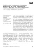
to the matrices. The effluent was applied to CM-Sephadex
C-50 column chromatography (Fig. 1). The salt-active
acharan sulfate lyase activity fraction was then further
purified to homogeneity by a series of hydroxyapatite
Ultrogel chromatography and finally phosphocelluose col-
umn chromatography (Fig. 2). The specific activity and
total activity at each purification step are summarized in
Table 1.
The specific activity of the purified acharan sulfate lyase
activity had 80.33 UÆmg
)1
proteinwithayieldof7.4%.The
purified acharan sulfate lyase was apparently homogeneous
by SDS/PAGE and its molecular mass was identically
estimatedtobe94000Da(Fig.2).
Characterization of salt-active acharan sulfate lyase
The molecular weight of salt-active acharan sulfate lyase
under nondenaturing conditions was determined by gel
filtration (data not shown). Acharan sulfate lyase was
estimated to be about 94 000 Da. It suggests that acharan
sulfate lyase is composed of one subunit. The optimal pH
of acharan sulfate lyase was determined to be 7.2–7.3 for
acharan sulfate, heparin and heparan sulfate (Fig. 3), and
the optimum temperature for the maximal activity was
shown at 40 °C (Fig. 4).
Fig. 1. Elution profile of CM-Sephadex C-50 ion exchange (A),
hydroxyapatite ultrogel (B) and phosphocellulose (C) column chromato-
graphies. s, acharan sulfate lyase activity without KCl; m, acharan
sulfate lyase activity with 50 m
M
KCl;
8
n, heparin lyase activity; simple
line, absorbance at 280 nm.
Fig. 2. SDS/PAGE of the purified salt-active acharan sulfate lyase at
various steps of purification. Lanes 1 and 8, marker; lane 2, preparation
after crude extract; lane 3, preparation after QAE column chroma-
tography; lane 4, preparation after DEAE-cellulose column chroma-
tography; lane 5, preparation after CM-Sephadex C-25 column
chromatography; lane 6, preparation after hydroxyapatite ultragel
column chromatography; and lane 7, purified salt-active heparin lyase
II after phosphocellulose column chromatography.
3170 S W. Hong et al. (Eur. J. Biochem. 270) Ó FEBS 2003
The purified acharan sulfate lyase was activated 5.3-fold
by the strength of salts, such as KCl and NaCl (Fig. 5).
However, divalent cations CaCl
2
, and MgCl
2
, did not
activate this enzyme compared to KCl and NaCl. The salt-
active acharan sulfate lyase activity was slightly increased by
addition of Mn
2+
, whereas they were severely inhibited
by Cu
2+
, Ni
2+
and Zn
2+
(Table 2). The purified enzyme
was inhibited by PCMS
3
and butane-1,3-diol. Particularly,
PCMS potently inhibited salt-active acharan sulfate lyase
4
,
but little inhibited by the other agents that modify histidine
and cysteine residues (Table 3).
Amino acid composition analysis revealed that the salt-
active acharan sulfate lyase contains a large proportion of
lysine (data not shown). The pI value of the purified salt-
active acharan sulfate lyase was 8.5, but slightly lower than
those of the previously purified Flavobacterial heparin
lyases range from 8.9 to 10.1. We analyzed the internal
sequences of a peptide obtained by digestion of each enzyme
with trypsin (Table 4). The internal sequence of the salt-
active acharan sulfate lyase showed homology of 50% to
Flavobacterial heparin lyases I and II previously reported,
but did not showed homology to Flavobacterial heparin
lyase III [10,20].
Fig. 3. Effect of pH on the activity of salt-active acharan sulfate lyase.
The enzyme activity was assayed in 50 m
M
sodium phosphate buffer at
the indicated pH. d, activity for acharan sulfate; j, activity for hep-
aran sulfate; m, activity for heparin.
Fig. 4. Effect of temperature on the activity of salt-active acharan sul-
fate lyase. The enzyme activity was assayed in 50 m
M
sodium phos-
phate.
Fig. 5. Effect of KCl on the activity of salt-active acharan sulfate lyase.
d, salt-active acharan sulfate lyase II purified from Fr-s in Fig. 1; m,
heparin lyase II-1 (acharan sulfate lyase 1) purified from Fr-a in Fig. 1
[16]; j, heparin lyase II-2 (acharan sulfate lyase 2) purified from Fr-a
in Fig. 1 [16].
Table 1. Purification summary of salt-active acharan sulfate lyase. One
unit (U) is the activity forming 1 lmol disaccharides from acharan
sulfate per minute. Numbers in parentheses indicate the activities for
heparin used as a substrate.
Stage
Total
activity
(U)
Total
protein
(mg)
Specific
activity
(UÆmg
)1
)
Crude extract 165.4 5250.12 0.03
QAE cellulose column
chromatography
65.2 1407.18 0.05
DEAE-cellulose column
chromatography
90.3 747.04 0.12
CM Sephadex C-50 column
chromatography
37.2 13.67 2.72
Hydroxyapatite ultrogel
column chromatography
34.5 1.82 18.96
Phosphocellulose column
chromatography
12.2 (1.3) 0.15 81.33 (8.67)
Table 2. Effect of divalent metal ions on salt-active acharan sulfate lyase
activity.
Metal ion
a
Residual activity
b
(%)
Control 100
Mg
2+
94.4
Cu
2+
0
Ni
2+
7.7
Co
2+
67.3
Mn
2+
102.6
Ca
2+
85.4
Zn
2+
14.5
Pb
2+
93.3
EDTA 107
a
Final concentration, 1 m
M
.
b
0.03 U of homogenously purified
enzyme activity was taken as 100%.
5
Ó FEBS 2003 Bacteroidal salt-active acharan sulfate lyase (Eur. J. Biochem. 270) 3171
Substrate specificity of purified salt-active acharan
sulfate lyase
The purified salt-active acharan sulfate lyase degraded
heparin and heparan sulfate as well as acharan sulfate
(Table 5). The salt-active acharan sulfate lyase was the most
active to acharan sulfate. When the salt-active acharan
sulfate lyase activity for heparan was taken as 100%, the
enzyme activities for acharan sulfate and heparan sulfate
were 952.3 and 149.5%, respectively. However, all types of
chondroitin sulfates were not also substrates for the enzyme.
Kinetic constants of purified salt-active acharan
sulfate lyase
Michaelis–Menten constants were determined under the
optimum reaction conditions in experiments designed to
calculate reaction velocities at each substrate concentration
(Table 6). K
m
and V
max
of acharan sulfate on salt-active
acharan sulfate lyase were estimated at 65.4 lgÆmL
)1
and
131.2 lmolÆmin
)1
Æmg
)1
, respectively. As for heparin on salt-
active acharan sulfate lyase, K
m
and V
max
values of heparin
and heparan sulfate were calculated at 18.5 lgÆmL
)1
,
8.7 lmolÆmin
)1
Æmg
)1
and 40.7 lgÆmL
)1
, 13.1 lmolÆ
min
)1
Æmg
)1
, respectively.
Discussion
In the present report, we have purified salt-active acharan
sulfate lyase specifically acting on acharan sulfate from Fr-s
fraction of CM-Sephadex C-50 chromatography, which
efficiently resolved GAGs degrading lyases of B. stercoris
HJ-15. As Fr-b and Fr-c fractions showed a higher
specificity to heparan sulfate and heparin, they were
considered to be similar to heparin lyase III and heparin
Table 3. Effect of divalent metal ions on salt-active acharan sulfate lyase
activity. Homogenously purified enzyme activity (0.03 U) was taken as
100%.
Chemical modifying agent IC
50
(m
M
)
Control >1
N-Tosyl-
L
-phenylalanine chloromethyl ketone >1
Butane-1,4-diol 0.4
Paraoxon >1
2-Mercaptoethanol >1
dl-Dithiothreitol >1
Phenylmethylsulfonyl fluoride >1
Iodoacetic acid >1
Sodium-p-tosyl-
L
-lysine chloromethyl ketone >1
p-Chloromercuric sulfonic acid 0.02
Table 4. Internal amino acid sequence of salt-active acharan sulfate
lyase from Bacteroides stercoris HJ-15.
Enzyme
Internal amino
acid sequence
Homology
(%)
Salt-active acharan sulfate lyase …GTIQYG…
Flavobacterial heparin lyase I
a
213 50
…GKITYV…
Flavobacterial heparin lyase II
a
157 50
…GAIVYD…
Flavobacterial heparin lyase III
a
567 33
…LMIQSL…
a
Data from [10,20].
Table 5. Substrate specificity of acharan sulfate lyase and the previously reported heparin lyases. Activity on heparin (or heparan sulfate in
Flavobacterial heparin lyase III) as the substrate was set at 100%.
6
Hep., heparin lyase.
Substrate
Activity (%)
Bacteroidal Favobacterial
a
Salt-active Hep II Hep II-1 (ASL1)
b
HepII-2
b
(ASL2) Hep III
b
Hep I Hep II Hep III
Heparin 100 100 100 100 100 100 0
Heparan sulfate (porcine) 149.5 128 121.7 610 30 172 100
Acharan sulfate 952.3 549.5 339 0 0 100
c
0
Chondroitin sulfate A 0 0 0 0 0 0 0
Chondroitin sulfate B 0 0 0 0 0 0 0
Chondroitin sulfate C 0 0 0 0 0 0 0
a
Data from [1,4,5,13,21].
b
Data from [15,16].
c
Unpublished data.
Table 6.
7
K
m
and V
max
values of salt-active acharan sulfate lyase.
Enzyme
K
m
(lgÆmL
)1
) V
max
(UÆmg protein
)1
)
Acharan sulfate Heparin Heparin sulfate Acharan sulfate Heparin Heparin sulfate
Salt-active acharan sulfate lyase 65.4 18.5 40.7 131.2 8.7 13.1
Acharan sulfate lyase 1
a
28.1 8.8 7.5 65.0 11.6 14.3
Acharan sulfate lyase 2
a
42.2 20.6 16.4 107.6 22.9 31.3
a
Data from [16].
3172 S W. Hong et al. (Eur. J. Biochem. 270) Ó FEBS 2003
lyase I prepared from F. heparinum, respectively [15,16].
Two acharan sulfate lyases previously purified from Fr-a
fraction showed the different substrate specificity compared
to those of Fr-b, Fr-c and the previously reported heparin
lyases [16]. These enzymes were highly specific to acharan
sulfate compared to heparin and heparan sulfate. The salt-
active acharan sulfate lyase purified from Fr-s fraction
showed a different substrate compared heparin lyase I and
III, but exhibited a similar substrate specificity of two
acharan sulfate lyase previously purified from Fr-a. Acha-
ran sulfate was the best substrate for the purified present
enzyme. Particularly, the present enzyme was significantly
activated by salts, KCl and NaCl, although two acharan
sulfate lyases were not activated by salts. Several attempts at
N-terminal analysis failed to yield sequence information
suggesting the N-terminus to be blocked. Therefore, we
analyzed the internal sequences of a peptide obtained by
digestion with trypsin. The internal sequence of the peptide
showed poor homology to Flavobacterial heparin lyases I
and II. Although the substrate specificities of heparin lyase I
and III are well understood, the structural requirement for
the cleavage of heparin and heparan sulfate by heparin lyase
II is not well characterized. This could be due to a structural
complexity of heparin and heparan sulfate and a lack of
homogeneous polysaccharide. A recently characterized
GAG, acharan sulfate, from African giant snail Achatina
fulica can provide the criteria to classify the heparin lyase II
among the specificity of heparin lyases.
In conclusion, the substrate specificity as well as the
characterization of salt-active acharan sulfate lyase are
different from those of the previous reported heparin lyases
(heparin lyase I and III prepared from F. heparinum), but is
similar to those of Flavobacterial heparin lyase II. There-
fore, we suggest that the salt-active acharan sulfate lyase
may belong to heparin lyase II.
Acknowledgements
This work was supported by KOSEF grant 1999-2-209-010-5.
References
1. Jackson, R.L., Busch, S.J. & Cardin, A.D. (1991) Glycosamino-
glycans: molecular properties, protein interactions, and role in
physiological processes. Physiol. Rev. 71, 481–539.
2. Griffin, C.C., Linhardt, R.J., Van Gorp, C.L., Toida, T., Hileman,
R.E., Schubert, R.L. & Brown, S.E. (1995) Isolation and char-
acterization of heparan sulfate from crude porcine intestinal
mucosal peptidoglycan heparin. Carbohydr. Res. 276, 183–197.
3. Kim, Y.S., Jo, Y.Y., Chang, I.M., Toida, T., Park, Y. & Linhardt,
R.J. (1996) A new glycosaminoglycan from the giant African snail
Achatina fulica. J. Biol. Chem. 271, 11750–11755.
4. Lohse, D. & Linhardt, R. (1992) Purification and characterization
of heparin lyases from Flavobacterium heparinum. J. Biol. Chem.
267, 24347–24355.
5. Desai, U.R., Wang, H.M. & Linhardt, R.J. (1993) Substrate
specificity of the heparin lyases from Flavobacterium heparinum.
Arch. Biochem. Biophys. 306, 461–468.
6. Desai, U.R., Wang, H.M. & Linhardt, R.J. (1993) Specificity
studies on the heparin lyases from Flavobacterium heparinum.
Biochemistry 32, 8140–8145.
7. Bourin, M C. & Lindahl, U. (1993) Glycosaminoglycans and the
regulation of blood coagulation. Biochem. J. 289, 313–330.
8. Folkman, J. & Shing, Y. (1992) Control of angiogenesis by
heparin and other sulfated polysaccharides. Adv. Exp. Med. Biol.
313, 355–364.
9. Castellot, J.J. Jr, Choay, J., Lormeau, J.C., Petitou, M., Sache, E.
& Karnovsky, M.J. (1986) Structural determinants of the capacity
of heparin to inhibit the proliferation of vascular smooth muscle
cells. II. Evidence for a pentasaccharide sequence that contains a
3-O-sulfate group. J. Cell. Biol. 102, 1979–1984.
10. Godavarti, R., Davis, M., Venkataraman, G., Cooney, C.,
Langer, R. & Sasisekharan, R. (1996) A comparative analysis of
the primary sequences and characteristics of heparinases I, II, and
III from Flavobacterium heparinum. Biochem. Biophy. Res. Com-
mun. 225, 751–758.
11. Bellamy, R.W. & Horikoshi, K. (1992) Heparinase Produced by
Microorganism Belonging to the Genus Bacillus in US Patent.
Research Development Corporation of Japan.
12. Watanabe, M., Tsuda, H., Yamada, S., Shibata, Y., Nakamura,
T. & Sugahara, K. (1998) Characterization of heparinase from an
oral bacterium Prevotella heparinolytica. J. Biochem. (Tokyo) 123,
283–288.
13. Ahn,M.Y.,Shin,K.H.,Kim,D.H.,Jung,E.A.,Toida,T.,Lin-
hardt, R.J. & Kim, Y.S. (1998) Characterization of a Bacteroides
species from human intestine that degrades glycosaminoglycans.
Can. J. Microbiol. 44, 423–429.
14. Kim, B T., Kim, W S., Kim, Y S., Linhardt, R.J. & Kim, D H.
(2000) Purification and characterization of a novel heparinase
from Bacteroides stercoris HJ-15. J. Biochem. 128, 323–328.
15. Hong, S H., Kim, B T., Shin, H Y., Kim, W S., Lee, K S.,
Kim, Y S. & Kim, D H. (2002) Purification and characterization
of novel chondroitin ABC and AC lyases from Bacteroides ster-
coris HJ-15, a human intestinal anaerobic bacterium. Eur. J.
Biochem. 269, 2934–2940.
16. Kim, B.T., Hong, S.H., Kim, W.S., Kim, Y.S. & Kim, D H.
(2001) Purificatio and characterization of acharan sulfate lyases,
two novel heparinases, from Bacteroides stercoris HJ-15. Eur. J.
Biochem. 268, 2635–2641.
17. Linhardt, R.J. (1994) Analysis of glycosaminoglycans with poly-
saccharide lyase. In Curr. Prot. Mol. Biol. (Varki, A., ed.), pp.
17.13.17–17.13.32. Wiley Interscience, New York.
18. Bradford, M.M. (1976) A rapid and sensitive method for the
quantitation of microgram quantities of protein utilizing the
principle of protein dye binding. Anal. Biochem. 72, 248–254.
19. Laemmli, U.K. (1970) Cleavage of structural proteins during the
assembly of the head of bacteriphage T4. Nature 227, 680–685.
20. Su, H., Blain, F., Musil, R.A., Zimmermann, J.J.F., Gu, K. &
Bennett, D.C. (1996) Isolation and expression in Escherichia coli of
hepB and hep C, genes coding for the glycosaminoglycan-
degrading enzymes heparinase II and heparinase III, respectively,
from Flavobacterium heparinum. Appl. Environ. Microbiol. 62,
2723–2734.
21. Riley, T.V. & Mee, B.J. (1984) Heparinase production by Bac-
teroides species. Microbios Lett. 25, 141–149.
Ó FEBS 2003 Bacteroidal salt-active acharan sulfate lyase (Eur. J. Biochem. 270) 3173