Báo cáo khoa học: The phosphorylation pattern of human as1-casein is markedly different from the ruminant species potx
Bạn đang xem bản rút gọn của tài liệu. Xem và tải ngay bản đầy đủ của tài liệu tại đây (193.13 KB, 5 trang )
The phosphorylation pattern of human a
s1
-casein is markedly different
from the ruminant species
Esben S. Sørensen, Lise Møller, Maria Vinther, Torben E. Petersen and Lone K. Rasmussen*
Protein Chemistry Laboratory, Department of Molecular Biology, University of Aarhus, Denmark
Caseins are highly phosphorylated milk proteins assembled
in large colloidal structures termed micelles. In the milk of
ruminants, a
s1
-casein has been shown to be extensively
phosphorylated. In this report we have determined the
phosphorylation pattern of human a
s1
-casein by a combi-
nation of matrix-assisted laser desorption mass spectrometry
and amino acid sequence analysis. Three phosphorylation
variants were identified. A nonphosphorylated form, a
variant phosphorylated at Ser18 and a variant phosphory-
lated at Ser18 and Ser26. Both phosphorylation sites are
located in the amino acid recognition sequence of the
mammary gland casein kinase. Notably, no phosphoryla-
tions were observed in the conserved region covering resi-
dues Ser70–Glu78, which is extensively phosphorylated in
the ruminant a
s1
-caseins.
Keywords: a
s1
-casein; human milk; mammary gland casein
kinase; phosphorylation.
Caseins are the predominant milk proteins of most
mammalian species [1]. In ruminants, about 75% of the
milk protein content is constituted of caseins. The
corresponding figure for human milk is only about 40%
[2]. In the milk of ruminants, caseins interact with calcium
phosphate forming large stable colloidal particles termed
micelles. These micellar complexes make it possible to
maintain a supersaturated calcium phosphate concentra-
tion in milk, providing the newborn with sufficient
calcium phosphate for the mineralization of the rapidly
growing calcified tissues. In this context, the phosphory-
lation of the individual caseins plays a significant role in
the interaction with calcium phosphate and thereby the
organization of the micelles. The ruminant caseins, which
are the most intensely studied, comprise a
s1
-, a
s2
-, b-and
j-casein. Their phosphorylation pattern has been the basis of
many studies and the general feature is that they are highly
phosphorylated proteins, phosphorylated by the mammary
gland casein kinase [3–7]. The primary requirement for
phosphorylation by this kinase is a glutamate, a phospho-
serine or an aspartate two residues to the C-terminal side of
the phosphoacceptor site (S-x-E/Sp/D) [8,9].
Compared with ruminants, human milk contains a very
low concentration of calcium phosphate and the function
of casein in delivering calcium to the neonate is therefore
muted in this species. In human milk, the predominant
caseins are j-andb-casein, which differ from its ruminant
counterpart by a lower degree of phosphorylation [10]. For
many years it was generally accepted that a
s1
-casein was
absent or present in only very small amounts in human
milk [2]. In the mid-1990s, two groups isolated and
sequenced a minor 27-kDa casein component that was
identified as being the human counterpart of a
s1
-casein
[11,12]. In addition, it was shown that this a
s1
-casein
component forms disulfide-bonded heteromultimers with
j-casein in human milk [12]. The molecular cloning and
sequencing of mRNA transcripts revealed the presence of
three forms of a
s1
-casein in human milk [13,14]. In the
present study, we report the phosphorylation pattern of
human a
s1
-casein.
Materials and methods
Materials
Trypsin (EC 3.4.21.4) was obtained from Worthington
Biochemical Corporation (Freehold, NJ, USA). Vydac C
4
and C
18
reverse-phase resins were from The Separations
Group (Hesperia, CA, USA) and the RP C
2
/C
18
column
was from Amersham Biosciences AB (Uppsala, Sweden).
Reagents used for sequencing were from Applied Biosys-
tems (Foster City, CA, USA). All other reagents were of
analytical reagent grade.
Purification of human a
s1
-casein
Human a
s1
-casein was purified from human milk as
described [12]. During this procedure, the protein was
reduced and alkylated to dissociate the disulfide-linked
complex consisting of a
s1
-andj-casein. To remove small
residual amounts of j-casein, the protein was subjected to
reverse-phase chromatography on a Vydac C
4
reverse-phase
column. The purity of the resulting a
s1
-casein was verified
by SDS/PAGE and N-terminal amino acid sequence
analysis.
Correspondence to E. S. Sørensen, Protein Chemistry Laboratory,
Department of Molecular Biology, University of Aarhus, Science
Park, Gustav Wieds Vej 10, DK-8000 Aarhus, Denmark.
Fax: + 45 8 6136597, Tel.: + 45 8 9425092,
E-mail:
Enzyme: Trypsin (EC 3.4.21.4)
*Present address: Symphogen A/S, DK-2800 Denmark.
(Received 5 May 2003, revised 14 July 2003,
accepted 16 July 2003)
Eur. J. Biochem. 270, 3651–3655 (2003) Ó FEBS 2003 doi:10.1046/j.1432-1033.2003.03755.x
Generation, separation and characterization of peptides
Approximately 300 lg of reduced and alkylated human
a
s1
-casein was digested with trypsin using a ratio of enzyme
to substrate of 1 : 100 (w/w) in 0.1
M
ammonium bicar-
bonate, pH 8.1, at 37 °C for 6 h. Separation of the peptides
was carried out by reverse-phase HPLC on a Vydac C
18
column and detected in the effluent by measuring the
absorbency at 226 nm (as described in the legend to Fig. 1).
Fraction 35 (Fig. 1) was rechromatographed by reverse-
phase HPLC on a SMART-system equipped with a
2.1 · 100 mm C
2
/C
18
RPC column using a gradient of
acetonitrile in 0.05% heptafluorobutyric acid at 25 °C.
Peptides were characterized by mass spectrometric- and
amino acid sequence analysis. Mass spectrometric analyses
of the peptides were performed using a MALDI-TOF mass
spectrometer (Voyager DE PRO, Applied Biosystems Inc.).
Theoretical peptide masses were calculated using the
GPMAW
program (Lighthouse Data, Odense, Denmark).
Amino acid sequence analysis was performed on an
automated amino acid sequencer (ABI 477A/120A; Applied
Biosystems Inc.). To locate phosphoserines in the sequence,
phosphopeptides were treated with ethanethiol to convert
phosphoserine into S-ethylcysteine [15] which can be
identified by amino acid sequence analysis as PTH-
S-ethylcysteine after its release in the corresponding cycle.
PTH-S-ethylcysteine eluted just before the diphenylthiourea
peak in the system used [16].
Results and discussion
Human a
s1
-casein was purified in a reduced and carboxy-
methylated state as described [12]. The protein was digested
with trypsin and the resulting peptides were separated
by reverse-phase chromatography (Fig. 1). Fractions were
collected and the peptides were characterized by mass
spectrometric- and sequence analysis. The combined results
are shown in Table 1. The amino acid sequence of human
a
s1
-casein is shown in Fig. 2. Peptides identified by mass
spectrometric analysis and/or sequence analysis are under-
lined. As seen in the Fig. 2, peptides covering the entire
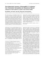
Fig. 1. Reversed-phase separation of a trypsin
digest of human a
s1
-casein. Human a
s1
-casein
was digested with trypsin as described in
Materials and methods. Peptides were eluted
with a gradient of 80% acetonitrile in 0.1%
trifluoroacetic acid (dotted line) on a Vydac
C
18
(10 lm) column (4 · 250 mm). The col-
umn was operated at 40 EC and the flow rate
was 0.85 mLÆmin
)1
. Peptides were detected in
the effluent by recording the absorbance at
226 nm (solid line), collected manually and
characterized as described in the text.
Table 1. Characterization of peptides from the tryptic digest of human
a
s1
-casein. Peak numbers designations correspond to those of Fig. 1.
The amino acid sequence was identified by sequence analysis and/or
MALDI-TOF MS. Calculated MH
+
, calculated protonated mono-
isotopic masses; observed MH
+
, molecular monoisotopic protonated
mass determined by MALDI-TOF MS.
Peak number Sequence Calculated MH
+
Observed MH
+
11 45–50 733.37 733.29
16 8–11 564.28 564.25
17 43–50 990.51 990.55
19 45–53 1105.55 1105.5
23 1–7 879.59 879.6
25 84–90 972.36 972.29
25 54–67 1605.71 1605.77
26 39–42 515.33 515.27
26 28–36 1143.46 1143.38
28 4–7 498.34 498.24
35–1
a
12–27+2P 1942.82 1942.78
35–2
a
68–83 1718.75 1718.75
36 4–11 1043.6 1043.62
37 12–27+1P 1862.86 1862.78
38 12–27 1782.89 1782.86
41 12–36+2P 3067.34 3067.26
43 12–36+1P 2987.34 2987.26
44 91–109 2267.16 2267.15
49 164–171 904.44 904.38
53 142–163 2580.21 2580.44
54 111–132 2591.24 2591.18
54 133–141 1170.57 1170.5
62 133–163 3731.75 3731.12
65 111–163 6303.97 6303.61
a
Peaks from rechromatography of fraction 35 from Fig. 1.
3652 E. S. Sørensen et al. (Eur. J. Biochem. 270) Ó FEBS 2003
sequence of human a
s1
-casein have been identified and
characterized in this study.
Glycosylation
Three asparagine residues in human a
s1
-casein (Asn14,
Asn54, Asn154) are located in the putative glycosylation
sequence Asn-X-Ser/Thr. In the case of Asn14 and Asn154,
a neighbouring proline residue in position X corrupts the
glycosylation sequence and renders it unfit for glycosylation.
Regarding Asn54, this study did not show any evidence for
glycosylation of this residue in human a
s1
-casein. Mass
spectrometric analysis of peak 25 (Fig. 1) containing the
peptide Asn54–Lys67 showed a mass of 1605.77 Da which
corresponds to the calculated protonated monoisotopic
mass (1605.71 Da) of the unmodified peptide sequence,
thereby showing that Asn54 was not glycosylated in human
a
s1
-casein.
Likewise in this study, we observed no O-glycosylations
in human a
s1
-casein.
Phosphorylation
Human a
s1
-casein contains 16 serines and four threonines,
where nine of the serines and one threonine are located
in the recognition sequence of the mammary gland casein
kinase [8,9]. The recognition sequence (Ser/Thr-X-Glu/
Ser(P)/Asp), comprises an acidic residue, glutamic acid,
aspartic acid or a phosphorylated residue, as the second
amino acid to the C-terminal side of the serine or threonine
to be targeted. Especially interesting, human a
s1
-casein
contains a serine rich region, SSISSSSEE(70–78), where five
of the serines are located in the recognition sequence of
the mammary gland casein kinase. This region is highly
conserved among all species with known a
s1
-casein
sequences (for alignment see [13]), and in all analyzed
species a high degree of phosphorylation has been observed
in this region [3–5].
Our laboratory has much experience of employing
MALDI-TOF mass spectrometric analysis for identification
and localization of phosphorylation sites in proteins [16–18].
MALDI-TOF mass spectrometric analysis of a phospho-
peptide results in a spectrum with an easily identifiable
fragmentation pattern which is characteristic for phospho-
rylated serines. These spectra contain a series of peaks
separated by approximately 98 Da, which represents the
fragmentation of a phosphoserine to dehydroalanine.
In this work, we have analyzed all fractions from the
reverse-phase HPLC separation of the tryptic digest of
human a
s1
-casein (Fig. 1) by MALDI-TOF mass spectro-
metric and N-terminal sequence analysis. We identified four
fractions with the characteristic fragmentation pattern of
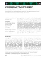
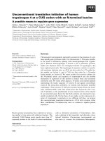
peaks at 35, 37, 41 and 43. A representative MALDI-TOF
spectrum of peak 41 showing the characteristic fragmenta-
tion of a phosphopeptide is shown in Fig. 3. Peak 35 was
found to contain two peptides which potentially could be
phosphorylated, thus the fraction was rechromatographed
by reversed-phase HPLC on a SMART HPLC system to
separate the two components, 35–1 and 35–2. Peak 35–2
gave a mass spectrum with only one mass at 1718.75 Da
which is identical with the calculated protonated mass for
the tryptic peptide covering residues 68–83, thereby showing
that this peptide is not phosphorylated in human a
s1
-casein.
This observation was confirmed by Edman sequencing of
the peptide, which showed normal yields of PTH-serine in
all relevant cycles. Mass analysis of peak 35–1 showed a
mass of 1942.78 Da, as well as two populations of ions at
approximately )98 Da and )196 Da. This triplet of ions,
each separated by approximately 98 Da, indicates that peak
35–1 contains a phosphopeptide with two phosphoserines.
Furthermore, the observed mass at 1942.78 Da correlates
with the calculated protonated mass (1942.82 Da) for the
tryptic peptide covering residues 12–27 and containing two
phosphorylations (159.93 Da). Mass analysis of peak 37
showed a mass of 1862.78 Da, and a single fragmentation
ion at )98 Da was observed, indicating the presence of a
single phosphorylation in the peptide. Furthermore, the
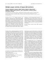
Fig. 2. Localization of phosphorylations in human a
s1
-casein. The
amino acid sequence was deduced from the cDNA sequence [11]. Solid
lines indicate isolated and characterized peptides (Table 1). P denotes
identified phosphorylation. Peptides are numbered according to the
reversed-phase elution profile in Fig. 1.
Fig. 3. MALDI-TOF MS of peak 41 from Fig. 1. The protonated
mass at m/z 3067.26 corresponds to the peptide 12–36 including to
phosphorylations. The characteristic fragmentation pattern confirms
the presence of two phosphorylations in the peptide.
Ó FEBS 2003 Phosphorylation of human a
s1
-casein (Eur. J. Biochem. 270) 3653
mass 1862.78 Da correlates with the calculated protonated
mass of residues 12–27 containing a single phosphorylation
(1862.86 Da). Finally, mass analysis of peak 38 showed a
mass which correlates with the mass of the peptide covering
residues 12–27 without any modifications. In conclusion, we
have observed the peptide 12–27 in three different forms,
with zero, and one and two phosphate groups attached.
Peaks 41 and 43 represent peptide 12–36 with two and one
phosphorylated groups, respectively. These peptides, result-
ing from incomplete cleavage at Arg27, do not contain any
additional serines or threonines compared with peaks 35
and 37, and thus they were not characterized further.
The peptide, LQNPSESSEPIPLESR(12–27) (peak
35–1), contains three serines located in the recognition
sequence of the mammary gland casein kinase (Ser16, Ser18
and Ser26). To determine which of the serines are in fact
phosphorylated, we subjected the two peptides to an
ethanethiol treatment followed by Edman sequencing as
outlined in Materials and methods. The ethanethiol treat-
ment converts the labile phosphoserine residues into
S-ethylcysteine, which is more stable and able to withstand
the relatively harsh Edman chemistry during automated
sequencing [15]. Furthermore, PTH-S-ethylcysteine elutes in
an open window just before the diphenylthiourea peak in
the on-line HPLC system used in these studies. Sequence
analysis of peptide 35–1 succeeding the ethanethiol treat-
ment revealed PTH-S-ethylcysteine in cycles 7 and 15,
corresponding to Ser18 and Ser26 in human a
s1
-casein.
Likewise, sequence analysis of peptide 37, after the
ethanethiol treatment gave PTH-S-ethylcysteine in sequence
cycle 7, corresponding to Ser18 in human a
s1
-casein. The
yields of PTH-serine in cycles corresponding to Ser16 and
Ser19 were as expected, indicating that these residues were
not phosphorylated.
As a control experiment (data not shown) human and
bovine b-casein were purified, tryptic digests were generated
and these were separated by reversed-phase HPLC using the
system and column described in Materials and methods. In
MALDI-TOF MS analyses of the human b-casein digest,
two peptides with protonated masses of 2407.85 and
2327.80 were identified. These masses correspond to the
expected masses of the peptide 1–18 of human b-casein with
four phosphorylations (2408.00) and three phosphoryla-
tions (2328.00), respectively. Likewise in the digest of bovine
b-casein, a peptide with a protonated mass of 3122.27 was
observed, corresponding to the peptide 1–25 of bovine
b-casein with four phosphorylations (3122.40 Da). These
results indicate that the protocol used for identification of
phosphorylation sites is capable of handling highly phos-
phorylated peptides. Furthermore, the methods used in the
present study have previously been used for identification
of phosphorylation sites in several milk proteins in our
laboratory, most prominently the 28 phosphorylation sites
in bovine milk osteopontin [16]. Therefore it is not likely
that the lack of identification of a highly phosphorylated
peptide in a
s1
-casein is due to limitations of the techniques
used.
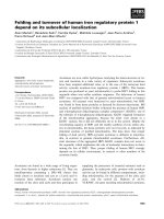
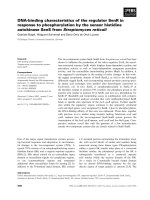
Finally, MALDI-TOF MS analysis of native human
a
s1
-casein showed ions corresponding to a mass of approxi-
mately 20 232 Da, which correlates well with the calculated
mass of human a
s1
-casein including two phosphate groups
(20246 Da) (Fig. 4).
In conclusion, these studies show that human a
s1
-casein
exists in three phosphorylation variants. A nonphosphory-
lated form, a variant containing a single phosphorylation
at Ser18 and a variant phosphorylated at Ser18 and Ser26.
It is difficult to determine the quantitative relation
between the three phosphorylation variants, but judged by
the reversed-phase HPLC trace in Fig. 1, the variant
containing a single phosphorylation at Ser18 is the major
variant ( 50%), followed by the nonphosphorylated form
( 30%) and the doubly phosphorylated variant ( 20%).
The degree of identity between human and other known
a
s1
-casein sequences is overall low (alignment of sequences is
shown in [6,13]). The phosphorylations have been charac-
terized in ovine (Ovis aries), caprine (Capra hircus), bovine
(Bos taurus), water buffalo (Bubalus bubalis)andcamel
(Camelus dromedarius) a
s1
-casein. Generally, all of these
species have been reported to have a significant higher
number of phosphorylations than shown to be the case for
human a
s1
-casein in this study. Bovine a
s1
-casein is phos-
phorylated at up to nine positions depending on the genetic
variants [3], the ovine a
s1
-casein is phosphorylated at up to
11 positions [5], the water buffalo a
s1
-casein is phospho-
rylated at 6–8 positions [6], the caprine counterpart is
phosphorylated at 9–10 positions [4], and camel a
s1
-casein is
phosphorylated at up to six serines [7]. The most striking
difference in the phosphorylation pattern between human
a
s1
-casein and its ruminant counterparts is the shortage of
phosphorylation in the serine-rich region consisting of
residues Ser70–Glu78 in the human sequence. This region
has been shown to be highly phosphorylated in all the above
mentioned species. In this study, we have isolated the tryptic
peptide covering residues Met68–Lys83, which contains the
serine-rich region, in a nonphosphorylated form, and no
traces of a phosphorylated form of this peptide were
observed.
The lack of phosphorylation at other positions reported
to be modified in ovine, caprine and bovine a
s1
-casein
(serines 41, 46, 48, 75 and 115 in all species, and Ser12 in
ovine a
s1
-casein, all numbers referring to the ruminant
sequences), can simply be explained by sequence substitu-
tions at these positions, leaving no hydroxyamino acids to
be phosphorylated at these positions in human a
s1
-casein.
The region containing the phosphorylated residues, Ser18
Fig. 4. MALDI-TOF MS of intact human a
s1
-casein. The peak at m/z
20233 corresponds well to the calculated mass of human a
s1
-casein
including two phosphate groups (20246 Da). The peak at m/z 10121
represents the doubly protonated species (M2H
+
).
3654 E. S. Sørensen et al. (Eur. J. Biochem. 270) Ó FEBS 2003
and Ser26, in human a
s1
-casein is not especially well
conserved among the other analyzed species. The sequence
containing Ser18 in the human a
s1
-casein is part of exon
3 in the human a
s1
-casein gene, which is not present in the
ruminant species [13]. Ser26, situated in exon 5 of the human
a
s1
-casein gene, is not conserved in any other species except
the wallaby, in which the phosphorylation pattern has not
been determined [19].
During the review of these results, we were puzzled by the
lack of phosphorylation in the conserved region Ser70–
Glu78, which is so extensively phosphorylated in the
ruminant species. To test whether our results were repre-
sentative, milk from three different women was analyzed.
a
s1
-Casein was purified and the reversed-phase traces of
tryptic digests of the protein were compared and found to be
identical in all cases, thereby showing similar phosphoryla-
tion of the protein in different individuals. The phosphory-
lation pattern of human a
s1
-casein described here, and
especially the lack of phosphorylations in the region Ser70–
Glu78, is therefore unlikely to be a result of intra-species
post-translational polymorphism in the protein. However, it
should be emphasized that it is more difficult to show the
absence of a modification convincingly than its presence;
hence the existence of minor species partially phosphory-
lated at the region discussed can not be entirely excluded.
The deletion of 11 amino acids at positions 59–69 and of
37 amino acids at positions 59–95 in caprine a
s1
-casein leads
to the variants D and F. In both cases these deletions, which
start at the same position of the polypeptide chain, include
the major phosphorylation site of the protein [20]. In
ruminant milk, a
s1
-casein, as well as the other three caseins
a
s2
-, b-andj-casein is present in micellar structures
responsible for the calcium transport to the neonates.
Compared with ruminant milk, the milk of primates holds a
much lower concentration of calcium and a function of a
s1
-
casein in calcium transport in human milk is not likely.
Recent studies of caprine a
s1
-casein suggest that the protein
interacts with the other caseins in the rough endoplasmic
reticulum and that the formation of this complex is required
for their efficient export to the Golgi apparatus [21].
Whether a similar scenario exists in the human system
remains to be elucidated.
Acknowledgments
Special thanks to H. Breinholt and K E. Højbjerg, Department of
Obstetrics and Gynaecology, University Hospital of Aarhus, for
providing the individual milk samples.
References
1. Jennes, R. & Holt, C. (1987) Casein and lactose in milk of 31
species are negatively correlated. Experentia 43, 1015–1018.
2. Kunz, C. & Lo
¨
nnerdal, B. (1990) Casein and casein subunits in
preterm milk, colostrum, and mature human milk. J. Pediatr.
Gastroenterol. Nutr. 10, 454–461.
3. Mercier, J.C., Grosclaude, F. & Ribadeau-Dumas, B. (1971)
Structure primaire de la case
´
ine a
s1
-bovine. Eur. J. Biochem. 23,
41–51.
4. Ferranti, P., Addeo, F., Malorni, A., Chianese, L., Leroux, C. &
Martin, P. (1997) Differential splicing of pre-messenger RNA
produces multiple forms of mature caprine a
s1
-casein. Eur. J.
Biochem. 249, 1–7.
5. Ferranti, P., Malorni, A., Nitti, G., Laezza, P., Pizzano, R., Chi-
anese, L. & Addeo, F. (1995) Primary structure of ovine a
s1
-
caseins: localization of phosphorylation sites and characterization
of genetic variants A, C and D*. J. Dairy Res. 62, 281–296.
6. Ferranti, P., Scaloni, A., Caira, S., Chianese, L., Malorni, A. &
Addeo, F. (1998) The primary structure of water buffalo a
s1
-and
b-casein: characterization of a novel b-variant. J. Protein Chem.
17, 835–844.
7. Kappeler, S., Farah, Z. & Puhan, Z. (1998) Sequence analysis of
Camelus dromedarius milk caseins. J. Dairy Res. 65, 209–222.
8. Mercier, J.C. (1981) Phosphorylation of caseins, present evidence
for an amino acid triplet code posttranslationally recognized by
specific kinases. Biochimie (Paris) 63, 1–17.
9.Lasa-Benito,M.,Marin,O.,Meggio,F.&Pinna,L.A.(1996)
Golgi apparatus mammary gland casein kinase: monitoring by a
specific peptide substrate and definition of specificity determi-
nants. FEBS Lett. 382, 149–152.
10. Greenberg, R., Groves, M.L. & Dower, H.J. (1984) Human beta-
casein: amino acid sequence and identification of phosphorylation
sites. J. Biol. Chem. 259, 5132–5138.
11. Cavaletto, M., Cantisani, A., Gluffrida, G., Napolitano, L. &
Conti, A. (1994) Human a
s1
-casein like protein: purification and
N-terminal sequence determination. Biol. Chem. Hoppe-Seyler
375, 149–151.
12. Rasmussen, L.K., Due, H.A. & Petersen, T.E. (1995) Human a
s1
-
casein: purification and characterization. Comp. Biochem. Physiol.
111B, 75–81.
13. Johnsen, L.B., Rasmussen, L.K., Petersen, T.E. & Berglund, L.
(1995) Characterization of three types of human a
s1
-casein mRNA
transcripts. Biochem. J. 309, 237–242.
14. Martin,P.,Brignon,G.,Furet,J.P.&Leroux,C.(1996)Thegene
encoding a
s1
-casein is expressed in human mammary epithelial
cells during lactation. Lait 76, 523–535.
15. Meyer, H.E., Hoffmann-Posorske, E., Korte, H. & Heilmyer,
M.G. Jr (1986) Sequence analysis of phosphoserine-containing
peptides: modification for picomolar sensitivity. FEBS Lett. 204,
61–66.
16. Sørensen, E.S., Højrup, P. & Petersen, T.E. (1995) Posttransla-
tional modifications of bovine osteopontin: identification of
twenty-eight phosphorylation and three O-glycosylation sites.
Protein Sci. 4, 2040–2049.
17.Sørensen,E.S.&Petersen,T.E.(1994)Identificationoftwo
phosphorylation motifs in bovine osteopontin. Biochem. Biophys.
Res. Commun. 198, 200–205.
18. Rasmussen, L.K., Sørensen, E.S., Petersen, T.E., Nielsen, N.C. &
Thomsen, J.K. (1997) Characterization of phosphate sites in
native ovine, caprine and bovine casein micelles and their case-
inomacropeptides: a solid-state
31
P NMR and sequence and mass
spectrometric study. J. Dairy Sci. 80, 607–614.
19. Ginger, M.R., Piotte, C.P., Otter, D.E. & Grigor, M.R. (1999)
Identification, characterisation and cDNA cloning of two caseins
from the common brushtail possum (Trichosurus vulpecula)1.
Biochim. Biophys. Acta 1427, 92–104.
20. Brignon, G., Mahe, M.F., Ribadeau-Dumas, B., Mercier. J.C. &
Grosclaude, F. (1990) Two of the three genetic variants of goat
alpha s1-casein which are synthesized at a reduced level have an
internal deletion possibly due to altered RNA splicing. Eur. J.
Biochem. 193, 237–241.
21. Chanat, E., Martin, P. & Ollivier-Bousquet, M. (1999) a
s1
-casein is
required for the efficient transport of b-andj-casein from the
endoplasmic reticulum to the Golgi apparatus of the mammary
epithelial cells. J. Cell Sci. 112, 3399–3412.
Ó FEBS 2003 Phosphorylation of human a
s1
-casein (Eur. J. Biochem. 270) 3655