Báo cáo khoa học: Binding affinity of nonsteroidal ecdysone agonists against the ecdysone receptor complex determines the strength of their molting hormonal activity pot
Bạn đang xem bản rút gọn của tài liệu. Xem và tải ngay bản đầy đủ của tài liệu tại đây (419.23 KB, 10 trang )
Binding affinity of nonsteroidal ecdysone agonists against
the ecdysone receptor complex determines the strength
of their molting hormonal activity
Chieka Minakuchi
1
, Yoshiaki Nakagawa
1
, Manabu Kamimura
2
and Hisashi Miyagawa
1
1
Division of Applied Life Sciences, Graduate School of Agriculture, Kyoto University, Kyoto, Japan;
2
National Institute of
Agrobiological Sciences, Tsukuba, Japan
N-tert-Butyl-N,N¢-dibenzoylhydrazine and its analogs are
nonsteroidal ecdysone agonists that exhibit insect molting
hormonal and larvicidal activities. The interaction mode of
those ecdysone agonists with the heterodimer of the ecdy-
sone receptor and ultraspiracle has not been fully elucidated.
We expressed the ecdysone receptor B1 and the ultraspiracle
of the lepidopteran, Chilo suppressalis,usinganin vitro
transcription/translation system and confirmed, using gel-
shift assays, that the proteins function as ecdysone receptors.
We also analyzed their ligand-binding affinity. A potent
ecdysteroid, ponasterone A, specifically bound to the ecdy-
sone receptor with low affinity (K
D
¼ 55 n
M
), and the spe-
cific binding was dramatically increased (K
D
¼ 1.2 n
M
)in
the presence of the ultraspiracle. For seven nonsteroidal
ecdysone agonists and five ecdysteroids, the binding activity
to the in vitro-translated ecdysone receptor–ultraspiracle
complex was linearly correlated with the binding activity to
the inherent receptor protein in the cell-free preparation of
C. suppressalis integument. The binding to the ecdysone
receptor–ultraspiracle complex for a series of compounds
was highly correlated with their molting hormonal activity,
indicating that the binding affinity of nonsteroidal ecdysone
agonists to the ecdysone receptor–ultraspiracle complex
primarily determines the strength of their molting hormonal
activity.
Keywords: ecdysone receptor; dibenzoylhydrazines; nuclear
receptor; receptor binding; dissociation constant.
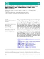
Insect molting is triggered by the binding of 20-hydroxy-
ecdysone (Fig. 1I) to its receptor protein. Significant effort
has been made to purify the receptor proteins for
20-hydroxyecdysone [1–3], but the isolation and purification
of these receptors from whole insects has, to date, been
unsuccessful owing to their instability and low yield. How-
ever, cDNAs for the ecdysone receptor (EcR) and the
ultraspiracle (USP) have been cloned from Drosophila mel-
anogaster [4–7], and it was later shown that EcR and USP
constitute a heterodimer which functions as the receptor for
20-hydroxyecdysone [8–10]. It was also made clear that
20-hydroxyecdysone binds to EcR, and that USP is an
essential partner for the binding of 20-hydroxyecdysone
[10]. The 20-hydroxyecdysone–EcR–USP complex binds to
the ecdysone response element (EcRE), located upstream of
other genes that are involved in molting and metamorpho-
sis, and regulates the transcription of those genes. Both EcR
and USP belong to the nuclear receptor superfamily, which
is generally composed of six distinct regions (A–F), and the
predicted amino acid sequences of C (DNA binding) and E
(ligand binding) regions are conserved among insects [11].
Little is known about the interactions between the EcR–
USP complex and ligands, because the EcR 3-D structure
has not yet been elucidated, while the crystal structures of
USPs were recently published for D. melanogaster and
Heliothis virescens [12,13].
N-tert-Butyl-N,N¢-dibenzoylhydrazine (RH-5849; Fig. 1II:
X
n
¼ Y
n
¼ H) [14–16] and its analogs are nonsteroidal
ecdysone agonists that, via binding to the EcR–USP
complex, cause incomplete molting in insects leading to
death. Among these nonsteroidal ecdysone agonists,
tebufenozide (RH-5992; Fig. 1II: X
n
¼ 3,5-CH
3
,Y
n
¼
4-C
2
H
5
), methoxyfenozide (RH-2485; Fig. 1II: X
n
¼ 3,5-
CH
3
,Y
n
¼ 2-CH
3
-3-OCH
3
), halofenozide (RH-0345;
Fig. 1II: X
n
¼ H, Y
n
¼ 4-Cl) and chromafenozide (ANS-
118; Fig. 1III) are currently on the market as safer
insecticides with reduced mammalian toxicity [17–21].
Previously, we evaluated the larvicidal and molting
hormonal activities of nonsteroidal ecdysone agonists in
the lepidopteran rice stem borer, C. suppressalis [22–29]. In
these studies, we analyzed the substituent effects of non-
steroidal ecdysone agonists on the larvicidal and molting
hormonal activities by using classical quantitative structure–
activity relationship procedures [30] to identify the import-
ant physicochemical properties for their biological activity.
However, the binding activity of ecdysone agonists against
the receptor proteins of C. suppressalis has not yet been
investigated. Quantitative structure–activity relationship
Correspondence to Y. Nakagawa, Division of Applied Life Sciences,
Graduate School of Agriculture, Kyoto University, Kyoto 606-8502,
Japan. Fax: + 81 75 753 6123, Tel.: + 81 75 753 6117,
E-mail:
Abbreviations: ANS-118, chromafenozide; EC
50
, 50% effective
concentration; EcR, ecdysone receptor; EcRE, ecdysone response
element; IC
50
, 50% inhibitory concentration; pIC
50
, reciprocal
logarithmic value of the IC
50
; RH-0345, halofenozide; RH-2485,
methoxyfenozide; RH-5849, N-tert-butyl-N,N¢-dibenzoylhydrazine;
RH-5992, tebufenozide; USP, ultraspiracle.
(Received 9 June 2003, revised 30 July 2003,
accepted 21 August 2003)
Eur. J. Biochem. 270, 4095–4104 (2003) Ó FEBS 2003 doi:10.1046/j.1432-1033.2003.03801.x
studies for the receptor-binding activity would be useful to
elucidate their mode of action.
For D. melanogaster, three EcR isoforms (EcR-A, EcR-
B1, EcR-B2) and one USP isoform have been cloned [4–7].
Subsequently, EcR and USP genes were cloned from several
insects [11]. Recently we also cloned cDNAs for EcR-A,
EcR-B1 and USP from C. suppressalis [31,32]. In this study,
we prepared EcR-B1 and USP for C. suppressalis by an
in vitro transcription/translation reaction to analyze their
functions. We also evaluated the binding activity of a
number of ecdysone agonists against the expressed EcR–
USP complex to obtain information on the ligand–receptor
interactions.
Experimental procedures
Chemicals
Ponasterone A was purchased from Invitrogen Corp.
(Carlsbad, CA, USA), and ecdysone and 20-hydroxyecdy-
sone were from Sigma Chemical Co. (St Louis, MO, USA).
Chromafenozide was a gift from Sankyo Agro and Nippon
Kayaku Co. Ltd. Other ecdysteroids and nonsteroidal
ecdysone agonists were from our stock samples [23–26,28,
29,33–35].
3
H-Labeled ponasterone A (150 CiÆmmol
)1
)was
purchased from American Radiolabeled Chemicals Inc.
(St Louis, MO, USA).
In vitro
transcription/translation and gel mobility
shift assay
The full coding regions of two splicing variants of EcR-B1,
encoding 547 aa or 542 aa (EcR-547aa and EcR-542aa,
respectively), and USP, were cloned into pBluescript II
vector (Stratagene, La Jolla, CA, USA), as described
previously [31,32]. Coupled in vitro transcription/translation
of these constructs was performed using a TNT Quick
Coupled Transcription/Translation System (Promega,
Madison, WI, USA), under control of the T7 promoter,
according to the manufacturer’s instructions.
Gel mobility shift assays were conducted using in vitro-
translated EcR-547aa, EcR-542aa, USP and the
32
P-labeled
Pal1 EcRE (5¢-GATCTAGAGAGGTCAATGACCTCG
TCC-3¢) [36] as a probe, as previously reported [31,32].
Briefly, in vitro-translated proteins were incubated in 20 m
M
modified Hepes (pH 7.5) buffer on ice for 30 min, then 1 ng
of
32
P-labeled EcRE probe was added. The mixture was
incubated for another 30 min at 25 °C. The proteins were
separated by electrophoresis on a nondenaturing polyacryl-
amide gel and analyzed using a BAS-2000 bioimaging
analyzer (Fuji Photo Film Co., Ltd, Tokyo, Japan).
Ligand binding to
in vitro
-translated EcR and USP
proteins
Ligand-binding assays were performed according to pub-
lished methods [37,38]. Briefly, 4 lLofin vitro-translated
EcR and/or USP was placed in a siliconized tube (BM
Equipment Co. Ltd, Tokyo, Japan) in low-salt buffer
(20 m
M
Hepes, 20 m
M
NaCl, 20% glycerol, 1 m
M
EDTA,
1m
M
2-mercaptoethanol, pH 7.9, containing 1 lgÆmL
)1
aprotinin, 1 lgÆmL
)1
pepstatin and 1 lgÆmL
)1
leupeptin)
with
3
H-labeled ponasterone A (25 000 d.p.m., final con-
centration 5 n
M
) and a test compound. To estimate the
nonspecific binding, a 500-fold excess of unlabeled pona-
sterone A was added to the incubation mixture. The total
volume of the incubation mixture was 16 lL. The final
concentration of solvent (ethanol or dimethylsulfoxide) in
the incubation mixture was less than 1%, which did not affect
the ligand–receptor binding. After a 60-min incubation at
25 °C, sample tubes were transferred to ice, and the reaction
mixture was filtered immediately through nitrocellulose
membrane NC45 (Schleicher & Schuell, Einbeck, Germany)
with the aid of a vacuum filtration apparatus (KGS-25
microanalysis holder; Advantec Toyo, Tokyo, Japan), as
described below. The incubation mixture was transferred to
the filtration apparatus with NC45 membrane, loading 1 mL
of ice-cold washing buffer (i.e. low salt buffer with 10%
glycerol and no protease inhibitors). The whole solution on
the membrane was immediately filtered by applying vacuum.
After washing the membrane three times with 1.5 mL of
ice-cold washing buffer, the radioactivity collected on the
membrane was measured in Aquasol-2 (PerkinElmer Life
Sciences, Wellesley, MA, USA) using a liquid scintillation
counter LSC-1000 (Aloka Co., Ltd, Tokyo, Japan).
Ligand binding to the cell-free preparation from
C. suppressalis
integument
Rice stem borer larvae were reared on rice seedlings at 28 °C
under a long-day photoperiod (16-h light : 8-h dark).
Integuments of six larvae in the wandering stage were
collected and sonicated in 6 mL of EcR40 buffer (40 m
M
KCl, 25 m
M
Hepes, pH 7.0, 10% glycerol, 1 m
M
EDTA,
1m
M
dithiothreitol, 10 m
M
Na
2
S
2
O
5
, 500 l
M
phenyl-
methanesulfonyl fluoride, 1 l
M
leupeptin and 1 l
M
pepst-
atin) according to a previously reported protocol [39–41].
The sonication was performed using an ultrasonic disruptor
UR-200P (Tomy Seiko, Tokyo, Japan) at an output of 4
(80 W), with eight cycles of 5-s pulses under cold conditions.
Hard cuticle, which remains in the homogenate, was
removed by use of forceps. The protein concentration of
this cell-free preparation was determined to be 1.0 mgÆmL
)1
by the Bradford method [42].
The cell-free preparation of C. suppressalis integument
(300 lL) was placed in a disposable glass tube
(12 · 75 mm) containing 1 lL of a dimethylsulfoxide or
ethanol solution of each compound and 45 lLofEcR40
buffer. After incubating the tubes for 5–10 min on ice, 4 lL
Fig. 1. Structures of ecdysone agonists. (I) 20-Hydroxyecdysone, (II)
dibenzoylhydrazine analogs, (III) chromafenozide.
4096 C. Minakuchi et al. (Eur. J. Biochem. 270) Ó FEBS 2003
of an ethanol solution of
3
H-labeled ponasterone A
(1.0 · 10
5
d.p.m., final concentration 1.0 n
M
) was added
to each tube, and the mixture was incubated for 30 min at
25 °C. The reaction mixture was then filtered rapidly
through a Whatman GF/F glass-fiber filter presoaked with
0.1% polyethylenimine, and the filter was washed three
times with 2 mL of water. The radioactivity collected on the
filter was measured in Aquasol-2 using a liquid scintillation
counter (Aloka LSC-1000).
Data analyses
The dissociation constant, K
D
, was evaluated from the
saturation curve for radioligand binding using nonlinear
regression analysis in the
GRAPHPAD PRISM
program
(GraphPad Software, San Diego, CA, USA). The K
D
value
was also calculated from Scatchard plots. From the
concentration–response curve for the binding of
3
H-labeled
ponasterone A, the 50% inhibitory concentration (IC
50
)
was evaluated by probit transformation [43,44] for each
compound. pIC
50
(the reciprocal logarithmic value of the
IC
50
) was used as an index of the binding activity.
Results
Gel mobility shift assay of
in vitro
-translated protein
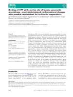
As shown in Fig. 2, none of EcR-547aa, EcR-542aa or USP
alone bound to the Pal1 probe (lanes 1, 2 and 3). However,
both EcR-547aa and EcR-542aa bound to the Pal1 probe in
the presence of USP (lanes 4 and 5). Although the density of
the bands was slightly different between EcR-547aa–USP
(lane 4) and EcR-542aa–USP (lane 5), this may be caused
by differences in protein concentration.
Specific binding of ponasterone A to
in vitro
-translated
protein
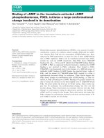
Ponasterone A bound to EcR-547aa specifically, but did
not bind to USP (Fig. 3). When EcR-547aa and USP were
mixed, specific binding of ponasterone A to EcR-547aa
increased dramatically. Similar results were observed using
EcR-542aa instead of EcR-547aa [EcR-542 alone,
203 ± 6 d.p.m. (control) vs. 111 ± 15 d.p.m. (with pona-
sterone A); EcR-542aa–USP, 754 ± 19 d.p.m. (control) vs.
105 ± 4 d.p.m. (with ponasterone A)]. The amount of
nonspecific binding of ponasterone A to each protein
(Fig. 3, lanes 2, 4 and 6) was equal to the radioactivity
captured in the nitrocellulose membrane without proteins
(data not shown). Specific binding to the co-expressed EcR-
547aa–USP was equivalent to that to the mixture of EcR-
547aa and USP expressed in the separate tubes (data not
shown).
Dissociation constant (
K
D
) for the binding
of ponasterone A to receptor proteins
As stated above,
3
H-labeled ponasterone A bound specific-
ally to EcR alone or to the EcR–USP complex. In order to
examine the kinetics for the ligand–receptor binding, we
calculated the dissociation constants (K
D
) of ponasterone A
against three different in vitro-translated proteins: EcR-
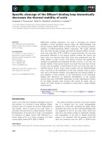
547aa; EcR-547aa–USP; and EcR-542aa–USP. As shown in
Fig. 4C,E, ponasterone A bound to EcR-547aa–USP and
EcR-542aa–USP with high affinity. The K
D
values of
ponasterone A to these complexes were 1.2 n
M
and 1.0 n
M
,
respectively. These values are comparable to those for
in vitro-translated EcR–USP of D. melanogaster (0.9 n
M
)
[10] and Bombyx mori (1.1 n
M
) [45]. On the other hand, the
K
D
value for EcR-547aa protein alone was 55 n
M
(Fig. 4A).
The K
D
value for EcR-542aa alone was not measured in this
study. The K
D
value of ponasterone A to inherent receptor
proteins in the cell-free preparation of C. suppressalis
integument was determined to be 6.9 n
M
by nonlinear
regression analysis (Fig. 5A). Scatchard plots for the binding
of ponasterone A to expressed proteins (Fig. 4) and inherent
receptor proteins (Fig. 5) were linear, indicating only one
type of binding site. The K
D
values evaluated from
Scatchard plots (Figs 4B,D,F and 5B) were similar to those
from the nonlinear regression model (Figs 4A,C,E and 5A).
Receptor-binding activity of ecdysone agonists
pIC
50
values of ecdysone agonists (ponasterone A and
tebufenozide) were very similar between EcR-547aa–USP
and EcR-542aa–USP, and each value was highly reprodu-
cible with only a small standard deviation (Table 1). In the
Fig. 2. Binding of the ecdysone receptor–ultraspiracle (EcR–USP)
complex to the ecdysone response element (EcRE). In vitro-translated
EcR-547aa, EcR-542aa and USP were incubated with
32
P-labeled Pal1
EcRE, and then analyzed following electrophoresis on a nondena-
turing polyacrylamide gel.
Ó FEBS 2003 Receptor binding affinity of ecdysone agonists (Eur. J. Biochem. 270) 4097
following binding assay, EcR-547aa–USP was used, and the
activity data were obtained from each single binding assay.
Binding activities of a series of ecdysone agonists did not
vary markedly between in vitro-translated EcR-547aa–USP
and the cell-free preparation (Table 2). As shown in
Table 2, some of the 3,5-dimethylbenzoyl analogs, such as
tebufenozide, methoxyfenozide and chromafenozide, bound
to the EcR–USP complex with very high affinity, being
200-fold higher than that of 20-hydroxyecdysone. By
introducing a C
2
H
5
group at the para-position of the B-ring
moiety, the binding activity against the EcR–USP complex
was enhanced 30 times (no. 1 vs. no. 2; see Table 2 for a
description of the compounds represented by the numbers),
while a Cl atom was not as effective as a C
2
H
5
group (no. 1
vs. no. 3; Table 2). With respect to the A-ring moiety,
introduction of a Cl atom at the ortho-position and methyl
groups at both of the meta-positions enhanced the activity
sixfold (no. 1 vs. no. 4; Table 2) and eightfold (no. 2 vs.
no. 6; Table 2), respectively. Three commercially available
insecticides, having a 3,5-dimethyl substitution pattern at
the A-ring moiety (no. 6, no. 7, no. 9; Table 2), are very
potent irrespective of the substitution pattern of the B-ring
moiety. However, by replacing the C
2
H
5
group at the para-
position of tebufenozide with an n-C
4
H
9
group, the activity
decreased 10-fold (no. 6 vs. no. 8; Table 2).
Among the ecdysteroids, the order of the binding
activity was ponasterone A > 20-hydroxyecdysone ‡
cyasterone > makisterone A > ecdysone, against both
the in vitro-translated EcR–USP complex and the cell-free
preparation. Ponasterone A (no. 10; Table 2) showed a
high binding activity against the in vitro-translated EcR–
USP complex, but it was 10-fold less potent than
chromafenozide and about sixfold less potent than
tebufenozide and methoxyfenozide. The binding activities
of 20-hydroxyecdysone (no. 11; Table 2) and ecdysone
(no. 15; Table 2) against the in vitro-translated EcR–USP
and inherent receptor proteins were 1/26 and 1/1000–
1/2000 lower than that of ponasterone A, being compar-
able to their molting hormonal activities against C. sup-
pressalis [25].
Discussion
In a previous study, we cloned two cDNA variants from
C. suppressalis [31]; these variants encoded EcR-547aa and
EcR-542aa, with the 15-bp difference in the D region
located between the C (DNA binding) and E (ligand
binding) regions. The presence of two homologous EcR
splicing variants with a 15-bp difference in the D region has
also been reported in Manduca sexta [46], but the functional
difference between these two variants has not yet been
investigated. Perera et al. observed no ligand binding in the
mutated EcR of the Lepidoptera Choristoneura fumiferana,
in which the D region was completely deleted [47]. Recently,
Grebe et al. suggested that the C-terminal part of the D
region of D. melanogaster EcR contributes to the ligand
binding and the dimerization with USP, even though the
N-terminal part is not essential for ligand binding [48]. In
this study, we showed that the lack of five amino acids
(LECLQ) in the D region of EcR did not affect the ligand–
receptor binding, the heterodimerization of EcR and USP,
or the binding of the EcR–USP heterodimer towards EcRE
(Figs 2 and 4 and Table 1). As previously reported, this
sequence is not located in the C-terminus of the D region of
CsEcR, but in the middle part [31]. Although the role of
these five amino acids is still unknown, the amino acids in
the middle part of the D region probably do not affect the
ligand–receptor binding.
Our results, demonstrating that ponasterone A specifi-
cally bound to EcR but not to USP, and that the specific
binding of ponasterone A to EcR was remarkably enhanced
by adding USP, are consistent with those observed for
D. melanogaster EcR and USP proteins [10,48]. In the case
of Chironomus tentans, the specific binding of ponaster-
one A was not observed for either EcR or USP, whereas
specific binding was observed for the EcR–USP complex
[38,49]. Recently, Grebe and co-workers showed that the
binding of ponasterone A to D. melanogaster EcR is
increased by 10-fold in the presence of D. melanogaster
USP [38]. They also suggested that the binding ability of
EcR to ligands in the absence of USP might be species
specific [48]. In this study, we have shown that the binding of
ponasterone A to C. suppressalis EcR is increased by
eightfold in the presence of C. suppressalis USP (Fig. 3),
which is consistent with the report by Grebe et al.on
D. melanogaster. We have clearly shown that the binding
affinity of ponasterone A to C. suppressalis EcR
(K
D
¼ 55 n
M
) was also enhanced 50-fold by adding USP
(K
D
¼ 1.2 n
M
). These results indicate that allosteric inter-
action between EcR and USP would change the confor-
mation of the ligand-binding pocket of EcR. The K
D
value
of ponasterone A for the expressed C. suppressalis EcR–
USP complex (K
D
¼ 1.2 n
M
) was not far from that for the
inherent receptor proteins in the cell-free preparation of
C. suppressalis integument (6.9 n
M
). It is to be expected that
Fig. 3. Binding of ponasterone A (PoA) to the in vit ro-translated ecdy-
sone receptor (EcR)-547aa (lanes 1 and 2), ultraspiracle (USP) (lanes 3
and 4), and a mixture of EcR-547aa and USP (lanes 5 and 6). In vitro-
translated EcR-547aa and/or USP were incubated with
3
H-labeled
PoA (5 n
M
), in the presence or absence of excess PoA, and filtered
through a nitrocellulose membrane. The radioactivity collected in the
filter was counted using a liquid scintillation counter. T, total binding;
N, nonspecific binding. The vertical bars show the standard deviation
of three replications. *P <0.01(Student’st-test).
4098 C. Minakuchi et al. (Eur. J. Biochem. 270) Ó FEBS 2003
these K
D
values vary slightly because the cell-free prepar-
ation contains various proteins that can affect binding.
The binding activity of ecdysone agonists to the inherent
receptor proteins was highly correlated (r ¼ 0.98) with that
measured against the in vitro-expressed EcR–USP complex
(Fig. 6). We therefore conclude that the EcR–USP complex
expressed in vitro in this study is a functional and useful
material for using in binding assays of ecdysone agonists.
In a previous study, we evaluated the molting hormonal
activity of different ecdysteroids and nonsteroidal ecdysone
agonists by measuring the induction of chitin synthesis in
the cultured integument of C. suppressalis larvae; the 50%
effective concentration (EC
50
) for the induction of the chitin
synthesis in the cultured integument of C. suppressalis
larvae was determined from the concentration–response
curve [25,27–29,50]. As shown in Fig. 7, the binding activity
(pIC
50
) is highly correlated with the molting hormonal
activity (pEC
50
), indicating that the strength of the hormo-
nal activity of ecdysone agonists is primarily determined
at the step of their binding to the EcR–USP complex.
Fig. 4. Binding of ponasterone A (PoA) to the in vitro-translated ecdysone receptor (EcR)-547aa (A, B), EcR-547aa–ultraspiracle (USP) (C, D), and
EcR-542aa–USP (E, F). In vitro-translated EcR and/or USP were incubated with different concentrations of
3
H-labeled PoA, in the presence or
absence of excess PoA, and filtered through a nitrocellulose membrane. The radioactivity collected in the filter was counted using a liquid
scintillation counter. Saturation radioligand-binding data (A, C, E) and Scatchard plots (B, D, F) are shown.
Ó FEBS 2003 Receptor binding affinity of ecdysone agonists (Eur. J. Biochem. 270) 4099
Furthermore, we reported previously that the compounds
possessing high molting hormonal activity are potent
insecticides for a series of dibenzoylhydrazine analogs
[28,29]. We therefore concluded that, regarding nonsteroidal
ecdysone agonists, the binding activity to the EcR–USP
complex results in potent larvicidal activity as well as potent
molting hormonal activity. On the other hand, regarding
ecdysteroids, the binding activity to EcR–USP is not
correlated to larvicidal activity: topical application of
20-hydroxyecdysone at 52 nmol resulted in no mortality
of C. suppressalis (Y. Nakagawa, Kyoto, Japan, unpub-
lished data). The topically applied ecdysteroids could not
easily permeate insect epidermis because of their low
hydrophilicity, or ingested ecdysteroids would be easily
metabolized and excreted from the insect body. It has been
reported that tomato moth larvae are able to feed on a diet
containing 400 p.p.m. 20-hydroxyecdysone without any
adverse effects on growth and development, while ingestion
of nonsteroidal ecdysone agonists, such as RH-5849 and
tebufenozide, induces a premature and lethal molt, indica-
ting that the ingested 20-hydroxyecdysone was metabolized
and rapidly excreted [51]. Although it has been shown that
nonsteroidal ecdysone agonists would also be metabolized
and excreted to some extent [52,53], we assumed that the
metabolism and excretion of nonsteroidal ecdysone agonists
in C. suppressalis would be less significant because piperonyl
butoxide, an inhibitor of oxidative metabolism, was used to
measure the larvicidal activity [23,24].
In this study, the binding activity of ecdysone agonists
was highly correlated with the molting hormonal activity
measured in C. suppressalis integument (Fig. 7). We had
expected that the physicochemical properties (such as
hydrophobicity) of compounds might affect their uptake
into the target cells. However, no physicochemical proper-
ties were taken into consideration for the correlation of the
binding activity to the molting hormonal activity of these
compounds. In fact, the partition coefficient (P) between
1-octanol and water, an index of hydrophobicity, varied by
7 000 000-fold among the compounds tested, as listed in
Table 2. We therefore concluded that hydrophobicity does
not affect compound cellular uptake in the C. suppressalis
integument.
Fig. 5. Binding of ponasterone A (PoA) to the inherent receptor proteins in the cell-free preparation of Chilo suppressalis integument. The cell-free
preparations were incubated with different concentrations of
3
H-labeled PoA, in the presence or absence of excess PoA, and filtered through a glass-
fiber filter (GF/F). The radioactivity collected in the filter was measured using a liquid scintillation counter. (A) Saturation radioligand-binding
data. (B) Scatchard plot.
Table 1. Binding activity [reciprocal logarithmic value of the 50% inhibitory concentration (pIC
50
)] of ponasterone A (PoA) and tebufenozide against
in vitr o-translated ecdysone receptor EcR-547aa–ultraspiracle (USP) or EcR-542aa–USP. The mean value ± SD of two duplicate experiments is
shown.
4100 C. Minakuchi et al. (Eur. J. Biochem. 270) Ó FEBS 2003
It is of great interest how the EcR interacts with
nonsteroidal ecdysone agonists, such as dibenzoylhydrazine
analogs, whose structures are totally different from that of
20-hydroxyecdysone. Wurtz and co-workers constructed the
ligand-binding domain of Chir. tentans EcR, in which ligand-
binding domains of the retinoic acid and vitamin D receptors
were used as templates, and predicted the binding modes of
20-hydroxyecdysone or unsubstituted RH-5849 to EcR [54].
Kumar and co-workers also modeled the ligand-binding
domain of Chor. fumiferana EcR [55]. They performed point
mutation analysis on the Chor. fumiferana EcR to identify
some of the amino acid residues essential for ligand binding
(ponasterone A and methoxyfenozide). Grebe and
co-workers created mutants of the D. melanogaster EcR by
site-directed mutagenesis and elucidated the function of
amino acid residues involved in the ligand binding to EcR
and the heterodimerization to USP [48]. Even though these
homology-modeling and point-mutation studies help to
clarify the interaction mode of ecdysone agonists to EcR–
USP, the detailed mechanism remains unknown.
In conclusion, we functionally expressed EcR and USP of
C. suppresalis in vitro, and showed that the dissociation
constant (K
D
) of ponasterone A to EcR was enhanced
50-fold by the addition of USP. The K
D
value of
ponasterone A to the EcR–USP complex was determined
to be 1n
M
, which is consistent with such values reported
for other insect EcR–USPs. The binding activity of
ecdysone agonists to the inherent receptor proteins in the
Fig. 6. Relationship between the binding
activities of the in vitro-translated and inherent
receptors. The binding activity [reciprocal
logarithmic value of the 50% inhibitory con-
centration (pIC
50
)] of ecdysone agonists
against in vitro-translated ecdysone receptor
(EcR)-547aa–ultraspiracle (USP) and the
binding activity (pIC
50
) against inherent
receptor proteins from Chilo suppressalis
integument.
Table 2. Binding activities [reciprocal logarithmic value of the 50% inhibitory concentration (pIC
50
)] of ecdysone agonists against the in vitro -translated
ecdysone receptor–ultraspiracle (EcR–USP) complex and inherent receptor proteins from Chilo suppressalis integument.
a
Mean ± SD. Values in parentheses indicate the number of replications. ND, not determined.
b
Single data.
c
Experimentally measured
[23,24].
d
Estimated empirically [23,24].
e
From Table 1.
f
Calculated using the CLOGP method [56].
g
From [32].
Ó FEBS 2003 Receptor binding affinity of ecdysone agonists (Eur. J. Biochem. 270) 4101
cell-free preparation of C. suppressalis integument was
highly correlated with that of the in vitro-expressed EcR–
USP complex. These results suggest that the EcR–USP
complex expressed in vitro in this study is useful for binding
assays of ecdysone agonists. The binding activity of a
number of steroidal and nonsteroidal ecdysone agonists was
linearly correlated to their molting hormonal activity with a
high correlation coefficient. Thus, we conclude that the
binding affinity of nonsteroidal ecdysone agonists to the
EcR–USP complex primarily determines the strength of
their biological activities.
Acknowledgements
We are thankful to Dr Craig Wheelock of the University of California
Davis for carefully reviewing this manuscript. We also express our
sincere gratitude to Drs Margarethe Spindler-Barth and Marco Grebe
(University of Ulm), and Drs Shuichiro Tomita and Atsushi Seino
(National Institute of Agrobiological Sciences), for their helpful
comments for the binding assay. We thank Sumitomo Chemical
Takeda Agro Co. Ltd for the gift of eggs of the rice stem borer, and
Sankyo Agro and Nippon Kayaku Co. Ltd for the gift of chromafe-
nozide. Part of this study was performed in the RI center of Kyoto
University. This investigation was supported, in part, by a grant-in-aid
for Scientific Research by the Ministry of Education, Science, and
Culture of Japan (09660117, 10161207) and Research Fellowships from
the Japan Society for the Promotion of Science for Young Scientists.
References
1. Lehmann, M. & Koolman, J. (1988) Ecdysteroid receptors of the
blowfly Calliphora vicina: partial purification and characterization
of ecdysteroid binding. Mol. Cell. Endocrinol. 57, 239–249.
2. Bidmon, H.J. & Sliter, T.J. (1990) The ecdysteroid receptor. Invert.
Reprod. Dev. 18, 13–27.
3. Sobek, L., Bohm, G A. & Penzlin, H. (1993) Ecdysteroid
receptors in last instar larvae of the wax moth Galleria mellonella
L. Insect Biochem. Mol. Biol. 23, 125–129.
4. Koelle, M.R., Talbot, W.S., Segraves, W.A., Bender, M.T.,
Cherbas, P. & Hogness, D.S. (1991) The Drosophila EcR gene
encodes an ecdysone receptor, a new member of the steroid
receptor superfamily. Cell 67, 59–77.
5.Henrich,V.C.,Sliter,T.J.,Lubahn,D.B.,Maclntyre,A.&
Gilbert, L.I. (1990) A steroid/thyroid hormone receptor super-
family membrane in Drosophila melanogaster that shares extensive
sequence similarity with a mammalian homologue. Nucleic Acids
Res. 18, 4143–4148.
6. Oro, A.E., McKeown, M. & Evans, R.M. (1990) Relationship
between the product of the Drosophila ultraspiracle locus and the
vertebrate retinoid X receptor. Nature 347, 298–301.
7. Shea, M.J., King, D.L., Conboy, M.J., Mariani, B.D. & Kafatos,
F.C.(1990)ProteinsthatbindtoDrosophila chorion cis-regulatory
elements: a new C
2
H
2
zinc finger protein and a C
2
C
2
steroid
receptor-like component. Genes Dev. 4, 1128–1140.
8. Yao, T P., Segraves, W.A., Oro, A.E., McKeown, M. & Evans,
R.M. (1992) Drosophila ultraspiracle modulates ecdysone receptor
function via heterodimer formation. Cell 71, 63–72.
9. Thomas, H.E., Stunnenberg, H.G. & Stewart, A.F. (1993) Het-
erodimerization of the Drosophila ecdysone receptor with retinoid
X receptor and ultraspiracle. Nature 362, 471–475.
10. Yao, T P., Forman, B.M., Jiang, Z., Cherbas, L., Chen, J.D.,
McKeown, M., Cherbas, P. & Evans, R.M. (1993) Functional
ecdysone receptor is the product of EcR and ultraspiracle genes.
Nature 366, 476–479.
11. Riddiford, L.M., Cherbas, P. & Truman, J.W. (2000) Ecdysone
receptors and their biological actions. Vitam. Horm. 60, 1–73.
12. Billas, I.M., Moulinier, L., Rochel, N. & Moras, D. (2001) Crystal
structure of the ligand-binding domain of the ultraspiracle protein
USP, the ortholog of retinoid X receptors in insects. J. Biol. Chem.
276, 7465–7474.
13. Clayton, G.M., Peak-Chew, S.Y., Evans, R.M. & Schwabe, J.W.
(2001) The structure of the ultraspiracle ligand-binding domain
reveals a nuclear receptor locked in an inactive conformation.
Proc.NatlAcad.Sci.USA98, 1549–1554.
14. Wing, K.D. (1988) RH 5849, a nonsteroidal ecdysone agonist:
effects on a Drosophila cell line. Science 241, 467–469.
15. Wing, K.D., Slawecki, R.A. & Carlson, G.R. (1988) RH-5849: a
nonsteroidal ecdysone agonist: effects on larval lepidoptera.
Science 241, 470–472.
16. Hsu, A.C T. (1991) 1,2-Diacyl-1-alkylhydrazines, a new class of
insect growth regulators. In Synthesis and Chemistry of Agro-
chemicals II (Moberg, W.K., ed.), pp. 478–490. American Chem-
ical Society, Washington DC.
17. Hsu, A.C T., Fujimoto, T.T. & Dhadialla, T.S. (1997) Structure–
activity study and conformational analysis of RH-5992, the first
commercialized nonsteroidal ecdysone agonist. In Phytochemicals
for Pest Control. (Thompson, D.G., ed.), pp. 206–219. American
Chemical Society, Washington DC.
18. Dhadialla, T.S., Carlson, G.R. & Le, D.P. (1998) New insecticides
with ecdysteroidal and juvenile hormone activity. Annu. Rev.
Entomol. 43, 545–569.
Fig. 7. Relationship between the binding activ-
ity and molting hormonal activity. The binding
activity [reciprocal logarithmic value of the
50% inhibitory concentration (pIC
50
)] of
ecdysone agonists against in vitro-translated
ecdysone receptor (EcR)-547aa–ultraspiracle
(USP) and the molting hormonal activity
[reciprocal logarithmic value of the 50%
effective concentration (pEC
50
)] against
Chilo suppressalis integument.
4102 C. Minakuchi et al. (Eur. J. Biochem. 270) Ó FEBS 2003
19. Tanaka, K., Tsukamoto, Y., Sawada, Y., Kasuya, A., Hotta, H.,
Ichinose, R., Watanabe, T., Toya, T., Yokoi, S., Kawagishi, A.,
Ando, M., Sadakane, S., Katsumi, S. & Masui, A. (2001) Chro-
mafenozide: a novel lepidopteran insect control agent. Annu.
Report Sankyo Res. Lab. 53, 1–49.
20. Toya, T., Fukasawa, H., Masui, A. & Endo, Y. (2002) Potent and
selective partial ecdysone agonist activity of chromafenozide in Sf9
cells. Biochem. Biophys. Res. Commun. 292, 1087–1091.
21. Sawada, Y., Yanai, T., Nakagawa, H., Tsukamoto, Y., Tama-
gawa, Y., Yokoi, S., Yanagi, M., Toya, T., Sugizaki, H., Kato, Y.,
Shirakura, H., Watanabe, T., Yajima, Y., Kodama, S. & Masui,
A. (2003) Synthesis and insecticidal activity of benzoheterocyclic
analogues of N-benzoyl-N-(tert-butyl) benzohydrazide: Part 3.
Modification of N-tert-butylhydrazine moiety. Pest Manag. Sci.
59, 49–57.
22. Oikawa, N., Nakagawa, Y., Soya, Y., Nishimura, K., Kurihara,
N., Ueno, T. & Fujita, T. (1993) Enhancement of N-acetyl-
glucosamine incorporation into the cultured integument of Chilo
suppressalis by molting hormone and dibenzoylhydrazine
insecticides. Pestic. Biochem. Physiol. 47, 165–170.
23. Oikawa, N., Nakagawa, Y., Nishimura, K., Ueno, T. & Fujita, T.
(1994) Quantitative structure–activity analysis of larvicidal
1-(substituted benzoyl)-2-benzoyl-1-tert-butylhydrazines against
Chilo suppressalis. Pestic. Sci. 41, 139–148.
24. Oikawa, N., Nakagawa, Y., Nishimura, K., Ueno, T. & Fujita, T.
(1994) Quantitative structure–activity studies of insect growth
regulators. X. Substituent effects on larvicidal activity of 1-tert-
butyl-1-(2-chlorobenzoyl)-2-(substituted benzoyl) hydrazines
against Chilo suppressalis and design synthesis of potent deriva-
tives. Pestic. Biochem. Physiol. 48, 135–144.
25. Nakagawa, Y., Nishimura, K., Oikawa, N., Kurihara, N. &
Ueno, T. (1995) Activity of ecdysone analogs in enhancing
N-acetylglucosamine incorporation into the cultured integument
of Chilo suppressalis. Steroids 60, 401–405.
26. Nakagawa, Y., Hattori, K., Shimizu, B., Akamatsu, M.,
Miyagawa, H. & Ueno, T. (1998) Quantitative structure–activity
studies of insect growth regulators. XIV. Three dimensional
quantitative structure–activity relationship of ecdysone agonists
including dibenzoylhydrazine analogs. Pestic. Sci. 53, 267–277.
27. Nakagawa, Y., Hattori, K., Minakuchi, C., Kugimiya, S. & Ueno,
T. (2000) Relationships between structure and molting hormonal
activity of tebufenozide, methoxyfenozide, and their analogs in
cultured integument system of Chilo suppressalis Walker. Steroids
65, 117–123.
28. Shimizu, B., Nakagawa, Y., Hattori, K., Nishimura, K., Kuri-
hara, N. & Ueno, T. (1997) Molting hormonal and larvicidal
activities of aliphatic acyl analogs of dibenzoylhydrazine
insecticides. Steroids 62, 638–642.
29. Nakagawa, Y., Soya, Y., Nakai, K., Oikawa, N., Nishimura, K.,
Ueno, T., Fujita, T. & Kurihara, N. (1995) Quantitative structure–
activity studies of insect growth regulators. XI. Stimulation and
inhibition of N-acetylglucosamine incorporation in a cultured
integument system by substituted N-tert-butyl-N,N¢-dibenzoyl-
hydrazines. Pestic. Sci. 43, 339–345.
30. Hansch,C.&Fujita,T.(1964)q-r-, p. Analysis. A method for the
correlation of biological activity and chemical structure. J. Am.
Chem. Soc. 86, 1616–1626.
31. Minakuchi, C., Nakagawa, Y., Kiuchi, M., Tomita, S. & Kami-
mura, M. (2002) Molecular cloning, expression analysis and
functional confirmation of two ecdysone receptor isoforms from
the rice stem borer Chilo suppressalis. Insect Biochem. Mol. Biol.
32, 999–1008.
32. Minakuchi, C., Nakagawa, Y., Kiuchi, M., Seino, A., Tomita, S.
& Kamimura, M. (2003) Molecular cloning and expression ana-
lysis of ultraspiracle (USP) from the rice stem borer Chilo sup-
pressalis. Insect Biochem. Mol. Biol. 33, 41–49.
33. Nakagawa, Y., Smagghe, G., Kugimiya, S., Hattori, K., Ueno,
T., Tirry, L. & Fujita, T. (1999) Quantitative structure–activity
studies of insect growth regulators. XVI. Substituent effects
of dibenzoylhydrazines on the insecticidal activity to Colorado
potato beetle Leptinotarsa decemlineata. Pestic. Sci. 55,
909–918.
34. Nakagawa, Y., Minakuchi, C. & Ueno, T. (2000) Inhibition of
[
3
H]ponasterone a binding by ecdysone agonists in the intact Sf-9
cell line. Steroids 65, 537–542.
35. Nakagawa, Y., Minakuchi, C., Takahashi, K. & Ueno, T. (2002)
Inhibition of [
3
H]ponasterone A binding by ecdysone agonists in
the intact Kc cell line. Insect Biochem. Mol. Biol. 32, 175–180.
36. Antoniewski, C., Mugat, B., Delbac, F. & Lepesant, J.A. (1996)
Direct repeats bind the EcR/USP receptor and mediate ecdyster-
oid responses in Drosophila melanogaster. Mol. Cell. Biol. 16,
2977–2986.
37. Turberg, A. & Spindler, K.D. (1992) Properties of nuclear and
cytosolic ecdysteroid receptors from an epithelial cell line from
Chironomus tentans. J. Insect Physiol. 38, 81–91.
38. Grebe, M. & Spindler-Barth, M. (2002) Expression of ecdysteroid
receptor and ultraspiracle from Chironomus tentans (Insecta,
Diptera) in E. coli and purification in a functional state. Insect
Biochem. Mol. Biol. 32, 167–174.
39. Mikitani, K. (1996) A new nonsteroidal chemical class of ligand
for the ecdysteroid receptor 3,5-di-tert-butyl-4-hydroxy-N-iso-
butyl-benzamide shows apparent insect molting hormone
activities at molecular and cellular levels. Biochem. Biophys. Res.
Commun. 227, 427–432.
40. Mikitani, K. (1996) An automated ecdysteroid receptor binding
assay using a 96-well microplate. J. Seric. Sci. Jpn 65, 141–144.
41. Minakuchi, C., Nakagawa, Y. & Miyagawa, H. (2003) Validity
analysis of a receptor binding assay for ecdysone agonists using
cultured intact insect cells. J. Pestic. Sci. 28, 55–57.
42. Bradford, M.M. (1976) A rapid and sensitive method for the
quantification of microgram quantities of protein utilizing the
principle-dye binding. Anal. Biochem. 72, 248–254.
43. Finney, D.J. (1952) Probit Analysis. Cambridge University Press,
Cambridge.
44. Sakuma, M. (1998) Probit analysis of preference data. Appl. Ent.
Zool. 33, 339–347.
45. Swevers, L., Cherbas, L., Cherbas, P. & Iatrou, K. (1996) Bombyx
EcR (BmEcR) and Bombyx USP (BmCF1) combine to form a
functional ecdysone receptor. Insect Biochem. Mol. Biol. 26,
217–221.
46. Fujiwara, H., Jindra, M., Newitt, R., Palli, S.R., Hiruma, K. &
Riddiford, L.M. (1995) Cloning of an ecdysone receptor homolog
from Manduca sexta and the developmental profile of its mRNA
in wings. Insect Biochem. Mol. Biol. 25, 845–856.
47. Perrera, S.C., Sundaram, K., Krell, P.J., Retnakaran, A., Dhad-
ialla, T.S. & Palli, S.R. (1999) An analysis of ecdysone receptor
domains required for heterodimerization with ultraspiracle. Arch.
Insect Biochem. Physiol. 41, 61–70.
48. Grebe, M., Przibilla, S., Henrich, V.C. & Spindler-Barth, M.
(2003) Characterization of the ligand-binding domain of the
ecdysteroid receptor from Drosophila melanogaster. Biol. Chem.
384, 105–116.
49. Vogtli, M., Imhof, M.O., Brown, N.E., Rauch, P., Spindler-
Barth, M., Lezzi, M. & Henrich, V.C. (1999) Functional
characterization of two Ultraspiracle forms (CtUSP-1 and CtUSP-
2) from Chironomus tentans. Insect Biochem. Mol. Biol. 29,
931–942.
50. Watanabe, B., Nakagawa, Y. & Miyagawa, H. (2003) Synthesis of
a castasterone/ponasterone hybrid compound and evaluation of
its molting hormone-like activity. J. Pestic. Sci. 28, 188–193.
51. Blackford, M. & Dinan, L. (1997) The tomato moth
Lacanobia oleracea (Lepidoptera: Noctuidae) detoxifies ingested
Ó FEBS 2003 Receptor binding affinity of ecdysone agonists (Eur. J. Biochem. 270) 4103
20-hydroxyecdysone, but is susceptible to the ecdysteroid agonists
RH-5849 and RH-5992. Insect Biochem. Mol. Biol. 27, 167–177.
52. Smagghe, G. & Degheele, D. (1993) Metabolism, pharmaco-
kinetics, and toxicity of the first nonsteroidal ecdysteroid agonist
RH-5849 to Spodoptera exempta (Walker), Spodoptera exigua
(Hubner), and Leptinotarsa decemlineata (Say). Pestic. Biochem.
Physiol. 46, 149–160.
53. Smagghe, G. & Degheele, D. (1994) The significance of pharma-
cokinetics and metabolism to the biological activity of RH-5992
(Tebufenozide) in Spodoptera exempta and Leptinotarsa decemli-
neata. Pestic. Biochem. Physiol. 49, 224–234.
54. Wurtz, J.M., Guillot, B., Fagart, J., Moras, D., Tietjen, K. &
Schindler, M. (2000) A new model for 20-hydroxyecdysone and
dibenzoylhydrazine binding: a homology modeling and docking
approach. Protein Sci. 9, 1073–1084.
55. Kumar, M.B., Fujimoto, T., Potter, D.W., Deng, Q. & Palli, S.R.
(2002) A single point mutation in ecdysone receptor leads to
increased ligand specificity: implications for gene switch applica-
tions. Proc. Natl Acad. Sci. USA 99, 14710–14715.
56. Hansch, C. & Leo, A.J. (1995) Exploring QSAR: Fundamentals
and Applications in Chemistry and Biology. American Chemical
Society, Washington DC.
4104 C. Minakuchi et al. (Eur. J. Biochem. 270) Ó FEBS 2003