Báo cáo khoa học: The activation of gelsolin by low pH The calcium latch is sensitive to calcium but not pH docx
Bạn đang xem bản rút gọn của tài liệu. Xem và tải ngay bản đầy đủ của tài liệu tại đây (289.97 KB, 8 trang )
The activation of gelsolin by low pH
The calcium latch is sensitive to calcium but not pH
Emeline Lagarrigue
1
, Diane Ternent
2
, Sutherland K. Maciver
2
, Abdellatif Fattoum
3
, Yves Benyamin
1
and Claude Roustan
1
1
UMR 5539 (CNRS) Laboratoire de motilite
´
cellulaire (Ecole Pratique des Hautes Etudes), Universite
´
de Montpellier 2, Montpellier
Cedex 5, France;
2
Genes and Development Group, Department of Biomedical Sciences, University of Edinburgh, Hugh Robson
Building, George Square, Edinburgh, Scotland;
3
Centre de Recherches de Biochimie Macromole
´
culaire, UPR 1086 (CNRS),
Montpellier Cedex 5, France
Gelsolin is a multidomain and multifunction protein that
nucleates the assembly of filaments and severs them. The
activation of gelsolin by calcium is a multistep process
involving many calcium binding sites that act to unfold the
molecule from a tight structure to a more loose form in
which three actin-binding sites become exposed. Low pH is
also known to activate gelsolin, in the absence of calcium and
this too results in an unfolding of the molecule. Less is
known how pH-activation occurs but we show that there are
significant differences in the mechanisms that lead to acti-
vation. Crucially, while it is known that the bonds between
G2 and G6 are broken by co-operative occupancy of calcium
binding sites in both domains [Lagarrique, E., Maciver,
S. K., Fattoum, A., Benyamin, Y. & Roustan, C. (2003)
Eur. J. Biochem. 270, 2236–2243.], pH values that activate
gelsolin do not result in a weakening of the G2-G6 bonds.
We report the existence of pH-dependent conformational
changes within G2 and in G4–6 that differ from those
induced by calcium, and that low pH overrides the require-
ment for calcium for actin-binding within G4–6 to a modest
extent so that a K
d
of 1 l
M
is measured, compared to
30–40 n
M
in the presence of calcium. Whereas the pH-
dependent conformational change in G2 is possibly different
from the change induced by calcium, the changes measured
in G4–6 appear to be similar in both calcium and low pH.
Keywords: gelsolin; actin-binding protein; cytoskeleton;
microfilament.
Actin microfilaments are responsible for much of the
structure of cells and many aspects of their motility. Actin
microfilaments in cells are regulated by a host of actin-
binding proteins [1] that act together to model them into a
large variety of structures. Gelsolin is one of these; it is a
calcium-activated microfilament severing and actin filament
nucleating protein that is expressed widely in vertebrates [2].
The protein is composed of six repeated segments that are
similar in both sequence [3], and structure [4,5]. The actin
binding functions of gelsolin result from three independent
actin-binding sites [6], two of which (G1 and G2) bind the
same actin monomer [7], and a third within G4 [8].
Although the primary function of gelsolin seems likely to
be actin modulation, the protein has a number of seemingly
unconnected functions such as acting as a crystallin in the
eye of fish [9], regulation of phospholipase D [10], and it is
thought to be involved in apoptosis [11]. While the clearest
function of gelsolin is to scavenge actin and actin filaments
from the serum [12,13], its involvement in cell motility is
most vividly illustrated by the fact that its expression levels
determine the rate of cell locomotion [14], that motility is
reduced when cells are treated with specific gelsolin inacti-
vating peptides [15], and fibroblasts isolated from gelsolin
nullmicemovemoreslowly[16].
In order for the various actin-binding activities of
gelsolin to become apparent, the molecule has to be
activated. This involves the transformation of the com-
pactly folded molecule to a more open conformation.
Activation of gelsolin by calcium has most often been
studied and this occurs by the binding of six or more
calcium ions. The C-terminal half (G4–6) of gelsolin
endows the whole molecule with calcium sensitivity
[17,18], through two high affinity sites [8] one in G5 and
the other in G6 [19,20]. The structure of the inactive
(calcium free) gelsolin molecule has been solved [4] but only
the C-terminal half (G4–6) has been solved both in the
presence [19] and absence [20] of bound actin in the active
(plus calcium) configuration. Strikingly, the structures of
calcium-bound G4–6 are very similar in the presence or the
absence of actin, meaning that this C-terminal half adopts
an actin-binding compatible shape as soon as it binds the
calcium ions [20]. In addition to the widely characterized
calcium activation, gelsolin is also activated by low pH [21].
Correspondence to C. Roustan, UMR 5539(CNRS) UM2 CC107,
Place E. Bataillon, 34095 Montpellier Cedex 5, France.
Fax: + 33 0467144927; E-mail:
Abbreviations: G1–6, The six repeated domains of gelsolin; IPTG,
isopropyl thio-b-
D
-galactoside; FITC, fluorescein isothiocyanate;
1,5-I-AEDANS, N,-iodoacetyl-N¢-(sulfo-1-naphthyl)-ethylenedi-
amine; G-actin, monomeric actin; F-actin, filamentous actin.
Note: web pages are available at
/>
/>(Received 30 May 2003, revised 19 August 2003,
accepted 22 August 2003)
Eur. J. Biochem. 270, 4105–4112 (2003) Ó FEBS 2003 doi:10.1046/j.1432-1033.2003.03803.x
Like calcium activation, low pH causes an increase in the
hydrodynamic size of the molecule while potentiating actin
filament severing and nucleating activities [21].
Here, we show that although gelsolin can be activated by
low pH in an apparently similar manner to that induced
by calcium, important differences exist. The most striking
difference is that the calcium ÔlatchÕ between G2 and 6 that
is released by calcium is not released by lowered pH. Also,
the actin-binding site in G4 that is high affinity (30 n
M
)in
calcium-activated gelsolin is low affinity (1 l
M
)in
pH-activated gelsolin. It is proposed [20] that calcium
binding especially in G4–6, sets off a cascade of exchanged
ion-pairs that leads to the disruption of the interdomain
bonds most notably those between G2 and G6 [5,22]. We
propose that low pH sets off a similar but distinct set of ion-
pair exchanges, presumably initiated at histidine residues,
that also disrupts interdomain bonds but not those formed
through the G2–6 interface.
Methods
Proteins and peptides
Rabbit skeletal muscle actin was isolated from acetone
powder [23] and stored in buffer G (2 m
M
Tris [pH 7.5],
0.1 m
M
CaCl
2
,0.1m
M
ATP). Actin was labeled at Cys374
by pyrenyl iodoacetamide [24]. All recombinant proteins in
this study were expressed in Escherichia coli BL21 deriva-
tives using the pMW172 vector [25]. Human gelsolin
domain 2 (G2) (residues 151–266 of human serum gelsolin)
was produced in E. coli BL21(pLysS) following induction of
expression with isopropyl thio-b-
D
-galactoside (IPTG) from
the soluble fraction of the bacteria [26]. G1–3 (residues
1–407) was expressed in BL21(de3) cells and purified as
described previously from the soluble fraction of the
bacteria [27]. G4–6 (residues 407–755) [27] was produced
in BL21(de3) cells and purified from inclusion bodies.
Whole gelsolin was produced in E. coli BL21(de3). Soluble
protein was dialyzed against 10 m
M
Tris (pH 8.0), 1 m
M
EGTA, 1 m
M
sodium azide, 50 m
M
NaClandaddedtoa
DE52 column equilibrated with the same buffer. Pure
gelsolin was eluted off the column with 10 m
M
Tris
(pH 8.0), 2 m
M
CaCl
2
,1m
M
sodium azide, 50 m
M
NaCl
[28].
Synthetic peptides derived from gelsolin sequences
159–193 and 203–225 [29] were prepared on a solid
phase support using a 9050 Milligen PepSynthesizer
(Millipore) according to the Fmoc/tBu system. The crude
peptides were deprotected and thoroughly purified by
preparative reverse-phase HPLC. The purified peptides
were shown to be homogenous by analytical HPLC.
Electrospray mass spectra, carried out in the positive ion
mode using a Trio 2000 VG Biotech mass spectrometer
(Altrincham, UK), were in line with the expected
structures.
Gelsolin G4–6 domain was labeled by FITC or Oregon
green 488 isothiocyanate as described elsewhere [30].
Biotinylation of the G2 domain by biotinamidocaproate
N-hydroxyl-succinimide ester was performed as reported
previously [31]. Excess reagents were eliminated by chro-
matography on a PD10 column (Pharmacia) in 0.1
M
NaHCO
3
buffer pH 8.6.
Immunological techniques
The ELISA technique [32], was used to monitor interaction
of gelsolin domains, peptides to gelsolin domains and to
actin as described previously [22,33]. Polyclonal antibodies
to domains G4–6 were elicited in rabbits as described
previously [33]. The binding parameters (apparent dissoci-
ation constant K
d
and the maximal binding A
max
)were
determined by nonlinear fitting:
A ¼ A
max
½L=ðK
d
þ½LÞ ð1Þ
where, A is the absorbance at 405 nm and L the ligand
concentration, by using the
CURVE FIT
software devel-
oped by K. Raner, Mt Waverley, Victoria, Australia.
Details on the different experimental conditions are
given in the figure legends.
Fluorescence measurements
Fluorescence experiments were conducted with a LS 50
Perkin-Elmer luminescence spectrometer. Spectra for FITC
or Oregon green isothiocyanate labeled proteins were
obtained with the excitation wavelength set at 470 nm.
Fluorescence changes were deduced from the area of the
emission spectra between 510 and 530 nm. Emission spectra
for the intrinsic tryptophan chromophore were obtained
with a wavelength of excitation at 280 nm. The parameters
K
d
(apparent dissociation constant) and A
max
(maximum
effect) were calculated by nonlinear fitting of the experi-
mental data points as for ELISA (Eqn 1) or by using the
following equation:
F ¼ 1=2A
max
½E
À1
ðð½Eþ½Lþ½K
d
Þ
À fð½Eþ½Lþ½K
d
Þ
2
À 4½E½Lg
0:5
Þ
where, [E] is the concentration of the fluorescent protein.
The maximum fluorescence change (A
max
) at infinite
substrate concentration expressed as percentage vari-
ation from initial fluorescence: F
8
–F °/F ° · 100 was
calculated by the relation F
8
) F °/F ° ¼ 0. A
max
/F °
where F ° and F
8
are fluorescence intensities for zero and
infinite ligand concentrations, respectively.
Collisional quenching of fluorophore such as tryptophan
in our study is described by the Stern–Volmer equation,
F °/F ¼ 1+KD· [Q]whereF and F ° are the fluorescence
intensities in the presence and in the absence of the
quencher, Q, respectively, and KD the Stern–Volmer
constant [34]. The constant, KD, depends upon the lifetime
of fluorescence without quencher, and the bimolecular rate
constant for the quencher. In this study iodine (I
–
)and
acrylamide were chosen as the quenchers.
Analytical methods
Protein concentrations were determined by UV absor-
bency using a Varian MS 100 spectrophotometer.
Gelsolin domain concentrations were determined spectro-
photometrically using values of A
280nm
(1 cm
)1
) ¼
15.5 l
M
for G4–6, 21.0 l
M
for G1–3, 79 l
M
for G2 and
8.93 l
M
for whole gelsolin. These extinction coefficients
were calculated by tryptophan, tyrosine and cysteine
content [35].
4106 E. Lagarrigue et al. (Eur. J. Biochem. 270) Ó FEBS 2003
Results
Effect of pH on the gelsolin binding to G-actin
A detailed study [21] reported that the calcium concentra-
tion required for gelsolin activity is reduced when the pH
value is lowered below 7.0. At less than pH 6.0 and in the
absence of calcium, the authors showed that gelsolin severed
actin filaments, nucleated actin polymerization and bound
G-actin.
In a previous paper [36], we showed by ELISA that the
binding of gelsolin to G-actin was similar for various actin
iso-forms [rabbit alpha skeletal, bovine alpha cardiac,
bovine aortic and scallop (Pecten) muscles], and estab-
lished conditions under which the ELISA assay faithfully
reflected actin binding. The binding of gelsolin to coated
G-actin at pH 7.5 was abolished in the presence of excess
EGTA (Fig. 1). This finding illustrates the specificity of
binding of gelsolin under the conditions of our ELISA
system. If the pH value is lowered to 6.8 and EGTA is
present, we observe no gelsolin binding (data not shown).
In contrast, at pH 5.7 we determined a saturation curve
for gelsolin interaction in the presence of 5 m
M
EGTA,
which is similar to that found at pH 7.5 in the presence of
calcium (Fig. 1).
Activation of gelsolin at low pH is expected to
involve an opening of the molecule as it does during
calcium activation, allowing the N-terminal half (G1–3)
to interact with actin. A pH induced increase in
hydrodynamic size of gelsolin has been found in support
of the notion that the molecules Ôopens upÕ [21]. The
assumption that G1–3 could bind actin at low pH
values after the site became available was tested by
incubating G1–3 with coated G-actin in the presence of
EGTA at pH 5.7. A tight interaction was observed
under these conditions and a similar binding for this
domain was also found at pH 7.5 (Fig. 2) in agreement
with earlier studies [27].
Conformational changes in gelsolin induced
by lowering the pH
Binding of calcium to gelsolin induces large conformational
changes [17]. The C-terminal half of the molecule in
particular is implicated in this regulation [18,22,37]. We
have reported previously [22] that the environment of an
extrinsic chromophore (FITC) is calcium sensitive. Two
transitions exist, first, a fluorescence quenching at % 0.1 l
M
[calcium]; second, at % 1 l
M
an increase in fluorescence
intensity. These conformational changes can be correlated
with the occurrence of the two constitutive binding sites in
G5 and G6 [22] that are involved in the calcium induced
activation of this half of gelsolin.
We tested the possible pH-induced conformational
changes in G4–6 required for the activation as there seemed
to be similarities between calcium- and pH- induced
activation. These changes were tested as below. First, the
intrinsic tryptophan fluorescence of G4–6 domain was
measured at various pH values (between 5.7 and 6.5). An
increase of pH induced a rise in fluorescence intensity
(Fig. 3) correlating with a red-shift of the maximum
wavelength. These spectral effects are compatible with
changes in the ionization of amino acids in the vicinity of
tryptophan residues. In a second experiment, conforma-
tional changes were detected by the extrinsic fluorescence
measurements of Oregon green-labeled G4–6 domains. In
both experiments, maximum fluorescence changes were
observed for a pH value of about 6.0 (Fig. 3).
In addition, quenching experiments were performed to
test the accessibility of the tryptophan at pH 5.7 and 6.8 in
the presence of EGTA compared with the conformation at
pH 6.8 in the presence of calcium. No effect was observed
using iodine as a quenching molecule in accordance with
the poor accessibility of the tryptophan suggested by the
position of the maximum wavelength of the fluorescence
(336 nm at pH 5.7 and 342 at pH 6.8 in EGTA). The results
obtained using the less bulky molecule, acrylamide (Fig. 4),
show that the tryptophans of domains G4–6 are at pH 5.7
in EGTA or pH 6.8 in calcium and somewhat less shielded
from the solvent than at pH 6.8 in EGTA as the apparent
Fig. 1. Effect of pH on the interaction of gelsolin with coated G-actin
monitored by ELISA. Gelsolin was incubated at pH 7.5 in 0.15
M
NaCl, 0.2 m
M
ATP, 50 m
M
Tris buffer containing either 4 m
M
CaCl
2
(d)or5 m
M
EGTA (j)orpH 5.7in0.1 m
M
ATP/100 m
M
Mes buffer
containing 5 m
M
EGTA (h) in the presence of coated G-actin. The
gelsolin interaction was monitored at 405 nm.
Fig. 2. Effect of pH on the interaction of gelsolin G1-3 domain with
coated G-actin monitored by ELISA. G1–3 domains were incubated in
0.1 m
M
ATP, 5 m
M
EGTA, 100 m
M
Mes buffer at pH 5.7 (j)or
pH 6.8 (h) in the presence of coated G-actin. The interaction was
monitored at 405 nm.
Ó FEBS 2003 Low pH activation of gelsolin (Eur. J. Biochem. 270) 4107
K
d
is less for the last condition (K
d
¼ 0.025
M
)1
) than for
the other two (K
d
¼ 0.042
M
)1
) or for a small tryptophan
peptide (21 amino acids) used as model (K
d
¼ 0.060
M
)1
).
A conformational change due to a pH-shift in the G2
domain was also observed. As shown in Fig. 5, an
enhancement of tryptophan fluorescence was observed
when the pH value increased from 5.8 to 6.5. A small red-
shift (about 4 nm) was also linked to this conformational
transition (not shown).
Interaction of the G2 segment with G4–6 domains
Previously, we have shown that calcium binding to gelsolin
domains G2, 5 and 6 induced conformational changes in
these domains that altered the interaction between domains
G2 and G4–6 [22]. Therefore, we investigated the possibility
of a pH sensitivity in the interaction of the G4–6 domains
with the G2 domain by two independent methods.
In ELISA experiments, G4–6 domains were coated onto
plastic and the binding of biotinylated G2 domain was
revealed by using alkaline phosphatase labeled streptavidin.
Figure 6 shows that binding occurs with similar affinity at
pH 5.7 and at pH 6.8, in the presence of EGTA. These data
were confirmed by studies in solution using fluorescence
measurements (Table 1). The G4–6 domains were labeled
by Oregon green isothiocyanate and increasing concentra-
tions of G2 were added. An apparent K
d
of 0.5 l
M
was
obtained at pH 5.7, a value similar to that reported
previously for experiments at pH 7.5 [22]. These values
and those obtained from ELISA, reported above, show a
similar interaction of G2 with G4–6 domains in the presence
of EGTA at the acidic or neutral pH. The differences in the
absolute values observed between the two methods are
probably explained by the heterogeneous phases used in
ELISA.
Fig. 3. pH-induced changes in the G4-6 tryptophan and Oregon green
isothiocyanate labeled G4-6 fluorescence emission. Aliquots of a phos-
phate solution (pH 9) were added successively to unlabeled G4–6 or
labeled G4–6 in 10 m
M
Mes buffer (pH 5.7) in the presence of 5 m
M
EGTA to increase pH to a value of 6.25. (A) Log of fluorescence
intensities corresponding to tryptophan emission (j) or Oregon green
emission (h) is plotted vs. pH. (B) The maximum wavelength of
tryptophan fluorescence emission is plotted vs. pH.
Fig. 4. Quenching of tryptophan fluorescence of G4–6 domain by
acrylamide. Stern–Volmer plot for the quenching of G4–6 domain in
0.1
M
Mes buffer pH 5.7 in the presence of 5 m
M
EGTA (h)orpH 6.8
in the presence of 5 m
M
EGTA (j)or1m
M
calcium (d). Quenching
of a small peptide that contains one tryptophan (sequence 355–375 of
actin) was reported as a control (n). F °/F were determined as des-
cribed in experimental procedure section. The excitation wavelength
was set at 280 nm.
Fig. 5. pH induced changes in the G2 tryptophan fluorescence emission.
pH of G2 in 10 m
M
Mes buffer in the presence of 5 m
M
EGTA was
varied between 5.7 and 6.50. Log of fluorescence intensities corres-
ponding to tryptophan emission (h) are plotted vs. pH.
4108 E. Lagarrigue et al. (Eur. J. Biochem. 270) Ó FEBS 2003
The model of the closed state of gelsolin shows that two
segments of the G2 domain are located in the G2/G4–6
interface [4]. The interaction of these two fragments (159–
193 and 203–225 sequences) was tested by fluorescence
using Oregon green labeled G4–6 domains. At pH 5.7 and
in the presence of EGTA, changes in the fluorescence
intensity (Fig. 7) were obtained with the two fragments. A
maximum fluorescence change of )15% and )17% were
calculated for 159–193 and 203–225 peptides, respectively.
The apparent K
d
(Table 1) are similar to those obtained at
pH 7.5. It appears that the interaction between G2 and
G4–6 is calcium but not pH sensitive.
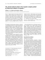
Does G4–6 interact with actin at acidic pH?
The above results suggest that the conformation of G4–6 at
acidic pH is significantly different from that induced by
calcium binding. Therefore, we tested the binding of G4–6
with G-actin by ELISA and fluorescence. G-actin was
coated onto plastic and increasing concentrations of G4–6
(between 0 and 0.6 l
M
) were added. Binding was monitored
by using specific G4–6 directed antibodies. We observed a
tight binding at pH 6.8 in the presence of 1 m
M
calcium
(apparent K
d
¼ 30 n
M
), and in the presence of 1 m
M
EGTA, no binding occured. In contrast, at pH 5.7 and
with 5 m
M
EGTA, a weak interaction was observed and a
K
d
of about 1 l
M
was estimated (Fig. 8).
Fig. 6. Binding of G2 domain to coated G4-6 monitored by ELISA.
Biotinylated G2 domain (0–0.8 l
M
)wasincubatedin5m
M
EGTA,
0.1
M
Mes buffer pH 5.7 (h)orpH6.8(j) in the presence of coated
G4–6. The interaction was monitored at 405 nm.
Table 1. Interaction of G4-6 with G-actin, G2 and derived fragments. ND, not determined; NI, no interaction; NS, no spectrum.
K
d
(l
M
)
EGTA (pH 5.7) EGTA (pH 6.8) EGTA (pH 7.5) Ca
2+
(pH 7.5)
Fluo(OG) ELISA Fluo(OG) ELISA Fluo(FITC) ELISA Fluo(FITC) ELISA
G2 0.5 0.15 NS 0.2 0.5
a
0.3
a
3
a
1
a
159–193 0.5 0.7 NS 0.7 1
a
1
a
2–3
a
2
a
197–226 2 ND NS ND 3
a
3
a
NI
a
NI
a
G-actin 1 1 NS NI NI NI 40 n
M
30 n
M
a
[22].
Fig. 8. Effect of pH on the binding of G4-6 to coated G-actin monitored
by ELISA. G4–6 was incubated at pH 7.5 in 0.1 m
M
ATP, 10 m
M
Tris
buffer containing either 1 m
M
CaCl
2
(d)or5m
M
EGTA (j)orat
pH 5.7 in 0.1 m
M
ATP, 100 m
M
Mes buffer containing 5 m
M
EGTA
(h) in the presence of coated G-actin. The interaction was monitored
at 405 nm.
Fig. 7. Binding of G4–6 domain with 159–193 and 197–226 fragments
derived from gelsolin G2 domain monitored by fluorescence measure-
ments. Interaction of Oregon green-labeled G4–6 domain (0.7 l
M
)
with Synthetic peptides 159–193 and 197–226 was carried out in 10 m
M
Mes buffer pH 5.7 in the presence of 5 m
M
EGTA. Change in fluor-
escence emission spectra of Oregon green was recorded at various
peptides concentrations: 0–7 l
M
for the 159–193 peptide (j)and
0–11 l
M
for the 197–226 peptide (h).
Ó FEBS 2003 Low pH activation of gelsolin (Eur. J. Biochem. 270) 4109
These data were confirmed by studies in solution using
fluorescence measurements. G4–6 domains were labeled by
Oregon green ITC and increasing concentrations of G-actin
were added (data not shown) we observed a decrease in the
fluorescence intensity of labeled G4–6 domains at pH 5.7 in
the presence of EGTA. Analysis of these data shows that
the fluorescence intensity decrease, extrapolated to infinite
concentration, is about 15%. An estimation of the apparent
K
d
(1 l
M
) can be obtained (Table 1). The same experiment
conducted in the presence of calcium at pH 6.8 shows that
the interaction of G-actin with Oregon green labeled G4–6
does not induce any spectral change. Consequently, further
analyses, using FITC-labeled G4–6, were carried out at
pH 7.5. Labeled G4–6 was incubated with increasing
concentrations of G-actin (between 0 and 0.2 l
M
)andthe
changes in fluorescence were monitored. Saturation curves
were observed in the presence of calcium and an apparent
K
d
¼ 40 n
M
was determined (Table 1). No binding was
observed at this pH when EGTA was present. These values
and those obtained from ELISA show a more pronounced
interaction of G-actin at neutral pH.
Studying the inhibitory effect of G4–6 on actin polymeri-
zation substantiated this important result. As shown in
Fig. 9A and B, equimolar addition of G4–6 to actin at
pH 5.7 produces only a small inhibition of the polymeriza-
tion process when compared with the control condition
(monitored at pH 7.5). These results suggest that the
conformation induced by lowering pH does not allow a
tight interaction of G4–6 with G-actin.
Discussion
The pH-dependence of the G2/G4–6 interface
Activation of gelsolin by calcium and/or low pH has been
found to be necessary for the binding of actin [21] and
similarly for the binding of tropomyosin [38]. Solving the
crystallographic solution of the whole gelsolin molecule in
its inactive state [4] led to the Ôhelix latchÕ hypothesis [5] in
which it is suggested that the C-terminal helix of gelsolin’s
G6 domain binds G2 and that this contact is released upon
the binding of calcium. We have further shown that residues
203–225 and 159–193 within G2 form the G6 binding site,
and that occupancy of a calcium binding site in G2 induces
conformational changes through this interface with a
calcium binding site in G6 that results in the release of the
latch [22]. A general unfolding of the molecule concomitant
with the release of the ÔlatchÕ between G2 and G4–6 is seen
as gelsolin is activated by calcium. During activation of
gelsolin by low pH treatment, a similar unfolding can be
recorded using dynamic light scattering [21] and it might be
assumed that a low pH also detaches the G2 to G4–6 latch.
The helix constitutes only a part of the switch that results in
full activation of gelsolin. Deletion of the last 23 residues
from the C-terminus of gelsolin for example, reduces the
requirement for calcium in activation but does not abolish it
totally [39]. Also, it is known that adseverin, a gelsolin
family member that naturally lacks the C-terminal helix has
a similar calcium requirement as the gelsolin mutant lacking
the helix [40]. However, both adseverin and gelsolin mutants
lacking the C-terminal 23 residues are equally activated by
lowered pH. Together, these observations suggest that the
calcium induced release of the helix latch may occur with
other rearrangements between domains caused either by the
occupancy by other calcium ions, or by the presence of
protons. We have shown that the helix latch and the G2 to
G4–6 interaction in general is not reduced by low pH. It is
possible therefore that in pH-activated gelsolin, the helical
latch may be the last event to occur being triggered not by
direct disruption of the interface but as a consequence of
the K
d
being rather large. At low pH, the helical latch
could be the rate limiting step in the kinetics of activation.
Alternatively, pH induced conformational changes within
or between other domains of gelsolin may strain the
G2/G4–6 interaction.
pH-dependent conformational changes in G2
Using tryptophan fluorescence as a probe we have been able
to show calcium dependent conformational changes with
G2 [22]. Using this same probe, we have now found a
pH-dependent conformational change in G2, however, this
change is in the opposite direction! This indicates that
although a pH-sensitive conformational change does occur
in G2 it is probably different than that induced by calcium.
This is in line with our finding that pH does not affect the
Fig. 9. Effect of G4-6 on actin polymerization. Pyrenyl actin (3 l
M
)was
mixedwith0.1mKCl,2m
M
MgCl
2
(A) in 5 m
M
EGTA, 10 m
M
Mes
(pH 5.7) or (B) in 0.4 m
M
CaCl
2
,10m
M
Tris buffer (pH 7.5) and the
polymerization was followed vs. time in the absence (––) or the pres-
ence (- - -) of 3 l
M
G4–6.
4110 E. Lagarrigue et al. (Eur. J. Biochem. 270) Ó FEBS 2003
G2/G4–6 interface, as we have previously found evidence
for a connectivity between the G2 calcium binding site
and the G6 site through the interface. Low pH-induced
conformational changes within G2 possibly reflect altered
association with G1 and/or G3, rather than G6 as occurs
in the presence of calcium.
pH-dependent conformational changes in G4–6
Some experimental data indicate that the calcium and
pH-induced conformational changes in G4–6 are similar.
The tryptophan fluorescence of G4–6 is lowered by
increasing pH value. Between pH values 5.7 and 6.2
qualitatively similar data were found across the same range
of pCa values [22]. Also, an increase in fluorescence of
FITC-labelled G4–6 [22] and of Oregon green-labelled
G4–6 was found in this range. Quenching experiments
(Fig. 4) show that tryptophans are similarly accessible in the
presence of calcium and at low pH (while tryptophans were
less exposed at neutral pH in EGTA), which also suggests
similar conformation.
pH dependence of gelsolin-actin binding
The gelsolin molecule forms a direct complex with two actin
monomers (GA
2
), through three distinct actin binding sites.
The GA
2
complex may not be equivalent to the geometry at
the barbed end of the capped actin filament, as the actins in
GA
2
are antiparallel [41]. We [29] and others [4,5,7,20], have
proposed various models for this interaction in which the
N-terminal gelsolin half (G1–3) binds a monomer through
sites in G1 and G2 and the C-terminal half (G4–6) binds a
second monomer in an analogous manner to G1 [42]. In
agreement with earlier work [43], we find that G4–6 inhibits
actin polymerization. In the presence of calcium, the
structure of G4–6 alone and G4–6 plus actin are so similar
[20] that it is suggested that calcium primes G4–6 for actin
binding, we have found that low pH removes (to some
extent) the requirement for calcium and so predict that at
low pH, G4–6 will adopt a similar structure to that found in
both the G4–6-actin structure and G4–6 in calcium alone.
The large difference in affinity for actin binding by pH-
activated (K
d
1m
M
) and calcium-activated (K
d
30–40 n
M
)
gelsolin is probably due to the type I calcium site coordi-
nated by both G4 and actin monomer [19]. These findings
explain why the formation of GA
2
isfastestatlowerpHin
thepresenceofcalcium[44].
A model for pH activation
Calcium binding by gelsolin domains generally breaks
interdomain salt bridge structures along the connecting
b-sheets and this results in domains moving a small
distance from each other having the overall effect of
enlarging the space occupied by the molecule [20]. In some
instances, pH changes may affect changes in the domain
through similar ion-pair swapping cascades proposed for
calcium binding [20], thereby activating gelsolin in a
similar manner to calcium. We suspect that one or more
of the many histidines in the core of the gelsolin molecules
may sense low pH conditions and initiate an ion-pair
swapping cascade. We have found however, that pH
changes cannot substitute for every calcium site, as
important differences exist between pH- and calcium-
induced conformational changes. If this model is correct,
pH is expected to affect calcium binding, as for some sites,
low pH should result in ion-pair swapping that would
substitute for calcium binding.
Although intracellular pH is held close to neutral in most
cell types, cell signalling involving significant changes in
global intracellular pH are well documented [45,46], and
these are often exaggerated in cell subdomains [47]. In
addition to gelsolin, other actin-binding proteins such as the
ADF/cofilins [48] and EF1a [49] are strongly pH-dependent
so it seems probable that pH transients that occur in cells
may act in part through the actin cytoskeleton.
We have shown that there is a clear difference between
calcium- and pH-induced activation of gelsolin. Future
challenges are to determine how low pH-induced conform-
ational change compares to calcium-induced conforma-
tional change and if there are differences in the properties of
pH and calcium activated gelsolin. Also it is necessary to
determine if low pH affects the various calcium binding sites
in gelsolin.
Acknowledgements
This research was supported by grants from AFM. We thank
Dr Paul McLaughlin ICMB, University of Edinburgh for very helpful
discussion.
References
1. dos Remedios, C.G., Chhabra, D., Kekic, M., Dedova, I.V.,
Tsubakihara, M., Berry, D.A. & Noseworthy, N.J. (2003) Actin
binding proteins: regulation of cytoskeletal microfilaments. Phy-
siol. Rev. 83, 433–473.
2. Sun, H.Q., Yamamoto, M., Mejillano, M. & Yin, H.L. (1999)
Gelsolin, a multifunctional actin regulatory protein. J. Biol. Chem.
274, 33179–33182.
3. Way, M. & Weeds, A.G. (1988) Nucleotide sequence of pig
plasma gelsolin. Comparison of protein sequence with human
gelsolin and other actin-severing proteins shows strong homo-
logies and evidence for large internal repeats. J. Mol. Biol. 203,
1127–1133.
4. Burtnick,L.D.,Koepf,E.K.,Grimes,J.,Jones,E.Y.,Stuart,D.I.,
McLaughlin,P.J.&Robinson,R.C.(1997)Thecrystalstructure
of plasma gelsolin: Implications for actin severing, capping and
nucleation. Cell. 90, 661–670.
5. Robinson, R.C., Mejillano, M., Le, V.P., Burtnick, L.D., Yin,
H.L. & Choe, S. (1999) Domain movement in gelsolin: a calcium-
activated switch. Science. 286, 1939–1942.
6. Bryan, J. (1988) Gelsolin has three actin-binding sites. J. Cell Biol.
106, 1553–1562.
7. Pope, B., Way, M. & Weeds, A.G. (1991) Two of the three actin-
binding domains of gelsolin bind to the same subdomain of actin.
FEBS Lett. 280, 70–74.
8. Pope, B., Maciver, S. & Weeds, A.G. (1995) Localization of the
calcium-sensitive actin monomer binding site in gelsolin to seg-
ment 4 and identification of calcium-binding sites. Biochemistry.
34, 1583–1588.
9. Xu, Y S., Kantorow, M., Davis, J. & Piatigorsky, J. (2000) Evi-
dence for gelsolin as a corneal crystallin in zebrafish. J. Biol. Chem.
275, 24645–24652.
Ó FEBS 2003 Low pH activation of gelsolin (Eur. J. Biochem. 270) 4111
10. Steed, P.M., Nagar, S. & Wennogle, L.P. (1996) Phospholipase D
regulation by a physical interaction with the actin-binding protein
gelsolin. Biochemistry. 35, 5229–5237.
11. Ohtsu, M., Sakai, N., Fujita, H., Kashiwagi, M., Gasa, S., Shi-
mizu, S., Eguchi, Y., Tsujimoto, Y., Sakiyama, Y., Kobayashi, K.
& Kuzumaki, N. (1997) Inhibition of apoptosis by the actin-
regulatory protein gelsolin. EMBO J. 16, 4650–4656.
12. Harris, H.E., Bamburg, J.R. & Weeds, A.G. (1980) Actin filament
disassembly in blood plasma. FEBS Lett. 123, 49–53.
13. Haddad, J.G., Harper, K.D., Guoth, M., Pietra, G.G. & Sanger,
J.W. (1990) Angiogenic consequences of saturating the plasma
scavenger system for actin. Proc. Nat. Acad. Sci. 87, 1381–1385.
14. Cunningham, C.C., Stossel, T.P. & Kwiatkowski, D.J. (1991)
Enhanced motility in NIH 3T3 fibroblasts that overexpress gel-
solin. Science. 251, 1233–1236.
15. Cunningham, C.C., Vegners, R., Bucki, R., Funaki, M., Korde,
N., Hartwig, J.H., Stossel, T.P. & Janmey, P.A. (2001) Cell per-
meant polyphsophoinositide-binding peptides that block cell
motililty and actin assembly. J. Biol. Chem. 276, 43390–43399.
16. Witke, W., Sharpe, A.H.H., Artwig, J.H., Azuma, T., Stossel, T.P.
& Kwiatkowski, D.J. (1995) Hemostatic, inflammatory and
fibroblast responses are blunted in mice lacking gelsolin. Cell. 81,
41–51.
17. Patkowski, A., Seils, J., Hinssen, H. & Dorfmuller, T. (1990) Size,
shape parameters and calcium-induced conformational change of
the gelsolin molecule. A dynamic light scattering study. Biopoly-
mers. 30, 427–435.
18. Hellweg, T., Hinssen, H. & Eimer, W. (1993) The Ca
2+
-induced
conformational change of gelsolin is located in the carboxyl-ter-
minalhalfofthemolecule.Biophys. J. 65, 799–805.
19. Choe, H., Burtnick, L.D., Mejillano, M., Yin, H.L., Robinson,
R.C. & Choe, S. (2002) The calcium activation of gelsolin: Insights
from the 3A
˚
structure of the G4–G6/actin complex. J. Mol. Evol.
324, 691–702.
20. Kolappan, S.P., Gooch, J.T., Weeds, A.G. & McLaughlin, P.J.
(2003) Gelsolin domains 4–6 in active, actin free conformation
identifies sites of regulatory calcium ions. J. Mol. Biol. 329, 85–92.
21. Lamb, J.A., Allen, P.G., Tuan, B.Y. & Janmey, P.A. (1993)
Modulation of gelsolin function – activation at low pH overrides
Ca
2+
requirement. J. Biol. Chem. 268, 8999–9004.
22. Lagarrique, E., Maciver, S.K., Fattoum, A., Benyamin, Y. &
Roustan, C. (2003) Co-operation of domain-binding and calcium-
binding sites in the activation of gelsolin. Eur. J. Biochem. 270,
2236–2243.
23. Spudich, J.A. & Watt, S. (1971) The regulation of rabbit skeletal
muscle contraction. Biochemical studies of the interaction of the
tropomyosin-troponin complex with actin and the proteolytic
fragments of myosin. J. Biol. Chem. 246, 4866–4871.
24. Kouyama, T. & Mihashi, K. (1981) Fluorimetry study of N-(1-
pyrenyl) iodoacetamide-labelled F-actin. Eur. J. Biochem. 114,
33–38.
25. Way,M.,Pope,B.,Gooch,J.,Hawkins,M.&Weeds,A.G.(1990)
Identification of a region of segment 1 of gelsolin critical for actin
binding. EMBO J. 9, 4103–4109.
26. Way, M., Pope, B. & Weeds, A.G. (1992) Evidence for functional
homology in the F-actin binding domains of gelsolin and alpha-
actinin: implications for the requirements of severing and capping.
J. Cell Biol. 119, 835–842.
27. Way, M., Gooch, J., Pope, B. & Weeds, A.G. (1989) Expression of
human plasma gelsolin in E. coli and dissection of actin binding
sites by segmental deletion mutagenesis. J. Cell Biol. 109, 593–605.
28. Kurokawa, H., Fujii, W., Ohmi, K., Sakurai, T. & Nonomura, Y.
(1990) Simple and rapid purification of brevin. Biochem. Biophys.
Res. Comm. 168, 451–457.
29. Renoult, C., Blondin, L., Fattoum, A., Ternent, D., Maciver,
S.K., Raynaud, F., Benyamin, Y. & Roustan, C. (2001) Binding of
gelsolin domain 2 to actin. An actin interface distinct from that of
gelsolin domain 1 and from ADF/cofilin. Eur. J. Biochem. 268,
6165–6175.
30. Miki, M. & dos Remedios, C.G. (1988) Fluorescence quenching
studies of fluorescein attached to Lys-61 or Cys-374 in actin:
effects of polymerization, myosin subfragment-1 binding, and
tropomyosin-troponin binding. J. Biochem. 104, 232–235.
31. Papa, I., Astier, C., Kwiatek, O., Lebart, M C., Raynaud, F.,
Benyamin, Y. & Roustan, C. (1999) Use of a chaotropic anion
iodide in the purification of Z-line proteins: isolation of CapZ.
from fish white muscle. Prot. Expr Purif. 17,1–7.
32. Engvall, E. (1980) Enzyme immunoassay ELISA and EMIT.
Meth. Enzymol. 70, 419–439.
33. Me
´
jean, C., Lebart, M.C., Poyer, M., Roustan, C. & Benyamin,
Y. (1992) Localization and identification of actin structures
involved in the filamin–actin interaction. Eur. J. Biochem. 209,
555–562.
34. Lakowicz, J.R. (1983) Principles of Fluorescence Spectroscopy.
Plenum Publishing Corp, New York.
35. Gill, S.C. & von Hippel, P.H. (1989) Calculation of protein
extinction coefficients from amino acid sequence data. Anal. Bio-
chem. 182, 319–326.
36. Houmeida, A., Hanin, V., Feinberg, J., Benyamin, Y. & Roustan,
C. (1991) Definition of a Ca
2+
-sensitive interface in the plasma
gelsolin-actin complex. Biochem. J. 274, 753–757.
37. Lin, K M., Mejillano, M. & Yin, H.L. (2000) Ca
2+
regulation of
gelsolin by its C-terminal tail. J.Biol. Chem. 275, 27746–27752.
38. Maciver, S.K., Ternent, D. & McLaughlin, P.J. (2000) Domain 2
of gelsolin binds directly to tropomyosin. FEBS Lett. 473, 71–75.
39. Kwiatkowski, D.J., Janmey, P.A. & Yin, H.L. (1989) Identifica-
tion of critical functional and regulatory domains in gelsolin.
J. Cell Biol. 108, 1717–1726.
40. Lueck, A., Yin, H.L., Kwiatkowski, D.J. & Allen, P.G. (2000)
Calcium regulation of gelsolin and adseverin: a natural test of the
helix latch hypothesis. Biochemistry. 39, 5274–5279.
41. Hesterkamp, T., Weeds, A.G. & Mannherz, H.G. (1993) The actin
monomers in the ternary gelsolin: 2 actin complex are in anti-
parallel orientation. Eur. J. Biochem. 218, 507–513.
42. McLaughlin, P.J., Gooch, J.T., Mannherz, H.G. & Weeds, A.G.
(1993) Structure of gelsolin segment-1-actin complex and the
mechanism of filament severing. Nature. 364, 685–692.
43. Chaponnier, C., Janmey, P.A. & Yin, H.L. (1986) The actin fila-
ment-severing domain of plasma gelsolin. J. Cell Biol. 103,
1473–1481.
44. Selve, N. & Wegner, A. (1987) pH-dependent rate of formation of
the gelsolin-actin complex from gelsolin and monomeric actin.
Eur. J. Biochem. 161, 111–115.
45. Watanabe, K., Hamaguchi, M.S. & Hamaguchi, Y. (1997) Effects
of intracellular pH on the mitotic apparatus and mitoitc stage in
the sand dollar egg. Cell Mot. Cytoskel. 37, 263–270.
46. Van Duijn, B. & Inouye, K. (1991) Regulation of movement speed
by intracellular pH during Dictyostelium discoideum chemotaxis.
Proc.Nat.Acad.Sci.88, 4951–4955.
47. Schwiening, C.J. & Willoughby, D. (2002) Depolarization-induced
pH microdomains and their relationship to calcium transients in
isolated snail neurones. J. Physiol. 538, 371–382.
48. Yonezawa, N., Nishida, E. & Sakai, H. (1985) pH control of actin
polymerization by cofilin. J. Biol. Chem. 260, 14410–14412.
49. Edmonds, B.T., Murray, J. & Condeelis, J. (1995) pH regulation
of the F-actin binding properties of Dictyostelium elongation
factor 1a. J. Biol. Chem. 270, 15222–15230.
4112 E. Lagarrigue et al. (Eur. J. Biochem. 270) Ó FEBS 2003