A linear epitope coupled to DsRed provides an affinity ligand for the capture of monoclonal antibodies
Bạn đang xem bản rút gọn của tài liệu. Xem và tải ngay bản đầy đủ của tài liệu tại đây (2.47 MB, 10 trang )
Journal of Chromatography A, 1571 (2018) 55–64
Contents lists available at ScienceDirect
Journal of Chromatography A
journal homepage: www.elsevier.com/locate/chroma
A linear epitope coupled to DsRed provides an affinity ligand for the
capture of monoclonal antibodies
C. Rühl a , M. Knödler a , P. Opdensteinen a , J.F. Buyel a,b,∗
a
b
Fraunhofer Institute for Molecular Biology and Applied Ecology IME, Forckenbeckstraße 6, 52074 Aachen, Germany
Institute for Molecular Biotechnology, Worringerweg 1, RWTH Aachen University, 52074 Aachen, Germany
a r t i c l e
i n f o
Article history:
Received 3 June 2018
Received in revised form 19 July 2018
Accepted 5 August 2018
Available online 7 August 2018
Keywords:
Affinity chromatography
Design of experiments
Fluorescent protein carrier
HIV-neutralizing monoclonal antibody
Plant molecular farming
Transient protein production
a b s t r a c t
Monoclonal antibodies (mAbs) dominate the market for biopharmaceutical proteins because they provide
active and passive immunotherapies for many different diseases. However, for most mAbs, two expensive
manufacturing platforms are required. These are mammalian cell cultures for upstream production and
Protein A chromatography for product capture during downstream processing. Here we describe a novel
affinity ligand based on the fluorescent protein DsRed as a carrier for the linear epitope ELDKWA, which
can capture the HIV-neutralizing antibody 2F5. We produced the DsRed-2F5-Epitope (DFE) in transgenic
tobacco (Nicotiana tabacum) plants and purified it using a combination of heat treatment and immobilized
metal-ion affinity chromatography, resulting in a yield of 24 mg kg−1 at 90% purity. Using a design-ofexperiments approach, we coupled up to 15 mg DFE per mL Sepharose. The resulting affinity resin was
able to capture 2F5 from the clarified extract of N. benthamiana plants, achieving a purity of 97%, a
recovery of >95% and an initial dynamic binding capacity at 10% product breakthrough of 4 mg mL−1 after
a contact time of 2 min. The resin capacity declined to 15% of the starting value within 25 cycles when
1.25 M magnesium chloride was used for elution. We confirmed the binding activity of the 2F5 product
by surface plasmon resonance spectroscopy. DFE is not yet optimized, and a cost analysis revealed that
boosting DFE expression and increasing its capacity by fourfold will make the resin cost-competitive with
some Protein A counterparts. The affinity resin can also be exploited to purify idiotype-specific mAbs.
© 2018 The Author(s). Published by Elsevier B.V. This is an open access article under the CC
BY-NC-ND license ( />
1. Introduction
Antibodies dominate the biopharmaceutical market, with more
than 50 approved products and more than 300 candidates in the
development pipeline [1]. The total sales volume was more than
D 40 billion in 2013, which is about 33% of all biopharmaceutical
protein sales. Most products are monoclonal antibodies (mAbs) that
are typically produced in mammalian cells, such as Chinese hamster
ovary (CHO) cells, with titers regularly exceeding ∼5 g L−1 in the
culture supernatant [2]. Despite these high product titers, upstream
Abbreviations: CV, column volume; DoE, design of experiments; IMAC, immobilized metal-ion affinity chromatography; SPR, surface plasmon resonance; TSP, total
soluble protein.
∗ Corresponding author at: Fraunhofer Institute for Molecular Biology and Applied
Ecology IME, Forckenbeckstraße 6, 52074 Aachen, Germany.
E-mail addresses: (C. Rühl),
,
(M. Knödler), (P. Opdensteinen),
(J.F. Buyel).
production in mammalian cells is expensive due to the cost of media
and the need for sterile conditions. Alternative expression systems
are therefore being investigated, including yeast such as Pichia pastoris [3] and plants, the latter offering a scalable and safe production
platform [4]. Plant-derived mAbs have already been tested in clinical trials, including the HIV-neutralizing mAb 2G12 [5].
Regardless of the expression host, another major cost driver for
mAb manufacturing is the reliance of most processes on a Protein A
capture step, which has become the gold standard for initial purification [6]. Although the production of this protein-based affinity
ligand in bacterial systems is cost-effective, the resin is nevertheless expensive given the need for qualification before its use in
processes that comply with good manufacturing practices (GMP)
and also the substantial margin which reflects the lack suitable
alternatives. Depending on the production scale, the costs for the
resin alone can amount to 10 million euros (assuming 6 × 15,000-L
bioreactors, and a 10-ton output of mAb product per year) [7]. This
corresponds to more than 25% of the total process costs [8]. The
impact of the Protein A resin on the cost of goods is one reason for
the high market prices, often exceeding 2000 euros per g purified
/>0021-9673/© 2018 The Author(s). Published by Elsevier B.V. This is an open access article under the CC BY-NC-ND license ( />4.0/).
56
C. Rühl et al. / J. Chromatogr. A 1571 (2018) 55–64
mAb [9]. Such prices are a major burden for healthcare systems and
can be prohibitive in developing countries, especially if large doses
of product are required. For example, up to 12 g of mAb per patient
is required for a lymphoma therapy [10], and up to 3 g annually per
person for a prophylactic anti-HIV treatment [5]. Therefore, several
inexpensive non-protein ligands have been developed that could in
principle replace Protein A [11]. Many of them preferentially target the constant regions of mAbs, e.g. the MEP ligand binds to the
CH2 domain [12], facilitating rapid process development due to the
uniform elution conditions [13]. However, the performance of such
alternative resins in terms of recovery and purity has been inconsistent compared to Protein A, e.g. both high and low purities have
been reported following mAb elution from MEP [14–16], whereas
>95% purity is typically achieved when using Protein A [17,18].
Here we have developed an alternative approach for the affinity purification of mAbs based on the use of linear epitopes, in this
case ELDKWA (one-letter amino acid code) for the HIV-neutralizing
antibody 2F5 [19,20]. We fused this epitope to the fluorescent
protein DsRed [21] as a carrier, generating the fusion protein
DsRed-2F5-Epitope (DFE). We then produced DFE in transgenic
tobacco (Nicotiana tabacum) plants and purified it by singlestep immobilized metal-ion affinity chromatography (IMAC). We
optimized the coupling of DFE to a Sepharose resin using a designof-experiments (DoE) approach, resulting in a novel affinity resin
which we used to purify mAb 2F5 (transiently expressed in N. benthamiana) from clarified leaf extracts. We discuss the optimization
of elution conditions and provide an initial cost evaluation, compared with a Protein A-based process counterpart.
construct using either the vacuum infiltration method [29] or manual injection into leaves [30]. Whole plants or leaf sections were
infiltrated with A. tumefaciens (OD600nm = 1.0) in infiltration buffer
(0.5 g L−1 Fertilizer MEGA 2 (Planta Düngemittel GmbH, Regenstauf,
Germany), 200 M acetosyringone, pH 5.6) and cultivated for a
further 5 days before harvesting [30].
2.4. Protein extraction and clarification
Proteins were extracted from plants by blade-based homogenization in 3 mL extraction buffer (50 mM sodium phosphate,
500 mM sodium chloride, 10 mM sodium bisulfite, pH 8.0) per gram
wet biomass, followed by clarification using a sequence of bag,
depth and sterile filters [31]. Tobacco extracts containing DFE were
heat treated before clarification [28].
2.5. Immobilized metal-ion affinity chromatography
DFE was purified by immobilized metal-ion affinity chromatography (IMAC) on an ÄKTApure system (GE Healthcare, Little
Chalfont, UK) using an XK-26 column containing 53 mL of chelating Sepharose fast flow IMAC resin loaded with nickel ions. After
loading the clarified extract onto a conditioned column (extraction
buffer without sodium bisulfite), the resin was washed with 10 column volumes (CVs) of buffer without imidazole followed by elution
in buffer containing 300 mM imidazole at a flow rate of 50 cm h−1 .
The concentrations of protein and nucleic acid were monitored at
280 and 260 nm, respectively.
2. Materials and methods
2.1. Design of experiments
2.6. Coupling DFE to Sepharose resin
Design Expert v10 (Stat-Ease, Minneapolis, MN, USA) was used
to set up and evaluate all experimental designs. The factors and
levels are presented in the supplementary data (Table S1), and the
detailed DoE method is discussed elsewhere [22].
The purified DFE affinity ligand was immobilized on HiTrap
NHS-activated [32] Sepharose HP columns (GE Healthcare) with a
bed volume of 1 mL. Before coupling, the columns were washed
with 6 mL ice-cold 1 mM hydrochloric acid at a flow rate of
<1 mL min−1 . Immediately after washing, 1.5 CVs of affinity ligand solution (0.15–15 mg mL−1 ) were injected using a 2-mL syringe
(Braun, Melsungen, Germany), and the flow-through fractions
were monitored using a TE6101 precision scale (Sartorius, Göttingen, Germany). The columns were then sealed and incubated for
15–45 min at 22 ◦ C, followed by thorough washing to remove residual NHS esters. This involved three cycles of washing, first with
6 mL of deactivation solution (0.5 M ethanolamine, 0.5 M sodium
chloride, pH 8.3) injected at a flow rate of <1 mL min−1 followed
by 6 mL of a low-pH solution (0.1 M sodium acetate, 0.5 M sodium
chloride, pH 4.0). The columns were left for 15 min after the third
washing cycle and were then stored in 0.05 M disodium phosphate
containing 0.1% (m/v) sodium azide (pH 7.0) at 4 ◦ C. For the simultaneous washing of multiple columns, an Ismatec IPC 24-channel
peristaltic pump (Cole-Parmer GmbH, Wertheim, Germany) was
used at a constant flow rate of 0.6 mL min−1 . The coupling procedure required ∼2 h in total.
2.2. Expression vectors and bacterial cultures
The nucleotide sequence of DsRed (a red fluorescent protein
from Discosoma sp. [23]) was extended by PCR using appropriate primers to add the sequence encoding the ELDKWA epitope
(to which mAb 2F5 binds) at the 3 end. The resulting construct
was transferred to vector pTRA for expression [24], yielding the
DFE fusion protein consisting of DsRed, the 2F5 epitope, a His6 tag
and a KDEL sequence for retention in the endoplasmic reticulum
(Fig. S1). The coding sequences for the heavy and light chains of
mAb 2F5 [19] were cloned as individual expression cassettes and
were also introduced into pTRA [25]. Accordingly, the expression
of all polypeptides was driven by the double enhanced Cauliflower
mosaic virus 35S promoter. The vectors for DFE and mAb 2F5
were introduced separately into Agrobacterium tumefaciens strain
GV3101:pMP90RK by electroporation. The DFE construct was used
to generate transgenic tobacco (N. tabacum) plants and the 2F5 construct was used for transient expression in N. benthamiana leaves
as described below. A homology model of the 3D structure of DFE
based on 1ZGO [26] was built using 3D-JIGSAW (ck.
ac.uk/∼populus/) [27].
2.3. Plant material, infiltration and expression
Transgenic tobacco plants expressing DFE were generated as
previously described [28]. For transient expression, N. benthamiana plants were infiltrated with A. tumefaciens carrying the 2F5
2.7. Affinity resin characterization and purification of mAb 2F5
DFE-coupled columns were mounted on an ÄKTApure system
and equilibrated with 5 CVs of equilibration buffer at a flow rate
of 1 mL min−1 . Up to 80 mL of extract containing 2F5 was loaded
onto the column at a rate of 0.5 mL min-1 ensuring a contact time of
2 min. The columns were washed with 6 CVs of equilibration buffer
before eluting 2F5 in 5 CVs of elution buffer with low pH (0.05 M
citrate, 0.05 M sodium chloride, pH 4.0–3.25) or slightly alkaline pH
(1.0–4.0 M magnesium chloride, 0.1 M HEPES, pH 8.0). The theoret-
C. Rühl et al. / J. Chromatogr. A 1571 (2018) 55–64
ical static binding capacity of the affinity resin was calculated based
on the immobilized amount of DFE using Eq. (1).
SBC theor. =
Mw,mAb
mDFE
×
Mw,DFE
Vresin
(1)
where SBCtheor is the theoretical static binding capacity [g L−1 ],
Mw,mAb is the molar mass of mAb 2F5 (154.6 kDa), Mw,DFE is he
molar mass of the DFE monomer (28.4 kDa), mDFE is the immobilized mass of DFE (3–10 mg), and Vresin is the column volume
(1 mL).
Approximately 80 mL of clarified plant extract containing 2F5
was loaded under the same conditions as above to obtain sigmoidal
breakthrough curves. The volume at which 10% of the plateau product concentration was detected in the flow-through fraction was
multiplied by the product concentration in the load to determine
the dynamic binding capacity at 10% product breakthrough.
2.8. Protein quantitation and activity testing
The concentration of total soluble protein (TSP) was determined
using a microtiter version of the Bradford method as described
before [33] and the sample protein composition was analyzed
by staining lithium dodecylsulfate (LDS) polyacrylamide gels with
Coomassie Brilliant Blue [29]. DFE and 2F5 were quantified by
fluorescence spectroscopy and surface plasmon resonance (SPR)
spectroscopy, respectively [34]. The amount of protein per gram
wet biomass was calculated as described elsewhere [35]. The
binding of DFE-purified 2F5 (eluted by pH shift or the addition
of magnesium chloride) to the 13.5-kDa trimeric HIV-1 fusion
inhibitor Fuzeon (enfuvirtid) containing the 2F5 epitope (Roche,
Basel, Switzerland) was used to assess the binding activity of 2F5.
Approximately 270 response units (RU) of 2F5 were captured on a
Protein A chip using a BIAcore T200 instrument (GE Healthcare) at
25 ◦ C in HEPES-buffered saline containing 0.05% (v/v) Tween-20 as
a running buffer. Eight dilutions of Fuzeon in the 0.16–20.00 nM
range were injected individually and captured by 2F5 bound to
Protein A. The kinetic binding constants ka , kd and kD were calculated based on a 1:1 stoichiometric model using the BIAevaluation
software (GE Healthcare).
3. Results and discussion
3.1. The DFE fusion protein is expressed at high levels in plants
and can be purified easily
The 28.4-kDa fusion protein DFE (Fig. S1) was expressed with
a yield of ∼120 mg kg−1 leaf biomass, equivalent to ∼42 mg L−1
extract (Fig. 1A), which is in the middle range compared to
other recombinant proteins expressed in transgenic tobacco, e.g.
0.9 mg kg−1 for mAb CO17-1 A [36], ∼500 mg kg−1 for mAb M12,
and ∼400 mg kg−1 of unmodified DsRed [31]. The purity of DFE in
the crude extract was <5% of TSP, but our DoE approach revealed
that blanching the tobacco leaves at 70 ◦ C for 1.5 min before extraction increased the purity to almost 40% because most of the host
cell proteins (HCPs) were precipitated (Fig. 1B, Fig. S2, Table S2).
Approximately 50% of the product was lost, regardless of the
blanching temperature, resulting in the recovery of ∼65 mg kg−1
(∼22 mg L−1 ). These results were in good agreement with previous studies using heat precipitation, indicating that more than
90% of the TSP can be removed by blanching prior to chromatography [28,37]. Removing HCPs early in a process can prevent
product degradation, as shown for other fusion proteins transiently
expressed in N. benthamiana [29]. We therefore used blanching for
all subsequent DFE purifications despite the product loss and the
availability of an affinity-based purification step (IMAC), given the
57
latter can also capture nonspecific plant HCPs [35,38]. After homogenization and the removal of coarse particles using a polypropylene
needle-felt bag filter, a PDH4 two-layer depth filter (nominal pore
sizes of ∼10 m and ∼1 m) was used to clarify the plant extract,
achieving an average capacity of 135 ± 36 L m-2 (±SD, n = 3) and a
product recovery of ∼70% up to this step, which was equivalent to
45 mg kg−1 biomass (15 mg L−1 ). These values were in good agreement with previous studies, which reported capacities of ∼70 L m-2
and recoveries of ∼75% [31]. The use of filter layers lacking diatomaceous earth may improve DFE recovery, as previously shown for
a multi-domain fusion protein [39]. Subsequent DFE purification
by IMAC on a resin containing Ni2+ increased the purity of DFE to
almost 90% (Fig. 1A), a typical purity achieved for plant-derived
recombinant proteins when using this technique [40–42]. The target protein concentration in the elution fraction was 20-fold higher
than in the load, but the recovery (based on fluorescence analysis)
was only 55%, corresponding to an overall yield of 23.5 mg kg−1 and
substantial fluorescence was observed in the flow-through fractions. However, western blots of these fractions (Fig. 1B) did not
reveal detectable amounts of DFE when using a primary antibody
directed against the C-terminal His6 tag of the fusion protein. We
speculate that at least the C-terminal His6 and KDEL parts of the
fusion protein were cleaved off either in planta or after extraction,
which explains the presence of DFE variants in the flow-through
fractions because they will not have been able to bind the IMAC
resin. Similar degradation effects have been reported for mAbs and
vaccine candidates expressed in plants [29,39,43] and we are currently investigating this phenomenon in more detail.
3.2. DFE can be coupled to Sepharose resin at a loading of up to
7 mg mL-11
In an initial screen, we determined the quantity of DFE that can
be coupled to NHS-activated HiTrap columns and found that the
coupling efficiency declined from 80 to 90% to <70% when we used
more than 15 mg DFE per milliliter resin (Fig. 1C). Interestingly,
we found that HEPES buffer, instead of the bicarbonate buffer recommended by the manufacturer, increased the average coupling
efficiency from 78 ± 9% (±SD, n = 3) to 89 ± 6 (±SD, n = 3) at pH 8.3.
We also observed a more intense red color at the top of the column when HEPES was used instead of bicarbonate, indicating that
the coupling capacity became saturated with less DFE ligand in the
presence of bicarbonate (Fig. S3A). The pka values of carbonic acid
are 3.6 and 10.3 [44], implying that at pH 8.3 most of the bicarbonate
buffer molecules should be present in the hydrogen carbonate form
(HCO3 –) and only a small amount in the carbonate form (CO3 2– ).
We speculate that the free electron pairs in these species may allow
them to act as nucleophiles, which compete with the amino groups
of the protein for interaction with the activated NHS esters as previously reported for other functional groups [45]. HEPES buffer was
therefore used in all subsequent experiments.
We then used a DoE approach to optimize the conditions for DFE
coupling (Table S1) and found that the amount of fusion protein
bound to the column increased as more DFE was brought into contact with the resin, reaching a plateau at ∼15 mg DFE per milliliter
resin and resulting in ∼10 mg of bound DFE, or ∼0.35 mol mL−1
(Fig. 2A). However, if more than 10 mg DFE was brought into contact with the resin, the coupling efficiency dropped from ∼90%
to less than 50%, depending on the pH (Fig. 2B). Also, increasing
the amount of coupled DFE increased the cost per column because
more purified fusion protein was consumed (Fig. 2C). We therefore
used the numerical optimization tool built into the DoE software
to identify the ideal conditions for DFE coupling, i.e. the conditions
combining high coupling efficiency, the greatest quantity of coupled DFE and the lowest costs, giving each optimization criterion
an equal weighting. These conditions were best met by coupling
58
C. Rühl et al. / J. Chromatogr. A 1571 (2018) 55–64
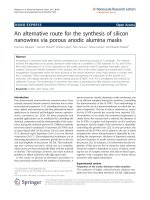
Fig. 1. DFE expression, purification and coupling. (A) DFE concentration and purity as a fraction of the total soluble protein in untreated plant extracts (control) and after
blanching of the leaf material (Hom, homogenate) as well as in the subsequent clarification and purification steps (Adj – pH adjusted, Bag – bag filtrate, DF – depth filtrate,
Load – filter-sterilized extract loaded onto the IMAC column, FT start – initial flow-through fraction, FT pool – pooled flow-through fractions). (B) LDS-PAGE analysis (top)
and western blot (bottom) of samples from panel A. The dominant plant host cell proteins (RuBisCO large and small subunits) are highlighted by green arrows whereas
the DFE product is indicated by red arrows. Note the apparent oligomerization of DFE despite the denaturing and reducing conditions. (C) Coupling efficiency of DFE to
NHS-activated Sepharose HP as a function of the injected amount of purified DFE. DFE concentrations were determined based on fluorescence analysis (circles) and Bradford
assay (diamonds) results for verification purposes.
7.0 mg of DFE at pH 9.0 for 45 min. We identified a broad and largely
pH-independent desirability plateau in the range 6–12 mg mL−1
resin DFE loading (Fig. 2D), which made the coupling a robust process. Interestingly, the fusion protein retained its red color even
after coupling and the inactivation of unused interaction sites, and
the color correlated with the absolute amount of DFE bound to the
resin (Fig. 2E). This indicated that DFE was present in the native
tetrameric state of DsRed despite the low-pH inactivation step
(pH 4.0) which was previously found to cause the denaturation of
DsRed and a near permanent loss of fluorescence [46]. The color of
the resin could therefore be used for quality control during later
manufacturing stages.
Based on the tetrameric structure of DFE [21], its molecular mass
of 28,411 g mol−1 and the coupled mass of up to ∼10 g L−1 resin, we
calculated the ligand density of the resulting affinity resin using
Eq. (1). The predicted value of 0.35 mol mL−1 was about 0.4% of
the 50–250 mol mL−1 reported for ion-exchange resins [47], but
was similar in magnitude to other affinity resins such as Protein A
(2–11 g L−1 ) [48]. The effective number of 2F5-epitope domains on
the fusion protein that are available for 2F5 binding may be lower
due to (i) steric hindrance resulting from the binding orientation
of the coupled DFE molecules, (ii) the direct involvement of the
epitope’s lysine residue in the coupling reaction, and (iii) shielding
of the epitopes by mAbs bound to adjacent ligands.
One option to reduce column costs in the future, especially
when high concentrations of DFE are needed for coupling to NHSactivated resin, is the recycling of uncoupled DFE recovered during
resin inactivation after coupling. For example we recovered ∼2 mg
(∼13%) of DFE when loading 15 mg of the fusion protein per
milliliter resin.
3.3. Magnesium chloride is a suitable replacement for the low-pH
elution of 2F5
We used DFE columns with ∼7 mg coupled fusion protein to
capture mAb 2F5 from a clarified plant extract (Fig. S3B). We then
tested a low-pH elution approach as used with Protein A and found
that greater quantities of 2F5 were released as the pH fell below
4.5 (Fig. 3A). The highest antibody recovery of ∼35% (91% purity)
was achieved at pH 3.25 according to western blot analysis and
densitometry, but when we analyzed the same samples by SPR
spectroscopy we found that the mAb eluted at this pH was unable
to bind to the Protein A surface of the sensor chip. We concluded
that 2F5 was probably irreversibly denatured during elution at pH
3.25 but that higher-pH elution conditions were uneconomical due
to the even lower mAb recovery. Furthermore, we observed that
the distinct red color of the column resulting from DFE coupling
faded as the elution pH fell below 5.0 (Fig. 3B). We attributed this
effect to the denaturation of the fusion protein, which has been
reported for DsRed at pH < 4.0 [21,49]. Although the 2F5 epitope
is linear [19] and should therefore be detected by 2F5 even after
denaturation, we speculate that the conformational change might
reduce the binding capacity of the resin because polypeptide chains
of the DFE tetramer that had not been covalently linked to the resin
matrix may dissociate into the liquid phase, reducing the number
of epitope ligands in the column. Indeed we found that the resin
capacity fell to zero after three cycles of elution at pH 3.0.
C. Rühl et al. / J. Chromatogr. A 1571 (2018) 55–64
59
Fig. 2. DFE coupling efficiency to NHS-activated Sepharose resin. (A) Absolute amount of coupled DFE, showing the dependence on pH and the DFE mass brought in contact
with the activated resin. (B) Coupling yield of DFE calculated as the fraction of fluorescence remaining on the column, showing the dependence on pH and the DFE mass
brought in contact with the activated resin. (C) Column costs based on the amount of immobilized DFE and the manufacturing costs for the affinity ligand as well as the
activated resin. (D) Desirability of coupling conditions, showing the dependence on pH and the DFE mass. The optimization target was a combination of a large quantity
of coupled DFE, a high coupling efficiency, and low costs, with each optimization criterion given equal weighting. The optimal condition is highlighted by a red dot. (E)
Photographs of columns containing the DFE affinity resin after coupling. The numbers beneath the photographs correspond to the conditions highlighted in panels A C.
Table 1
Kinetic parameters and absolute binding capacity of mAb 2F5 transiently expressed in N. benthamiana and purified by DFE or Protein A affinity chromatography.
Purification
Units
Protein A
DFE (pH)
DFE (magnesium chloride)
Protein A (tobacco)a
Protein A (CHO)a
RmAb
RFuzeon
Mr,mAb
Mr,Fuzeon
Absolute activity
kon
koff
KD
KD
[RU]
[RU]
[Da]
[Da]
[-]
[M−1 s−1 ]
[s−1 ]
[M]
[pM]
272.0
45.7
154,600
13,476
0.96
2.93 × 10⁶
2.24 × 10⁻³
7.63 × 10⁻¹⁰
763
270.0
45.1
154,600
259.0
47.1
154,600
326.5
54.0
159,383
331.5
55.8
150,814
0.95
2.97 × 10⁶
2.51 × 10⁻³
8.43 × 10⁻¹⁰
843
1.04
2.86 × 10⁶
2.26 × 10⁻³
7.91 × 10⁻¹⁰
791
0.98
6.30 × 10⁶
1.80 × 10⁻³
2.94 × 10⁻¹⁰
294
0.94
5.40 × 10⁶
1.90 × 10⁻³
3.66 × 10⁻¹⁰
366
a
values according to [25].
We therefore tested magnesium chloride as an alternative elution agent because it has been used to elute antibodies from other
affinity resins [50–52]. In an initial test, we found that 1.0 M magnesium chloride predominantly eluted nonspecifically bound HCPs,
whereas 2.0 M magnesium chloride was sufficient for the complete
elution of 2F5 (Fig. 3C). Interestingly, 4.0 M magnesium chloride
for elution caused similar color fading as observed for the lowpH elution (Fig. 3D). In a subsequent refinement we observed that
even 1.25 M magnesium chloride was sufficient to elute 2F5 from
the DFE columns and the antibody was consistently detected by
western blotting and SPR spectroscopy (Fig. 4A). Under these conditions, we achieved 105 ± 11% recovery (±SD, n = 3) and 97 ± 3%
purity (±SD, n = 3) (Fig. S3C). Although we achieved a similar purity
(∼96%) using Protein A, the recovery of 2F5 dropped to only ∼50%
when it was eluted in citrate buffer at pH 3.0. However, recoveries
of ∼90% [53] and purities of >95% [17] have been reported for other
antibodies. We assumed that 2F5 is sensitive to acidic denaturation,
and therefore determined the binding constants for the interaction between 2F5 and the synthetic trimeric peptide Fuzeon, which
contains the 2F5 epitope [25], following the purification of 2F5 by
conventional Protein A chromatography, DFE affinity chromatography with elution at pH 4.0, and the same technique with elution in
1.25 M magnesium chloride. The absolute activity in all three preparations was high (Table 1) and similar to those reported previously
[25]. Values above unity may reflect protein glycosylation, which
we did not investigate in this study, hence their exclusion from our
calculations. In contrast, the kon we observed was only half of that
reported for mAb 2F5 expressed in either CHO cells or tobacco, possibly reflecting the different host species and expression platform.
However, all three preparations showed similar kinetic parameters, so we concluded that the purification methods did not have a
negative impact on the functionality of 2F5.
60
C. Rühl et al. / J. Chromatogr. A 1571 (2018) 55–64
Fig. 3. Elution of 2F5 from the DFE affinity resin using low-pH buffer or magnesium chloride. (A) Total soluble protein and 2F5 concentrations in pH elution fractions after
DFE affinity purification as determined using the Bradford assay and SPR spectroscopy, respectively. (B) Photographs of DFE columns following exposure to different pH
buffers and for repeated bind-and-elute cycles. (C) Total soluble protein and 2F5 concentrations in magnesium chloride elution fractions after DFE affinity purification as
determined using the Bradford assay and SPR spectroscopy, respectively. (D) Photographs of DFE columns after exposure to different magnesium chloride concentrations
and for repeated bind-and-elute cycles.
DFE affinity capture also achieved a log reduction value of ∼3 for
HCPs, similar to the value reported for Protein A [53]. This may indicate that DFE and Protein A resins have similar levels of selectivity.
However, the HCP concentration in our load was 59 mg mg−1 mAb
and thus 200-fold higher than for typical CHO-based processes for
the manufacturing of mAbs [17].
3.4. The dynamic binding capacity remains at 15% after 25
bind-and-elute cycles
The initial specific DBC10% of the affinity resin for mAb 2F5 was
0.70 mg 2F5 per immobilized mg of DFE, or ∼4 mg mL−1 resin. This
was determined using optimized elution conditions combined with
loading at pH 7.5 in 0.05 M phosphate buffer, a conductivity of
∼48 mS cm−1 , a residence time of 2 min, and a linear flow rate of
75 cm h−1 . The DBC10% value corresponded to ∼12.5% of the theoretical static binding capacity calculated based on the amount of
coupled DFE (Fig. 4B) and was ∼13% of the 25–60 mg mL−1 recently
reported for novel Protein A resins under similar conditions [17,48]
but similar to the 0.76–4.80 mg mL−1 observed for other custom
resins [54]. Over the course of 25 cycles, the DBC10% of the DFE
resin declined linearly (adj. R2 = 0.99) to 0.10 mg mg−1 (∼15% of the
initial value) (Fig. 4C).
We speculate that the observed loss in DBC10% was due to the
loss of DFE molecules that were not covalently bound to the base
matrix but were only retained on the column through association with other DFE molecules forming the characteristic DsRed
tetramer [21]. This limitation could therefore be addressed by using
a monomeric derivative of DsRed as an epitope carrier.
3.5. The potential benefits of linear epitope ligands can outweigh
the current drawbacks compared to Protein A
We used our DFE expression levels and the simple onestep purification procedure as input parameters for a previously
reported cost model [34] to estimate the production effort for DFE,
and combined these results with the costs of affinity resin manufacturing to enable a cost comparison with Protein A. The current cost
per run for the DFE affinity resin was found to be ∼170-fold higher
than a conventional Protein A resin, particularly reflecting the lower
DBC10% and fewer re-use cycles (Table 2). However, the Protein A
resin selected for comparison represents more than 45 years of
intensive development [55,56]. We therefore performed an effect
analysis for the DFE resin costs including potential improvements
to the resin that seemed within reach given the current body of data.
Based on the latest reports of high level protein expression in plants
[57], we predict that DFE expression can be increased to 2.0 g kg−1
biomass, which will reduce the production costs for the ligand by
more than 85% per unit mass. The costs can also be reduced through
an increase in DFE recovery during purification, which could be
achieved by optimizing the blanching procedure to reduce proteolytic degradation or thermal denaturation [39], both of which
we observed for DFE (Fig. 1). We predict that these measures
would increase the DFE recovery factor from 0.5 to 0.7. Furthermore, increasing the ligand density can in some cases improve the
DBC10% as shown for ion-exchange resins [58,59]. However, when
we investigated the size of the DFE–2F5 complex compared to the
typical pore diameter of ∼80 nm reported for Sepharose HP resin
[60], we found that the complex is ∼29 nm in diameter in its most
C. Rühl et al. / J. Chromatogr. A 1571 (2018) 55–64
61
Fig. 4. DFE resin characteristics. (A) Typical chromatogram of a bind-and-elute cycle for a DFE affinity column used to capture mAb 2F5 from a clarified plant extract. The
axis dimensions of the inset are the same as in the main panel. (B) Breakthrough curves of mAb 2F5 using DFE affinity resin after multiple bind-and-elute cycles. (C) Dynamic
binding capacity for 10% product breakthrough compared to the load referring to the amount of immobilized DFE. (D) Schematic representation of the DFE (red) 2F5 (green)
complex at full extension in an idealized pore with circular perimeter and a pore radius (rpore ) of 40 nm. The 2F5 epitope (orange) is indicated by an orange arrow, and the
theoretical minimal effective remaining pore radius (rmin,eff ) is shown by a size bar. The resulting minimal pore size is shown as a gray circle.
Table 2
Calculation of DFE affinity resin costs compared to Protein A, including two hypothetical scenarios for feasible improvements of the DFE setup assuming the immobilization
of 7 mg DFE per milliliter of resin.
Setup
Protein A
DFE
Parameter
Unit
Current
Moderate improvement
Substantial improvement
Expression level
Recovery
Ligand production costs
Base resin costs
Coupling costs
Ligand costs
Affinity resin costs
DBC10% b
Resin volume for 1 g mAb purification
Resin costs
Resin life time
Resin costs per run
[g kg−1 ]
[-]
[D g−1 ]
[D L−1 ]
[D L−1 ]
[D L−1 ]
[D L−1 ]
[g L−1 ]
[L]
[D ]
[cycles]
[D g−1 mAb]
0.12
0.50
1960
6800
220
13,720
20,740
3
0.25
5185
6
864
0.50
0.60
495
3400
220
3468
7089
10
0.10
709
20
35
2.00
0.70
198
1700
220
1389
3309
15
0.07
221
50
4
a
b
15,000a
30a
0.03
500
100
5
Values according to [69].
DBC10% – dynamic binding capacity at 10% product breakthrough.
extended state, leaving only an effective minimal pore radius of
∼11 nm for protein diffusion into and out of the resin pores, which
would be too small for additional antibodies to pass (Fig. 4D). Even
though the orientation of the complex is flexible and not all complexes will be present in the most extended form, this may limit the
effective binding capacity. Others have reported a pore blocking
effect for ion-exchange ligand densities exceeding 400 mol g−1
[59] and we assume that such an effect would occur at lower
densities for DFE due to the larger size of the affinity ligand. Furthermore, increasing the ligand density above 50 mol mL−1 does
not improve the DBC10% [47]. Therefore, densities in the 2–11 g L−1
range as for Protein A are more likely to be effective [48] and matrices with larger pore sizes for DFE affinity resin preparation may
help to improve ligand access and thus the binding capacity. The
use of recently-developed monomeric variants of the DsRed carrier protein [61,62] might reduce the loss of DFE ligands due to
the wash-out of non-covalently bound molecules from tetrameric
DFE, which we speculate is one reason for the declining capacity
we observed over several bind-and-elute cycles. These monomeric
variants of the DsRed carrier have also been designed for minimal
cytotoxicity, enabling them to be used widely for the analysis of
protein localization and interaction in living cells, so they should
62
C. Rühl et al. / J. Chromatogr. A 1571 (2018) 55–64
not trigger regulatory concerns in the context of process-related
impurities [63,64]. Protein engineering may also facilitate rational
increases in the stability of DFE, as achieved for Protein A [65,66],
and may alter the preferred coupling orientation of the DFE ligand
[67]. The latter can increase the likelihood that the 2F5 epitope is
exposed to the center of the resin pores and may thus facilitate
antibody binding, resulting in a higher binding capacity. A similar effect could be achieved by increasing the number of repeats
of the 2F5 epitope on the DFE C-terminus, as demonstrated for
Protein A [53]. Furthermore, increasing the current contact time
from 2 to 4 min could double the DBC10% as reported for several
Protein A resins [68]. We speculate that these modifications could
cumulatively increase the DBC10% from currently 4 g L−1 to 15 g L−1
(which is about half of the DBC10% of Protein A [69]) and facilitate
50 instead of 6 cycles of the affinity resin. By gradually incorporating these modifications in our cost calculations, we find that the
DFE resin can become competitive with a Protein A-based counterpart (Table 2). Even with moderate DFE production cost savings and
small increases in column performance, the price for the base resin
was the major cost driver (Fig. S4). We predict that bulk production of the affinity resin would reduce the base matrix price by up
to 75%, which would reduce the cost of goods for the DFE resin to
D 35 per gram of antibody for the moderate improvement scenario
and to
A. In addition to the direct resin costs, DFE may also be economically advantageous because the amount of 2F5 recovered was about
twice that achieved during conventional Protein A chromatography.
Cost benefits aside, the DFE resin has the general advantage
that only mAbs specific for the epitope will be purified. This
feature could be exploited to facilitate the purification of certain idiotype-specific antibodies from a polyclonal mixture or to
improve in-process quality by ensuring that only mAb isoforms
with a functional antigen-binding moiety are enriched. Furthermore, antibody derivatives that lack the Fc component (e.g. the
scFv, Fab and diabody formats) can be purified using this new
approach, and by combining two epitope-based affinity ligands in a
two-stage bind-and-elute process, bispecific antibodies could also
be purified from a bulk extract or cell culture supernatant containing a mixture of monospecific and bispecific mAbs. Additionally,
mAbs containing Fc domains which exhibit only a weak interaction with Protein A or do not bind to the resin (e.g. human IgA and
IgG3 or mouse IgG1) can easily be purified using DFE or similar
ligands carrying the according epitope.
So far, we have shown that DFE has the potential compete with
Protein A or provide novel purification modes. It will be interesting
to investigate how well the epitope-fusion approach can be transferred to other mAbs with linear epitopes, given that the expression
levels of new affinity ligand proteins may vary depending on the
nature of the epitope sequence. However, we have worked with
several DsRed fusion proteins in the past 20 years, and have regularly achieved expression levels exceeding 100 mg kg−1 biomass
[70], making it likely that novel epitope fusion proteins can be
expressed at similarly high levels. Furthermore, given that transient protein expression in plants has a gene-to-product timescale
of only 2–4 weeks [71], it should be possible to prepare individual resins for mAbs binding to different epitopes. The sequence of
the linear epitope must be known in order to generate such novel
affinity–ligand fusion proteins, but this should not require further
work because sequence characterization is typically required as
part of regular product and process development, not only due
to regulatory requirements but also to ensure freedom to operate
and to prevent legal issues [72,73]. Even if epitope characterization
is not part of the process development, a DsRed–epitope fusion
protein library can be generated rapidly using techniques such as
random-primer PCR combined with appropriate scaffolds to identify suitable affinity ligands.
4. Conclusions
We have shown that the fluorescent protein DsRed can be
used as a carrier for antibody epitopes, resulting in fusion protein
expression levels exceeding 0.1 g kg−1 biomass. The subsequent
purification of DFE was simplified by the incorporation of blanching and IMAC steps, facilitating the cost-effective production of a
novel affinity ligand. The optimized coupling procedure ensured
a DBC10% that was only one order of magnitude lower than the
well-established industry standard Protein A. Moderate improvements in expression, purification and coupling could make DFE
economically competitive with Protein A, and its engagement with
epitope-specific contacts (paratopes) on the antibody means that
DFE and similar ligands would be particularly beneficial when
dealing with mixtures of different antibodies, such as those encountered during the manufacturing of bispecific mAbs. Our future work
will focus on the further improvement of DFE stability, epitope
density and binding affinity.
Acknowledgements
The authors acknowledge Ibrahim Al Amedi for cultivating the
plants used in this investigation and Dr. Thomas Rademacher for
providing the pTRA vector. We are grateful to Markus Sack for fruitful discussions on the DFE ligand structure. We wish to thank Dr.
Richard M Twyman for editorial assistance. This work was funded
by the Fraunhofer-Gesellschaft Internal Programs, Germany under
Grant No. Attract 125-600164. The authors have no conflicts of
interest to declare.
Appendix A. Supplementary data
Supplementary material related to this article can be found, in
the online version, at doi: />08.014.
References
[1] D.M. Ecker, S.D. Jones, H.L. Levine, The therapeutic monoclonal antibody
market, mAbs 7 (2015) 9–14.
[2] R.A. Rader, E.S. Langer, 30 years of upstream productivity improvements,
Bioprocess Int. 13 (2015) 10–14.
[3] O. Purcell, P. Opdensteinen, W. Chen, K. Lowenhaupt, A. Brown, M. Hermann,
J. Cao, N. Tenhaef, E. Kallweit, R. Kastilan, A.J. Sinskey, P. Perez-Pinera, J.F.
Buyel, T.K. Lu, Production of functional anti-ebola antibodies in pichia
pastoris, ACS Synth. Biol. 6 (2017) 2183–2190.
[4] J.F. Buyel, R.M. Twyman, R. Fischer, Very-large-scale production of antibodies
in plants: the biologization of manufacturing, Biotechnol. Adv. 35 (2017)
458–465.
[5] J.K. Ma, J. Drossard, D. Lewis, F. Altmann, J. Boyle, P. Christou, T. Cole, P. Dale,
C.J. van Dolleweerd, V. Isitt, D. Katinger, M. Lobedan, H. Mertens, M.J. Paul, T.
Rademacher, M. Sack, P.A. Hundleby, G. Stiegler, E. Stoger, R.M. Twyman, B.
Vcelar, R. Fischer, Regulatory approval and a first-in-human phase I clinical
trial of a monoclonal antibody produced in transgenic tobacco plants, Plant
Biotechnol. J. 13 (2015) 1106–1120.
[6] C.M.C. The, Biotech Working, Group, A-Mab: a case study in bioprocess
development, A-Mab: a Case Study in Bioprocess Development, CASSS - An
International Separation Science Society (2009) 1–278.
[7] B. Kelley, Very large scale monoclonal antibody purification: the case for
conventional unit operations, Biotechnol. Prog. 23 (2007) 995–1008.
[8] M. Pathak, G. Ma, D.G. Bracewell, A.S. Rathore, Re-use of protein a resin fouling
and economics, BioPharm International, BioPharm Int. 28 (2015) 28–33.
[9] B. Kelley, Industrialization of mAb production technology: the bioprocessing
industry at a crossroads, mAbs 1 (2009) 443–452.
[10] P. Chames, M. Van Regenmortel, E. Weiss, D. Baty, Therapeutic antibodies:
successes, limitations and hopes for the future, Br. J. Pharmacol. 157 (2009)
220–233.
[11] S. Kabir, Immunoglobulin purification by affinity chromatography using
protein a mimetic ligands prepared by combinatorial chemical synthesis,
Immunol. Invest. 31 (2002) 263–278.
C. Rühl et al. / J. Chromatogr. A 1571 (2018) 55–64
[12] D.Q. Lin, H.F. Tong, H.Y. Wang, S.J. Yao, Molecular insight into the ligand-IgG
interactions for 4-mercaptoethyl-pyridine based hydrophobic
charge-induction chromatography, J. Phys. Chem. B 116 (2012) 1393–1400.
[13] S. Ghose, M. Allen, B. Hubbard, C. Brooks, S.M. Cramer, Antibody variable
region interactions with Protein A: implications for the development of
generic purification processes, Biotechnol. Bioeng. 92 (2005) 665–673.
[14] V.B. Brochier, V. Ravault, High throughput development of a non protein A
monoclonal antibody purification process using mini-columns and bio-layer
interferometry, Eng. Life Sci. 16 (2015) 152–159.
[15] T. Arakawa, M. Futatsumori-Sugai, K. Tsumoto, Y. Kita, H. Sato, D. Ejima, MEP
HyperCel chromatography II: binding, washing and elution, Protein Express.
Purif. 71 (2010) 168–173.
[16] S. Ghose, B. Hubbard, S.M. Cramer, Evaluation and comparison of alternatives
to Protein A chromatography Mimetic and hydrophobic charge induction
chromatographic stationary phases, J. Chromatogr. A 1122 (2006) 144–152.
[17] T.M. Pabst, R. Palmgren, A. Forss, J. Vasic, M. Fonseca, C. Thompson, W.K.
Wang, X. Wang, A.K. Hunter, Engineering of novel Staphylococcal Protein A
ligands to enable milder elution pH and high dynamic binding capacity, J.
Chromatogr. A 1362 (2014) 180–185.
[18] R.L. Fahrner, D.H. Whitney, M. Vanderlaan, G.S. Blank, Performance
comparison of protein A affinity-chromatography sorbents for purifying
recombinant monoclonal antibodies, Biotechnol. Appl. Biochem. 30 (Pt 2)
(1999) 121–128.
[19] T. Muster, F. Steindl, M. Purtscher, A. Trkola, A. Klima, G. Himmler, F. Rüker, H.
Katinger, A conserved neutralizing epitope on gp41 of human
immunodeficiency virus type 1, J. Virol. 67 (1993) 6642–6647.
[20] C.E. Parker, L.J. Deterding, C. Hager-Braun, J.M. Binley, N. Schulke, H. Katinger,
J.P. Moore, K.B. Tomer, Fine definition of the epitope on the gp41 glycoprotein
of human immunodeficiency virus type 1 for the neutralizing monoclonal
antibody 2F5, J. Virol. 75 (2001) 10906–10911.
[21] G.S. Baird, D.A. Zacharias, R.Y. Tsien, Biochemistry, mutagenesis, and
oligomerization of DsRed, a red fluorescent protein from coral, Proc. Natl.
Acad. Sci. U. S. A. 97 (2000) 11984–11989.
[22] J.F. Buyel, R. Fischer, Characterization of complex systems using the design of
experiments approach: transient protein expression in tobacco as a case
study, J. Vis. Exp. 1 (2014) e51216.
[23] M.V. Matz, A.F. Fradkov, Y.A. Labas, A.P. Savitsky, A.G. Zaraisky, M.L. Markelov,
S.A. Lukyanov, Fluorescent proteins from nonbioluminescent Anthozoa
species, Nat. Biotechnol. 17 (1999) 969–973.
[24] M. Sack, T. Rademacher, H. Spiegel, A. Boes, S. Hellwig, J. Drossard, E. Stoger, R.
Fischer, From gene to harvest: insights into upstream process development
for the GMP production of a monoclonal antibody in transgenic tobacco
plants, Plant Biotechnol. J. 13 (2015) 1094–1105.
[25] D.M. Floss, M. Sack, J. Stadlmann, T. Rademacher, J. Scheller, E. Stoger, R.
Fischer, U. Conrad, Biochemical and functional characterization of anti-HIV
antibody-ELP fusion proteins from transgenic plants, Plant Biotechnol. J. 6
(2008) 379–391.
[26] J.L. Tubbs, J.A. Tainer, E.D. Getzoff, Crystallographic structures of Discosoma
red fluorescent protein with immature and mature chromophores: linking
peptide bond trans-cis isomerization and acylimine formation in
chromophore maturation, Biochemistry-Us 44 (2005) 9833–9840.
[27] P.A. Bates, L.A. Kelley, R.M. MacCallum, M.J. Sternberg, Enhancement of
protein modeling by human intervention in applying the automatic programs
3D-JIGSAW and 3D-PSSM, Proteins (Suppl. 5) (2001) 39–46.
[28] J.F. Buyel, H.M. Gruchow, A. Boes, R. Fischer, Rational design of a host cell
protein heat precipitation step simplifies the subsequent purification of
recombinant proteins from tobacco, Biochem. Eng. J. 88 (2014) 162–170.
[29] S. Menzel, T. Holland, A. Boes, H. Spiegel, J. Bolzenius, R. Fischer, J.F. Buyel,
Optimized blanching reduces the host cell protein content and substantially
enhances the recovery and stability of two plant-derived malaria vaccine
candidates, Front. Plant Sci. 7 (2016) 1–15.
[30] J.F. Buyel, T. Kaever, J.J. Buyel, R. Fischer, Predictive models for the
accumulation of a fluorescent marker protein in tobacco leaves according to
the promoter/5’UTR combination, Biotechnol. Bioeng. 110 (2013) 471–482.
[31] J.F. Buyel, R. Fischer, Scale-down models to optimize a filter train for the
downstream purification of recombinant pharmaceutical proteins produced
in tobacco leaves, Biotechnol. J. 9 (2014) 415–425.
[32] P. Cuatrecasas, I. Parikh, Adsorbents for affinity chromatography. Use of
N-hydroxysuccinimide esters of agarose, Biochemistry-Us 11 (1972)
2291–2299.
[33] J.F. Buyel, R. Fischer, Flocculation increases the efficacy of depth filtration
during the downstream processing of recombinant pharmaceutical proteins
produced in tobacco, Plant Biotechnol. J. 12 (2014) 240–252.
[34] J.F. Buyel, R. Fischer, Predictive models for transient protein expression in
tobacco (Nicotiana tabacum L.) can optimize process time, yield, and
downstream costs, Biotechnol. Bioeng. 109 (2012) 2575–2588.
[35] B.B. Gengenbach, C.R. Müschen, J.F. Buyel, Expression and purification of
human phosphatase and actin regulator 1 (PHACTR1) in plant-based systems,
Protein Express. Purif. 151 (2018) 46–55.
[36] K. Ko, Z. Steplewski, M. Glogowska, H. Koprowski, Inhibition of tumor growth
by plant-derived mAb, Proc. Natl. Acad. Sci. U. S. A. 102 (2005) 7026–7030.
[37] J.F. Buyel, J. Hubbuch, R. Fischer, Comparison of tobacco host cell protein
removal methods by blanching intact plants or by heat treatment of extracts,
J. Vis. Exp. e54343 (2016) 9.
[38] J.A. Bornhorst, J.J. Falke, Purification of proteins using polyhistidine affinity
tags, Methods Enzymol. 326 (2010) 245–254.
63
[39] S. Menzel, T. Holland, A. Boes, H. Spiegel, R. Fischer, J.F. Buyel, Downstream
processing of a plant-derived malaria transmission-blocking vaccine
candidate, Protein Expr. Purif. 27 (July (152)) (2018) 122–130, .
org/10.1016/j.pep.2018.07.012 [Epub ahead of print] PMID: 30059744.
[40] J.F. Buyel, J.A. Bautista, R. Fischer, V.M. Yusibov, Extraction, purification and
characterization of the plant-produced HPV16 subunit vaccine candidate E7
GGG, J. Chromatogr. B 880 (2012) 19–26.
[41] F. Sainsbury, P.V. Jutras, J. Vorster, M.C. Goulet, D. Michaud, A chimeric affinity
tag for efficient expression and chromatographic purification of heterologous
proteins from plants, Front. Plant Sci. 7 (2016) 141.
[42] Y. Kim, G. Babnigg, R. Jedrzejczak, W.H. Eschenfeldt, H. Li, N. Maltseva, C.
Hatzos-Skintges, M.Y. Gu, M. Makowska-Grzyska, R.Y. Wu, H. An, G. Chhor, A.
Joachimiak, High-throughput protein purification and quality assessment for
crystallization, Methods 55 (2011) 12–28.
[43] M. Benchabane, C. Goulet, D. Rivard, L. Faye, V. Gomord, D. Michaud,
Preventing unintended proteolysis in plant protein biofactories, Plant
Biotechnol. J. 6 (2008) 633–648.
[44] N.N. Greenwood, A. Earnshaw, Chemistry of the Elements, 2 ed., Elsevier, New
York, NY, 1997.
[45] S. Madler, C. Bich, D. Touboul, R. Zenobi, Chemical cross-linking with NHS
esters: a systematic study on amino acid reactivities, J. Mass Spectrom. 44
(2009) 694–706.
[46] A. Sacchetti, V. Subramaniam, T.M. Jovin, S. Alberti, Oligomerization of DsRed
is required for the generation of a functional red fluorescent chromophore,
FEBS Lett. 525 (2002) 13–19.
[47] A.M. Hardin, C. Harinarayan, G. Malmquist, A. Axen, R. van Reis, Ion exchange
chromatography of monoclonal antibodies: effect of resin ligand density on
dynamic binding capacity, J. Chromatogr. A 1216 (2009) 4366–4371.
[48] A.D. Tustian, L. Laurin, H. Ihre, T. Tran, R. Stairs, H. Bak, Development of a
novel affinity chromatography resin for platform purification of bispecific
antibodies with modified protein a binding avidity, Biotechnol. Prog.
(Feburary) (2018), [Epub ahead of print]
PMID: 29464924.
[49] P.V. Vrzheshch, N.A. Akovbian, S.D. Varfolomeyev, V.V. Verkhusha,
Denaturation and partial renaturation of a tightly tetramerized DsRed protein
under mildly acidic conditions, FEBS Lett. 487 (2000) 203–208.
[50] V.C. Tsang, P.P. Wilkins, Optimum dissociating condition for immunoaffinity
and preferential isolation of antibodies with high specific activity, J. Immunol.
Methods 138 (1991) 291–299.
[51] A. Ben-David, M.A. Firer, Immunoaffinity purification of monoclonal
antibodies In search of an elution buffer of general applicability, Biotechnol.
Tech. 10 (1996).
[52] M.A. Firer, Efficient elution of functional proteins in affinity chromatography,
J. Biochem. Biophys. Methods 49 (2001) 433–442.
[53] E. Muller, J. Vajda, Routes to improve binding capacities of affinity resins
demonstrated for Protein A chromatography, J. Chromatogr. B 1021 (2016)
159–168.
[54] T. Besselink, M. Liu, M. Ottens, R. van Beckhoven, A.E. Janssen, R.M. Boom,
Comparison of activated chromatography resins for protein immobilization, J.
Sep. Sci. 36 (2013) 1185–1191.
[55] H. Hjelm, K. Hjelm, J. Sjöquist, Protein A from Staphylococcus aureus. Its
isolation by affinity chromatography and its use as an immunosorbent for
isolation of immunoglobulins, FEBS Lett. 28 (1972) 73–76.
[56] G.R. Bolton, K.K. Mehta, The role of more than 40 years of improvement in
protein A chromatography in the growth of the therapeutic antibody
industry, Biotechnol. Prog. 32 (2016) 1193–1202.
[57] J. Zischewski, M. Sack, R. Fischer, Overcoming low yields of plant-made
antibodies by a protein engineering approach, Biotechnol. J. 11 (2015)
107–116.
[58] A. Franke, N. Forrer, A. Butte, B. Cvijetic, M. Morbidelli, M. Johnck, M. Schulte,
Role of the ligand density in cation exchange materials for the purification of
proteins, J. Chromatogr. A 1217 (2010) 2216–2225.
[59] H.L. Lu, D.Q. Lin, M.M. Zhu, S.J. Yao, Effects of ligand density and pore size on
the adsorption of bovine IgG with DEAE ion-exchange resins, J. Sep. Sci. 35
(2012) 2131–2137.
[60] B.C. To, A.M. Lenhoff, Hydrophobic interaction chromatography of proteins. I.
The effects of protein and adsorbent properties on retention and recovery, J.
Chromatogr. A 1141 (2007) 191–205.
[61] R.E. Campbell, O. Tour, A.E. Palmer, P.A. Steinbach, G.S. Baird, D.A. Zacharias,
R.Y. Tsien, A monomeric red fluorescent protein, Proc. Natl. Acad. Sci. U. S. A.
99 (2002) 7877–7882.
[62] T.M. Wannier, M.M. Moore, Y. Mou, S.L. Mayo, Computational Design of the
beta-Sheet Surface of a Red Fluorescent Protein Allows Control of Protein
Oligomerization, PLoS One 10 (2015), e0130582.
[63] R.L. Strack, D.E. Strongin, D. Bhattacharyya, W. Tao, A. Berman, H.E.
Broxmeyer, R.J. Keenan, B.S. Glick, A noncytotoxic DsRed variant for
whole-cell labeling, Nat. Methods 5 (2008) 955–957.
[64] I.I. Shemiakina, G.V. Ermakova, P.J. Cranfill, M.A. Baird, R.A. Evans, E.A.
Souslova, D.B. Staroverov, A.Y. Gorokhovatsky, E.V. Putintseva, T.V.
Gorodnicheva, T.V. Chepurnykh, L. Strukova, S. Lukyanov, A.G. Zaraisky, M.W.
Davidson, D.M. Chudakov, D. Shcherbo, A monomeric red fluorescent protein
with low cytotoxicity, Nat. Commun. 3 (2012) 1204.
[65] K. Minakuchi, D. Murata, Y. Okubo, Y. Nakano, S. Yoshida, Remarkable alkaline
stability of an engineered protein A as immunoglobulin affinity ligand: C
domain having only one amino acid substitution, Protein Sci. 22 (2013)
1230–1238.
64
C. Rühl et al. / J. Chromatogr. A 1571 (2018) 55–64
[66] S. Hober, K. Nord, M. Linhult, Protein A chromatography for antibody
purification, J. Chromatogr. B 848 (2007) 40–47.
[67] O. Koniev, A. Wagner, Developments and recent advancements in the field of
endogenous amino acid selective bond forming reactions for bioconjugation,
Chem. Soc. Rev. 44 (2015) 5495–5551.
[68] R. Hahn, P. Bauerhansl, K. Shimahara, C. Wizniewski, A. Tscheliessnig, A.
Jungbauer, Comparison of protein A affinity sorbents II. Mass transfer
properties, J. Chromatogr. A 1093 (2005) 98–110.
[69] B. Lain, Protein a, Bioprocess Int. 11 (2013) 29–38.
[70] A. Boes, H. Spiegel, N. Voepel, G. Edgue, V. Beiss, S. Kapelski, R. Fendel, M.
Scheuermayer, G. Pradel, J.M. Bolscher, M.C. Behet, K.J. Dechering, C.C.
Hermsen, R.W. Sauerwein, S. Schillberg, A. Reimann, R. Fischer, Analysis of a
multi-component multi-stage malaria vaccine candidate–tackling the cocktail
challenge, Plos One 10 (2015), e0131456.
[71] Y. Shoji, J.A. Chichester, M. Jones, S.D. Manceva, E. Damon, V. Mett, K.
Musiychuk, H. Bi, C. Farrance, M. Shamloul, N. Kushnir, S. Sharma, V. Yusibov,
Plant-based rapid production of recombinant subunit hemagglutinin vaccines
targeting H1N1 and H5N1 influenza, Hum. Vaccin. Immunother. 7 (2011)
41–50.
[72] X. Deng, U. Storz, B.J. Doranz, Enhancing antibody patent protection using
epitope mapping information, mAbs 10 (2018) 204–209.
[73] C.G. Sandercock, U. Storz, Antibody specification beyond the target: claiming
a later-generation therapeutic antibody by its target epitope, Nat. Biotechnol.
30 (2012) 615–618.