Application of steric exclusion chromatography on monoliths for separation and purification of RNA molecules
Bạn đang xem bản rút gọn của tài liệu. Xem và tải ngay bản đầy đủ của tài liệu tại đây (1.86 MB, 10 trang )
Journal of Chromatography A, 1574 (2018) 50–59
Contents lists available at ScienceDirect
Journal of Chromatography A
journal homepage: www.elsevier.com/locate/chroma
Application of steric exclusion chromatography on monoliths for
separation and purification of RNA molecules
Alesia Levanova, Minna M. Poranen ∗
Molecular and Integrative Biosciences Research Programme, Faculty of Biological and Environmental Sciences, University of Helsinki, Biocenter 1, PL 56
(Viikinkaari 9), 000140 University of Helsinki, Finland
a r t i c l e
i n f o
Article history:
Received 4 June 2018
Received in revised form 22 August 2018
Accepted 31 August 2018
Available online 31 August 2018
Keywords:
Nucleic acids
RNA
Steric exclusion chromatography
CIM monolithic columns
Polyethylene glycol (PEG)
a b s t r a c t
Steric exclusion chromatography (SXC) is a method for separation of large target solutes based on their
association with a hydrophilic stationary phase through mutual steric exclusion of polyethylene glycol
(PEG). Selectivity in SXC is determined by the size or shape (or both) of the solutes alongside the size and
concentration of PEG molecules. Elution is achieved by decreasing the PEG concentration. In this study,
SXC applicability for the separation and purification of single-stranded (ss) and double-stranded (ds)
RNA molecules was evaluated for the first time. The retention of ssRNA and dsRNA molecules of different
lengths on convective interaction media (CIM) monolithic columns was systematically studied under
variable PEG-6000 and NaCl concentrations. We determined that over 90% of long ssRNAs (700–6374
nucleotides) and long dsRNAs (500–6374 base pairs) are retained on the stationary phase in 15% PEG6000 and ≥0.4 M NaCl. dsDNA and dsRNA molecules of the same length were partially separated by
SXC. Separation of RNA molecules below 100 nucleotides from longer RNA species is easily achieved
by SXC. Furthermore, SXC has the potential to separate dsRNAs from ssRNAs of the same length. We
also demonstrated that SXC is suitable for the enrichment of ssRNA (PRR1 bacteriophage) and dsRNA
(Phi6 bacteriophage) viral genomes from contaminating cellular RNA species. In summary, SXC on CIM
monolithic columns is an appropriate tool for rapid RNA separation and concentration.
© 2018 The Authors. Published by Elsevier B.V. This is an open access article under the CC BY license
( />
1. Introduction
Ribonucleic acid (RNA) and deoxyribonucleic acid (DNA) are
macromolecules built from nucleotides, which consist of a heterocyclic nitrogenous base, a phosphate, and a pentose sugar. In
aqueous solutions, duplex DNA is primarily in B-conformation,
while RNA is mostly a single-stranded molecule adopting complex
three-dimensional structures. When RNA duplexes are observed,
they are predominantly in an A-form [1]. DNA and RNA differ
in one of four bases; DNA contains thymine, while RNA contains
uracil, a demethylated derivative of thymine. Furthermore, the
pentose sugar in RNA is ribose but deoxyribose in DNA, which is
derived from ribose by the loss of a 2’ oxygen atom [2]. This loss
provides DNA with the stability necessary to fulfill its main biological function as a keeper of genetic information [3]. One of the
numerous functions of RNA molecules is to serve as a mediator
in the flow of genetic information from DNA to proteins. Furthermore, RNA itself carries genetic information in many viruses
either as a single-stranded (ssRNA) or a double-stranded (dsRNA)
∗ Corresponding author.
E-mail address: minna.poranen@helsinki.fi (M.M. Poranen).
molecule. Biochemical and structural studies of viral RNA biosynthesis require large amounts of pure RNA molecules. For some
applications, ssRNA and dsRNA can be simply synthesized in vitro
[4] and purified from the reaction components. However, viral
RNAs are heavily modified [5]. Accordingly, to study the effect of
these modifications RNA species must be isolated directly from host
cells with subsequent removal of cellular RNA, separation of viral
ssRNA from dsRNA, and size-separation of RNA molecules.
There are a number of biochemical methods and their modifications developed for RNA isolation, purification, and separation
[6]. However, all of these methods have certain limitations.
While isopycnic centrifugation yields RNA molecules of high
purity [7], the method is laborious, time-consuming, requires
expensive equipment, and hence is rarely used. The most
commonly used laboratory method for RNA isolation is acid guanidinium thiocyanate-phenol-chloroform extraction [8,9], which is
unsurpassed in terms of pure RNA yields and provides excellent protection from RNases due to protein denaturation by
guanidinium thiocyanate. However, the method has serious disadvantages as it is time-consuming and requires noxious reagents.
Although fast and easy, solid phase extraction by immobilizing RNA
on a specific support (silica, glass, magnetic beads, polysterenelatex) in the presence of chaotropic salts generally results in low
/>0021-9673/© 2018 The Authors. Published by Elsevier B.V. This is an open access article under the CC BY license ( />
A. Levanova, M.M. Poranen / J. Chromatogr. A 1574 (2018) 50–59
yields of RNA and requires DNase treatment [6]. Recently, two
alternative methods of RNA isolation have been proposed, namely
RNAsnap [10] and RNASwift [11]. Both methods do not involve toxic
reagents and total RNA can be recovered by isopropanol precipitation after a single round of differential centrifugation of cell lysates.
Furthermore, amino-acid based affinity chromatography (histidine
affinity chromatography or arginine affinity chromatography) can
be successfully applied to isolate total RNA or certain fractions of
small RNAs [12–14].
All the methods listed above lead to isolation of total RNA,
which is a subject of further fractionation. Preparative polyacrylamide gel electrophoresis (PAGE) allows RNA separation with
single-base resolution for ssRNA molecules up to approximately
1000 nt in denaturing conditions [15]. However, the method is
extremely lengthy and provides only low yields of RNA molecules
with acrylamide contaminants, which are difficult to remove from
the sample. The RNA purified under denaturing conditions must be
refolded, which is not always possible or might result in unproductive conformations with the loss of biological activity [16,17]. LiCl
fractionation can selectively separate long and short RNA as well
as ssRNA and dsRNA molecules from impurities [18]. However, the
procedure is not very efficient and might require additional steps
to obtain RNA of required purity and to remove LiCl, which might
interfere with downstream applications. A separation of dsRNA
from ssRNA and DNA can be achieved on CF11 cellulose using
ethanol in the elution buffer [19]. This chromatographic method
is commonly used to isolate viral dsRNA from plants and fungi.
Nonetheless, the procedure is laborious, lengthy, and requires
considerable amounts of initial sample. Furthermore, it does not
provide separation of viral dsRNA segments of different lengths.
Fast processing and superior resolution of RNA molecules of
different lengths can be achieved by ion-pair reversed-phase chromatography [20,21]. In this chromatography mode an ion-pairing
reagent forms ion pairs with phosphates of nucleic acid. The resulting complex is strongly retained in the column via hydrophobic
interactions [22]. However, to achieve a good separation of different RNA species, this technique requires denaturing conditions that
can result in irreversible loss of RNA secondary structure. While
ion-pair reversed-phase chromatography is an excellent analytical tool, its application for preparative purposes is limited as
some ion-pairing reagents and toxic solvents (acetonitrile) used
in the procedure are difficult to remove from the purified sample.
Furthermore, resins have only moderate loading capacity. Anion
exchange chromatography (AEX) is based primarily on reversible
electrostatic interactions between negatively charged phosphates
of nucleic acids and the positively charged stationary phase. Stationary phases are also designed to have some hydrophobicity,
which contribute to the separation. AEX is a robust and widely
applied preparative method to obtain pure RNA molecules [23]. In
our laboratory, we have developed a non-denaturing AEX protocol
to purify enzymatically produced siRNA molecules on a monolithic
strong anion exchange QA column [24]. According to our observations [24], it was not possible to resolve RNA molecules longer
than 1000 base pairs (bp) by AEX. Since viral RNA genomes range
in size from 1700 nucleotides (nt; human hepatitis D virus [25]) to
32 000 nt (coronoviruses [26]), we sought alternative methods to
separate RNA molecules larger than 1000 nt under non-denaturing
conditions.
A novel mode of steric exclusion chromatography (SXC)
was recently described as a tool to separate and purify large
biomolecules [27]. It should be noted that the term “SXC” was
previously used to refer to size exclusion chromatography (SEC)
[28]. Although both SXC and SEC do not rely on any direct chemical interactions and the separation of sample components is based
on differences in their size or shape (or both), the SXC method
developed by Lee et al. in 2012 substantially differs from SEC
51
since it is suitable only for large solutes whose retention strongly
depends on the mobile phase composition and is feasible only in
hydrophilic stationary phase. In this approach, a sample is mixed
with a specific size and concentration of polyethylene glycol (PEG)
and is immediately loaded onto a hydroxyl-functionalized monolithic column. The mutual steric exclusion of PEG from both the
large target solutes (proteins, virus particles) and the hydrophilic
stationary phase results in their association without direct chemical
interactions, while lower molecular weight molecules are washed
away. The elution is achieved by reducing PEG concentration [27].
This method is mechanistically similar to the size fractionation
of nucleic acids [29] and proteins [30] with PEG, in which the
effectiveness of PEG increases along with the size of the polymer
and larger molecules precipitate at lower PEG concentrations. The
phenomenon is based on the steric exclusion of chemically nonreactive solutes [31,32]. According to this theory, PEG and biological
solutes are sterically excluded from each other, which results in
the formation of a PEG-deficient zone around a solute and creates
discontinuity between the PEG-deficient zone and high-PEG bulk
solution. This discontinuity results in an unfavorable increase in
free energy. Grouping of the solutes followed by their precipitation leads to reduction in the contact area between PEG-deficient
and high-PEG bulk solvents, and thus decreases the discontinuity and free energy of the system [27,31,32]. In the case of SXC,
biomolecules accrete to the inert hydrophilic support instead of
forming precipitates.
It has been demonstrated that monolithic columns can be successfully applied for SXC since their performance is minimally
affected by viscosity (due to the large size of the interconnected
pores) and convective mass transfer [33]. The described mode of
SXC was applied for the purification of immunoglobulin M [27]
and immunoglobulin G monoclonal antibodies [34], virus particles
[27,35], and for the separation of serum proteins [36]. However, the
potential of SXC to separate nucleic acid molecules has not been
investigated.
In this study, we evaluated the binding and elution behavior of
nucleic acid molecules during SXC with a focus on RNA, we determined the conditions suitable for separation of ssRNA and dsRNA
molecules of different lengths, and purified viral RNA genomes.
2. Material and methods
2.1. Steric exclusion chromatography
A Convective Interaction Media (CIM) OH 1 ml tube monolithic column in polypropylene housing was obtained from BIA
Separations (Ajdovˇscˇ ina, Slovenia). This polyglycidyl methacrylateco-ethylene dimethacrylate-based monolith has an average pore
size of 1.3 m, outer diameter 18.6 mm, inner diameter 6.7 mm,
length 4.2 mm, and bed volume 1.0 ml. The matrix is highly
hydrophilic due to the high density of hydroxyl groups that originate from hydrolysis of epoxy ligands.
All chromatography experiments were performed at room temperature using an ÄKTA Purifier 10 UPC liquid chromatography
system (GE Healthcare) operated by Unicorn 5.2 software (GE
Healthcare). The chromatography system consisted of pump P-900,
mixer M-925, monitor UPC-900, and fraction collector Frac-920.
The absorbance at 260 nm was monitored during chromatography
and 0.5-ml fractions corresponding to peak areas were collected
automatically.
All reagents used in this study were of analytical grade. PEG4000 and PEG-8000 were from Fluka and PEG-6000 was purchased
from Ubichem. We observed that to obtain reproducible results and
minimize data variability between different experiments, it was
required to use PEG from the same manufacturer. Trizma base,
52
A. Levanova, M.M. Poranen / J. Chromatogr. A 1574 (2018) 50–59
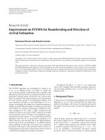
Fig. 1. Chromatography system setup for the in-line sample injection.
Tris-HCI, and NaCI were purchased from Sigma-Aldrich. Freshly
prepared autoclaved Milli-Q (Millipore) water was used for buffer
preparation. This water was determined to be nuclease-free by our
internal laboratory tests.
The buffer A used for chromatography was composed of 50 mM
Tris-HCl (pH 8.0). Buffer B contained different concentrations of PEG
and NaCl in 50 mM Tris-HCl (pH 8.0). All buffers were vacuum filtered through 0.22-m bottle-top filters (ThermoFisher Scientific)
and degassed by sonication for 18 min prior to use. Fresh buffers
were prepared every week and used within 4–5 days.
A basic chromatographic procedure used previously [27] was
adapted as described below: (1) System equilibration. The standard
setup of the ÄKTA Purifier was changed to make it suitable for
SXC so that only buffer A flew to the injector valve, while buffer
B bypassed it and went directly to the mixer (Fig. 1). The column
was placed in-line and equilibrated with 20% buffer A:80% buffer
B. Unless otherwise indicated, the buffer A:buffer B ratio was kept
the same for simplicity, while the concentrations of PEG and NaCl
in buffer B were optimized to achieve sufficient retention of nucleic
acids. Further in the text, only the final concentrations of two chemicals are indicated (i.e. those in a mixture of 20% buffer A and 80%
buffer B). To calculate the initial concentrations of PEG and NaCl
in buffer B, the target final concentrations were divided by 0.8.
(2) Sample loading. Samples for chromatography were diluted with
buffer A and centrifuged (11,000 × g, 10 min) to remove particulate matter. The injection volume was 0.25 ml and flow rate was
2.5 ml/min or 3 ml/min depending on the sample amount and PEG
concentration in the system. Flow rates below 2.5 ml/min resulted
in pre-column sample precipitation and chromatography failure.
(3) Washing. The system was washed until the UV absorbance of
column effluent reached baseline. (4) Elution. The buffer A:buffer B
ratio was reduced stepwise or linearly. (5) Cleaning in place (CIP).
Removal of residual RNA from the column matrix was achieved by
washing with 1 M NaOH solution followed by 1 M NaCl in 50 mM
Tris-HCl (pH 8.0). CIP was performed after each chromatography
experiment to avoid cross-contamination of subsequent analyses.
Each chromatography experiment was repeated two to six times.
To calculate the retention of nucleic acids on the CIM-OH matrix,
the molecules were precipitated from the fractions and flowthrough (see 2.5 for details). Concentration was measured with a
NanoDrop 2000c (Thermo Fisher Scientific) and the percentage of
RNA or DNA eluted by the gradient was then calculated using the
b
× 100, where NA is the fraction of nucleic acid
formula: NA, % = a+b
retained in the column (%); a is the amount of unbound sample in
the flow-through solution (g), and b is the amount of the sample
eluted under gradient conditions (g). The recovery of nucleic acids
Sl
was calculated according to the formula: Recovery, % = Se
× 100%,
where Sl is the amount of loaded sample (g) and Se is the amount
of eluted sample (g). The average recovery ± standard deviation
(S.D.) was calculated in Excel based on the data from multiple
experiments.
2.2. Preparation of nucleic acid molecules for chromatography
Bacteriophage genomic DNA (48 502 bp) was obtained from
Fermentas and dissolved in nuclease-free Milli-Q water. DNA
molecules ranging in length from 88 to 1800 bps were pre-
pared by PCR amplification using Phusion HF DNA polymerase
(Finnzymes), deoxynucleotide triphosphates (Thermo Fisher Scientific), and pLM659 plasmid containing a complementary DNA copy
of Pseudomonas phage Phi6 genome segment S [37]. The forward
primers for PCR amplification contained the T7 polymerase promoter sequence (5’-TAATACGACTCACTATAGGG-3’) followed by a
sequence of 17 to 21 nt complementary to the Phi6 S-segment at
positions 80, 100, 200, 500, 700, or 1800 nt as counted from the 3’
end. The reverse primer was complementary to the very 3’ end of
the Phi6 S-segment (5’-GGAAAAAAAGAGAGAGAGCCCCCGAAGG3’) and contained the Phi6 polymerase promoter sequence at the
5’end (underlined). A list of primer sequences has been published [38]. Plasmids pLM659 [37], pLM656 [39], and pLM687
[40] were used as templates for the production of full-length
complementary DNA copies of phage Phi6 S (2948 bp), M (4065
bp), and L (6374 bp) genome segments, respectively. The reverse
primer was the same as described above and the forward primer
contained the T7 polymerase promoter sequence followed by a
homologous sequence (5’-GGAAAAAAACTTTATATA-3’) present at
the 5’-end of all three genome segments of Phi6 [41]. PCR products of the correct size were excised from the gel and purified
with NucleoSpin ExtractII kit (Macherey-Nagel) according to the
manufacturer’s instructions. Pure DNA molecules were eluted in
30 l Milli-Q water. The PCR-generated DNA molecules were used
as templates for RNA synthesis. ssRNAs were produced by in vitro
transcription with T7 RNA polymerase (Thermo Fisher Scientific)
as previously described [42]. dsRNA molecules were also generated in vitro using a single-tube transcription and replication
reaction catalyzed by the T7 and Phi6 RNA polymerases [4,42].
The recombinant Phi6 polymerase was expressed and purified as
described [42]. Nucleoside 5’-triphosphates were obtained from
Thermo Fisher Scientific. Enzymatically synthesized ssRNA and
dsRNA molecules were isolated with TRIzure reagent (Bioline)
and chloroform (Merck) according to the manufacturer’s instructions, followed by precipitation of T7-generated transcripts in 3 M
sodium acetate (Merck) or stepwise fractionation of dsRNA with
LiCl (Merck) (see below). All RNAs were washed with 70% ethanol
and dissolved in sterile nuclease-free water.
2.3. LiCl precipitation
Contaminating ssRNA molecules were removed from the dsRNA
synthesis reactions by LiCl precipitation. ssRNA molecules were
first precipitated by incubation at −20 ◦ C for 30 min in 2 M LiCl followed by 20 min centrifugation at 13,000×g at 4 ◦ C. dsRNA was
collected from the resulting supernatant by repeating the procedure in 4 M LiCl. The dsRNA pellet was washed twice with 70%
ethanol and dissolved in sterile nuclease-free water. In case of
unsatisfactory separation of dsRNAs from ssRNAs, the procedure
described above was repeated.
2.4. Preparation of bacteriophage genomes for chromatography
Pseudomonas phage Phi6 [43] was propagated and purified as
previously described [44]. The same protocol was used to recover
Pseudomonas phage PRR1 [45]. Briefly, virions were collected from
the lysates of infected Pseudomonas syringae HB10Y or Pseudomonas
aeruginosa PAO1 by precipitation with 10% PEG-6000 and 0.5 M
NaCl. To open the viral capsids, half of the precipitate was treated
with proteinase K (Thermo Fisher Scientific) at a concentration of
1 mg/ml in 10 mM Tris-HCl (pH 7.5) buffer containing 5 mM EDTA
(Merck) and 1% sodium dodecyl sulfate (SDS, Merck) at 50 ◦ C for
1 h. From the remainder of the precipitate, total RNA was extracted
using TRIzure reagent (Bioline) according to the manufacturer’s
instructions.
A. Levanova, M.M. Poranen / J. Chromatogr. A 1574 (2018) 50–59
53
2.5. Analysis of dsRNA integrity and purity
3.2. Suitability of CIM-OH column for nucleic acid separation
RNA was recovered from the collected 0.5-ml chromatography
fractions corresponding to the peak areas on chromatograms by
overnight precipitation at −20 ◦ C in 0.3 M sodium acetate (pH 6.5)
and 67% ethanol, followed by centrifugation at 10,000×g for 30 min.
The RNA pellets were washed with 70% ethanol, air dried, and dissolved in 10 l of sterile nuclease-free water. A 5-l sample was
mixed with 2 × U loading buffer (10 mM EDTA [pH 8.0], 0.2% SDS,
0.05% bromphenol blue, 0.05% xylene cyanol, 6% [v/v] glycerol, 8 M
urea). RNA molecules were separated in an 0.8% agarose gel by
electrophoresis in Tris-borate-EDTA (TBE) buffer (6.6 g Trizma base,
3.9 g boric acid, 0.93 g EDTA). For electrophoresis, an EPS 301 electrophoresis power supply (GE Healthcare) and an Owl EasyCast B2
mini gel electrophoresis system (Thermo Fisher Scientific) were
used. A detailed protocol has been described previously [46].
High-capacity binding of nucleic acids under SXC conditions has
not been observed under the conditions used previously [27] and
suitability of this chromatography mode for nucleic acid purification remained unclear. Therefore, the initial experiments were
performed with dsDNA molecules, which are more robust and easier to prepare than RNA molecules. We used three different dsDNA
species, namely 500 bp, 1800 bp, and 48 502 bp in length (see 2.2
for the origin of dsDNAs). We established that retention of nucleic
acids on the column matrix required the presence of at least 0.4 M
NaCl (Fig. S1) and was negligible (0.5–3%) when buffer B contained
only PEG-6000 (up to 20% PEG-6000 was tested; data not shown).
This observation is consistent with the PEG fractionation method,
in which 15% PEG6000 at 0.55 M NaCl precipitates essentially all
DNA in the size range of 100 to 46 500 bp [29]. The shorter dsDNA
molecules (500 bp and 1800 bp) were eluted almost completely
from the column at reduced PEG-6000 and NaCl concentrations.
However, about 25% of the 48 502-bp long bacteriophage DNA
was irreversibly retained on the column.
The requirement of NaCl for efficient retention of nucleic acids
under SXC conditions is likely connected to the modification of
steric effects by the repulsive forces between negatively charged
chemical groups of the nucleic acid molecules [31,48,49]. Although
PEG changes electrostatic interactions in solutions and the molar
conductivity of NaCl-containing buffer decreases with increases in
PEG concentration, all nucleic acid molecules become hydrated. In
this aqueous phase, NaCl ionizes and forms ion pairs with phosphates of nucleic acids. Therefore, to neutralize the negative charge
and to decrease the electrostatic repulsion forces between nucleic
acid molecules, we used NaCl-containing buffers in all subsequent
experiments. This resulted in substantially better binding of nucleic
acids to the column matrix (see below). The requirement to neutralize charges for optimal SXC performance was also observed
previously for protein samples; the best results were achieved
when proteins were near their isoelectric point [27].
We also initially tested PEG molecules of different molecular
weights (4000, 6000, and 8000 g × mol−1 ). PEG-6000 performed
better in terms of RNA binding and elution and in generated backpressure. Therefore, in our subsequent experiments we used only
PEG-6000.
2.6. Treatment of the chromatography fractions with RNase or
DNase
The fractions collected after the separation of DNA and dsRNA
on CIM-OH column (Section 2.1) were precipitated as described
(Section 2.5). 5.5 l from the combined fractions 1 and 2; 6 l from
the combined fractions 3 and 4; 5 l from the combined fractions 5
and 6; and 6.5 l from the combined fractions 7 to 9 were subjected to RNase A treatment, DNase treatment, or no treatment
(control). The reaction volume was 20 l. For RNase treatment,
1 g of nucleic acid was incubated with 0.1 g of RNase A (Fermentas) in 0.1 × SSC buffer (3 M sodium chloride, 0.3 M sodium
citrate [pH 7.0]) for 15 min at room temperature. DNase treatment
was performed using RQ1 DNase (Promega; 1 unit/g nucleic acid)
at 37 ◦ C for 30 min. All samples (including controls) were desalted
with Illustra MicroSpin G25 columns according to the manufacturer’s instructions (GE Healthcare) and resolved with agarose gel
electrophoresis in TBE buffer (see Section 2.5).
3. Results and discussion
3.1. Setup for steric exclusion chromatography for nucleic acid
molecules
The monolithic chromatographic support is characterized by
very high porosity, exceptional chemical stability, and flow characteristics, which makes it a valuable tool for separation or
purification of large biomolecules [47]. CIM monolithic columns,
a trademark of BIA Separations (Slovenia), are available in different formats with the same structure of the monolith and ligand
density, which ensure scalability. In our work, we used a CIM-OH
column with a column volume of 1 ml. A chromatographic procedure used previously for protein purification [27] was adopted here
for separation of nucleic acid molecules.
To keep the pressure below 1.8 MPa (the pressure limit for
the CIM-OH 1 ml column), we applied a maximum flow rate of
3 ml/min and the final concentration of PEG did not typically exceed
15%. To minimize PEG-induced precipitation of nucleic acids before
entrance into the column, we used the in-line dilution technique
where sample was injected in buffer A followed by mixing with a
PEG-containing buffer B directly in the M-925 mixer (Fig. 1). The
gradient delay volume (the volume between the mixer and the
column) was also kept as low as possible (here 29 l). Thus, at a
3 ml/min flow rate it took only 34.8 s for the sample to enter the
column.
3.3. Optimizing SXC for ssRNA and dsRNA molecules
3.3.1. Influence of NaCl concentration on RNA retention in
CIM-OH column matrix
After successful experiments with dsDNA, we tested the possibility to use SXC for the separation of RNA molecules and the
influence of NaCl and PEG-6000 concentrations on RNA retention
and subsequent elution. We used 300-bp, 1800-bp or 6374-bp
dsRNA molecules and initially kept the PEG-6000 concentration
constant (15%) while systematically varying the salt content of the
buffers (Fig. 2A). Small fractions of the applied RNAs were retained
on the CIM-OH column matrix in the presence of 0.2 M NaCl. However, about 75% of the 300-bp dsRNA and 50% of the 1800-bp
and 6374-bp dsRNAs eluted in the flow-through (Fig. 2A). After
increasing the NaCl concentration to 0.4 M or above, no RNA was
detected by agarose gel electrophoresis in the flow-through samples of the 1800-bp and 6374-bp dsRNAs. For the 300-bp dsRNA, an
even higher salt concentration (0.8–1 M) was required for efficient
binding to the column matrix. At higher salt concentrations, PEG
molecules are known to adopt a more compact, coiled structure
[50]. This decreases the hydrodynamic radius of PEG and consequently its steric exclusion effects, which should lead to reduced
retention of nucleic acids on the column matrix [50]. In protein
SXC [27], retention of IgM diminished rapidly to zero when the NaCl
54
A. Levanova, M.M. Poranen / J. Chromatogr. A 1574 (2018) 50–59
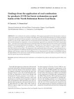
was observed in 11% PEG-6000. However, even in 15% PEG-6000,
less than 80% of ssRNAs of 500 to 100 nt was retained.
Small percentage of dsRNA molecules in the range of 700–6374
bp accreted to the column matrix already in 8% PEG-6000, while
retention of 86%–100% was achieved in 12% PEG-6000 (Fig. 2C).
dsRNA molecules of 500 bp started to accrete to the column matrix
in 10% PEG-6000; full retention was accomplished in 15% PEG-6000.
Complete retention of dsRNA shorter than 500 bp was not achieved
under the conditions tested (Fig. 2C).
In the case of longer dsRNA molecules corresponding to phage
Phi6 genome segments, we did not observe a clear relationship
between molecule size and the PEG concentration required for
binding. We calculated the lengths of these dsRNA molecules,
assuming that 1 bp of dsRNA corresponds to 0.29 nm [51]. Thus,
the estimated length of Phi6 L-, M-, and S-segments (6374, 4065,
and 2948 bp, respectively) were approximately 1.8 m, 1.2 m, and
854 nm, respectively. The structure of dsRNA molecules is more
rigid compared to ssRNAs, and dsRNAs might not condense to the
same extent as ssRNAs of the same length in PEG/NaCl solution. This
could hamper the migration of the long dsRNA molecules through
the pores of the column matrix, which are only 1.3 m in diameter.
Alternatively, cryogel monoliths [36] with superporous structures
(10–100 m) could be tested for the separation of the longer dsRNA
molecules.
The dynamic binding capacity for RNA samples could not be
determined, as upon loading >250 g of dsRNA in 13% PEG-6000 the
backpressure increased beyond acceptable limits. The RNA recovery under optimal gradient conditions was 80 ± 9%. Our preliminary
experiments demonstrated that for effective elution of RNA species,
a high-amplitude rapid change in PEG concentration is required.
Conversely with long linear gradients (>18 CV), RNA molecules
were spread in numerous fractions, which resulted in low ultimate RNA recovery and poor separation (Fig. S2). Thus, to achieve
satisfactory results a short linear or a stepwise gradient should preferentially be applied (see Figs. 3–6 for the examples of optimized
gradient conditions).
Fig. 2. NaCl (A) and PEG-6000 (B, C) dependence of RNA binding to the CIM-OH column matrix and effect of RNA size on retention. (A) dsRNA of 300 bp, 1800 bp, or
6374 bp was loaded onto the column in 15% PEG-6000 and variable NaCl concentrations. Data are shown as mean ± SD of three independent experiments. (B) ssRNA
molecules of 100–6374 nt or (C) dsRNA molecules of 88–6374 bp were loaded onto
the column in 0.8 M NaCl and variable concentrations of PEG-6000. 15 g of each
RNA was applied to the column.
concentration exceeded certain threshold values (0.4 M NaCl at 8%
PEG-6000 or 0.9 M NaCl at 10% PEG-6000). However, in our experiments a higher PEG-6000 concentration and up to 1 M NaCl did not
show a substantial negative effect on the nucleic acid retention.
3.3.2. Influence of PEG concentration on ssRNA and dsRNA
retention in CIM-OH column matrix
In the next series of experiments, we kept the NaCl concentration constant (0.8 M) and followed how changes in PEG-6000
concentration affected the binding and elution behavior of ssRNA
and dsRNA molecules (Fig. 2B and 2C). Irrespective of the ssRNA or
dsRNA length, no binding was achieved when the PEG-6000 concentration was 7% or less. Since the volume of the PEG-deficient
zone must be proportional to the size of the target biomolecules
[31], larger species of ssRNA molecules bound at lower PEG concentrations (Fig. 2B). Accordingly, 35% of 6374-nt, 20% of 4065-nt,
and 7% of 2948-nt ssRNAs were retained on the column in 8% PEG6000. None of these RNA species were detected in the flow-through
in 11% or 12% PEG-6000. ssRNAs of 1800 nt and 700 nt required at
least 10% PEG-6000 for binding to the column matrix. About 95% of
these ssRNA species were retained in 15% PEG-6000. Some retention of the shorter ssRNA molecules used in the study (≤500 nt)
3.4. Separation of DNA and dsRNA molecules
dsDNA and dsRNA species of the same length could be partially
resolved by SXC on a CIM-OH column (Fig. 3). dsDNA was eluted first
while the majority of dsRNA molecules were retained longer and
eluted in the second peak. Similar results were obtained for nucleic
acid molecules of 1800 bp and 500 bp. Data on the separation of
1800-bp dsRNA and dsDNA are shown in Fig. 3. In addition to the
size of the nucleic acid molecule (Fig. 2), differences in their configuration and chemistry might play an important role in sample
retention. Compared to the B-form of dsDNA, the A-form of dsRNA
duplex is shorter and wider with a deeper major groove. Monovalent and divalent cations penetrate into the major grooves of dsRNA,
which results in more efficient shielding of dsRNA charge compared
with dsDNA [52]. Intermolecular and intramolecular repulsion vanishes at a lower cation concentration and, therefore, dsRNA might
associate with each other and the column matrix more efficiently
than dsDNA.
3.5. Separation of RNA molecules of different sizes
We did not achieve any satisfactory separation of RNA molecules
of different sizes (either ssRNA or dsRNA) despite significant (2
times or greater) differences in size (data not shown). Only separation of RNA molecules shorter than 100 nt from longer RNA
species was easily achieved by SXC (Fig. S3). This is because the
short molecules are not retained on the column matrix, whereas
the longer ones are retained under the conditions applied (Fig. 2).
A. Levanova, M.M. Poranen / J. Chromatogr. A 1574 (2018) 50–59
55
Fig. 3. Chromatographic separation of dsRNA and dsDNA molecules of 1800 bp using a CIM-OH column. A sample containing 15 g of dsRNA and 15 g of dsDNA molecules of
1800 bp long was loaded onto the CIM-OH column pre-equilibrated with 80% buffer B (50 mM Tris-HCl [pH 8.0], 18.75% PEG-6000, 0.5 M NaCl) to obtain a final concentration
of 15% (w/v) PEG-6000 and 0.4 M NaCl. A linear gradient (80% to 0% buffer B in 15 CV) and flow rate 2.5 ml/min was applied to separate the nucleic acid molecules. (Left
panel) Absorbance profile (260 nm; black line) and conductivity (gray line) of the eluate. (Right panel) Agarose gel electrophoresis analysis of the eluted fractions without any
treatment or after RNAse A or DNase treatment.
3.6. Separation of dsRNA and ssRNA molecules of the same length
We have developed a method for in vitro production of dsRNA
molecules of different lengths and sequences [4]. For subsequent
biochemical applications, the dsRNA molecules must be purified
from contaminating ssRNAs of the same length, abortive transcripts, NTPs, and enzymes. A stepwise LiCl precipitation can be
applied for routine use. However, this method does not provide
efficient removal of ssRNAs. When ssRNA molecules interfere with
a subsequent application, an AEX can be used to purify dsRNA
[24]. We applied SXC as an alternative means to separate dsRNA
from contaminating ssRNA. Since the difference in electrophoretic
mobility between ssRNA and dsRNA species is substantial only for
long molecules (in our study ≥700 nt), we tested RNA molecules
of 700 nt, 1800 nt, and 4063 nt (Fig. 4) to verify the efficiency of
SXC separation using non-denaturing agarose gel electrophoresis
analysis of the samples.
Generally, the separation of ssRNA and dsRNA molecules by SXC
improved as the size of the molecules increased; the best separation and highest purity was obtained for dsRNAs of 1800 bp and
4063 bp, whereas after SXC dsRNA of 700 bp contained a substantial amount of ssRNA of 700 nt. In all experiments, ssRNA molecules
were retained on the column matrix more strongly than dsRNAs of
equal length (Fig.4). Thus, on the basis of agarose gel electrophoresis analysis of the elution fractions, a pure fraction of dsRNA was
attainable from a mixture of dsRNA and ssRNA using SXC. However,
subsequent fractions always contained dsRNA molecules in addition to ssRNA. This might be due to the presence of dsRNA in the
interstices of the densely packed ssRNA precipitates on the column
stationary phase surface.
The observed separation efficiency of SXC is comparable to
that obtained by AEX [24]. Moreover, the whole process took only
30 min (20 min for system preparation and 10 min for sample injection and elution). Achievement of similar resolution with AEX
requires the use of substantially longer gradients (100 CV) [24].
Accordingly, for the separation of long dsRNA molecules from a
mixture of ssRNA and dsRNA, SXC is an effective and efficient alternative to AEX.
3.7. Purification of ssRNA and dsRNA virus genomes by SXC
3.7.1. Purification of phage PRR1 genome from host cell lysate
We applied both linear and stepwise gradients to separate the
3574 nt-long genomic ssRNA of Pseudomonas phage PRR1 directly
from lysate of infected bacterial cells (Fig. 5A and B). Short bacterial ssRNA molecules (<300 nt) did not bind to the column and
were recovered in the flow-through. A decrease in PEG concentration resulted in the elution of the phage genome together with
contaminating host RNAs. Host plasmid DNA was also detected in
some of the fractions containing the phage genome (Fig. 5A and
B). Moreover, some proteins co-eluted with the viral RNA (Fig. S4),
which was expected since the separation is size-dependent and
large impurities can co-precipitate with the target molecules. To
increase the purity of the viral genome, we extracted the total RNA
from the bacterial lysate by TRIzure reagent to remove proteins and
cellular DNA molecules from the sample (see 2.4). Using a stepwise
gradient of PEG-6000 we obtained 85% pure phage PRR1 genome,
as determined by agarose gel electrophoresis analysis (Fig. 5C).
The CIM-OH column concentrated viral RNA molecules so that
even minor RNA species could be detected in some of the fractions
by agarose gel electrophoresis (Fig. 5A). This confirmed previous
observations [27] that unlike PEG precipitation, binding efficiency
during SXC is unaffected by low target concentration. Thus, SXC
could potentially be used as an analytical tool for characterization
of complex RNA mixtures.
3.7.2. Purification of phage Phi6 genome from host cell lysate
A Pseudomonas phage Phi6-infected bacterial lysate was used
to evaluate the possibility to purify a viral dsRNA genome from
infected cells and to separate the individual genome segments. We
first applied the lysate from a Phi6-infected bacterial culture onto a
CIM-OH column after proteinase K and SDS treatment (see 2.4). In
this case, the best separation was achieved with a stepwise gradient (Fig. 6A). However, bacterial plasmid DNA co-eluted with
both dsRNA and ssRNA species. With this approach it was possible to obtain fractions significantly enriched with Phi6 genomic
dsRNA directly from the host lysate without major protein contaminants (Fig. S4). However, we were unable to separate contaminating
ssRNA molecules from the viral genome. We were also unable to
separate the three viral genome segments from each other.
To improve the SXC-based purification of the dsRNA genome
of bacteriophage Phi6, we isolated the total RNA from the lysate of
Phi6-infected bacteria using TRIzure reagent. A linear gradient (12%
PEG-6000, 0.6 M NaCl to 0% PEG-6000 in 10 CV) provided good separation of the dsRNA genome from contaminating ssRNA molecules
(Fig. 6B). Pure dsRNA genome was eluted at 7.5% PEG-6000 and
0.38 M NaCl.
The recovery and efficiency of dsRNA purification with a CIMOH column was compared with the LiCl fractionation routinely
used in our laboratory. Six identical samples of total RNA after
phenol-chloroform extraction from P. syringae lysates were prepared. Three samples were applied onto a CIM-OH column and the
56
A. Levanova, M.M. Poranen / J. Chromatogr. A 1574 (2018) 50–59
Fig. 4. Chromatographic separation of dsRNA from contaminating ssRNA of the same size using a CIM-OH column and a linear (A, C, E) or stepwise (B, D, F) PEG-6000 gradient.
Products of an in vitro dsRNA synthesis reaction containing 30 g of dsRNA and ssRNA molecules of 700 nt (A, B), 1800 nt (C, D), or 4068 nt (E, F) in 150 l volume were loaded
onto the CIM-OH column pre-equilibrated with 75% buffer B (50 mM Tris-HCl [pH 8.0], 17% PEG-6000, 1.07 M NaCl) to obtain a final concentration of 13% (w/v) PEG-6000
and 0.8 M NaCl. Elution was performed using a linear gradient of 75% to 0% buffer B in 13 (A, E) or 18 (C) CV or a two-step gradient of 50% buffer B in 7 CV and 0% buffer B in
5 CV (B), 50% buffer B and 0% buffer B, both in 5 CV (D), or 48% buffer B and 0% buffer B, both in 5 CV (D). The flow rate was 3 ml/min. (Left panel) Absorbance profile (260 nm;
black line) and conductivity (gray line) of the eluate. (Right panel) Agarose gel electrophoresis analysis of selected elution fractions.The mobility of the dsRNA and ssRNA
molecules is indicted on the right. L – DNA ladder, S – sample, FT – flow-through, P – peak, Fr. – fractions, kbp – kilo base pairs.
A. Levanova, M.M. Poranen / J. Chromatogr. A 1574 (2018) 50–59
57
Fig. 5. Separation of Pseudomonas phage PRR1 ssRNA genome from contaminating cellular nucleic acid molecules. Bacterial lysate containing PRR1 virions was treated with
proteinase K and SDS and applied to the CIM-OH column (A, B) or the total RNA extracted from the bacterial lysate with TRIzol/chloroform extraction (100 g) was applied
(C). The column was equilibrated before sample loading with 80% buffer B (50 mM Tris-HCl [pH 8.0], 16% [w/v] PEG-6000, 0.8 M NaCl) to provide initial concentrations of
13% (w/v) PEG-6000 and 0.64 M NaCl. Elution was performed using (A) a linear gradient of 80% to 0% buffer B in 13 CV, (B) a stepwise gradient of 45% buffer B in 5 CV and
0% buffer B in 5 CV, or (C) a stepwise gradient of 48%, 38%, and 0% buffer B, each in 4 CV. Flow rate was 3 ml/min. (Left panel) Absorbance profile (260 nm; black line) and
conductivity (gray line) of the eluate. (Right panel) Agarose gel electrophoresis analysis of the eluted fractions.
remaining samples were precipitated with LiCl. After LiCl precipitation, 7.05 ± 1% of the total RNA was precipitated as dsRNA. While
slightly less RNA (5.4 ± 1.6%) was recovered in the dsRNA-enriched
fractions eluted from the column, the purity significantly surpassed
that obtained using a single cycle of LiCl fractionation (Fig. 6B).
4. Conclusions
We evaluated the suitability of SXC for the separation and purification of ssRNA and dsRNA molecules of different lengths. We
determined the conditions under which efficient retention and
elution of nucleic acids (both DNA and RNA) could be achieved
(Fig. 2, Fig. S1). Retention of nucleic acids required up to 1 M NaCl
depending on the molecule length and above 7% PEG-6000. We
demonstrated that SXC on CIM monolithic columns can be applied
to separate dsRNA from ssRNA and that the resolution is better for
longer (>700 bp) dsRNA molecules (Fig. 4). Nevertheless, the use
of SXC for the separation of RNAs of different lengths is limited,
and only short RNA molecules (<100 nts) can be easily resolved
from longer RNA species (Fig. S3). SXC on a CIM-OH column has
the potential to separate dsDNA and dsRNA molecules of the same
length (Fig. 3) due to the structural differences between these
molecules.
Although separation of viral genome segments was not
achieved, SXC could separate and purify whole viral ssRNA and
dsRNA genomes from contaminating cellular RNAs (Fig. 5 and 6). In
terms of recovery, SXC surpassed AEX on CIM-QA, CIM-DEAE, and
Gen-Pak FAX columns by at least 25%. Furthermore, SXC is of general utility for concentrating RNA virus genomes. This is especially
useful for low-abundance RNA species, such as viral replicative
forms and mutualistic viruses.
58
A. Levanova, M.M. Poranen / J. Chromatogr. A 1574 (2018) 50–59
Fig. 6. Separation of Pseudomonas phage Phi6 dsRNA genome from contaminating cellular nucleic acid molecules. (A) Bacterial lysate containing Phi6 virions was treated
with proteinase K and SDS and applied to the CIM-OH column equilibrated with 80% buffer B to provide an initial concentration of 13% (w/v) PEG-6000 and 0.64 M NaCl
(buffer A, 50 mM Tris-HCl [pH 8.0]; buffer B, 50 mM Tris-HCl [pH 8.0], 16% [w/v] PEG-6000, 0.8 M NaCl). The elution was performed using a stepwise gradient of 45% buffer
B in 5 CV and 0% buffer B in 5 CV at flow rate 2.5 ml/min. (B) Total RNA extracted from the bacterial lysate with TRIzol/chloroform extraction (120 g) in 350 l of 50 mM
Tris-HCl (pH 8.0) was loaded onto the column pre-equilibrated with 75% buffer B to obtain a final concentration of 12% (w/v) PEG-6000 and 0.6 M NaCl. A linear gradient of
75% to 0% buffer B in 10 CV was applied for elution at flow rate 3 ml/min. An identically prepared RNA sample was used for stepwise LiCl precipitation. (Left panel) Absorbance
profile (260 nm; black line) and conductivity (gray line) of the eluate. (Right panel) Agarose gel electrophoresis analysis of selected elution fractions and LiCl-precipitated
ssRNA and dsRNA species. The position of the Phi6 genome, composed of three dsRNA molecules, is indicated on the right.
Declarations of interest
None.
Acknowledgements
We thank Dr. Sebastijan Peljhan for his valuable advice on SXC
setup of a chromatography system and Tanja Westerholm and
Hirnou Scott for excellent technical assistance. This work was supported by the Academy of Finland [grant 272507], the Sigrid Jusélius
Foundation, Helsinki, Finland, the Jane and Aatos Erkko Foundation,
Helsinki, Finland (to M.M.P), and the Finnish Cultural Foundation,
Helsinki, Finland (to A.L.). The authors acknowledge the use of the
University of Helsinki Instruct-HiLIFE Biocomplex unit (member
of the Biocenter Finland and Instruct-FI) and Academy of Finland
support [grant 1306833] for the unit.
Appendix A. Supplementary data
Supplementary material related to this article can be found, in
the online version, at doi: />08.063.
References
[1] A. Sedova, N.K. Banavali, RNA approaches the B-form in stacked single strand
dinucleotide contexts, Biopolymers 105 (2016) 65–82.
[2] M.M. Cox, D.L. Nelson, Nucleotides and nucleic acids, in: Lehninger Principles
of Biochemistry, W.H. Freeman and Company, New York, 2005, pp. 273–305.
[3] A. Travers, G. Muskhelishvili, DNA structure and function, FEBS J. 282 (2015)
2279–2295.
[4] A.P. Aalto, L.P. Sarin, A.A. van Dijk, M. Saarma, M.M. Poranen, U. Arumae, D.H.
Bamford, Large-scale production of dsRNA and siRNA pools for RNA
interference utilizing bacteriophage 6 RNA-dependent RNA polymerase,
RNA 13 (2007) 422–429.
[5] E.M. Kennedy, D.G. Courtney, K. Tsai, B.R. Cullen, Viral epitranscriptomics, J.
Virol. (2017) 91.
[6] R. Martins, J.A. Queiroz, F. Sousa, Ribonucleic acid purification, J. Chromatogr.
A 1355 (2014) 1–14.
[7] V. Glisin, R. Crkvenjakov, C. Byus, Ribonucleic acid isolated by cesium chloride
centrifugation, Biochemistry 13 (1974) 2633–2637.
[8] P. Chomczynski, N. Sacchi, Single-step method of RNA isolation by acid
guanidinium thiocyanate-phenol-chloroform extraction, Anal. Biochem. 162
(1987) 156–159.
[9] P. Chomczynski, N. Sacchi, The single-step method of RNA isolation by acid
guanidinium thiocyanate-phenol-chloroform extraction: twenty-something
years on, Nat. Protoc. 1 (2006) 581–585.
[10] M.B. Stead, A. Agrawal, K.E. Bowden, R. Nasir, B.K. Mohanty, R.B. Meagher, S.R.
Kushner, RNAsnap: a rapid, quantitative and inexpensive, method for
isolating total RNA from bacteria, Nucleic Acids Res. 40 (2012), e156.
[11] A.O. Nwokeoji, P.M. Kilby, D.E. Portwood, M.J. Dickman, RNASwift: a rapid,
versatile RNA extraction method free from phenol and chloroform, Anal.
Biochem. 512 (2016) 36–46.
[12] R. Martins, C.J. Maia, J.A. Queiroz, F. Sousa, A new strategy for RNA isolation
from eukaryotic cells using arginine affinity chromatography, J. Sep. Sci. 35
(2012) 3217–3226.
[13] R. Martins, J.A. Queiroz, F. Sousa, A new affinity approach to isolate Escherichia
coli 6S RNA with histidine-chromatography, J. Mol. Recogn. 23 (2010)
519–524.
[14] R. Martins, J.A. Queiroz, F. Sousa, Histidine affinity chromatography-based
methodology for the simultaneous isolation of Escherichia coli small and
ribosomal RNA, Biomed. Chromatogr. 26 (2012) 781–788.
A. Levanova, M.M. Poranen / J. Chromatogr. A 1574 (2018) 50–59
[15] A. Petrov, T. Wu, E.V. Puglisi, J.D. Puglisi, RNA purification by preparative
polyacrylamide gel electrophoresis, Methods Enzymol. 530 (2013) 315–330.
[16] J.S. Kieft, R.T. Batey, A general method for rapid and nondenaturing
purification of RNAs, RNA 10 (2004) 988–995.
[17] O.C. Uhlenbeck, Keeping RNA happy, RNA 1 (1995) 4–6.
[18] J.R. Diaz-Ruiz, J.M. Kaper, Isolation of viral double-stranded RNAs using a LiCl
fractionation procedure, Prep. Biochem. 8 (1978) 1–17.
[19] R.M. Franklin, Purification and properties of the replicative intermediate of
the RNA bacteriophage R17, Proc. Natl. Acad. Sci. U. S. A. 55 (1966) 1504–1511.
[20] A. Azarani, K.H. Hecker, RNA analysis by ion-pair reversed-phase high
performance liquid chromatography, Nucleic Acids Res. 29 (2001) E7.
[21] M.J. Dickman, Effects of sequence and structure in the separation of nucleic
acids using ion pair reverse phase liquid chromatography, J. Chromatogr. A
1076 (2005) 83–89.
[22] J.A. Thompson, R.D. Wells, HPLC in nucleic acids research, Nature 334 (1988)
87–88.
[23] J. Koubek, K.F. Lin, Y.R. Chen, R.P. Cheng, J.J. Huang, Strong anion-exchange
fast performance liquid chromatography as a versatile tool for preparation
and purification of RNA produced by in vitro transcription, RNA 19 (2013)
1449–1459.
[24] A. Romanovskaya, L.P. Sarin, D.H. Bamford, M.M. Poranen, High-throughput
purification of double-stranded RNA molecules using convective interaction
media monolithic anion exchange columns, J. Chromatogr. A 1278 (2013)
54–60.
[25] C.R. Huang, S.J. Lo, Evolution and diversity of the human hepatitis d virus
genome, Adv. Bioinformatics (2010), 323654.
[26] A.E. Gorbalenya, L. Enjuanes, J. Ziebuhr, E.J. Snijder, Nidovirales: evolving the
largest RNA virus genome, Virus Res. 117 (2006) 17–37.
[27] J. Lee, H.T. Gan, S.M. Latiff, C. Chuah, W.Y. Lee, Y.S. Yang, B. Loo, S.K. Ng, P.
Gagnon, Principles and applications of steric exclusion chromatography, J.
Chromatogr. A 1270 (2012) 162–170.
[28] Steric exclusion chromatography, in: P. N.A (Ed.), J. Chromatogr. Library, 1984,
pp. 253–283.
[29] J.T. Lis, R. Schleif, Size fractionation of double-stranded DNA by precipitation
with polyethylene glycol, Nucleic Acids Res. 2 (1975) 383–389.
[30] A. Polson, G.M. Potgieter, J.F. Largier, G.E. Mears, F.J. Joubert, The fractionation
of protein mixtures by linear polymers of high molecular weight, Biochim.
Biophys. Acta 82 (1964) 463–475.
[31] T. Arakawa, S.N. Timasheff, Mechanism of poly(ethylene glycol) interaction
with proteins, Biochemistry 24 (1985) 6756–6762.
[32] R. Bhat, S.N. Timasheff, Steric exclusion is the principal source of the
preferential hydration of proteins in the presence of polyethylene glycols,
Protein Sci. 1 (1992) 1133–1143.
[33] A. Strancar, P. Koselj, H. Schwinn, D. Josic, Application of compact porous
disks for fast separations of biopolymers and in-process control in
biotechnology, Anal. Chem. 68 (1996) 3483–3488.
[34] P. Gagnon, P. Toh, J. Lee, High productivity purification of immunoglobulin G
monoclonal antibodies on starch-coated magnetic nanoparticles by steric
exclusion of polyethylene glycol, J. Chromatogr. A 1324 (2014) 171–180.
[35] P. Marichal-Gallardo, M.M. Pieler, M.W. Wolff, U. Reichl, Steric exclusion
chromatography for purification of cell culture-derived influenza A virus
using regenerated cellulose membranes and polyethylene glycol, J.
Chromatogr. A 1483 (2017) 110–119.
59
[36] C. Wang, S. Bai, S.P. Tao, Y. Sun, Evaluation of steric exclusion chromatography
on cryogel column for the separation of serum proteins, J. Chromatogr. A 1333
(2014) 54–59.
[37] P. Gottlieb, J. Strassman, X. Qiao, M. Frilander, A. Frucht, L. Mindich, In vitro
packaging and replication of individual genomic segments of bacteriophage
6 RNA, J. Virol. 66 (1992) 2611–2616.
[38] M. Jiang, P. Osterlund, L.P. Sarin, M.M. Poranen, D.H. Bamford, D. Guo, I.
Julkunen, Innate immune responses in human monocyte-derived dendritic
cells are highly dependent on the size and the 5’ phosphorylation of RNA
molecules, J. Immunol. 187 (2011) 1713–1721.
[39] V.M. Olkkonen, P. Gottlieb, J. Strassman, X.Y. Qiao, D.H. Bamford, L. Mindich,
In vitro assembly of infectious nucleocapsids of bacteriophage 6: Formation
of a recombinant double-stranded RNA virus, Proc. Natl. Acad. Sci. U. S. A. 87
(1990) 9173–9177.
[40] L. Mindich, X. Qiao, S. Onodera, P. Gottlieb, M. Frilander, RNA structural
requirements for stability and minus-strand synthesis in the dsRNA
bacteriophage 6, Virology 202 (1994) 258–263.
[41] M. Szekeres, B.H. Brownstein, H.R. Revel, R. Haselkorn, Terminal sequences of
the bacteriophage 6 segmented dsRNA genome and its messenger RNAs,
Virology 142 (1985) 1–11.
[42] E.V. Makeyev, D.H. Bamford, Replicase activity of purified recombinant
protein P2 of double-stranded RNA bacteriophage 6, EMBO J. 19 (2000)
124–133.
[43] A.K. Vidaver, R.K. Koski, J.L. Van Etten, Bacteriophage phi6: a lipid-containing
virus of Pseudomonas phaseolicola, J. Virol. 11 (1973) 799–805.
[44] D.H. Bamford, P.M. Ojala, M. Frilander, L. Walin, J.K.H. Bamford, Isolation,
purification, and function of assembly intermediates and subviral particles of
bacteriophages PRD1 and 6, in: K.W. Adolph (Ed.), Methods in Molecular
Genetics, Academic Press, San Diego, 1995, pp. 455–474.
[45] R.H. Olsen, D.D. Thomas, Characteristics and purification of PRR1, an RNA
phage specific for the broad host range Pseudomonas R1822 drug resistance
plasmid, J. Virol. 12 (1973) 1560–1567.
[46] D.C. Rio, M. Ares Jr., G.J. Hannon, T.W. Nilsen, Nondenaturing agarose gel
electrophoresis of RNA, Cold Spring Harb. Protoc. (2010), pdb.prot5445.
[47] M. Krajacic, M. Ravnikar, A. Strancar, I. Gutierrez-Aguirre, Application of
monolithic chromatographic supports in virus research, Electrophoresis 38
(2017) 2827–2836.
[48] A.P. Minton, The influence of macromolecular crowding and macromolecular
confinement on biochemical reactions in physiological media, J. Biol. Chem.
276 (2001) 10577–10580.
[49] D.H. Atha, K.C. Ingham, Mechanism of precipitation of proteins by
polyethylene glycols. Analysis in terms of excluded volume, J. Biol. Chem. 256
(1981) 12108–12117.
[50] C. Tan, J.G. Albright, O. Annunziata, Determination of preferential interaction
parameters by multicomponent diffusion. Application to poly(ethylene
glycol)-salt-water ternary mixtures, J. Phys. Chem. B 112 (2008) 4967–4974.
[51] J.A. Abels, F. Moreno-Herrero, T. van der Heijden, C. Dekker, N.H. Dekker,
Single-molecule measurements of the persistence length of double-stranded
RNA, Biophys. J. 88 (2005) 2737–2744.
[52] S.A. Pabit, X. Qiu, J.S. Lamb, L. Li, S.P. Meisburger, L. Pollack, Both helix topology
and counterion distribution contribute to the more effective charge screening
in dsRNA compared with dsDNA, Nucleic Acids Res. 37 (2009) 3887–3896.