Báo cáo khoa học: A sucrose binding protein homologue from soybean exhibits GTP-binding activity that functions independently of sucrose transport activity pptx
Bạn đang xem bản rút gọn của tài liệu. Xem và tải ngay bản đầy đủ của tài liệu tại đây (411.82 KB, 11 trang )
A sucrose binding protein homologue from soybean exhibits
GTP-binding activity that functions independently of sucrose
transport activity
Carlos P. Pirovani
1
, Joci Neuby A. Mace
ˆ
do
2
, Luı
´
s Anto
ˆ
nio S Contim
2
, Fabiana S. V. Matrangolo
2
,
Marcelo E. Loureiro
1
and Elizabeth P. B. Fontes
2
Departments of
1
Biologia Vegetal and
2
Bioquı
´
mica e Biologia Molecular/BIOAGRO, Universidade Federal de Vic¸ osa, Brazil
The sucrose binding protein (SBP) has been implicated as an
important component of the sucrose uptake system in
plants. SBP-mediated sucrose transport displays unique ki-
netic features and the protein is not similar to other transport
proteins. Here, we report the characterization of a member
of the SBP family from soybean [Glycine max (L) Merrill]
designated S64 or SBP2. Subcellular fractionation and pre-
cipitation by GTP-agarose demonstrated that S64/SBP2 is a
membrane-associated protein that exhibits GTP binding
activity. Purified recombinant S64/SBP2 protein, expressed
as a histidine-tagged protein in Escherichia coli, exhibited
nucleotide-binding specificity to guanine nucleotides. The
GTP binding site was mapped to an imperfect Walker A
type-sequence, Ala279-Leu-Ala-Pro-Thr-Lys-Lys-Ser286,
by site-directed mutagenesis. Escherichia coli-produced wild-
type protein and a truncated version of the protein con-
taining the putative binding-sequence-bound GTP, although
not with the same efficiency. In contrast, replacement of
Thr283 and Lys284 residues to Leu and Glu residues pre-
vented GTP binding. The site directed mutant failed to bind
GTP but retained the ability to undergo oligomerization
and to promote growth of the susy7 yeast strain, deficient
in utilizing extracellular sucrose, on medium containing
sucrose as the sole carbon source. Our results indicate that
GTP binding and sucrose transport by SBP are separable
and function independently. The implications of our findings
with respect to the function and membrane topology of SBP
are discussed.
Keywords: sucrose transporter; soybean; yeast complemen-
tation assay; Glycine max.
In many higher plants, sucrose is the predominant form of
photoassimilate that is transported from mature leaves
(sourcetissues)tosinktissues,suchasseeds,stems,
reproductive organs and roots, via the vascular system [1].
Biochemical studies have demonstrated that sucrose uptake
kinetics in leaves is complex and consists of multiple
components; for example, in Vicia faba, two saturable
(high- and low-affinity) components and one linear,
low-affinity component have been described [2]. Our
understanding of sucrose translocation has advanced con-
siderably over the last decade with the molecular and
biochemical characterization of the sucrose transporter
(SUT) family of low- and high-affinity sucrose transporters
[1]. The SUT1 protein has been described as the proton-
motive-force-driven sucrose symporter that mediates
phloem loading and long-distance transport, the key
transport step in assimilate partitioning for many plants
[3–5]. SUT1 serves as a high-affinity transporter, whereas
SUT4, a second member of this sucrose transporter family,
corresponds to the low-affinity/high capacity saturable
component of sucrose uptake found in leaves [6]. A third
structurally related-member of the family has been identified
and designated SUT2 [7]. The SUT2 protein has been
proposed to act as a sugar sensor that controls sucrose
fluxes across the plasma membrane of sieve elements by
regulating expression, activity and turnover of SUT1 and
SUT4 [7]. This hypothesis was raised based on the lack of
transport activity of SUT2 and its colocalization with the
high and low-affinity sucrose transporter in sieve elements.
Nevertheless, direct evidence for a SUT2 sucrose sensor and
regulatory function has not been provided.
Earlier attempts to identify sucrose transporters resulted
in the identification of a sucrose binding protein from
soybean cotyledonary microsomal membrane fraction by
its capacity to bind to the sucrose analogue 6¢-deoxy-6¢-
(4-azido-2-hydroxy)-benzamido-sucrose [8]. Subsequent
progress in characterizing SBP led to the isolation of its
cDNA from an expression library prepared from cotyledon
mRNA [9]. Molecular characterization of the cDNA-
encoded product revealed that SBP was quite dissimilar
from the H
+
/sucrose symporter SUT. Despite the lack of
similarity between SBP and other known membrane
transport proteins, several lines of evidence have implicated
the SBP protein as the linear, low affinity component of
sucrose uptake system in plants. The SPB protein is
localized in the plasma membrane of cells that are actively
engaged in sucrose transport, such as mesophyll cells of
Correspondence to E. P. B. Fontes, DBB/BIOAGRO-Universidade
Federal de Vic¸ osa, Avenue. P.H. Rolfs s/n, 36571.000 Vic¸ osa MG,
Brazil.
Fax: + 55 31 38992864, Tel.: + 55 31 38992949,
E-mail:
Abbreviations: SUT, sucrose transporter; SBP, sucrose binding
protein; CaMV, cauliflower mosaic virus; rbcS, small subunit of
RUBISCO; ADH, alcohol dehydrogenase; DAF, days after flowering.
(Received 1 April 2002, revised 12 June 2002, accepted 2 July 2002)
Eur. J. Biochem. 269, 3998–4008 (2002) Ó FEBS 2002 doi:10.1046/j.1432-1033.2002.03089.x
young sink leaves, the companion cells of mature phloem
and the cells of cotyledons undergoing differentiation [9,10].
In the cotyledon, expression of the SBP gene is temporally
regulated and accumulation of the protein is coordinated
with active sucrose uptake [9]. In spinach, a SBP homologue
was immunolocalized in the plasma membrane of sieve
elements in fully expanded leaves, shoots and roots [11,12]
and in V. faba developing seeds, SBP was colocalized with
the H
+
/sucrose symporter in the plasma membrane of
transfer cells [13]. A SBP homologue was also detected in
the microsomal fraction of young leaves from Nicotiana
tabacum [14]. Direct evidence implicating SBP in sucrose
transport has been obtained with complementation studies
using a secreted-invertase-deficient mutant yeast strain,
incapable of growth on medium containing sucrose as the
only carbon source [15,16]. Ectopic expression of the SBP
cDNA alone reverses the mutant yeast phenotype and SBP-
mediated specific sucrose uptake in yeast displays linear,
nonsaturable kinetics up to 30 m
M
external sucrose, being
relatively insensitive to pH gradient across the membrane
[15,17]. These biochemical features closely resemble the
kinetics properties of the previously characterized linear
component of sucrose uptake in higher plants [18–20].
Recently, we have conducted overexpression and antisense
repression studies in transgenic tobacco (Nicotiana tabacum
L. Cv Havana) to analyze the function of SBP in the long-
distance sucrose transport [14]. The antisense transgenic
plants developed symptoms consistent with inhibition of
sucrose translocation and displayed a reduction in plant
growth and development. Furthermore, both antisense
repression and overexpression of a SBP homologue in
transgenic lines altered carbohydrate partitioning in mature
leaves. These results indicated that SBP might represent an
important component of the sucrose translocation pathway
in plants.
More recently, we have addressed the role of SBP in
plant cell sucrose transport by performing radiolabeled
sucrose uptake experiments with transgenic tobacco cell
lines expressing the SBP sense or antisense gene [21]. In
this condition, the level of a SBP homologue correlated
with the efficiency of radiolabeled uptake by the trans-
genic tobacco cells. Furthermore, manipulation of SBP
levels altered sucrose-cleaving activities in a metabolic
compensatory manner. Enhanced accumulation of SBP
caused an increase in intracellular sucrose synthase activity
with a concomitant decline in cell-wall invertase activity.
This alteration in sucrose-cleaving activities is consistent
with a metabolic adjustment of the sense cell lines caused
by its high efficiency of direct sucrose uptake as
disaccharide. Although these studies clearly demonstrated
that SBP is involved in sucrose translocation-dependent
physiological processes, still unresolved is whether the
underlying mechanism involves SBP-mediated sucrose
transport or SBP-mediated regulation of alternative car-
bohydrate uptake systems.
Despite the functional characterization of SBP, potential
post-translational modifications that could regulate its
function have not been examined. In this investigation, we
describe the identification of an isoform of soybean SBP,
designated S64 or SBP2, and we show that the SBP
homologue is a membrane-associated GTP binding protein.
We have generated mutants that blocked its GTP binding
activity but not interfered in its oligomerization property
and S64/SBP-mediated sucrose transport in yeast. These
mutants should be valuable tools for determining the
physiological role of SBP as a G-protein in vivo.
EXPERIMENTAL PROCEDURES
Isolation of a SBP homologue cDNA from soybean
DNA manipulations were performed essentially as
described previously [22]. The S64 cDNA (GeneBank
accession number AF191299) was unintentionally isolated
from a soybean seed expression library using an antibody
raised against a partially purified microsomal membrane
fraction from immature soybean seeds [14]. The positive
clones resulted from this screening were designated by the
letter S from soybean seeds followed by 1 : 1000 of the
estimated M
r
of the encoded product. The identity of this
particular S64 clone was obtained by sequence comparison
analysis using the
BLAST
program [23]. The computer
program
CLUSTALW
was used for sequence alignment. The
S64 deduced protein shares 86% sequence identity with the
sucrose binding protein (GeneBank accession number
L06038) and is also referred to as SBP homologue or SBP2.
Construction of plasmids and antibody production
The S64/SBP homologue insert was released from the k
recombinant DNA with EcoRI digestion and subcloned
into the EcoRI site of pUC118 to obtain the clone
pUFVS64. The S64 protein was expressed as a fusion
protein using the pET-16b vector (Novagen), which
provides an N-terminal His tag. For this purpose, an EcoRI
site immediately adjacent to the stop codon was created by
PCR using the Pfu DNA polymerase, the forward primer
S64XHOF 5¢-AAGAAAC
TCGAGGTCGAAGA-3¢
(coordinates 103–121, XhoI site underlined) and the reverse
primer SEF97R 5¢-ATACATTCCCC
GAATTCAGCCA
CCTCC-3¢ (positions 1498–1524, EcoRI site underlined).
The amplified sequence, spanning the entire protein-coding
region and lacking the putative peptide signal coding
sequence and the 3¢ untranslated sequences, was subcloned
into the EcoRI/SmaI-restricted pGEM7Zf(–) vector (Pro-
mega), and then moved as a XhoI insert into pET16b,
yielding pUFV120.
The construction was transformed into E. coli strain
BL21 (DE3) and the synthesis of the recombinant protein
was induced by isopropyl thio-b-
D
-galactoside (IPTG). The
induced protein was affinity-purified using Ni-chelating
Sepharose resin (Amersham Pharmacia Biotech.) and used
as an antigen to raise polyclonal antisera in rabbits, which
were immunized through subcutaneous injections during
2-week intervals. The specificity of the anti-S64 serum was
previously evaluated with protein extracts from transgenic
tobacco plants expressing the S64/SBP homologue cDNA
either in the sense or antisense orientation [14,21], in a yeast
expression system [14] and in a bacteria expression system
[14].
Truncated protein, mutagenesis and bacterial
overexpression
To produce a S64 truncated protein, an internal 916-bp
sequence of S64 cDNA was released from pUFVS64 with
Ó FEBS 2002 GTP binding of sucrose binding protein (Eur. J. Biochem. 269) 3999
Sau3AI digestion and inserted into the BamHIsiteof
pET16b to create pUFV50. The inserted sequence spans
nucleotides 122–1041 of the cDNA and encodes the amino
acid residues from position 36–343.
The putative GTP-binding site was mutated using a
PCR-based mutagenesis strategy, in which overlapping
upstream and downstream sequences of the site were
individually amplified with sets of primers to create an
internal XbaI site within the putative site. The sets of
primers used were SPI97F 5¢-TCCTCA
CTGCAG
TCACCATGGCGACCA-3¢ (coordinates 1–27, PstIsite
underlined) and SGTPXBAF 5¢- GGCCCC
TCTAGA
GAAAAGCTC-3¢ (coordinates 857–878, XbaIsite
underlined) for the S64 N-terminal encoding sequence as
well as SGTPXBAR 5¢-GCTTTTC
TCTAGAGGGGCC
AACG-3¢ (positions 853–876, XbaI site underlined)
and SEF97R 5¢-ATACATTCCCC
GAATTCAGCCACC
TCC-3¢ (positions 1498–1524, EcoRI site underlined) for the
adjacent C-terminal encoding sequence. The upstream-
amplified sequence was digested with PstIandXbaI,
whereas the downstream-amplified fragment was digested
with XbaIandEcoRI, and then they were inserted by triple
ligation into PstI–EcoRI sites of pUC118 to obtain
pUFV193. This restored the S64 coding region in which
an internal XbaI site was created and, as consequence, the
putative GTP-binding site Ala279-Leu-Ala-Pro-Thr-Lys-
Lys-Ser286 was mutated to Ala279-Leu-Ala-Pro-Leu-Glu-
Lys-Ser286. The mutations were confirmed by sequencing.
To transfer the mutated S64 sequence to pET16b, it was
amplified from pUFV193 with the sense primer S64XHOF
and the antisense primer SEF97R. The amplified sequence,
harboring the mutated protein-coding region and lacking
the putative peptide signal coding sequence and the
3¢ untranslated sequences, was subcloned into the EcoRI/
SmaI-restricted pGEM7Zf(–) vector (Promega), and then
moved as a XhoI insert into pET16b to obtain pUFV232.
Constructions in pET16b were expressed in E. coli
strain BL21 (DE3) LysS following induction by IPTG.
N-Terminal His-tagged SBP fusion proteins were purified
according to manufacturer’s instructions (Novagen) for
soluble proteins. For oligomerization studies, after a first
round of purification, the His tag was removed from the
E. coli-produced proteins by treatment with catalytic
amounts of Factor Xa (10 lgÆmg
)1
of recombinant protein)
in 100 m
M
NaCl, 50 m
M
Tris/HCl, pH 8.0, 1 m
M
CaCl
2
at
37 °C for 24 h.
Isolation of microsomal fraction
For microsomal membrane isolation, soybean cotyledons
were homogenized with 25 m
M
Tris/HCl, pH 7.0,
250 m
M
sucrose, 2.5 m
M
dithiothreitol, 10 m
M
MgSO
4
,
0.5% (w/v) gelatin and 0.5 m
M
phenylmethanesulfonyl
fluoride [8]. The homogenate was filtered and centrifuged
for 15 min at 13 000 g and 4 °C. Microsomal prepara-
tions were isolated by centrifugation at 80 000 g for
45 min [24].
Transient expression of S64/SBP homologue
in soybean suspension cells
The pUFVS64 clone was modified by site-directed muta-
genesis to create an EcoRI restriction site immediately
downstream of the stop codon, yielding pUFV32. A
plant expression cassette containing the S64/SBP homo-
logue gene was constructed by insertion of the S64
coding region that was released from pUFV32 with
EcoRI/BamHI digestion into pMON921 vector [25],
previously digested with BglII/EcoRI. The resulting
plasmid, pUFV52, harbors the S64 coding region in the
sense orientation placed between the CaMV 35S pro-
moter with a duplicated enhancer region and the 3¢ end
of the pea E9 rbcS gene. A soybean cell culture line was
generated and established as described previously [26].
Transient expression of S64 was performed by electro-
poration (380 V, 975 lF) of 10 lg of expression cassette
DNA and 40 lg of sheared salmon sperm DNA into
0.8 mL of cultured soybean cells in electroporation buffer
(80 m
M
KCl, 5 m
M
CaCl
2
,10m
M
Mes, pH 6.7, 0.425
M
mannitol). Prior to electroporation, soybean suspension
cells at 4 days after passage were recovered by centrifu-
gation at 200 g, washed three times and concentrated
twice with electroporation buffer, incubated with plasmid
and carrier DNA at 37 °C for 1 h and then at 0 °Cfor
10 min. The electroporated cells were diluted into 10 mL
of MS medium [27], supplemented with complex vitamin
B5, 0.2 mgÆmL
)1
2,4-dichlorophenoxyacetic acid, 6%
(w/v) sucrose and 15 m
M
glutamine, pH 5.7. Total
protein was isolated from cells 48 h after transfection as
described [24], separated by SDS/PAGE and immuno-
blotted with anti-S64 serum.
Gel electrophoresis and immunoblotting analysis
SDS/PAGE was carried out as described previously [28]
and the proteins were transferred from 10% SDS/
polyacrylamide gels to nitrocellulose membrane by elec-
troblotting. The membrane was blocked with 3% (w/v)
BSA in NaCl/Tris/Tween [100 m
M
Tris/HCl, pH 8.0,
150 m
M
NaCl, 0.05% (v/v) Tween-20]. S64/SBP homo-
logue was detected using polyclonal anti-S64 serum at a
1 : 1000 dilution, followed by a goat anti-(rabbit IgG) Ig
conjugated to alkaline phosphatase (Sigma) at a 1 : 5000
dilution. Alkaline phosphatase activity was assayed using
5-bromo-4-chloro-3-indolyl phosphate (Life Technologies,
Inc.) and p-nitroblue tetrazolium (Life Technologies,
Inc.).
Binding of S64/SBP homologue to GTP-agarose
Whole cell protein extracts were obtained from transgenic
tobacco cell lines expressing a soybean S64 transgene [21]
and from soybean suspension cells transiently trans-
formed with a S64 expression cassette. Protein extracts
were prepared by homogenization of the cells with lysis
buffer [100 m
M
Tris/HCl, pH 7.5, 50 m
M
KCl, 1 m
M
EDTA, 1% (v/v) Triton X-100, 1 m
M
phenyl-
methanesulfonyl fluoride, 0.1 m
M
dithiothreitol, 5 m
M
MgCl
2
]ataratioof1mgofcellsper2lL of buffer
and then clarified by centrifugation at 20 000 g for
20 min. The supernatant (2 mL) was incubated with
100 lL of 50% (v/v) GTP-agarose suspension in 50 m
M
Tris/HCl, pH 7.5, for 12 h under agitation at 4 °C[29].
The agarose beads were pelleted by centrifugation,
washed extensively with cold 50 m
M
Tris/HCl, pH 7.5
and resuspended in 40 lL of SDS/PAGE sample buffer.
4000 C. P. Pirovani et al. (Eur. J. Biochem. 269) Ó FEBS 2002
GTP bound proteins were fractionated by SDS/PAGE,
transferred to nitrocellulose and probed with anti-S64
serum, as described above.
Binding of S64/SBP homologue fusion protein
to nucleotide-agarose
The purified His-tagged S64 fusion protein (2 lg) was
incubated with either 50 lL of ATP-agarose, GTP-agarose
or Protein A-agarose suspension, previously equilibrated
with binding buffer [20 m
M
Tris/HCl pH 7.5, 100 m
M
NaCl, 0.01% (v/v) Triton X-100, 2 m
M
MgCl
2
]. After 1 h
at 4 °C, the beads were washed three times with 500 mL of
cold binding buffer and eluted in SDS/PAGE sample
buffer at 100 °C for 3 min. The eluted proteins were
separated by SDS/PAGE and stained with Coomassie Blue.
In competition assays, GTP, GDP, GTPcS (guanosine
5¢-O-3-thiotriphosphate), ATP, UTP or CTP were included
in the binding buffer at 2 m
M
andincubatedwiththe
recombinant protein (0.5 lg) for 30 min at 4 °Cpriortothe
binding reaction to GTP-agarose.
GTP-binding assay
The GTP-binding assay was performed as described
previously [30]. Briefly, E. coli-expressed His fusion proteins
were affinity-purified and blotted onto nitrocellulose mem-
brane using BIO-DOT
TM
(Bio-Rad), according to the
manufacture’s instructions. Alternatively, affinity-purified
recombinant proteins were fractionated by SDS/PAGE and
transferred to nitrocellulose by electroblotting. The mem-
branes were washed twice with binding buffer [50 m
M
NaH
2
PO
4,
pH 7.5, 10 m
M
MgCl
2
,2m
M
dithiothreitol,
0.3% (v/v) Tween-20 and 4 l
M
ATP] and then incubated
with binding buffer supplemented with 1 lCÆmL
)1
(or
0.33 n
M
)[a-
32
P]GTP (3000 CiÆmmol
)1
; Amersham/Phar-
macia) for 2 h. After the incubation period, the membranes
were washed at least six times with binding buffer and
subjected to autoradiography at )80 °C, using WOLF
L-PLUS 505504 LP intensifying screens (Sigma).
Yeast strain and plasmids
The generation of susy7 yeast strain has been described
previously [3]. It has the potato sucrose synthase gene stably
integrated into its genome but lacks an endogenous sucrose
transport system and invertase activity. Thus, susy7yeast
strain is incapable to grow on a medium containing sucrose
as the sole carbon source, unless a sucrose uptake system is
provided through ectopic expression. For complementation
assays in the mutant yeast strain, the intact S64 cDNA was
released from pUFVS64 with EcoRIandinsertedintothe
same site of the yeast expression vector 112AINE [3]. The
resulting plasmid, pUFV373, contains the S64 cDNA in the
right orientation placed between the ADH1 promoter and 3¢
end of ADH1 gene. A yeast expression cassette containing
the mutated S64 gene was constructed by insertion of the
GTP binding site mutated cDNA that was released from
pUFV193 with PstIandEcoRI digestion into the same
restriction sites of the 112AINE vector. The resulting
plasmid, pUFV375, harbors the mutated S64 coding region
in the sense orientation placed between the ADH1promoter
and the 3¢ end of the ADH1 gene.
The susy7 yeast strain was transformed with either
pUFV373 or pUFV375 by electroporation [31], resulting in
susy7-S64 or susy7-MS64, respectively. To monitor growth
on sucrose medium, 200 lL of a 24-h-old liquid culture of
either susy7-S64 or susy7-MS64 growing in complete
medium supplemented with 2% (w/v) glucose were used
to inoculate 20 mL of complete medium with 2% (w/v)
sucrose as the only carbon source. Relative growth was
monitored by taking the D
600
during 24-h intervals, as
indicated in the figure legend. For each DNA construct, at
least three independent transformants were monitored.
RESULTS
Isolation of a second member of the
SBP
gene family
from soybean
Based on structural homology and functional analogy, we
have isolated a sucrose binding protein (SBP) homologue
cDNA from soybean. The predicted encoded protein was
first designated S64, has an estimated M
r
of 55 834 and pI
of 6.32. Sequence comparison analysis revealed that the
predicted encoded protein was quite similar to the sucrose
binding protein, first identified in soybean cotyledon (86%
sequence identity). It also showed a significant amino-acid
sequence similarity to heterologous SBP sequences from
other plant species (Fig. 1). Analysis of the deduced amino-
acid sequence allowed us to predict a signal peptide and its
processing site, which suggests that the protein be targeted
to the secretory pathway. In fact, the S64 protein was
detected in microsomal fraction of soybean cotyledon
(Fig. 2).
In soybean cotyledon, the S64 antibody recognized two
cross-reacting polypeptides with slightly different electro-
phoretic mobility (Fig. 2B, lane ME). Because SBP has a
predicted M
r
of 60 522 and it is highly homologous to S64,
the reduced SDS/PAGE mobility polypeptide could repre-
sent SBP. Alternatively, the cross-reacting polypeptides
could be differentially processed forms of the same S64/SBP
homologue protein. The primary structure of the S64/SBP
homologue protein shows the presence of a consensus
sequence for nucleotide binding and a site for N-linked
glycosylation, as potential sites for post-translational mod-
ifications of the protein (Fig. 1). Furthermore, despite the
hydrophilic nature of SBP, solubilization and partitioning
studies of plasma membrane proteins have demonstrated
that 25% of SBPs are associated with a hydrophobic
portion of the plasma membrane [10]. This observation has
led to the suggestion that the putative leader peptide, which
corresponds to the only hydrophobic region of the protein,
is not quantitatively cleaved from the mature protein. The
presence of the leader peptide in a fraction of S64/SBP
homologue would explain the antibody cross-reactivity to
the higher molecular mass form. To examine these possi-
bilities, we transferred the S64 coding region to a plant
expression cassette and the recombinant protein was over-
synthesized in cultured soybean cells (Fig. 3, compare lanes
1 and 2). In the homologous system, the apparatus for
protein processing is expected to operate properly and with
similar specificity. As shown in Fig. 3, the S64/SBP
homologue protein was synthesized as single polypeptide
(lane 1) that comigrated with the faster migrating polypep-
tide detected in membrane fraction of soybean cotyledon
Ó FEBS 2002 GTP binding of sucrose binding protein (Eur. J. Biochem. 269) 4001
(lane 3). This result indicates that the reduced SDS/PAGE
mobility polypeptide may represent SBP, whereas the faster
migrating form corresponds to S64/SBP homologue. Con-
sistent with this observation, the predicted M
r
of SBP
(60 552) is slightly higher than that of S64 (55 834). Thus,
SBP and S64 cDNAs may correspond to nonallelic SBP
genes from soybean. Investigation of the genomic complex-
ity of the SBP genes by Southern blot analysis revealed a
pattern of cross-hybridizing bands consistent with the
argument that SBP is encoded by a small gene family in
soybean (data not shown). In view of this observation, the
S64 protein may also be designated SBP2 (isoform 2),
whereas the previously identified SBP [9] would be SBP1
(isoform 1).
The S64/SBP2 protein is a membrane-associated
GTP-binding protein
The S64/SBP2 deduced protein contains a predicted
nucleotide-binding site (Fig. 1) that harbors classical
Walker-type consensus sequence for the P-loop, [Ala/
Gly]x(4)Gly-Lys[Ser/Thr] [32]. Despite the fact that the
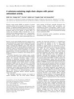
Fig. 2. SDS/PAGE and immunoblotting of membrane fractions from
soybean cotyledons. (A) Whole cell protein extracts (TE) and
microsomal membranes (ME) were isolated from soybean seeds at
20 days after flowering (DAF), fractionated by SDS/PAGE and
stained with Coomassie Brilliant Blue. M corresponds to molecular
mass markers indicated on the left in kDa. (B) SDS/PAGE fraction-
ated protein was transferred to nitrocellulose membranes and probed
with an anti-S64 serum.
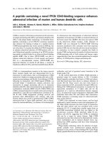
Fig. 1. Comparison of the amino acid sequence of S64 with SBP from soybean and other organisms. A multiple sequence alignment of the deduced
amino acid sequence of soybean S64 (pUFVS64, GeneBank accession number AF191299), soybean SBP (SOYSBP, GeneBank accession number
L06038), pea SBP homologue (p54, GeneBank accession number Y11207) and Vicia faba SBP homologue (VPSBP, GeneBank accession number
VFA292221) was obtained with the
CLUSTALW
program. The amino acid sequences are in the one-letter code and have been aligned by introducing
gaps (shown as dashes) to maximize identity. Dots represent identity to S64. The nucleotide-binding motif is boxed and the putative N-linked
glycosylation site is underlined. The open arrow indicates the putative signal peptide cleavage site of S64. Amino acid residues indicated below the
sequences correspond to highly conserved residues in equivalent positions of vicilin-like protein sequences that are important in maintaining their
three-dimensional structure.
4002 C. P. Pirovani et al. (Eur. J. Biochem. 269) Ó FEBS 2002
sequence Ala-Leu-Ala-Pro-Thr-Lys-Lys-Ser (position 279–
286) differs from the nucleotide binding consensus
sequence in the sixth position where a Lys replaces the
conserved Gly, we tested whether S64/SBP2 had the
capacity to bind GTP. Whole-cell protein extracts
obtained from cultured soybean cells transiently trans-
formed with S64/SBP2 cDNA under the control of the
35S promoter and from transgenic tobacco (Nicotiana
tabacum L. Cv Havana) cell lines expressing soybean
S64/SBP2 transgene [21] were allowed to bind to GTP-
agarose overnight. The bound proteins were then recov-
ered from the pelleted beads by boiling in SDS-sample
buffer and analyzed by SDS/PAGE followed by immu-
noblotting with antibodies to S64/SBP2 (Fig. 4A). The
recombinant protein synthesized in transiently trans-
formed soybean cells and in transgenic tobacco cell lines
bound to GTP-agarose (lanes 2 and 3). In control
cultured soybean cells, the endogenous protein was also
detected at 55–58 kDa in immunoblottings of GTP-
agarose precipitates (lane 1). The anti-S64 Ig cross-
reactive protein seemed to be specifically associated with
GTP, as it was not selected in control precipitates with
agarose resin alone (data not shown). The electrophoretic
mobility of the anti-S64 Ig cross-reactive GTP-agarose
bound protein together with its over-accumulation in
transgenic cell lines (compare lanes 2 and 3 with lane 1)
suggested that S64/SBP2 bound GTP. Nevertheless,
despite the presence of the nucleotide binding consensus
motif in SBP sequence (Fig. 1), our data did not allow us
to determine precisely if SBP (SBP1 isoform) also binds
GTP. Although SBP is immunologically related to S64/
SBP2, as the anti-S64 serum recognized both proteins in
microsome preparation from soybean seeds, it is synthe-
sized at very low levels in soybean suspension cells, and
in the majority of our assays SBP accumulation was
below the detection level. Attempts to increase the total
protein extract as starting material in the binding reaction
led to a remarkable increase in the background levels
compromising the quality and interpretation of the data.
The precipitation of S64/SBP2 by GTP-agarose beads
could reflect either direct binding of the protein to GTP or
Fig. 3. Transient expression of S64 in cultured cotyledon cells. Aplant
expression vector containing S64 cDNA under the control of
35SCaMV promoter and 3¢ end of rbcS gene was electroporated into
cultured soybean cells. Total protein from electroporated cells (1) and
control cells (2) was extracted 2 days postelectroporation, fractionated
by SDS/PAGE and immunoblotted with an anti-S64 serum. Lane 3 is
a microsome preparation from 20 DAF seeds. M corresponds to
prestaining molecular mass standards indicated on the right in kDa.
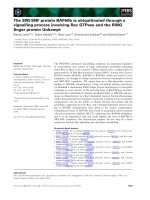
Fig. 4. Binding of S64/SBP homologue to nucleotides. (A) The S64/
SBP2 protein cosediments with a GTP-agarose resin. Whole cell pro-
tein extracts from cultured soybean cells either nontransformed (1) or
transiently transformed with S64 cDNA expression construct (2) and
transgenic tobacco cells expressing the soybean S64 transgene (3) were
incubated with GTP-agarose for 12 h. After rinsing the beads, bound
proteins were solubilized in Laemmli sample buffer and separated on a
10% SDS/PAGE gel under reducing conditions. After transferring to
nitrocellulose, proteins were detected with antibodies to S64/SBP2.
The migration positions of molecular mass standards are indicated on
the left in kDa. (B) Nucleotide binding assay of His-tagged S64/SBP2
protein. Purified His-S64 protein was incubated with GTP-agarose (1),
ATP-agarose (2) or Protein A-agarose (3) resins for 1 h at 4 °C. The
resins were washed with binding buffer and the bound proteins were
eluted by SDS/PAGE sample buffer at 100 °C. Samples were analyzed
by SDS/PAGE and Coomassie Blue staining. Lane P corresponds to
the purified His-tagged S64 fusion protein. The migration positions of
molecular mass standards are indicated on the left in kDa. (C) GTP
binding of S64/SBP2 in the presence of various nucleotides. Purified
His-S64 protein was incubated with binding buffer containing 2 m
M
competitor nucleotides, as indicated on the top of the figure, for 30 min
at 4 °C prior to the addition of GTP-agarose. Resin-bound proteins
were eluted as in (B), separated by SDS/PAGE and immunoblotted
using an anti-S64 serum. In lane M, the GTP-agarose binding assay
was performed in the absence of nucleotide competitor. The migration
positions of molecular mass standards are indicated on the left in kDa.
Ó FEBS 2002 GTP binding of sucrose binding protein (Eur. J. Biochem. 269) 4003
previous association of S64/SBP2 with GTP-binding pro-
teins present in the whole cell extracts. To examine these
possibilities, we analyzed the capacity of purified E. coli-
expressed His-tagged S64/SBP2 fusion protein to bind GTP
(Fig. 4B). A fraction of the starting material (lane 1) bound
to agarose-immobilized GTP (lane 2), whereas protein
binding to ATP-agarose resin was negligible (lane 3). The
S64/SBP2 recombinant protein also did not bind to protein
A-agarose resin (lane 3). The specificity of S64/SBP2
binding to guanine nucleotides was further confirmed in
competition assays (Fig. 4C). Incubation of the recombi-
nant protein with 100-fold molar excess of GTP (lane GTP),
GDP (lane GDP) and GTPcS (data not shown) prior to the
binding reaction prevented the recovery of a large fraction
of the protein through GTP-agarose resin. In contrast,
excess of ATP, UTP and CTP did not abolish S64/SBP2
binding to GTP-agarose resin. Taken together, our data
indicate that S64/SBP homologue exhibited a high degree of
selectivity to guanine nucleotides (GTP, GDP, GTPcS) over
adenine and pyrimidine nucleotide triphosphates.
The GTP binding activity of E. coli-produced protein
was also investigated by a filter-binding assay. Immobilized
His-wild-type S64/SBP2 fusion protein efficiently binds
GTP in the presence of 4 l
M
unlabelled ATP nonspecific
competitor (Fig. 5B, lane N and 5C, lane 1). A purified
truncated version of the protein, in which the putative signal
peptide and 149 amino acid residues from the C-terminus
were deleted (Fig. 5A, lane 2), retained the capacity to bind
GTP (Fig. 5B, lane T), albeit not to wild-type levels. The
putative GTP-binding site was further mapped by site-
directed mutagenesis. Replacement of Thr283 and Lys284
residues with Leu and Glu residues prevented GTP binding
(Fig. 5B, lane M; Fig. 5C, lane 2), indicating that these
residues are critical for binding.
Although there was some variation on protein amount,
the mutant recombinant protein and the fusion-truncated
protein accumulated to similar levels in the heterologous
expression system (Fig. 5A). Thus, the lack of GTP binding
of the mutant protein was not due to a decrease in protein
stability that could account for loss of protein integrity
during the dot blot assay. Previous experiments have shown
that SBP is organized in vivo as dimers and trimers whose
subunits interact to each other through disulfide linkage
[16]. The mutation on the GTP binding site should have no
effect on oligomerization if the proteins are properly
expressed and folded. To certify that the failure of the
GTP binding mutant to bind GTP was not due to global
misfolding, the mutant protein was assayed for its capacity
to form oligomers under nonreducing conditions (Fig. 6).
E. coli-produced wild-type, truncated and mutant proteins
were purified and separated by electrophoresis in the
presence (+2-mer) and absence ()2-mer) of 2-mercapto-
ethanol. Under reducing conditions (+2-mer), the wild-type
(lane 1) and truncated protein (lane 2) migrated at M
r
55 000 and 44 000, respectively, that correspond to the
predicted M
r
of their monomeric forms. The removal of the
2-mercaptoethanol caused a large fraction of both proteins
to migrate at an M
r
approximately twice greater than the
corresponding monomer ()2-mer, lanes 1 and 2). In
addition, a fraction of both proteins migrated as larger
complexes that may correspond to their trimeric forms.
These results indicate that both wild-type S64/SBP2 and the
truncated protein oligomerize as dimers and trimers, which
are stabilized by disulfide bounds. The identity of the
oligomers was confirmed by immunoblotting (data not
shown). The electrophoretic pattern of the mutated protein
in the presence and absence of the reducing agent was very
similar to that observed for the wild-type protein (compare
lanes 1 and 3). The presence of high molecular mass
migrating forms of the mutated protein in the absence of 2-
mercaptoethanol indicated that mutation in the GTP
binding sequence did not impair the capacity of the protein
to self-associate into dimers and trimers.
GTP binding is not required for S64/SBP2-mediated
sucrose transport in yeast
Functional complementation assays using the engineered
susy7 yeast have demonstrated that SBP mediates sucrose
uptake across the plasma membrane [17]. The SBP-medi-
ated sucrose transport in yeast has been characterized
biochemically and displays nonsaturable, linear uptake
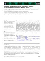
Fig. 5. GTP-binding of S64/SBP2. (A) N-Terminal His-tagged S64 fusion protein of the wild-type (His-S64) construct (1), truncated His-S64
protein (2) and Thr238Leu, Lys284Glu mutant His-S64 protein (3) were produced in E. coli, affinity-purified and separated by SDS/PAGE.
Molecular mass markers (kDa) are shown on the left. (B) Increasing amounts (1, 2.5, and 5 lg) of E. coli-produced wild-type recombinant protein
(N), Thr238Leu, Lys284Glu mutant protein (M) and truncated protein (T) were blotted onto nitrocellulose and reacted with [a-
32
P]-GTP as
described in methods. (C) Affinity-purified recombinant proteins produced in E. coli were separated by SDS/PAGE, electroblotted onto nitro-
cellulose and reacted with [a-
32
P]-GTP. Lane 1 corresponds to affinity-purified wild type fusion protein, lane 2 to Thr238Leu, Lys284Glu mutant
recombinant protein and lane 3 to an unrelated control protein (BSA).
4004 C. P. Pirovani et al. (Eur. J. Biochem. 269) Ó FEBS 2002
kinetics [15]. Heterologous expression of S64/SBP2 in susy7
yeast, which is deficient in utilizing extracellular sucrose,
restored the ability of this strain to grow on sucrose as the
sole carbon source, providing evidence that S64 and SBP are
functionally analogs (Fig. 7). To determine the effect of the
GTP binding site mutations on S64/SBP2-mediated sucrose
transport, we assayed the capacity of these mutants to
promote growth of the susy7 yeast strain on sucrose
medium. The susy7-S64 yeast and susy7-MS64 (transformed
with site-directed mutant S64 cDNA) displayed similar
growth rate when transferred to a medium supplemented
with sucrose as the only carbon source. Thus, the site-
directed mutant failed to bind GTP but not to mediate
sucrose transport in yeast.
DISCUSSION
The sucrose binding protein is a membrane-associated
protein that has been shown to mediate sucrose transport in
yeast [15,17]. We showed that a member of this family,
designated S64 or SBP2, binds GTP, although it is not
structurally related to other GTP binding proteins. The
heterotrimeric and monomeric G-proteins contain four
consensus GTP-binding motifs [A/GXXXXGK(S/T),
DXXG, NXXG, and (C/S)AX] that fold into a structurally
conserved GTP-binding site comprised of five a helices and
a central six-stranded b sheets [33,34]. In contrast, the
sucrose binding protein shows no structural homology with
other GTP-binding proteins, except for the presence of an
imperfect nucleotide binding A consensus Ala279-X-X-X-
X-Lys-Lys-Ser286, in which the conserved Gly is replaced
by a Lys284, and a second GTP binding motif D376-X-X-
G379. In the Walker-type A consensus sequence or P loop,
the loop formed between a b strand and an a helix interacts
with the phosphate groups of the nucleotide. In the S64/SBP
homologue, the single deviation (Gly/Lys) from the pattern
seems not to interfere with the nucleotide binding because
we have mapped by site-directed mutagenesis the GTP
binding site of S64/SBP2 to this imperfect A consensus
sequence. It is very likely that the positive charge of the
Lys284 residue could be involved in potential electrostatic
interactions between negatively charged phosphate groups
of GTP, as the introduction of negatively charged Glu
(Lys284Glu) residue abolished GTP binding (Fig. 5).
Alternatively, the imperfect Walker-type A consensus
sequence may not contribute specific contacts with GTP
but, instead, mutations in this sequence caused misfolding
of the protein, thereby affecting multiple functions. This
possibility was raised because the specific amino-acid
replacements made in the putative binding site (Thr283Leu
and Lys284Glu changes) do not represent structurally
neutral changes. However, two lines of evidence suggest that
incorrect folding cannot account for the failure of the
mutant protein to bind GTP. First, the site-directed
mutations did not impair S64/SBP2-mediated sucrose
transport, implying that the mutant protein retains the
capacity to bind sucrose and to assemble correctly with the
plasma membrane (Fig. 7). Second, mutation did not
prevent S64/SBP2 oligomerization, further indicating that
their effects were specific for GTP binding (Fig. 6).
Although definitive evidence for involvement of this
sequence in GTP binding will require further studies,
including the determination of the crystal structure of
GTP-bound S64/SBP2, the characterization of the site-
directed mutant presented here is consistent with the direct
involvement of the Lys284 residue in binding GTP.
Fig. 6. Oligomerization of Ecoli-produced S64/SBP2 proteins. E. coli-
produced wild-type recombinant protein (1), truncated protein (2) and
mutant protein (3) were affinity-purified, treated with Factor Xa,
solubilized in sample buffer prepared with (+2-Mer) and without
()2-Mer) 2-mercaptoethanol and separated on a 8% SDS/
polyacrylamide gel. Following electrophoresis, the gel was stained
with Coomassie Brilliant Blue. Molecular mass standards are shown in
M and their units are in kDa. TR denotes trimer; DI, dimer and MO,
monomer.
Fig. 7. Relative growth rate of susy7 transformed with either the wild
type cDNA construct (susy7-S64) or the mutated cDNA construct
(susy7-MS64) on sucrose-based medium. About 200 lL of a 24-h-old
liquid culture of susy7alone,susy7-S64 or susy7-MS64 growing in
complete medium with 2% (w/v) glucose were used to inoculate 20 mL
of yeast complete medium supplemented with 2% (w/v) sucrose. D
600
were taken on 1 mL aliquots at the indicated incubation time.
Ó FEBS 2002 GTP binding of sucrose binding protein (Eur. J. Biochem. 269) 4005
We also analyzed a possible role of the GTP-binding
activity of SBP2 on its capacity to transport sucrose in a
yeast deficient invertase strain. Complementation assays in
the mutant yeast strain incapable in utilizing extracellular
sucrose demonstrated that the GTP binding site of SBP2
was not essential for the sucrose transporting capacity
mediated by heterologous overexpression of the protein.
This result may indicate that the GTP binding and transport
activities of S64/SBP2 are separable and can function
independently.
Consistent with its function as a transport protein,
topological characterization of SBP in purified plasma
membrane vesicles has demonstrated that a proportion
(25%) of SBP behaves as a type II membrane protein,
which spans the bilayer once and has the bulk of the protein
exposed to the extracellular environment [10]. The remain-
ing protein (about 75%) is a peripheral associated-mem-
brane protein. The fraction of intrinsic membrane protein
has been proposed to be tethered to the plasma membrane
by its uncleavable putative N-terminal leader peptide, the
only hydrophobic region of the protein that could function
as a membrane-spanning domain (Fig. 1). Consistent with
this hypothesis, the leader peptide is not quantitatively
cleaved from in vitro-transcribed and -translated SBP and
S64/SBP2 in the presence of microsome ([10]; data not
shown). According to this topological model, the oligomer-
ization properties of the protein would provide the means
for assembling protein conduits across the membrane to
mediate sucrose transport. This possibility has been previ-
ously considered with the observation that SBP is structur-
ally related to vicilin-like storage proteins [16,35], which
assemble into trimers to form a 86–88 A
˚
toroid complex
with an internal hole of 18 A
˚
[36,37]. Conserved residues
that are involved in stabilizing the three-dimensional
structure of vicilin-like proteins are found at similar
positions in the SBP and S64 sequence (Fig. 1). As S64
and SBP share an extraordinary conservation of primary
structure that extends to include identical tertiary motifs, it
is very likely that the S64/SBP2 protein might also fit into
the canonical model proposed for the structure of the vicilin-
like protein family.
The proposed topology for SBP/S64, as an intrinsic
membrane transporter, would also predict that GTP
binding might not affect sucrose transport as nucleotides
are absent extracellularly. A consequence of this hypoth-
esis is that S64/SBP2-mediated sucrose transport functions
independently of its GTP binding activity (Fig. 7). In
contrast, any biological significance for the presence of a
functional GTP binding site would be strictly dependent
on the intracellular localization of the protein where high
nucleotide concentration is present. While previous result
based on NHS-biotin labeling membrane proteins dem-
onstrated that SBP was associated with the external
surface of the membrane [10], they did not exclude other
sites of cellular localization that would provide the
appropriate compartmentalization for a functional GTP
binding properties. Recently, a Vicia faba SBP-like protein
(VfSBPL) was found to accumulate predominantly in the
protein storage vacuole, but a small fraction of the protein
was also detected in the plasma membrane of cotyledo-
nary cells [38]. One possibility is that a proportion of the
soybean SBP is indeed localized extracellularly at the cell
surface as a type II membrane transport protein and a
fraction of correctly processed protein remains intracellu-
larly as membrane-associated protein where its capacity to
bind GTP and sucrose may implicate a regulatory role.
Further experiments will be necessary to confirm the
proposed topology for SBP and its subcellular localiza-
tion.
Many studies in plants have described sugar-mediated
changes in gene expression and recent research has
provided convincing evidence for a sucrose-dependent
signaling pathway, as an important regulatory step in
resource allocation [1,39,40]. The demonstration that S64/
SBP2 exhibits GTP binding activity together with its
capacity to bind sucrose specifically and reversibly [8]
raises the possibility that SBP may also serve a regulatory
role in sucrose translocation-dependent physiological pro-
cesses in plants. The resulting phenotypes from alteration
on SBP levels in transgenic plants may support such
function [14,21]. S64/SBP repression studies in tobacco
have indeed shown some of the typical phenotypes caused
by impairment of sucrose translocation [4,41,42], such as
accumulation of carbohydrates within source leaves,
inhibition of photosynthesis and stunted growth [14].
Nevertheless, the pattern of sugar accumulation in the S64
antisense leaves was not identical to that caused by
antisense repression of H
+
/SUT1 symporter [4]. This
observation suggests that SBP and SUT have distinct
functions in sucrose translocation and favors the argument
that SBP serves a regulatory role in the plant sucrose
uptake system. Consistent with this hypothesis, manipu-
lation of S64/SBP levels in transgenic plants and cultured
cells correlated inversely with cell-wall invertase activity
and directly with sucrose synthase activity [14,21].
Remarkably, the increase in S64/SBP levels had a stronger
effect on sucrose synthase activity than on sucrose uptake
[22]. These observations further support the idea that
S64/SBP functions in the sucrose translocation pathway
by regulating the expression or activity of alternative
carbohydrate uptake systems. In addition, they may
provide an alternative explanation for the capacity of
SBP to promote growth of susy7 yeast on sucrose as a
sole carbon source, as this strain harbors a potato sucrose
synthase gene integrated into its genome [3]. Thus, the
functional complementation assays in susy7 yeast for
sucrose transport processes have been conducted in the
presence of an intracellular plant sucrose cleaving activity.
Further studies will be necessary to discern whether
S64/SBP2 mediates sucrose transport or functions directly
as regulator of sucrose metabolizing enzymes or both.
The importance of the present study is twofold. First,
we have localized residues required for GTP-binding by
SBP and have shown that this binding probably involves a
novel GTP-binding folding protein. Second, this is the first
report of a sucrose binding protein that also binds GTP.
The demonstration that S64/SBP2 is a GTP binding
protein may provide new insight into the role of this
protein in sucrose translocation-dependent physiological
processes in plants. Nonetheless, much remains to be
learned about many aspects of post-translational modifi-
cation and regulation of SBP and even the most general
aspects of SBP-mediated transport in plants. The specific
mutant can now be used to dissect the physiological role
of SBP as a G-protein in transgenic plants and cultured
cells.
4006 C. P. Pirovani et al. (Eur. J. Biochem. 269) Ó FEBS 2002
ACKNOWLEDGEMENTS
We are grateful to Dr Wolf B. Frommer for kindly providing the susy7
yeast strain and to Dr Jo
¨
rg Riesmeier and Franciska Springer, MPI-
MOPPP, Golm, Germany for the 112AINE expression vector. This
research was supported by the Brazilian Government Agency,
PADCT/CNPq Grant 62.0272/97.0 (to E. P. B. F.) and FAPEMIG
Grant CBB 333/01 (to E. P. B. F.). C. P. P., J. N. A. M. and
L. A. S. C. were supported by CNPq graduate fellowships and
F. S. V. M. was supported by a CAPES graduate fellowship from
the Brazilian Government.
REFERENCES
1. Lalonde, S.L., Boles, E., Hellmann, H., Barker, L., Patrick, J.W.,
Frommer, W.B. & Ward, J.M. (1999) The dual function of sugar
carriers: transport and sugar sensing. Plant Cell 11, 707–726.
2. Delrot, S. & Bonnemain, J L. (1981) Involvement of protons as a
substrate for the sucrose carrier during phloem loading in Vicia
faba leaves. Plant Physiol. 67, 560–564.
3. Riesmeier, J.W., Willmitzer, L. & Frommer, W.B. (1992) Isolation
and characterization of a sucrose carrier cDNA from spinach by
functional expression in yeast. EMBO J. 11, 4705–4713.
4. Riesmeier, J.W., Willmitzer, L. & Frommer, W.B. (1994) Evidence
for an essential role of the sucrose transporter in phloem loading
and assimilate partitioning. EMBO J. 13, 1–7.
5. Bu
¨
rkle, L., Hibberd, J.M., Quick, W.P., Ku
¨
hn, C., Hirner, B. &
Frommer, W.B. (1998) The H
+
-sucrose cotransporter NtSUT1 is
essential for sugar export from tobacco leaves. Plant Physiol. 118,
59–68.
6. Weise, A., Barker, L., Ku
¨
hn, C., Lalonde, S., Buschmann, H.,
Frommer, W.B. & Ward, J.M. (2000) A new subfamily of
sucrose transporters, SUT4, with low affinity/high capacity
ocalized in enucleate sieve elements of plants. Plant Cell 12, 1345–
1355.
7. Barker, L., Ku
¨
hn, C., Weise, A., Schulz, A., Gebhardt, C., Hirner,
B., Hellmann, H., Schulze, W., Ward, J.M. & Frommer, W.B.
(2000) SUT2, a putative sucrose sensor in sieve elements. Plant
Cell 12, 1153–1164.
8. Ripp, K.G., Viitanen, P.V., Hitz, W.D. & Franceschi, V.R. (1988)
Identification of a membrane protein associated with sucrose
transport into cells of developing soybean cotyledons. Plant
Physiol. 88, 1435–1445.
9. Grimes, H.D., Overvoorde, P.J. & Hipp, K. (1992) A 62-kD
sucrose binding protein is expressed and localized in tissues
actively engaged in sucrose transport. Plant Cell 4, 1561–1574.
10. Overvoorde, P.J. & Grimes, H.D. (1994) Topographical analysis
of the plasma membrane-associated sucrose binding protein from
soybean. J. Biol. Chem. 269, 15154–15161.
11. Warmbrodt, R.D., Buckhout, T.J. & Hitz, W.D. (1989) Locali-
zation of a protein, immunologically similar to a sucrose-binding
protein from developing soybean cotyledons, on the plasma
membrane of sieve-tube members of spinach leaves. Planta 180,
105–115.
12. Warmbrodt, R.D., Vanderwoude, W.J. & Hitz, W.D. (1991)
Studies on the localization of a protein, immunologically similar to
a 62-kilodalton sucrose-binding protein isolated from developing
soybean cotyledons, in the shoot and root of spinach. New Phytol.
118, 501–511.
13. Harrington, G.N., Franceschi, V.R., Offler, C.E., Patrick, J.W.,
Tegeder, M., Frommer, W.B., Harper, J.F. & Hitz, W.D. (1997)
Cell specific expression of three genes involved in plasma mem-
brane sucrose transport in developing Vicia faba seed. Proto-
plasma 197, 160–173.
14. Pedra, J.H.F., Delu´ -Filho, N., Pirovani, C.P., Contim, L.A.S.,
Dewey, R.E., Otoni, W.C. & Fontes, E.P.B. (2000) Antisense and
sense expression of a sucrose binding protein homologue gene
from soybean in transgenic tobacco affects plant growth and
carbohydrate partitioning in leaves. Plant Sci. 152, 87–98.
15. Overvoorde, P.J., Frommer, W.B. & Spencer, D. (1996) A soy-
bean sucrose binding protein independently mediates nonsatur-
able sucrose uptake in yeast. Plant Cell 8, 271–280.
16. Overvoorde, P.J., Chao, W.S. & Grimes, H.D. (1997) A plasma
membrane sucrose-binding protein that mediates sucrose uptake
shares structural and sequence similarity with seed storage pro-
teins but remains functionally distinct. J. Biol. Chem. 272, 15898–
15904.
17. Grimes, H.D. & Overvoorde, P.J. (1996) Functional character-
ization of sucrose binding protein-mediated sucrose uptake in
yeast. J. Exp. Bot. 47, 1217–1222.
18. Lin, W., Schmitt, M.R., Hitz, W.D. & Giaquinta, R.T. (1984)
Sugar transport into protoplast isolated from developing soybean
cotyledons. Plant Physiol. 75, 936–940.
19. Maynard, J.W. & Lucas, W.J. (1982a) A re-analysis of the two-
component phloem loading system in Beta vulgaris. Plant Physiol.
69, 734–739.
20. Maynard, J.W. & Lucas, W.J. (1982b) Sucrose and glucose uptake
into Beta vulgaris leaf tissues. Plant Physiol. 70, 1436–1443.
21. Delu´ -Filho, N., Pirovani, C.P., Pedra, J.H.F., Matrangolo,
F.S.V., Maceˆ do, J.N.A., Otoni, W.C. & Fontes, E.P.B. (2000) A
sucrose binding protein homologue from soybean affects sucrose
uptake in transgenic tobacco suspension-cultured cells. Plant
Physiol. Bioch. 38, 353–361.
22. Sambrook, J., Fritsch, E.F. & Maniatis, T. (1989) Molecular
Cloning: a Laboratory Manual, 2th edn. Cold Spring. Harbor
Laboratory Press, Cold Spring Harbor, NY.
23. Altschul, S.F., Gish, W., Miller, W., Myers, E.W. & Lipman, D.J.
(1990) Basic local alignment search tool. J. Mol. Biol. 215,
403–410.
24. Cascardo, J.C.M., Almeida, R.S., Buzeli, R.A.A., Carolino,
S.M.B.,Otoni,W.C.&Fontes,E.P.B.(2000)Thephosphoryla-
tion state and expression of soybean BiP isoforms are differentially
regulated following abiotic stresses. J. Biol. Chem. 275, 14494–
14500.
25. Fontes, E.P.B., Eagle, P., Sipes, P. & Hanley-Bowdoin, L. (1994)
Interaction between a gemini virus replication protein and origin
DNA is essential for replication. J. Biol. Chem. 269, 8459–8465.
26. Alvim, F.C., Carolino, S.M.B., Cascardo, J.C.M., Nunes, C.C.,
Martinez, C.A., Otoni, W.C. & Fontes, E.P.B. (2001) Enhanced
accumulation of BiP in transgenic plants confers tolerance to
water stress. Plant Physiol. 126, 1042–1054.
27. Murashige, T. & Skoog. F. (1962) A revised medium for rapid
growth and bioassays with tobacco tissue cultures. Physiol. Plant
15, 473–497.
28. Laemmli, U.K. (1970) Cleavage of structural protein during the
assembly of the head of bacteriophage T4. Nature 227, 680–685.
29. Gaudry, C.A., Verderio, E., Aeschlimann, D., Cox, A., Smith, C.
& Griffin, M. (1999) Cell surface localization of tissue transglu-
taminase is dependent on a fibronectin-binding site in its
N-terminal b-sandwich domain. J. Biol. Chem. 274, 30707–30714.
30. Wagner, P., Hengst, L. & Gallwitz, D. (1992) Ypt proteins in
yeast. Methds Enzymol. 153, 3–11.
31. Ausubel, F.M., Brent, R., Kingston, R.E., Moore, D.D.,
Seidman, J.G., Smith, J.A. & Struhl, K. (1994) Basic local align-
ment search tool. Current Protocols in Molecular Biology.John
Wiley & Sons, Inc, New York.
32. Saraste, M., Sibbald, P.R. & Wittinghofer, A. (1990) The P-loop –
a common motif in ATP- and GTP-binding proteins. Trends
Biochem. Sci. 15, 430–444.
33. Hamm, H.E. (1998) The many faces of G protein signaling. J. Biol.
Chem. 273, 669–672.
34. Stenmark, H. & Olkkonen, V.M. (2001) The Rab GTPase family.
Genome Biol. 2, 3007.1–3007.3007.7.
Ó FEBS 2002 GTP binding of sucrose binding protein (Eur. J. Biochem. 269) 4007
35. Braun, H., Czihal, A., Shutov, A.D. & Ba
¨
umlein, H. (1996) A
vicilin-like seed protein of cycads: similarity to sucrose binding
proteins. Plant Mol. Biol. 31, 35–44.
36. Ko, T.P., Ng, J.D. & mcPherson, A. (1993) The three-dimensional
structure of canavalin from jack bean (Canavalia ensiformis). Plant
Physiol. 101, 729–744.
37. Lawrence, M.C., Izard, T., Beuchat, M., Blagrove, R.J. &
Colman, P.M. (1994) Structure of phaseolin at 2.2 A
˚
Resolution.
J. Mol. Biol. 238, 748–776.
38.Heim,U.,Wang,Q.,Kurz,T.,Borisjuk,L.,Golombek,S.,
Neubohn, B., Adler, K., Gahrtz, M., Sauer, N., Weber, H. &
Wobus, U. (2001) Expression patterns and subcellular localization
ofa55kDasucrose-bindingproteinhomologueofVicia faba
(VfSBPL) suggest different functions during development. Plant
Mol. Biol. 47, 461–474.
39. Gibson, S.I. (2000) Plant sugar- response pathways. Part of a
complex regulatory web. Plant Physiol. 124, 1532–1539.
40. Smeekens, S. (2000) Sugar-induced signal transduction in plants.
Annu. Rev. Plant Physiol. Plant Mol. Biol. 51, 49–81.
41. Ku
¨
hn, C., Quick, W.P., Schulz, A., Sonnewald, U. & Frommer,
W.B. (1996) Companion cell-specific inhibition of the potato
sucrose transporter SUT1. Plant Cell Environ. Plant Cell Environ.
19, 1115–1123.
42. Lemoine, R., Ku
¨
hn, C., Thiele, N., Delrot, S. & Frommer, W.B.
(1996) Antisense inhibition of the sucrose transporter in potato:
effects on amount and stability. Plant Cell Environ. 19, 1124–1131.
4008 C. P. Pirovani et al. (Eur. J. Biochem. 269) Ó FEBS 2002