Báo cáo Y học: Structural studies on the core and the O-polysaccharide repeating unit of Pseudomonas aeruginosa immunotype 1 lipopolysaccharide pot
Bạn đang xem bản rút gọn của tài liệu. Xem và tải ngay bản đầy đủ của tài liệu tại đây (590.39 KB, 10 trang )
Structural studies on the core and the O-polysaccharide repeating
unit of
Pseudomonas aeruginosa
immunotype 1 lipopolysaccharide
Olga V. Bystrova
1
, Aleksander S. Shashkov
1
, Nina A. Kocharova
1
, Yuriy A Knirel
1
, Buko Lindner
2
,
Ulrich Za¨ hringer
2
and Gerald B. Pier
3
1
N. D. Zelinsky Institute of Organic Chemistry, Russian Academy of Sciences, Moscow, Russia;
2
Research Center Borstel,
Center for Medicine and Biosciences, Borstel, Germany;
3
Channing Laboratory, Department of Medicine,
Brigham and Women’s Hospital, Harvard Medical School, Boston, MA, USA
The structure of the lipopolysaccharide (LPS) of Pseudo-
monas aeruginosa immunotype 1 was studied after mild acid
and strong alkaline degradations by MS and NMR spec-
troscopy. Three types of LPS molecules were found, inclu-
ding those with an unsubstituted glycoform 1 core (A) or an
isomeric glycoform 2 core substituted with one O-polysac-
charide repeating unit (B) or with a long-chain O-polysac-
charide. Therefore, of two core glycoforms, only glycoform 2
accepts the O-polysaccharide.
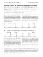
In the structures A and B, Kdo, Hep, Hep7Cm,
GalNAcAN3Ac, GalNFoAN, QuiNAc, GalNAla repre-
sent 3-deoxy-
D
-manno-octulosonic acid,
L
-glycero-
D
-manno-
heptose, 7-O-carbamoyl-
L
-glycero-
D
-manno-heptose, 2-acet-
amido-3-O-acetyl-2-deoxygalacturonamide, 2-formamido-
2-deoxygalacturonamide, 2-acetamido-2,6-dideoxyglucose
and 2-(
L
-alanylamino)-2-deoxygalactose, respectively; all
sugars are in the pyranose form and have the
D
configuration
unless otherwise stated. One or more phosphorylation sites
may be occupied by diphosphate groups. In a minority of the
LPS molecules, an O-acetyl group is present in the outer core
region at unknown position.
The site and the configuration of the linkage between the
O-polysaccharide and the core and the structure of the O-
polysaccharide repeating unit were defined in P. aeruginosa
immunotype 1. The QuiNAc residue linked to the Rha
residue of the core was found to have the b configuration,
whereas in the interior repeating units of the O-polysac-
charide this residue is in the a-configuration. The data
obtained are in accordance with the initiation of biosynthesis
of the O-polysaccharide of P. aeruginosa O6, which is closely
related to immunotype 1, by transfer of
D
-QuiNAc-1-P to
undecaprenyl phosphate followed by synthesis of the
repeating O-antigen tetrasaccharide.
Keywords: lipopolysaccharide; core oligosaccharide struc-
ture; repeating unit; O-polysaccharide; Pseudomonas
aeruginosa.
Correspondence to Y. A. Knirel, N. D. Zelinsky Institute of Organic Chemistry, Russian Academy of Sciences, Leninsky Prospekt 47, 119991
Moscow, GSP-1, Russia. Fax: + 7095 1355328. E-mail:
Abbreviations: aKdo, anhydro form of 3-deoxy-
D
-manno-octulosonic acid; Cm, carbamoyl; FT-ICR, Fourier transform ion cyclotron resonance;
Fo, formyl; Kdo, 3-deoxy-
D
-manno-oct-2-ulosonic acid; LPS, lipopolysaccharide; OS, oligosaccharide; Hep,
L
-glycero-
D
-manno-heptose; HexN,
hexosamine (GlcN or GalN); GalNA, 2-amino-2-deoxygalacturonic acid; DHexNA, 2-amino-2-deoxy-
L
-threo-hex-4-enuronic acid; QuiN,
2-amino-2,6-dideoxy-
D
-glucose; Und-P, undecaprenyl phosphate.
(Received 28 November 2001, revised 25 February 2002, accepted 11 March 2002)
Eur. J. Biochem. 269, 2194–2203 (2002) Ó FEBS 2002 doi:10.1046/j.1432-1033.2002.02875.x
Pseudomonas aeruginosa is an opportunistic human patho-
gen, which causes severe infections in hosts with weakened
defense mechanisms often as a result of thermal burns,
surgical operations, or another predisposing disease, such as
cystic fibrosis and cancer [1,2]. Lipopolysaccharide (LPS) is
the major surface antigen of P. aeruginosa, which plays an
important role in interaction of the bacterium with its host.
It is composed of lipid A, a core oligosaccharide, and an
O-chain polysaccharide built up of oligosaccharide repeat-
ing units. Lipid A and core are structurally conserved parts
of LPS, whereas the O-polysaccharide is highly variable in
composition and structure. The O-specific heteropolysac-
charides are synthesized by assembling individual mono-
saccharides into an oligosaccharide (the so called ÔbiologicalÕ
repeating unit) on an undecaprenyl phosphate (Und-P)
carrier followed by polymerization. In several P. aeruginosa
strains, the O-polysaccharide is structurally heterogeneous,
most likely, as a result of postpolymerization nonstoichio-
metric modifications, such as O-acetylation, amidation or
epimerization at C5 of uronic acids [3]. The structures of the
O-polysaccharides of all serologically distinguishable
smooth (S)-type strains have been determined [3], but the
biological repeating unit was defined only in P. aeruginosa
serogroup O5 [4]. Structures of the core [5–7] and lipid A
[8,9] of P. aeruginosa LPS have also been investigated. The
inner core region is composed of two residues of 3-deoxy-
D
-
manno-oct-2-ulosonic acid (Kdo) and two residues of
L
-glycero-
D
-manno-heptose (Hep), one of which is specific-
ally 7-O-carbamoylated. The inner core region is character-
ized by a high degree of phosphorylation but data on the
location of the phosphate groups are contradictory [5–7].
The outer core region contains up to four
D
-glucose
residues, one
L
-rhamnose residue, and one residue of
N-(
L
-alanyl)- or N-acetyl-
D
-galactosamine; it may include
also O-acetyl groups. Recently, it has been reported that
strain P. aeruginosa PAO1 and a cystic fibrosis isolate
P. aeruginosa 2192 produce two different glycoforms of the
LPS outer core [4,5].
Strains belonging to P. aeruginosa immunotype 1 (sero-
group O6) are frequently isolated from a variety of sources
[3]. Previously, the following structure of the O-polysac-
charide of immunotype 1 has been established [3,10–12]:
! 4Þ-a-d-GalpNAcAN3Ac-ð1 ! 4Þ-a-d-
GalpNFoAN-ð1 ! 3Þ-a-d-QuipNAc-ð1 ! 2Þ
-a-l-Rhap-ð1 !
where GalNAcAN and GalNFoAN stand for 2-acetamido-
and 2-formamido-2-deoxygalacturonamide, respectively;
QuiNAc stands for 2-acetamido-2,6-dideoxyglucose.
In this paper, we present new structural data on the LPS
of P. aeruginosa immunotype 1, including elucidation of the
core phosphorylation pattern, the O-polysaccharide biolo-
gical repeating unit, and the site and the mode of the
attachment of the O-polysaccharide to the core.
MATERIALS AND METHODS
Bacterium and cultivation
P. aeruginosa immunotype 1, strain 170041, was from the
Hungarian National Collection of Medical Bacteria
(National Institute of Hygiene, Budapest, Hungary). It
belongs to serogroup O6 of the international antigenic
typing system (IATS) and is characterized by an O-antigen
factor O6a according to the classification scheme of Lanyi &
Bergan [3]. Cells were grown in Roux flasks with solid agar
medium based on Hottinger broth at 37 °C for 18 h, then
washed in physiological saline, separated by centrifugation,
washed with acetone and dried.
Isolation of the lipopolysaccharide
LPS was isolated from dry bacterial cells by extraction with
aqueous 45% phenol (2 · 30 min) at 67 °C [13]. Cells were
removed by centrifugation (4000 g, 60 min). The superna-
tant was dialyzed against distilled water, nucleic acids were
precipitated using Cetavlon [13] and removed by centrifu-
gation (5000 g, 90 min). The supernatant was dialyzed
against distilled water and lyophilized.
Mild acid degradation of the lipopolysaccharide
LPS (200 mg) was dissolved in 0.1
M
sodium acetate buffer
pH 4.2 and heated for 13 h at 100 °C. The precipitate was
removed by centrifugation (12 000 g, 10 min), the superna-
tant fractionated by gel-permeation chromatography on a
column (80 · 2.5 cm) of Sephadex G-50 (Pharmacia-
Upjohn, Uppsala, Sweden) in pyridinium acetate buffer
pH 4.5 (4 mL pyridine and 10 mL HOAc in 1 L water) at
30 mLÆh
)1
, monitoring with a Knauer differential refrac-
tometer, and 5-mL fraction volume. Fractions 24–34 were
pooled to give a polysaccharide and fractions 41–54 an
oligosaccharide mixture (19.8 and 9.1% of the LPS mass,
respectively), the latter containing the core and the core with
one O-antigen repeating unit attached.
Alkaline degradation of the lipopolysaccharide
LPS (200 mg) was treated with anhydrous hydrazine
(4 mL) for 1 h at 37 °C, then 16 h at 20 °C, hydrazine
was flushed out in a stream of air at 30–33 °C, the residue
washed with cold acetone and dried in vacuum. The
O-deacylated LPS was dissolved in 4
M
NaOH (8 mL), the
solution was flushed with nitrogen for 1 h with stirring,
heated at 100 °C for 16 h, cooled, acidified with concentra-
ted HCl to pH 5.5, extracted twice with dichloromethane,
and the aqueous solution desalted by gel chromatography
on Sephadex G-50. The yield of the oligosaccharide fraction
was 16.1% of the LPS mass.
Composition analysis
Core oligosaccharide was hydrolyzed with 2
M
CF
3
CO
2
H
(120 °C, 2 h), monosaccharides were converted into the
alditol acetates and analyzed by GLC on a Hewlett-Packard
HP 5890 Series II chromatograph equipped with a 30-m
fused-silica SPB-5 column (Supelco), using a temperature
gradient of 160 °C (3 min) to 290 °Cat10°CÆmin
)1
.
Mass spectrometry
ESI MS was performed using a Fourier transform ion
cyclotron resonance (FT-ICR) mass analyser (ApexII,
Bruker Daltonics, USA) equipped with a 7-T actively
shielded magnet and an Apollo electrospray ion source.
Ó FEBS 2002 LPS of Pseudomonas aeruginosa immunotype 1 (Eur. J. Biochem. 269) 2195
Capillary skimmer dissociation was induced by increasing
the capillary exit voltage from )100 to )350 V. Samples
were dissolved in a 30 : 30 : 0.01 (v/v/v) mixture of
2-propanol, water, and triethylamine at a concentration of
20 ngÆlL
)1
and sprayed with a flow rate of 2 lLÆmin
)1
.
NMR spectroscopy
The NMR spectra were obtained on a Bruker DRX-500
spectrometer at 30 °C in 99.96% D
2
O. Prior to the
measurements, the samples were lyophilized twice from
D
2
O. Chemical shifts are referenced to internal acetone (d
H
2.225, d
C
31.45) or external aqueous 85% H
3
PO
4
(d
P
0.0).
Bruker software
XWINNMR
1.2 was used to acquire and
process the data. A mixing time of 200 or 100 ms was used
in 2D TOCSY and ROESY experiments, respectively.
RESULTS
The LPS was delipidated by mild acid hydrolysis at pH 4.2,
and the products were fractionated by gel-permeation
chromatography to give a high-molecular-mass O-polysac-
charide and an oligosaccharide mixture, both eluting as
wide peaks. Sugar analysis of the oligosaccharide product
revealed Glc, Rha, and
L
-glycero-
D
-manno-heptose (Hep) in
the ratios 4 : 2 : 1, respectively, as well as a trace amount
of GalN. These monosaccharides are typical components of
P. aeruginosa LPS core [5–7]; Rha is also present in the O-
polysaccharide repeating unit [11,12]. Most likely, a lower
content of Hep than expected is due to its phosphorylation
and poor release of GlaN is accounted for by its N-acylation
with
L
-alanine [5–7].
The oligosaccharide was then studied by capillary skim-
mer dissociation ESI FT-ICR MS (Fig. 1). The mass
spectrum showed an intense group of [M-H]
–
pseudo-
molecular ions for core oligosaccharides 6dHexHex
3
-
(HexNAla)Hep(HepCm)aKdoP
0)2
Ac
0)1
with a Kdo
residue in an anhydro form [14] and a variable number of
phosphate (P
0-2
) and O-acetyl (Ac
0)1
) groups (Fig. 1A).
The major ion peak at m/z 1590.41 corresponded to the
monophosphorylated non-O-acetylated derivative (the cal-
culated molecular mass 1591.48 Da). A similar series was
observed in the ESI mass spectrum of the core oligosac-
charide from the rough (R)-type LPS of P. aeruginosa 2192
[5]. In addition, a less intense series of [M-H]
–
pseudo-
molecular ions was present for the core with one O-poly-
saccharide repeating unit attached (Fig. 1B). Again,
heterogeneity of the oligosaccharides was associated with
nonstoichiometric phosphorylation (P
0-2
) and O-acetyla-
tion (Ac
0)2
) as well as with incomplete amidation of
GalNAcA or/and GalNAcFo residues resulting in a mass
difference of 1 or 2 Da. The major ion peak at m/z
2383.62 corresponded to the monophosphorylated bisam-
idated derivative containing one O-acetyl group (mostly
from the O-polysaccharide repeating unit) having the
calculated molecular mass 2384.75 Da. Finally, in the
mass spectrum there was a region of fragment ions from
the reducing end (Y and Z) induced by cleavage of the
linkage between two heptose residues (Fig. 1C). They
contained one or two phosphate groups and no O-acetyl
groups. Peaks for triple-charged [M-3H]
3–
pseudomolecu-
lar ions of the core oligosaccharides were in the same
spectral region.
The
1
H-NMR spectrum of the mild acid degradation
oligosaccharide product contained a number of signals for
the anomeric protons at d 4.64–5.74, methylene protons of
Kdo residues at d 1.9–2.2, methyl groups of 6-deoxy sugars
at d 1.26–1.35 and alanine at d 1.56,aswellasN-acetyland
O-acetyl groups. Analysis of the spectrum using 2D COSY
and TOCSY experiments confirmed the presence of com-
ponents of both core and O-polysaccharide repeating unit.
The position of the major GalNAcAN signals indicated
that, like in the O-polysaccharide, this residue is 3-O-
acetylated as followed from a downfield displacement of the
H3 signal to d 4.89, e.g. by 1 p.p.m., due to a deshielding
effect of the 3-O-acetyl group. An attempt to determine the
location of the second, minor O-acetyl group and phosphate
groups in the core by NMR spectroscopy failed owing to a
high degree of structural heterogeneity.
The LPS was O-deacylated by mild hydrazinolysis and
N-deacylated by strong alkaline hydrolysis [15]. The alkaline
degradation was accompanied by depolymerization of the
O-polysaccharide by b-elimination in 4-substituted GalNA
residues, which were converted into the corresponding hex-
4-enuronic acid (DHexNA) (Fig. 2). The negative ion mode
ESI FT-ICR mass spectrum of the product (Fig. 3) showed
the presence of the core-lipid A backbone oligosaccharide
6dHexHex
3
(HexN)
3
Hep
2
Kdo
2
P
5
and that with a DHexNA-
QuiN disaccharide remainder of the repeating unit of the O-
polysaccharide attached (the determined and calculated
molecular masses 2357.54 and 2357.51 Da for the former
and 2659.64 and 2659.61 Da for the latter compound,
respectively). In addition to the major pentakisphosphoryl-
ated compounds (P
5
), there were minor compounds con-
taining six (P
6
) and four (P
4
) phosphate groups.
The
1
H-NMR spectrum of the alkaline degradation
product (Fig. 4, Table 1) contained, among other things,
signals for anomeric protons at d 4.64–5.74, axial and
equatorial protons (H3) of Kdo residues at d 1.85–2.24,
three methyl groups (H6) of one QuiN and two rhamnose
residues at d 1.26–1.34, and a proton at the double bond
(H4) of DHexNA at d 5.98. Accordingly, the
13
C-NMR
spectrum (Table 2) showed signals for anomeric carbons at
d 92.9–105.7, five nitrogen-bearing carbons (C2 of GlcN
I
,
GlcN
II
,GalN,QuiN,andDHexNA) at d 51.8–56.7, methyl
groups of one QuiN and two rhamnose residues at d 17.6–
18.3, methylene groups (C3) of two Kdo residues at d 35.3
and 36.0, three carboxyl groups (C1 of two Kdo residues
and C6 of DHexNA) at d 174.2–174.7, C4 and C5 of
DHexNA at d 107.3 and 147.1. The
31
P-NMR spectrum of
the product contained signals for five monophosphate
monoesters at d )1.8, )0.1, 0.2, and 1.3 (two phosphorous).
The
1
H- and
13
C-NMR spectra of the alkaline degrada-
tion product were assigned using 2D shift-correlated NMR
experiments (COSY, TOCSY, ROESY, and H-detected
1
H,
13
C HMQC) (Tables 1 and 2). The monosaccharide spin
systems were assigned based on the coupling constant values
and those for amino sugars by correlation of the protons at
the nitrogen-bearing carbons to the corresponding carbons.
The configurations of the glycosidic linkages of the gluco
and galacto sugar residues (Glc, GlcN, GalN, QuiN, and
DHexNA) followed from the J
1,2
coupling constant values
and those of Rha, Hep
I
,Hep
II
,Kdo
I
,andKdo
II
from
typical
1
H NMR chemical shifts (compare published data
[7,16]). Two series of NMR signals were present for most
sugars in the outer core region, which consists of three
2196 O. V. Bystrova et al. (Eur. J. Biochem. 269) Ó FEBS 2002
glucose residues (Glc
I
–Clc
III
) and one residue each of
rhamnose and GalN (Fig. 4, Table 1). In contrast, Hep,
Kdo and GlcN residues in the inner core-lipid A backbone
region gave only one series of signals each. These findings
indicated the occurrence of the outer core as two glycoforms
(compare published data [4,5]).
Linkage and sequence analyses were performed using 2D
ROESY and HMBC experiments. The glycosylation pat-
tern in the core-lipid A backbone region was found to be the
same as in the LPS of P. aeruginosa O5 [4,7] and 2192 [5]
with two glycoforms 1 and 2 (Fig. 5A and B). As judged by
the signal intensities of the methyl groups of Rha and QuiN,
in P. aeruginosa immunotype 1 glycoforms 1 and 2 are
present in almost equal amounts.
The remainder of the degraded first repeating unit of the
O-polysaccharide was shown to be a b-DHexNA-(1 fi 3)-
b-
D
-QuiN disaccharide attached to position 3 of the
terminal Rha residue in glycoform 2 (Fig. 5B). This
followed from DHexNA H1/QuiN H3 and QuiN H1/Rha
H3 at d 5.65/4.20 and 5.06/4.01 in the ROESY spectrum, as
Fig. 1. Negative ion capillary skimmer disso-
ciation ESI FT-ICR mass spectrum of mild acid
degradation products of the LPS. Shown are
regions of [M-H]
–
pseudomolecular ions for
the core oligosaccharide [M, 6dHex-
Hex
3
(HexNAla)Hep(HepCm)aKdo] (A),
the core oligosaccharide with one O-polysac-
charide repeating unit [M
I
,6dHexNAc
(HexNAcAN)(HexNFoAN)(6dHex)
2
Hex
3
(HexNAla)Hep(HepCm) aKdo] (B), and
[M-3H]
3–
pseudomolecular ions and fragment
ions from the reducing end (C), which repre-
sent all essentials ion peaks found in the
complete spectrum, except for peaks for
doubly charged pseudomolecular ions. An
explanation of the fragments is shown at the
top of the region C. M
P1
,M
P1Ac1
, etc., refer to
the molecular ions with one phosphate group
and no or one O-acetyl group, Y
P1
,Z
P2
,etc.,
refer to the fragment ions with one and two
phosphate groups, respectively.
Ó FEBS 2002 LPS of Pseudomonas aeruginosa immunotype 1 (Eur. J. Biochem. 269) 2197
well as DHexNA H1/QuiN C3, QuiN H1/Rha C3, DHex-
NA C1/QuiN H3, and QuiN C1/Rha H3 correlations at d
5.65/80.1, 5.06/80.7, 97.4/4.20, and 101.1/4.01 in the HMBC
spectrum, respectively. Remarkably, the QuiN residue has
the b configuration (d
H1
5.06, J
1,2
8Hz),whereasinthe
interior repeating units of the O-polysaccharide, this sugar is
in the a-configuration [11,12]. The terminal Rha residue in
glycoform 1 is not substituted (Fig. 5A) as confirmed by the
C2–C6 chemical shifts (Table 2) being close to those in free
a-rhamnopyranose [17].
The positions of the phosphate groups were determined
using a
1
H,
31
P HMQC experiment (Fig. 6), which showed
three-bond correlations for the phosphorus signals with
the H1 signals of GlcN
I
,H4ofGlcN
II
,H2andH4of
Hep
I
, and H6 of Hep
II
at d )1.80/5.74, 0.18/3.85, )0.1/
4.53, 1.30/4.50, and 1.33/4.54, respectively. The assignment
of the Hep
I
P2/H2 and Hep
II
P6/H6 cross-peaks was
confirmed by Hep
I
P2/H3 and Hep
II
P6/H5 four-bond
correlations.
Therefore, the alkaline degradation products have struc-
tures shown in Fig. 5. Taking all of the data together, the
structures of the core and core with one O-polysaccharide
repeating unit in the LPS of P. aeruginosa immunotype 1
were established (Fig. 7).
DISCUSSION
Previously, the structure of the O-polysaccharide of
P. aeruginosa immunotype 1 LPS was elucidated [3,10–
12]. However, it cannot be ascertained from this structure
what are the first and last monosaccharides in the repeating
unit, nor which monosaccharide would be linked to the
LPS core. The actual biological repeating unit represents
the properly ordered oligosaccharide, which, after having
been preassembled on an Und-P carrier, is polymerized
into the O-polysaccharide. In this work, the structure of the
biological repeating unit was established by studies of the
LPS degradation products prepared using two different
Fig. 3. Charge deconvoluted negative ion ESI
FT-ICR mass spectrum of alkaline degradation
products. MandM
I
belong to the core oligo-
saccharide 6dHexHex
3
(HexN)
3
Hep
2
Kdo
2
P
5
and the core oligosaccharide with a remainder
of the O-polysaccharide repeating unit
DHexNA(6dHexN)6dHexHex
3
(HexN)
3
Hep
2
Kdo
2
P
5
, respectively.
Fig. 2. Alkaline degradation of the LPS. The
glycosidic linkage of QuiNAc is a between the
O-polysaccharide repeating units and b
between the O-polysaccharide and the core.
2198 O. V. Bystrova et al. (Eur. J. Biochem. 269) Ó FEBS 2002
approaches. One was mild acid degradation, which,
together with a long-chain polysaccharide, resulted in an
oligosaccharide mixture containing a core oligosaccharide
and that with one O-polysaccharide repeating unit at-
tached. The other was strong alkaline degradation, which
caused b-elimination in GalNA residues present in the O-
polysaccharide to give a core oligosaccharide with a
truncated single O-polysaccharide repeating unit from the
LPS species with both long-chain O-polysaccharide and
one repeating unit.
Analysis of the products with one O-polysaccharide
repeating unit or its remainder by ESI MS and NMR
spectroscopy enabled determination of not only the biolo-
gical repeating unit but also of the mode and the site of the
linkage between the O-polysaccharide and the core. It was
found that the QuiNAc residue, which occupies the
reducing end of the biological repeating unit, has the b con-
figuration when linking the O-polysaccharide to the core but
the a configuration when connecting the repeating units to
each other in the O-polysaccharide. This finding is in
Fig. 4.
1
H NMR spectrum of alkaline degradation products. Arabic numerals refer to protons in sugar residues. Designations for Glc
II
,Glc
III
,and
Rha in glycoforms 1 and 2 are not italicized and italicized, respectively.
Table 1.
1
H-NMR data (d, p.p.m.).
Sugar residue
H1
H3ax
H2
H3eq
H3
H4
H4
H5
H5
H6
H6(6a)
H7
H6b(7a)
H8a
H7b
H8b
Inner core-lipid A backbone
fi 6)-a-
D
-GlcpN
I
-1-P 5.74 3.47 3.92 3.63 4.11 4.28 3.82
fi 6)-b-
D
-GlcpN
II
-(1 fi 4.84 3.14 3.89 3.85 3.76 3.73 3.51
fi 4,5)-a-Kdop
I
-(2 fi 2.07 2.24 4.11 4.29 3.73 3.85 3.89 3.60
a-Kdop
II
-(2 fi 1.85 2.09 4.13 4.08 3.73 4.11 3.96 3.69
fi 3)-a-Hepp
I
2,4P-(1 fi 5.36 4.53 4.18 4.50 4.28 3.98 3.94 3.80
fi 3)-a-Hepp
II
6P-(1 fi 5.14 4.41 4.22 3.84 4.01 4.54 3.80 3.73
Outer core, glycoform 1
fi 3,4)-a-
D
-GalpN-(1 fi 5.58 3.84 4.45 4.40 4.20 3.88 3.88
fi 6)-b-
D
-Glcp
I
-(1 fi 4.64 3.27 3.53 3.28 3.68 3.90 3.80
fi 6)-a-
D
-Glcp
II
-(1 fi 4.99 3.51 3.70 3.61 4.17 3.89 3.78
a-
D
-Glcp
III
-(1 fi 4.97 3.57 3.68 3.46 3.65 3.85 3.78
a-
L
-Rhap-(1 fi 4.79 4.01 3.80 3.44 3.73 1.31
Outer core, glycoform 2
fi 3,4)-a-
D
-GalpN-(1 fi 5.58 3.84 4.59 4.40 4.20 3.88 3.88
fi 3,6)-b-
D
-Glcp
I
-(1 fi 4.66 3.49 3.65 3.53 3.68 3.90 3.90
a-
D
-Glcp
II
-(1 fi 5.03 3.51 3.76 3.52 4.03 3.84 3.84
a-
D
-Glcp
III
-(1 fi 5.00 3.56 3.70 3.40 3.67 3.86 3.73
fi 3)-a-
L
-Rhap-(1 fi 5.16 4.25 4.01 3.65 4.03 1.26
Remainder of the O-polysaccharide repeating unit
fi 3)-b-
D
-QuipN-(1 fi 5.06 3.32 4.20 3.51 3.62 1.34
b-
L
-DHexpNA-(1 fi 5.65 3.73 4.52 5.98
Ó FEBS 2002 LPS of Pseudomonas aeruginosa immunotype 1 (Eur. J. Biochem. 269) 2199
accordance with the biosynthesis pathway of O-polysac-
charides, which involves multiple enzymes that mediate the
formation of the QuiNAc glycosidic linkage. One of them,
glycosyltransferase WbpL, transfers
D
-QuiNAc-1-P from
UDP-
D
-QuiNAc to Und-P to initiate the O-polysaccharide
repeating unit biosynthesis in P. aeruginosa O6 [18], which is
closely related to P. aeruginosa immunotype 1. Another
enzyme, O-antigen polymerase Wzy, mediates polymeriza-
tion of the preassembled oligosaccharide attached to Und-
PP to form a long-chain O-polysaccharide. Finally, ligase
WaaL ligates the preassembled oligosaccharide or the long-
chain O-polysaccharide to the core-lipid A moeity.
Differences between O-polysaccharide structures of
P. aeruginosa immunotype 1 and related serotypes of
P. aeruginosa O6 are associated with the configuration
of the QuiNAc linkage (a or b)andthesiteofattachmentof
QuiNActoRha(atposition2or3)aswellaswith
O-acetylation and amidation of the GalNA derivatives
(Table 3). Because O-acetylation and amidation, which are
nonstoichiometric, are likely to be postpolymerization
modifications, it is possible that the oligosaccharide assem-
bled on Und-PP is the same in all the serogroup O6 strains
and its biosynthesis involves the same WbpL protein and
other glycosyltransferases (WbpT, WbpU, and WbpR for
D
-GalNAcA,
D
-GalNFoA, and Rha, respectively [18]). In
contrast, the O-antigen polymerase Wzy, or other putative
protein(s) that influence the activity of Wzy [18], must be
divisible into at least three types in order to adopt the
a1 fi 2-, a1 fi 3-, and b1 fi 3-linkages between QuiNAc
and Rha within the O-polysaccharide.
Previously, the structure of the biological repeating unit
of the O-polysaccharide was elucidated in P. aeruginosa O5
[4]. This O-polysaccharide includes
D
-FucNAc, which is
located at the reducing end of the biological repeating unit
Table 2.
13
C-NMR data (d, p.p.m.).
Sugar residue C1 C2 C3 C4 C5 C6 C7 C8
Inner core-lipid A backbone
fi 6)-a-
D
-GlcpN
I
-1-P 92.9 55.2 70.5 70.7 73.8 70.5
fi 6)-b-
D
-GlcpN
II
-(1 fi 100.2 56.7 72.8 75.5 75.0 63.7
fi 4,5)-a-Kdop
I
-(2 fi 174.2
a
100.1 35.3 72.2 69.5 73.1 70.3 64.9
a-Kdop
II
-(2 fi 174.2
a
102.4 36.0 66.8 67.7 73.2 70.3 64.3
fi 3)-a-Hepp
I
2,4P-(1 fi 98.6 75.6 75.3 74.2 73.3 71.8 64.4
fi 3)-a-Hepp
II
6P-(1 fi 103.2 70.5 78.9 67.7 72.6 71.8 62.7
Outer core, glycoform 1
fi 3,4)-a-
D
-GalpN-(1 fi 98.3 51.8 77.8 76.8 73.7 61.1
fi 6)-b-
D
-Glcp
I
-(1 fi 105.7 74.6 76.9 71.5 75.8 68.8
fi 6)-a-
D
-Glcp
II
-(1 fi 100.6 73.0 74.0 70.1 71.6 67.7
a-
D
-Glcp
III
-(1 fi 99.6 72.4 74.4 70.5 73.5 61.7
a-
L
-Rhap-(1 fi 102.3 71.4 71.6 73.3 70.0 18.3
Outer core, glycoform 2
fi 3,4)-a-
D
-GalpN-(1 fi 98.3 51.8 77.8 76.8 73.7 61.1
fi 3,6)-b-
D
-Glcp
I
-(1 fi 105.6 74.9 83.2 69.5 75.8 68.8
a-
D
-Glcp
II
-(1 fi 100.5 73.0 73.8 70.3 72.6 61.5
a-
D
-Glcp
III
-(1 fi 99.3 72.4 74.4 70.8 73.5 62.1
fi 3)-a-
L
-Rhap-(1 fi 101.7 71.7 80.7 72.3 70.1 17.6
Remainder of the O-polysaccharide repeating unit
fi 3)-b-
D
-QuipN-(1 fi 101.1 55.9 80.1 76.1 73.5 17.7
b-
L
-DHexpNA-(1 fi 97.4 53.3 63.3 107.3 147.1 174.7
a
a
Assignment could be interchanged.
Fig. 5. Structures of the major alkaline degra-
dation products of the LPS. Glycoform 1 core
is unsubstituted (A) and glycoform 2 core
substitued with a remainder of the O-poly-
saccharide repeating unit (B). All sugars are in
the pyranose form and have the
D
configur-
ation unless otherwise stated.
2200 O. V. Bystrova et al. (Eur. J. Biochem. 269) Ó FEBS 2002
and thus plays the same role in serogroup O5 as
D
-QuiNAc
in serogroup O6. Interestingly, WbpL that transfers
D
-FucNAc-1-P from UDP-
D
-FucNAc to Und-P in
P. aeruginosa O5 showed homology to WbpL in
P. aeruginosa O6 and both enzymes possess substrate
specificity for UDP-
D
-FucNAc and UDP-
D
-QuiNAc [18].
In strains of most other P. aeruginosa serogroups the
O-polysaccharide includes
D
-QuiNAc or/and
D
-FucNAc
[3]; therefore, the initiation of the O-polysaccharide biosyn-
thesis may proceed in a similar manner in these strains too.
The occurrence of two core glycoforms seems to be a
common feature of all P. aeruginosa LPS[4,5].Asinthe
LPS of P. aeruginosa O5 [4], in the LPS of immunotype 1
(serogroup O6) the terminal 1 fi 3-linked Rha residue of
the core oligosaccharide is the site of the attachment of the
O-polysaccharide. This residue only occurs in the core
glycoform 2, whereas the terminal 1 fi 6-linked Rha
residue in the other, isomeric glycoform 1 cannot accept
the O-polysaccharide. No unsubstituted core glycoform 2
was detected, nor a core glycoform with two Rha residues.
It could be thus suggested that the attachment of the 1 fi
6-linked Rha blocks the attachment of the 1 fi 3-linked
Rha, which is the acceptor of the O-polysaccharide. A
competition of the corresponding rhamnosyl transferases
may provide a mechanism for regulation of the content of
long-chain (S-type) and short-chain (R-type) LPS species on
the cell surface by an enhanced synthesis of the appropriate
glycoform. Like the O-polysaccharide chain length, the
content of the LPS species containing the core with one
O-polysaccharide repeating unit attached (SR-type LPS) is
controlled by the O-antigen chain length regulator Wzz [18],
which influences the functioning of O-antigen polymerase
and ligase by a mechanism that is not clearly understood.
As in P. aeruginosa strains studied previously [5–7,19,20],
the core of the LPS of P. aeruginosa immunotype 1 is
distinguished by a high degree of phosphorylation. Three
major phosphorylation sites were determined in the core,
two of which are at positions 2 and 4 of Hep
I
and one at
position 6 of Hep
II
. This finding is in agreement with the
phosphorylation pattern in P. aeruginosa strains H4 [6] and
2192 [5] but inconsistent with the data reported previously
for P. aeruginosa O5 and O6 strains [7]. According to the
latter data, all three phosphorylation sites are located at
Hep
I
at positions 2, 4, and 6, whereas Hep
II
is nonphos-
phorylated. Such a pattern seems to be unlikely because the
release of 7-O-carbamoylated Hep
II
is significantly increased
by dephosphorylation [21]; these discrepancies are unlikely
to be due to a strain difference because P. aeruginosa O6
and immunotype 1 are closely related.
Another feature of the LPS core of P. aeruginosa
immunotype 1 is O-acetylation. In P. aeruginosa, O-acety-
lation has been recently reported in the core of a rough,
serogroup O1-derived cystic fibrosis isolate, strain 2192,
Fig. 7. Structures of the major glycoform 1 core (A) and glycoform 2
core substitued with one O-polysaccharide repeating unit (B). All sugars
are in the pyranose form and have the
D
configuration unless otherwise
stated. It is not excluded that one or more phosphorylation sites are
occupied by diphosphate groups. In the minor products, the outer core
region includes one O-acetyl group at unknown position. O-Acetyla-
tation of GalNAcA and amidation of both GalNA derivatives in the
O-polysaccharide repeating unit are incomplete.
Fig. 6. 2D
1
H,
31
P HMQC spectrum of alka-
line degradation products. Three-bond and
four-bond correlations are shown by positive
and negative levels, respectively. Other four-
bondcorrelationsweretooweaktobedis-
tinguished from noise signals.
Ó FEBS 2002 LPS of Pseudomonas aeruginosa immunotype 1 (Eur. J. Biochem. 269) 2201
which produces an R-type LPS [5]. The outer core of this
strain has at least four O-acetylation sites, and the major
LPS species is mono-O-acetylated. A similar O-acetylation
pattern with up to five O-acetylation sites has been found in
thecoreofP. aeruginosa immunotype 5, O3a,3b,3c, and
O12 (O. V. Bystrova, A. S. Shashkov, N. A. Kocharova,
Y. A. Knirel, B. Lindner, U. Za
¨
hringer & G. B. Pier,
unpublished data). In immunotype 1, the outer core has at
least one O-acetylation site and the O-acetyl group is present
in the core of a minority of the LPS molecules. The position
of the O-acetyl groups in the core of P. aeruginosa LPS, as
well as their biological significance, remains to be deter-
mined.
ACKNOWLEDGEMENTS
This work was supported by the Civilian Research and Development
Foundation (CRDF, USA) grant RB1-2042 (to Y. A. K. and
G. B. P.), the Sonderforschungsbereich (SFB, Germany) 470 (project
B4) (to U. Z.), the Deutsche Forschungsgemeinschaft grant LI-448/1-1
(to B. L. and U. Z.), and NIH grants AI22535 and HL58398 (to
G. B. P.).
REFERENCES
1. Pollack, M. (1985) Pseudomonas aeruginosa.InPrinciples and
Practice of Infectious Diseases, Part III, Infectious Diseases and
Their Etiologic Agents (Mandell, G.L., Douglas, R.G. & Bennet,
J.E., eds), pp. 1236–1250. John Wiley, New York.
2. Pier, G.B. (2000) Peptides, Pseudomonas aeruginosa, polysac-
charides and lipopolysaccharides – players in the predicament of
cystic fibrosis patients. Trends Microbiol. 8, 247–250.
3. Knirel, Y.A. (1990) Polysaccharide antigens of Pseudomonas
aeruginosa. CRC Crit. Rev. Microbiol. 17, 273–304.
4. Sadovskaya, I., Brisson, J R., Thibault, P., Richards, J.C., Lam,
J.S. & Altman, E. (2000) Structural characterization of the outer
core and the O-chain linkage region of lipopolysaccharide from
Pseudomonas aeruginosa serotype O5. Eur. J. Biochem. 267, 1640–
1650.
5. Knirel, Y.A., Bystrova, O.V., Shashkov, A.S., Kocharova, N.A.,
Senchenkova, S.N., Moll, H., Lindner, B., Za
¨
hringer, U., Hatano,
K. & Pier, G.B. (2001) Structural analysis of the lipopoly-
saccharide core of a rough, cystic fibrosis isolate of Pseudomonas
aeruginosa. Eur. J. Biochem. 268, 4708–4719.
6. Sa
´
nchez-Carballo, P.M., Rietschel, E.T., Kosma, P. & Za
¨
hringer,
U. (1999) Elucidation of the structure of an alanine-lacking core
tetrasaccharide trisphosphate from the lipopolysaccharide of
Pseudomonas aeruginosa mutant H4. Eur. J. Biochem. 261,500–
508.
7. Sadovskaya, I., Brisson, J R., Lam, J.S., Richards, J.C. &
Altman, E. (1998) Structural elucidation of the lipopolysaccharide
core regions of the wild-type strain PAO1 and O-chain-deficient
mutant strains AK1401 and AK1012 from Pseudomonas aerugi-
nosa serotype O5. Eur. J. Biochem. 255, 673–684.
8. Bhat, U.R., Marx, A., Galanos, C. & Conrad, R.S. (1990)
Structural studies of lipid A from Pseudomonas aeruginosa PAO1:
occurrence of 4-amino-4-deoxyarabinose. J. Bacteriol. 172, 6631–
6636.
9. Kulshin, V.A., Za
¨
hringer, U., Lindner, B., Ja
¨
ger, K.E., Dmitriev,
B.A. & Rietschel, E.T. (1991) Structural characterization of the
lipid A component of Pseudomonas aeruginosa wild-type and
rough mutant lipopolysaccharides. Eur. J. Biochem. 198, 697–704.
10. Vinogradov, E.V., Knirel, Y.A., Shashkov, A.S. & Kochetkov,
N.K. (1987) Determination of the degree of amidation of 2-deoxy-
2-formamido-
D
-galacturonic acid in O-specific polysaccharides of
Pseudomonas aeruginosa O4 and related strains. Carbohydr. Res.
170, C1–C4.
11. Knirel, Y.A., Shashkov, A.S., Dmitriev, B.A. & Kochetkov, N.K.
(1984) Structural studies of the Pseudomonas aeruginosa
immunotype 1 antigen, containing the new sugar constituents:
2-acetamido-2-deoxy-
D
-galacturonamide and 2-deoxy-2-for-
mamido-
D
-galacturonic acid. Carbohydr. Res. 133, C12–C14.
12. Knirel, Y.A., Vinogradov, E.V., Shashkov, A.S., Dmitriev, B.A.,
Kochetkov, N.K., Stanislavsky, E.S. & Mashilova, G.M. (1985)
Somatic antigens of Pseudomonas aeruginosa. The structure of the
Table 3. Structures of the O-polysaccharides of P. aeruginosa immunotype 1 and serogroup O6 (Lanyi-Bergan classification). In serotypes O6a,6b
and O6a,6d, GalNFoA is amidated to a minor extent (10–20%) [3,10–12].
2202 O. V. Bystrova et al. (Eur. J. Biochem. 269) Ó FEBS 2002
O-specific polysaccharide chains of lipopolysaccharide of
P. aeruginosa serogroup O4 (Lanyi) and related serotype O6
(Habs) and immunotype 1 (Fisher). Eur. J. Biochem. 150, 541–550.
13. Westphal, O. & Jann, K. (1965) Bacterial lipopolysaccharides.
Extraction with phenol-water and further applications of the
procedure. Methods Carbohydr. Chem. 5, 83–91.
14. Olsthoorn, M.M.A., Haverkamp, J. & Thomas-Oates, J.E. (1999)
Mass spectrometric analysis of Klebsiella pneumoniae ssp. pneu-
moniae roughstrainR20(O1
–
:K20
–
) lipopolysaccharide prepa-
rations: Identification of novel core oligosaccharide components
and three 3-deoxy-
D
-manno-oct-2-ulopyranosonic artifacts.
J. Mass Spectrom. 34, 622–636.
15. Holst, O. (2000) Deacylation of lipopolysaccharides and isolation
of oligosaccharide phosphates. In Bacterial Toxins. Methods and
Protocols (Holst, O., ed.), pp. 345–353. Humana Press, Totowa,
New Jersey.
16. Knirel, Y.A., Grosskurth, H., Helbig, J.H. & Za
¨
hringer, U. (1995)
Structures of decasaccharide and tridecasaccharide tetraphos-
phates isolated by strong alkaline degradation of O-deacylated
lipopolysaccharide of Pseudomonas fluorescens strain ATCC
49271. Carbohydr. Res. 279, 215–226.
17. Bock, K. & Pedersen, C. (1983) Carbon-13 nuclear magnetic
resonance spectroscopy of monosaccharides. Adv. Carbohydr.
Chem. Biochem. 41, 27–66.
18. Rocchetta, H.L., Burrows, L.L. & Lam, J.S. (1999) Genetics of
O-antigen biosynthesis in Pseudomonas aeruginosa. Microbiol.
Mol. Biol. Rev. 63, 523–553.
19. Masoud, H., Altman, E., Richards, J.C. & Lam, J.S. (1994)
General strategy for structural analysis of the oligosaccharide
region of lipooligosaccharides. Structure of the oligosaccharide
component of Pseudomonas aeruginosa IATS serotype O6
mutant R5 rough-type lipopolysaccharide. Biochemistry 33,
10568–10578.
20. Masoud, H., Sadovskaya, I., De Kievit, T., Altman, E., Richards,
J.C. & Lam, J.S. (1995) Structural elucidation of the lipopoly-
saccharide core region of the O-chain-deficient mutant strain A28
from Pseudomonas aeruginosa serotype 06 (International Anti-
genic Typing Scheme). J. Bacteriol. 177, 6718–6726.
21. Beckmann, F., Moll, H., Ja
¨
ger, K E. & Za
¨
hringer, U. (1995) 7-
O-Carbamoyl-
L
-glycero-
D
-manno-heptose: a new core constituent
in the lipopolysaccharide of Pseudomonas aeruginosa. Carbohydr.
Res. 267, C3–C7.
Ó FEBS 2002 LPS of Pseudomonas aeruginosa immunotype 1 (Eur. J. Biochem. 269) 2203
