Báo cáo Y học: Engineering and mechanistic studies of the Arabidopsis FAE1 b-ketoacyl-CoA synthase, FAE1 KCS pot
Bạn đang xem bản rút gọn của tài liệu. Xem và tải ngay bản đầy đủ của tài liệu tại đây (278.22 KB, 9 trang )
Engineering and mechanistic studies of the
Arabidopsis
FAE1
b-ketoacyl-CoA synthase, FAE1 KCS
Mahin Ghanevati and Jan G. Jaworski
Department of Chemistry and Biochemistry, Miami University, Oxford, OH, USA
The Arabidopsis FAE1 b-ketoacyl-CoA synthase (FAE1
KCS) catalyzes the condensation of malonyl-CoA with long-
chain acyl-CoAs. Sequence analysis of FAE1 KCS predicted
that this condensing enzyme is anchored to a membrane by
two adjacent N-terminal membrane-spanning domains. In
order to characterize the FAE1 KCS and analyze its mech-
anism, FAE1 KCS and its mutants were engineered with a
His
6
-tag at their N-terminus, and expressed in Saccharomyces
cerevisiae. The membrane-bound enzyme was then solubi-
lized and purified to near homogeneity on a metal affinity
column. Wild-type recombinant FAE1 KCS was active with
several acyl-CoA substrates, with highest activity towards
saturated and monounsaturated C16 and C18. In the
absence of an acyl-CoA substrate, FAE1 KCS was unable to
carry out decarboxylation of [3–
14
C]malonyl-CoA, indica-
ting that it requires binding of the acyl-CoA for decarb-
oxylation activity. Site-directed mutagenesis was carried out
on the FAE1 KCS to assess if this condensing enzyme was
mechanistically related to the well characterized soluble
condensing enzymes of fatty acid and flavonoid syntheses. A
C223A mutant enzyme lacking the acylation site was unable
to carry out decarboxylation of malonyl-CoA even when
18:1-CoA was present. Mutational analyses of the conserved
Asn424 and His391 residues indicated the importance of
these residues for FAE1-KCS activity. The results presented
here provide the initial analysis of the reaction mechanism
for a membrane-bound condensing enzyme from any source
and provide evidence for a mechanism similar to the soluble
condensing enzymes.
Keywords: very long chain fatty acids; fatty acid elongation;
condensation mechanism.
Fatty acids with greater than 18 carbon atoms (very long
chain fatty acids, VLCFA) are precursors of many biolo-
gically important compounds such as sphingolipids [1,2],
waxes [3], and triacylglycerols in many seed oils [4].
Biosynthesis of VLCFA in plants and animals, is dependent
on the activity of a membrane-bound fatty acid elongation
system which consists of four component reactions similar
to fatty acid synthase. The first reaction of elongation
involves condensation of malonyl-CoA with a long chain
acyl substrate producing a b-ketoacyl-CoA. Subsequent
reactions are reduction to b-hydroxyacyl-CoA, dehydration
to an enoyl-CoA, followed by a second reduction to form
the elongated acyl-CoA [5]. In both animals and plants, the
initial condensation reaction is believed to be the rate-
limiting step [6,7].
In Arabidopsis, FAE1 codes for a b-ketoacyl-CoA
synthase (FAE1 KCS) which is expressed exclusively in
the seed and catalyzes the initial condensation step in the
elongation pathway [8]. Based on fatty acid profiles of
transgenic plants and yeast, it has been reported that FAE1
KCS has a substrate preference for C18:1, producing
eicosenoic (C20:1) acid as the major product and erucic acid
(C22:1) as a minor product [7].
A prominent feature of b-ketoacyl-CoA synthases
involved in VLCFA biosynthesis is their membrane-bound
nature. This makes them different from all other condensing
enzymes studied to date, which are soluble enzymes. These
include, for example, those involved in fatty acid and
polyketide synthesis. Amino-acid sequence analysis of the
Arabidopsis KCS1 [9] indicated that elongase KCS enzymes,
including FAE1 KCS, have two transmembrane-spanning
domains close to their N-terminus, thus suggesting that
these enzymes are anchored to the membrane.
Although fatty acid elongases and their b-ketoacyl-CoA
synthase component have been partially purified from a
number of sources [10–13] and studied using cellular
fractions [14,15], the information about KCS enzymes and
their kinetic properties is very limited. This is mainly due to
the complexity of the membrane fractions used as the
enzyme source and the presence of a high level of
background activities. Currently, there is only one report
of an extensively purified membrane-bound KCS, jojoba
KCS [13], and its characterization was limited to the
substrate specificity.
Despite their membrane-bound nature, some domains of
the elongase condensing enzymes have limited homology to
two soluble condensing enzymes: plant chalcone synthases
[16] and 3-ketoacyl-ACP synthase III (KAS III) from plants
[17] and Escherichia coli [18]. The reaction mechanism of the
soluble condensing enzymes has been extensively studied,
and recently the crystal structures of chalcone synthase [19]
and all isoforms of KASs from E. coli [20–23] have been
published. Site-directed mutagenesis studies, as well as
crystal structures, indicate that these soluble condensing
enzymes all utilize the same general reaction mechanism
(Fig. 1). This involves, successively, transfer of the acyl
Correspondence to J. G. Jaworski, Donald Danforth Plant Science
Center, 975 North Warson Road, St Louis, MO 63132, USA.
Fax: + 314 587 1721, Tel.: + 314 587 1621,
E-mail:
Abbreviations: VLCFA, very long chain fatty acid; FAE1 KCS, fatty
acid elongase 1 b-ketoacyl-CoA synthase; FAS, fatty acid synthase.
(Received 20 February 2002, revised 9 May 2002,
accepted 10 June 2002)
Eur. J. Biochem. 269, 3531–3539 (2002) Ó FEBS 2002 doi:10.1046/j.1432-1033.2002.03039.x
primer substrate to an active-site cysteine forming an acyl
thioester intermediate, decarboxylation of the donor malo-
nyl substrate to yield an acetyl carbanion intermediate, and
finally, nucleophilic attack of the carbanion on the carbonyl
carbon atom of the thioester intermediate, resulting in the
formation of the product. The FAE1 KCS mechanism has
not been characterized and is known to use malonyl-CoA
instead of malonyl-ACP. Nonetheless, the mechanism of the
fatty acid synthase condensing enzymes should serve as an
appropriate model for the FAE1 KCS.
In addition to an active-site cysteine, at least one histidine
residue is directly involved in catalysis by soluble condensing
enzymes. Crystal structures of both KAS I [21] and KAS II
[20] from E. coli reveal the presence of two histidines in close
proximity to their active site cysteine of which at least one is
assumed to be important for enzyme catalysis. In addition,
crystal structures of E. coli KAS III [22,23] and alfalfa
chalcone synthase [19] show a histidine and an asparagine
residue in the active site architecture. The role of these
residues in catalysis were subsequently confirmed in both
KAS III [23] and chalcone synthase [24] by in vitro
mutagenesis and were shown to be important catalytic
residues in the decarboxylation of the malonyl substrate.
Based on its limited sequence similarity to resveratrol
synthase, a closely related condensing enzyme to chalcone
synthase, Lassner et al. [13] have tentatively identified the
active-site cysteine of the jojoba elongase KCS. The
corresponding cysteine of FAE1 KCS is Cys223. Recently,
we confirmed this hypothesis by site-directed mutagenesis of
FAE1 KCS [25]. Furthermore, of all conserved histidine
and asparagine residues of FAE1 KCS only His391 and
N424 align with the active-site histidine and asparagine of
both KAS III and chalcone synthase-related enzymes [25]. It
is likely that these residues play similar role to those
involved in chalcone synthase and KAS III.
In order to characterize the mechanism of an elongase
KCS, an expression system that allowed facile purification
of enzyme was required. We report here one approach to
the expression and purification of FAE1 KCS. We
engineered a His
6
-tag at N-terminus of FAE1, expressed it
in yeast and isolated the recombinant protein from yeast
microsomal pellet using a metal affinity column. Partially
purified recombinant FAE1 KCS was assayed for conden-
sation, decarboxylation, and substrate specificity. Addition-
ally, this provided an opportunity to analyze the effect of
mutagenesis of His391 and Asn424 in FAE1 KCS. To our
knowledge, this is the first report of the analysis of the
mechanism of a membrane-bound condensing enzyme from
any source.
EXPERIMENTAL PROCEDURES
Materials
S. cerevisiae (InvSc1) and pYES2 plasmid were purchased
from Invitrogen (Carlsbad, CA, USA). pBluescript was
from Stratagene (La Jolla, CA, USA). Oligonucleotide
primers were purchased from Integrated DNA Technol-
ogies (Coralville, IA, USA). Vent DNA polymerase,
nucleotide triphosphates, and restriction endonucleases
were purchased from New England Biolabs (Beverly, MA,
USA). T4 DNA ligase was from Gibco BRL (Grand Island,
NY, USA). Ni
2+
-PDC was purchased from Affiland
(Affinity Methodology in Biotechnology, Belgium). Throm-
bin, acyl-CoAs, and galactose were purchased from Sigma.
All other chemicals for media culture were obtained from
Fisher. [3–
14
C]malonyl-CoA was prepared as described by
Roughan [26].
Engineering of FAE1 KCS and its mutants
The overlap extension method as described by Ho et al.[27]
was used to introduce a thrombin cleavage site into the
Arabidopsis FAE1 KCS. Four constructs containing the
thrombin cleavage sites near the membrane-spanning
domain were prepared. LVPRGS was inserted at residues
74, 101, 115, and in one construct, at residue 106, LVPRGS
was substituted for RKADTS. This method requires two
gene-flanking primers and two internal overlapping oligo-
nucleotides containing the sequence encoding the thrombin
cleavage site. As our starting template was FAE1 gene
subcloned in pYEUra-3 (Clontech, Palo Alto, CA, USA),
flanking primers used were an antisense 5¢-CGTCAAG
GAGAAAAAACCTCTAGCCGAAT-3¢ primer and an
universal T7 sense primer. To insert a thrombin cleavage site
at amino-acid residue 115 in FAE1 KCS (T115-FAE1
KCS) a sense primer 5¢-GAACGTG
TTGGTTCCGC
GTGGTAGCGCATGTGATGATCCGTCCTCG-3¢ and
an antisense primer 5¢-CACATGC
GCTACCACGCGG
AACCAACACGTTCCGTGAAGAAGTATC-3¢ were
used (underlined sequence encodes for thrombin cleavage
site). This led to a 32-bp overlap in the second round of
PCR amplification. A similar approach was taken to make
the three other thrombin cleavage site constructs. Each of
these constructs was subcloned in the yeast expression
vector, pYES2, in which expression is under control of the
GAL1 promoter. The constructs were sequenced to verify
the presence of thrombin cleavage sequence.
To generate FAE1 KCS construct with a N-terminus
His
6
-tag the sense primer 5¢-CGCGGATCCGCGATG
(CAT)
6
ACTTCCGTTAACGTTAAGCTCCTTTAC-3¢
and the antisense primer 5¢-CGCGGATCCGCGTTAG
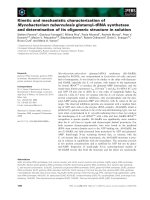
Fig. 1. Scheme of fatty acid synthase condensation reaction. The com-
mon reaction scheme of fatty acid synthase b-ketoacyl-ACP synthases
(KAS) involves: (1) acylation of an active-site cysteine; (2) binding of
malonyl-ACP followed by decarboxylation; and (3) attack on the acyl
group by the carbanion, producing a b ketoacyl-ACP.
3532 M. Ghanevati and J. G. Jaworski (Eur. J. Biochem. 269) Ó FEBS 2002
GACCGACCGTTTTGGACATGAGTCTT-3¢ were used.
To facilitate subcloning both primers were designed with a
terminal BamHI restriction site. A similar approach was
usedtopreparetheC-terminusHis
6
tag FAE1 KCS.
Previously prepared mutants of FAE1 KCS, C223A,
H391A, and H391K [25] were used as templates to generate
the His
6
-tagged recombinant mutants. To generate H391Q,
N424D, and N424H mutants, a set of overlapping muta-
genic oligonucleotides along with the flanking primers listed
above were used according to method by Ho et al. [27]. The
sense and antisense mutagenic primers were as follows (the
mutagenized codons are underlined): H391Q, sense
5¢-ATTTCTGTATTC
AAGCTGGAGGCAGAGCCGTG
AT-3¢,antisense5¢-CTGCCTCCAGC
TTGAATACAG
AAATG-3¢; N424D, sense 5¢-AGATTTGGG
GATAC
TTCATCTAGCTCAATTT-3¢,antisense5¢-AGATGAAGT
ATCCCCAAATCTATGTAACG-3¢; N424H, sense
5¢-AGATTTGGG
CATACTTCATCTAGCTCA-3¢,anti-
sense 5¢-AGATGAAGT
ATGCCCAAATCTATGTAA
CG-3¢. PCR reactions were carried out with Vent DNA
polymerase, and the amplified PCR products were sub-
cloned into the yeast expression vector, pYES2. These
constructs were sequenced to confirm the presence of
mutations and that no errors were introduced during the
PCR amplification or subcloning.
Expression and microsomal preparation
S. cerevisiae strain InvSc1 (Invitrogen) was transformed
with the pYES2 vector or the pYES2 constructs described
above using a lithium acetate procedure [28]. The trans-
formants were selected on synthetic complete media lacking
uracil (Cm-ura). Transformed yeast cells were grown
overnight in YPDA at 30 °C. The overnight cultures were
used to inoculate Cm-ura culture supplemented with 2%
galactose to give an initial D
600
¼ 0.02, and the cultures
were grown to D
600
¼ of 1.5.
Yeast microsomes were prepared as previously described
[25]. The microsomal pellet was resuspended in ice-cold IB
(80 m
M
Hepes/KOH, pH 7.2, 5 m
M
EGTA, 5 m
M
EDTA,
10 m
M
KCl, 320 m
M
sucrose, 2 m
M
dithiothreitol) contain-
ing 20% glycerol to give a final protein concentration of
2.5 mgÆmL
)1
. Protein concentrations were determined
according to the Bradford method using bovine serum
albumin as standard [29].
Solubilization and purification of recombinant
His
6
-FAE1 KCS
Microsomal proteins were solubilized in the presence of
0.32
M
NaCl and 0.5% Triton X-100 and a final protein
concentration of 2 mgÆmL
)1
. This yielded a detergent to
protein ratio of 2.5 : 1 (w/w), which was the optimal ratio
for solubilization of the FAE1 KCS protein. After incuba-
tion on ice for 2 h, the samples were centrifuged at
100 000 g for 60 min, and the supernatant fractions were
collected.
The supernatant fractions were diluted three fold with
buffer A (50 m
M
sodium phosphate buffer, pH 8.0, 0.5%
Triton X-100, 0.15
M
NaCl, 10% glycerol). A sample was
loaded onto a 200-lLNi
2+
-PDC column that had been
equilibrated with buffer A. The column was then washed
with 1.0 mL of buffer A followed by 1.0 mL of buffer B
(50 m
M
sodium phosphate buffer, pH 8.0, 0.5% Triton
X-100, 0.5
M
NaCl,10%glycerol,20m
M
imidazole), and
finally with 1.0 mL of buffer A. His
6
-FAE1 KCS and its
mutantswerethenelutedwith300lL of buffer A
containing 300 m
M
imidazole, and dithiothreitol was added
to a final concentration of 2 m
M
. The isolated recombinant
FAE1 KCS and its mutants were stored at )80 °Cand
remained stable.
Immunoblot analysis and silver staining
To a protein sample, trichloroacetic acid was added to a
final concentration of 10% (w/v). The sample was frozen at
)80 °C for 10 min, thawed and centrifuged, and the pellet
washed twice with 1% trichloroacetic acid followed by one
wash with 80% acetone. Precipitated protein was then
resuspended in sample buffer, and the sample run on a 10%
SDS/PAGE gel [30]. For Western blot analysis, proteins
were transferred to poly(vinylidene difluoride) membrane
by semidry transfer [31]. Western blot analysis was
performed according to standard protocols [32], and the
protein bands were detected using rabbit anti-(FAE1 KCS)
Ig (a gift from L. Kunst, University of British Columbia,
Canada) followed by alkaline phosphatase-conjugated goat
antirabbit IgG and color development. Silver staining of the
SDS/PAGE was carried out according to method by
Hochstrasser et al.[33].
Enzyme assays
FAE1 KCS condensation activity was routinely deter-
mined by the method of Garwin et al.[34].Theassay
contained 40 m
M
sodium phosphate buffer, pH 7.2, 15 l
M
18 : 1-CoA, 20 l
M
[1-
14
C]malonyl-CoA (35.7 lCiÆlmol
)1
),
and FAE1 KCS in a 25-lL reaction volume at 30 °C.
Reactions were stopped by addition of 0.5 mL of 0.1
M
K
2
HPO
4
,0.4
M
KCl, 30% tetrahydrofuran and
5mgÆmL
)1
NaBH
4
,heatedat37°C for 30 min, and
extracted twice with 0.8 mL of petroleum ether. The
extract was dried under N
2
gas, and
14
C product was
quantified by liquid scintillation counting.
The decarboxylation activity of FAE1 KCS and its
mutants was determined by measuring the release of
radiolabeled CO
2
from [3-
14
C]malonyl-CoA. Decarboxyla-
tion assays were carried out in a 15 · 45 mm glass vial,
sealed with a Mininert valve (Pierce). To capture the
released radiolabeled CO
2
,a6· 30-mm tube containing a
filter paper was placed in the 15 · 45 mm glass vial. A
50-lL reaction mixture, containing 40 m
M
sodium phos-
phate buffer, pH 7.2, 15 l
M
18 : 1-CoA, and 20 l
M
[3-
14
C]malonyl-CoA (30.5 lCiÆlmol
)1
), was placed in the
15 · 45 mm glass vial. The reaction was started by addition
of protein, and the mixture was incubated at 30 °C. The
reaction was stopped by the addition of trichloroacetic acid
to the reaction mixture to give a final concentration of 10%.
Immediately after trichloroacetic acid addition, 200 lLof
the CO
2
trapping solution (20% triethylamine in methanol)
was added to the 6 · 30-mm tube containing the filter paper
and incubated for 1 h at room temperature. After comple-
tion of
14
CO
2
absorption, the tube containing the trapping
solution was analyzed by liquid scintillation counter. An
absorption efficiency factor of 50% for the system was
determined using
14
C-labeled sodium bicarbonate.
Ó FEBS 2002 Mechanistic studies of FAE1 KCS (Eur. J. Biochem. 269) 3533
RESULTS
Structural analysis of FAE1 KCS protein
Hydropathy analysis (Kyte–Doolittle) of amino-acid
sequence of FAE1 KCS revealed several hydrophobic
domains, which constituted potential membrane spanning
domains (Fig. 2A). However, alignment of KCS1 and
several other putative KCSs [25] with FAE1 KCS and
analysis with the TMAP algorithm [35] predicted only two
N-terminal transmembrane domains. The first transmem-
brane domain corresponds to amino-acid residues 9–36, and
the second one spans residues 48–76 (Fig. 2B), suggesting
that the FAE1 KCS is anchored to the membrane. In
addition, FAE1 KCS and other elongase condensing
enzymes lack any known signal targeting sequence for
plant enzymes [36], and might suggest that these micro-
somal membrane proteins are targeted to the endoplasmic
reticulum.
Engineering FAE1 KCS
Earlier work in our laboratory to express FAE1 KCS in
E. coli was unsuccessful, resulting in inclusion bodies. In
contrast, expression of this protein in yeast yielded an active
enzyme and proved to be a reliable system for analysis of the
FAE1 KCS activity [7,9,25]. Two approaches were taken to
engineer FAE1 KCS to facilitate its purification. One
approach was to engineer in a thrombin cleavage site just
downstream from the putative transmembrane domains
with the aim to release an active soluble protein after the
thrombin cleavage. The second approach was to entail a
His-tagatC-orN-terminusofFAE1KCS,allowing
purification on a metal affinity column after solubilizing the
enzyme.
Out of the four FAE1 KCSs engineered with thrombin
cleavage site at different locations, only T115-FAE1 KCS
retained wild-type activity after expression in yeast (data not
shown). However, thrombin digestion of microsomal T115-
FAE1 KCS resulted in complete loss of activity (data not
shown). Although this approach failed to yield an active
soluble enzyme, it provided useful information regarding
the structure of FAE1 KCS. Immunoblot analysis revealed
that thrombin treatment of the microsomal T115-FAE1
KCS produced a fragment corresponding to the expected
size of 43 kDa (Fig. 3). However, this fragment was not
released from the membrane as it was still associated with
the pellet fraction after centrifugation at 100 000 g for 1 h
(Fig. 3, Lane 5 and 6). This indicated that there were
additional interactions, beyond amino-acid residue 115,
between this protein and the membrane. To determine the
nature of this interaction, microsomal pellet samples were
treated with 0.5% Triton X-100, 0.5% Triton X-100 plus
0.32
M
NaCl, or 2
M
NaCl after thrombin digestion. As we
had determined earlier for the native enzyme, treatment
with 0.5% Triton X-100 alone did not solubilize the cleaved
fragment completely (Fig. 3, Lane 7 and 8). However,
treatments with Triton X-100 in combination with 0.32
M
NaCl or treatment with 2
M
NaCl alone resulted in
complete release of the cleaved fragment (Fig. 3).
Engineering the His-tag at C-terminus of FAE1 KCS led
to a significant loss of activity of the recombinant protein
(data not shown). However, the microsomal pellet contain-
ing the N-terminus His-tagged FAE1 KCS retained
the same level of condensation activity (1.06 ± 0.04
nmolÆmin
)1
Æmg
)1
) as microsomal pellet containing wild-
type protein (1.06 ± 0.03 nmolÆmin
)1
Æmg
)1
).
Solubilization and purification of N-terminus
His-tagged FAE1 KCS
The optimal detergent to protein ratio for solubilization of
N-His
6
-FAE1 KCS protein was 2.5 : 1 (0.5% w/v Triton
X-100 with 2 mg proteinÆmL
)1
), and the presence of 0.32
M
salt was required for solubilization of recombinant protein.
After 2 h of treatment, microsomes were centrifuged at
100 000 g for 1 h, and the supernatant fractions were
assayed for FAE1 KCS activity. All of the activity was
Fig. 2. Hydropathy analysis of FAE1 KCS. (A) Hydropathy plot of
FAE1 KCS indicating the presence of several hydrophobic regions.
The position of the active-site cysteine, Cys223, is indicated by an
arrow. (B) Schematic representation of the putative transmembrane
domains of FAE1 KCS amino-acid sequence as predicted by TMAP
analysis [35]. Numbers shown inside the boxes correspond to the
residues of each domain in FAE1 KCS.
Fig. 3. Immunoblot analysis of thrombin-treated microsomal T115-
FAE1 KCS. Microsomal T115-FAE1 KCS was treated overnight at
4 °C with thrombin. After thrombin digestion, sample was divided into
four aliquots. Each aliquot was treated separately with 0.5% Triton
X-100, 0.5% Triton X-100 plus 0.32
M
NaCl, 2
M
NaCl, or no treat-
ment for 2 h at 4 °C. Samples were then centrifuged at 100 000 g for
60 min. The pellet (P) and supernatant (S) fractions of each sample
were separated on 10% SDS/PAGE gel, followed by immunoblot
analysis. Lanes 1: control yeast microsomes; Lanes 2, and 3: T115-
FAE1 KCS, and Thrombin-treated T115-FAE1 KCS microsomes,
respectively. Lanes 4 and 5, respectively, P and S fractions of untreated
thrombin digested microsomes. Lanes 6 and 7, respectively, P and S
fractions of 0.5% Triton X-100 treatment of thrombin digested
microsomes. Lane 8 and 9, respectively, P and S fractions of 0.5%
Triton X-100 plus 0.32
M
NaCl treatment of thrombin digested
microsomes. Lane 10 and 11, respectively, P and S fractions of 2
M
NaCl treatment of thrombin digested microsomes.
3534 M. Ghanevati and J. G. Jaworski (Eur. J. Biochem. 269) Ó FEBS 2002
recovered in the supernatant fraction indicating that the
enzyme has been solubilized. In all experiments, microsomes
from yeast transformed with the empty vector were used as
a negative control.
The supernatant fractions (0.4 mg protein) were purified
on a Ni
2+
-PDC column, and the eluants were assayed for
FAE1 KCS activity using 18 : 1-CoA substrate. The yield
for the purified recombinant FAE1 KCS was 3.5–4 lg, and
its activity was close to 100% of the activity loaded onto the
Ni
2+
-PDC column, indicating no loss of activity. No
condensation activity was detected in the Ni
2+
-PDC
purified control.
Silver stain analysis of the eluant for purified recombinant
protein indicated the presence of a major distinct band with
the apparent molecular mass of 56 kDa (Fig. 4). The
identity of this band as FAE1 KCS was confirmed by
Western blot analysis (data not shown). Furthermore,
Western blot analysis demonstrated the purified FAE1 KCS
comigrated with the membrane-bound FAE1 KCS (as
shown in Fig. 3) and thus confirmed that FAE1 KCS had
not undergone degradation during solubilization and puri-
fication. In addition to FAE1 KCS, several other minor
protein bands were present, indicating the sample was
highly enriched for the FAE1 KCS. The expression and
accumulation of FAE1 KCS was very low in these samples
as evidenced by the lack of a distinguishable FAE1 KCS
band in the solubilized microsomes prior to purification
(Fig. 4, lane 2). This one step purification resulted in
approximately 100-fold purification of the recombinant
FAE1 KCS with the specific activity increasing from 1.0
to 2.0 nmolÆmin
)1
Æmg protein
)1
to 150–200 nmolÆmin
)1
Æ
mg protein
)1
. Attempts to further purify the His-tagged
FAE1 KCS to homogeneity were not successful due to the
loss of activity in subsequent steps.
Optimization of assay conditions of the wild-type
recombinant FAE1 KCS
Measurement of the condensation activity of the isolated
recombinant FAE1 KCS in the pH range between 4.5 and
8.5 in sodium phosphate buffer indicated a pH optimum in
the range of 6.6–7.5. Addition of cofactors such as CoA,
NADPH and ATP had no effect on the condensation
activity of the recombinant FAE1 KCS. Condensation
activity, as measured by the incorporation of [1–
14
C]
malonyl-CoA, was linear for at least 15 min at low
concentration (35 ng) of protein (Fig. 5). All subsequent
condensation assays for FAE1 KCS were carried out at low
protein concentration for 10 min.
Substrate specificity of wild-type recombinant FAE1 KCS
Analysis of the substrate preference of isolated recombinant
FAE1 KCS showed that 18:1-CoA is the preferred substrate
for this enzyme (Fig. 6). However, FAE1 KCS was nearly
as active with 16:0, 16:1, and 18:0 and had 35% activity with
20:1. In contrast with its high activity with 18:0 and
18:1-CoAs, FAE1 KCS had no activity with polyunsatu-
rated C18:2 and C18:3. Little or no activity was detected
with acyl-CoAs having 22 carbons or longer in chain
length.
Decarboxylation activity
In order to assay the second partial reaction of the
condensation mechanism (Fig. 1), the decarboxylation of
malonyl-CoA was monitored by the release of
14
CO
2
.Yeast
microsomes exhibited high rates of decarboxylation activity,
such that the yeast control activity was equal to the
decarboxylation activity of the microsomal FAE1 KCS
(data not shown). Furthermore, these high rates of decarb-
oxylation were observed in the solubilized fraction of the
control microsomes, and this activity was 18 : 1-CoA
independent. The purification of the recombinant FAE1
KCS on the Ni
2+
-PDC column eliminated nearly all of this
background decarboxylation activity (Fig. 7). In addition,
decarboxylation of malonyl-CoA by the isolated recombin-
ant FAE1 KCS was reduced to the background activity
Fig. 4. SDS/PAGE analysis of isolated recombinant FAE1 KCS. Lane
1: 15 lg of the supernatant fraction of solubilized control microsomes;
Lane 2: 15 lg of supernatant fraction of solubilized microsomes con-
taining recombinant His-tag FAE1 KCS; Lane 3: 0.3 lgofNi
2+
-PDC
purification of solubilized control microsomes; Lane 4: 0.3 lgofNi
2+
-
PDC purified recombinant FAE1 KCS. The position of FAE1 KCS is
indicatedbyanarrow.
Fig. 5. Time course for condensation activity of FAE1 KCS. Isolated
recombinant FAE1 KCS was assayed for condensation activity as
described under Experimental procedures using either 35 ng (open
circle)or70 ng(closedcircle)ofproteinina25-lL reaction mixture for
indicated times.
Ó FEBS 2002 Mechanistic studies of FAE1 KCS (Eur. J. Biochem. 269) 3535
when 18 : 1-CoA substrate was excluded from the reaction
mixture (Fig. 7).
Site-directed mutagenesis of the conserved residues
To investigate the role of several conserved residues in the
reaction mechanism of the FAE1 KCS, several FAE1 KCS
mutants (C223A, H391A, H391K, H391Q, N424D,
N424H) were made with N-terminus His
6
-tag and expressed
in yeast cells. The His-tagged proteins were isolated on a
Ni
2+
-PDC column and analyzed for overall condensation
and decarboxylation activity. An initial progress curve was
established for both activities for all mutant proteins. All
subsequent measurements were carried out in quadruple at
a fixed time point in the linear region of progress curve.
Of the His391 mutants, the H391Q mutant remained the
most active, with 25% activity compared to the wild-type
for both condensation and decarboxylation reaction
(Table 1). Replacement of His391 with Ala abolished
condensation activity and Lys substitution resulted in
retaining of only 1% of condensation activity. Decarboxy-
lation activity of His391A mutant protein was at the
background level and H391K had decarboxylation activity
that was slightly above that of the background (Table 1).
Substitution of Cys223 with Ala abolished overall
condensation activity, as expected based on our earlier
study [25]. In addition, decarboxylation activity was reduced
to background activity for this mutant, indicating that
decarboxylation of malonyl-CoA is dependent on binding
of acyl-CoA substrate (Table 1).
Substitution of Asn424 with His produced inactive
enzyme, while its substitution with Asp led to only modest
80% and 70% reduction in activity for condensation and
decarboxylation reactions, respectively (Table 1).
DISCUSSION
Site-directed mutagenesis and crystal structure analysis of
soluble condensing enzymes involved in fatty acid and
polyketide biosynthesis have demonstrated that the reaction
catalyzed by these enzymes is tripartite and involves Cys,
His, His [20,21] or Cys, His, Asn [19,23] as catalytic triad. It
is now well documented that the active site cysteine acts as
the nucleophile and provides an attachment site for the acyl
substrate. Studies of both chalcone synthase [24] and KAS
III [23] have demonstrated the importance of active site
histidine and asparagine residues in decarboxylation of
malonyl substrate by stabilizing the carbanion intermediate
derived from decarboxylation.
Unlike soluble condensing enzymes, which have been well
characterized, little information is available on the structure
and mechanism of the membrane-bound condensing
enzymes. This is mainly due to the difficulties associated
in solubilization and purification of these enzymes. Nearly
all enzymatic studies of these membrane-bound condensing
enzymes have been carried out using microsomal membrane
or solubilized membranes, which precluded any analysis of
reaction mechanism [10–12,14,15].
To overcome this shortcoming, we attempted to engineer
the FAE1 KCS so that it could be rapidly isolated. In
Fig. 7. Decarboxylation activity of isolated N-His
6
-FAE1 KCS. Time
course decarboxylation of purified control and recombinant FAE1
KCS in the presence and absence of 15 l
M
18:1-CoA. Decarboxylation
activity was measured by release of CO
2
from [3-
14
C]malonyl-CoA as
described under Experimental procedures. (d) FAE1 KCS with
18:1-CoA; (s) FAE1 KCS without 18:1-CoA; (r)yeastcontrolwith
18:1-CoA; (m)yeastcontrolwithout18:1-CoA.
Fig. 6. Substrate specificity of recombinant FAE1 KCS. Substrate
preference of FAE1 KCS was determined as measurement of con-
densation activity using indicated acyl-CoA substrates at a final con-
centration of 15 l
M
in 25 lL reaction mixture. The condensation assay
was carried out as described in Experimental procedures. Reactions
were started by addition of protein and carried out for 10 min. The
activities are expressed as nmolÆmin
)1
Æmg protein
)1
, and they represent
amean±SDforn ¼ 3.
Table 1. Condensation and decarboxylation activity of purified mutant
proteins. The activities are expressed as nmolÆmin
)1
Æmg protein
)1
and
they represent a mean ± SD for n ¼ 4. ND; not detectable.
Condensation Decarboxylation
Vector ND 3.44 ± 0.87
FAE1-KCS 158 ± 23 67.0 ± 9.0
H391Q 37 ± 4.2 18.8 ± 3.8
N424D 36 ± 6.0 24.2 ± 3.5
H391K 1.0 ± 0.17 5.69 ± 0.83
C223A ND 2.24 ± 0.46
H391A ND 2.01 ± 0.52
N424H ND 1.04 ± 0.14
3536 M. Ghanevati and J. G. Jaworski (Eur. J. Biochem. 269) Ó FEBS 2002
addition, expression of this enzyme in yeast provided an
opportunity to further analyze the FAE1 KCS using site-
directed mutagenesis. In so doing, comparison of this
membrane-bound condensing enzyme to soluble conden-
sing enzymes became feasible.
KCSs are predicted by the TMAP algorithm to have
two transmembrane spanning domains close to their
N-terminus [35]. Our results presented here for T115-
FAE1 KCS confirmed this prediction. Treatment of the
thrombin-digested microsomal T115-FAE1 KCS by 2
M
salt alone was sufficient to solubilize the cleaved fragment,
suggesting that the interaction of FAE1 KCS beyond its
transmembrane domains with the membrane is mainly
ionic. These results therefore support a model in which
FAE1 KCS is anchored to the membrane by its trans-
membrane domains, and the region beyond the transmem-
brane domains constitutes the globular portion of this
enzyme.
Elongation of acyl substrates by fatty acid elongase
system has been shown to be dependent on the presence
of ATP and CoA [37,38]. However, it has not been
demonstrated whether the b-ketoacyl-CoA synthase com-
ponent of this elongase system requires cofactors for its
activity. We found that there was no requirement for
ATP, CoA, and NADPH for the activity of FAE1 KCS.
FAE1 KCS showed high activity with monounsaturated
and saturated C16 and C18 and no activity with
polyunsaturated C18:2 and C18:3. In addition, consistent
with previous observations [7], the level of activity on
saturated and monounsaturated C20 was substantially
lowerthanonC18.
Wild-type recombinant FAE1 KCS was unable to carry
out decarboxylation of malonyl-CoA in the absence of
18 : 1-CoA, thus suggesting that binding of the acyl-CoA to
the active-site cysteine is required for decarboxylation of
malonyl-CoA. Similarly, C223A recombinant FAE1 KCS
protein was unable to carry out the decarboxylation of
malonyl-CoA substrate, indicating that decarboxylation
activity is dependent on acylation of the enzyme. Replace-
ment of Cys223 with an alanine eliminates the binding site
required for covalent attachment of the acyl group,
therefore making this protein incapable of carrying the
decarboxylation reaction.
These results are consistent with the observations for
decarboxylation activity of soluble condensing enzymes
involved in fatty acid biosynthesis [39] and the b-ketoacyl
synthase domain of the multifunctional animal fatty acid
synthase [40] in which decarboxylation of malonyl
substrate is dependent on the binding of the acyl substrate
to the active-site cysteine. It is suggested that these
enzymes follow a ping pong mechanism, in which after
binding acyl-CoA, CoA is released before binding the
second substrate, malonyl-CoA. In contrast, recent muta-
tional studies of chalcone synthase have demonstrated that
decarboxylation of malonyl-CoA is independent of acyla-
tion of the active site cysteine [24]. In these studies,
substitution of the active-site cysteine to alanine did not
significantly reduce the decarboxylation activity of the
chalcone synthase, thus indicating that acylation of the
active-site cysteine is not essential for decarboxylation of
malonyl-CoA substrate. Therefore, despite its higher
degree of homology to chalcone synthase than to other
condensing enzymes, FAE1 KCS appears to be more
similar to soluble condensing enzymes involved in fatty
acid biosynthesis with regard to the effect of acylation on
decarboxylation activity.
To further analyze the relation of structure and activity
of FAE1 KCS, site-directed mutagenesis was also carried
out on the histidine and asparagine residues that were
conserved with chalcone synthase and KAS III. Both of
these latter enzymes have been crystallized and the effect
of mutagenesis on these conserved residues analyzed
[23,24]. For both chalcone synthase and KAS III, a
histidine to alanine substitution led to complete loss of
condensation and decarboxylation activity, whereas a
histidine to glutamine mutant of chalcone synthase
retained approximately 15% of both its condensation
and decarboxylation activity [24]. In the present study,
very similar results were obtained, with complete loss of
activity with the H391A mutant and retention of 25% of
condensation and decarboxylation activities by the H391Q
mutant.
Similar to chalcone synthase, high retention of activity for
H391Q mutant suggests that this residue is not involved in
proton abstraction from the active site Cys223. Recently,
kinetic studies of histidine mutants of chalcone synthase
have demonstrated the existence of a thiolate-imidazolium
ion pair at the chalcone synthase active site [41]. It is
reported that due to its potential to form hydrogen bond,
glutamine residue is still capable of stabilizing the thiolate of
theactivesitecysteine.Theloweractivity,comparedtothe
wild-type, of the histidine to glutamine mutant of chalcone
synthase has been attributed to an increase in pKa value of
the active site cysteine for this mutant. It is very likely, that
the slight decrease in activity for H391Q mutant of FAE1
KCS is due to a similar effect. Furthermore, as FAE1 KCS
is still very active at low pH of 4.5 it might suggest the
presence of a thiolate-imidazolium ion pair at its active site
similar to chalcone synthase.
The effect, on activity, of amino-acid substitutions for the
conserved asparagine residue in FAE1 KCS was also similar
to the effect of the same substitutions in chalcone synthase.
The chalcone synthase mutant N336H was completely
inactive, whereas the N336D mutant retained 0.06%
condensation activity and 0.3% of the decarboxylation
activity [24]. The N424H mutant of FAE1 KCS was also
completely inactive, whereas N424D mutant retained a
surprising 20% and 30% of the condensation and decarb-
oxylation activities, respectively. Although the N424D
mutant was much more active than the corresponding
mutant of chalcone synthase, it may be more significant that
in the case of both mutants, the substitution of an acidic
residue resulted in an active enzyme, whereas substitution of
basic histidine for the asparagine resulted in inactive
enzyme.
Taken together, the analysis of the decarboxylation
activity and characterization of the mutants of the putative
catalytic triad strongly support the hypothesis that the
membrane-bound FAE1 KCS shares the same basic
mechanism with the soluble condensing enzymes. Addi-
tional studies will determine the full extent of this similarity.
ACKNOWLEDGEMENTS
This work was supported by National Science Foundation Grant
MCB-9728786.
Ó FEBS 2002 Mechanistic studies of FAE1 KCS (Eur. J. Biochem. 269) 3537
REFERENCES
1. Lynch, D.V. (1993) Sphingolipids. In Lipid Metabolism in Plants
(Thomas, S, Moore, J., eds), pp. 286–308. CRC Press, Inc, Boca
Raton, FL.
2. Merrill, A.H.J., Schmelz, E.M., Wang, E., Dillehay, D.L., Rice,
L.G., Meredith, F. & Riley, R.T. (1997) Importance of sphingol-
ipids and inhibitors of sphingolipid metabolism as components of
animal diets. J. Nutrition 127, 830S–833S.
3. Post-Beittenmiller, D. (1996) Biochemistry and molecular biology
of wax production in plants. Ann. Rev. Plant Phys. Plant Mol. Biol.
47, 405–430.
4.Downey,R.K.&Ro
¨
bbelen, G. (1989) Brassica species. In Oil
Crops of the World (Ro
¨
bbelen,G.,Downey,R.KandAshri,A.,
eds) , pp. 339–362. McGraw-Hill, Inc, New York, NY.
5. Fehling, E. & Mukherjee, K.D. (1991) Acyl-CoA elongase from a
higher plant (Lunaria annua): metabolic intermediates of very-
long-chain acyl-CoA products and substrate specificity. Biochim.
Biophys. Acta. 1082, 239–246.
6. Suneja, S.K., Nagi, M.N., Cook, L. & Cinti, D.L. (1991)
Decreased long-chain fatty acyl coenzyme A elongation activity in
quaking and jimpy mouse brain: deficiency in one enzyme or
multiple enzyme activities? J. Neurochem. 57, 140–146.
7. Millar, A.A. & Kunst, L. (1997) Very-long-chain fatty acid
biosynthesis is controlled through the expression and specificity of
the condensing enzyme. Plant J. 12, 121–131.
8. James, J.D.W., Lim, E., Keller, J., Plooy, I., Ralston, E. &
Dooner, H.K. (1995) Directed tagging of the Arabidopsis fatty acid
elongation1 (FAE1) gene with maize transposon activator. Plant
Cel. 7, 309–319.
9. Todd, J., Post-Beittenmiller, D. & Jaworski, J.G. (1999) KCS1
encodes a fatty acid elongase 3-ketoacyl-CoA synthase affect-
ing wax biosynthesis in Arabidopsis thaliana. Plant J. 17,
119–130.
10. Lessire, R., Bessoule, J J. & Cassagne, C. (1985) Solubilization of
C18-CoA and C20-CoA elongases from Allium porrum L. epi-
dermal cell microsomes. FEBS Lett. 187, 314–320.
11. Fehling, E., Lessire, R., Cassagne, C. & Mukherjee, K.D. (1992)
Solubilization and Partial Purification of Constituents of Acyl-
CoA Elongase from Lunaria annua. Biochim. Biophys. Acta. 1126,
88–94.
12. Imai, H., Hlousek-Radojcic, A., Matthis, A. & Jaworski, J. (1994)
Elongation system involved in the biosynthesis of very long chain
fatty acids in Brassica napus seeds: characterization and solubili-
zation. In Plant Lipids Metabolism (Kader, J C & Mazliak, P.,
eds), pp. 118–120. Kluwer Academic Publishers, Dordrecht, the
Netherlands.
13. Lassner, M.W., Lardizabal, K. & Metz, J.G. (1996) A jojoba
b-ketoacyl-CoA synthase cDNA complements the canola fatty
acid elongation mutation in transgenic plants. Plant Cell 8,
281–292.
14. Evenson, K.J. & Post-Beittenmiller, D. (1995) Fatty acid-elonga-
ting activity in rapidly expanding leek epidermis. Plant Physiol.
109, 707–716.
15. Lessire, R., Bessoule, J J. & Cassagne, C. (1989) Involvement of a
b-ketoacyl-CoA intermediate in acyl-CoA elongation by an acyl-
CoA elongase purified from leek epidermal cells. Biochim. Biophys.
Acta 1006, 35–40.
16. Martin, C.R. (1993) Structure, function, and regulation of the
chalcone synthase. International Review of Cytology – a Survey of
Cell Biology, Vol. 147 (Jeon, K.W. & Jarvik, J., eds), pp. 233–284.
Academic Press Inc, San Diego, CA, USA.
17. Tai, H.Y. & Jaworski, J.G. (1993) 3-Ketoacyl-acyl carrier protein
synthase-iii from spinach (Spinacia oleracea)isnotsimilartoother
condensing enzymes of fatty acid synthase. Plant Physiol. 103,
1361–1367.
18. Tsay,J.T.,Oh,W.,Larson,T.J.,Jackowski,S &Rock,C.O.
(1992) Isolation and characterization of the b-ketoacyl-acyl carrier
protein synthase-III gene (fab H) from Escherichia coli K-12.
J. Biol. Chem. 267, 6807–6814.
19. Ferrer, J.L., Jez, J.M., Bowman, M.E., Dixon, R.A. & Noel,
J.P. (1999) Structure of chalcone synthase and the molecular
basis of plant polyketide biosynthesis. Nat. Struct. Biol. 6,775–
784.
20. Huang,W.J.,Jia,J.,Edwards,P.,Dehesh,K.,Schneider,G.&
Lindqvist, Y. (1998) Crystal structure of b-ketoacyl-acyl carrier
protein synthase II from E. coli reveals the molecular architecture
of condensing enzymes. EMBO J. 17, 1183–1191.
21. Olsen, J.G., Kadziola, A., von Wettstein-Knowles, P., Siggaard-
Andersen, M., Lindquist, Y. & Larsen, S. (1999) The X-ray crystal
structure of b-ketoacyl [acyl carrier protein] synthase I. FEBS Lett.
460, 46–52.
22. Qiu, X.Y., Janson, C.A., Konstantinidis, A.K., Nwagwu, S.,
Silverman, C., Smith, W.W., Khandekar, S., Lonsdale, J. &
Abdel-Meguid, S.S. (1999) Crystal structure of b-ketoacyl-acyl
carrier protein synthase III – a key condensing enzyme in bacterial
fatty acid biosynthesis. J. Biol. Chem. 274, 36465–36471.
23. Davies, C., Heath, R.J., White, S.W. & Rock, C.O. (2000) The 1.8
_ crystal structure and active-site architecture of b-ketoacyl-acyl
carrier protein synthase III (FabH) from Escherichia coli. Struc-
ture 8, 185–195.
24. Jez, J.M., Ferrer, J L., Bowman, M.E., Dixon, R.A. & Noel, J.P.
(2000) Dissection of malonyl-Coenzyme A decarboxylation from
polyketide formation in the reaction mechanism of a plant poly-
ketide synthase. Biochemistry 39, 890–902.
25. Ghanevati, M. & Jaworski, J.G. (2001) Active-site residues of a
plant membrane-bound fatty acid elongase b-ketoacyl-CoA syn-
thase, FAE1 KCS. Biochim. Biophys. Acta 1530, 77–85.
26. Roughan, G. (1994) A semi-preparative enzymic synthesis of
malonyl-CoA from [C-14]acetate and (CO
2
)-C-14: labelling in the
1, 2 or 3 position. Biochem. J. 300, 355–358.
27. Ho, S.N., Hunt, H.D., Horton, R.M., Pullen, J.K. & Pease, L.R.
(1989) Site-directed mutagenesis by overlap extension using the
polymerase chain reaction. Gene 77, 51–59.
28. Gietz, R.D. & Woods, R.A. (1994) High efficiency transformation
in yeast. In Molecular Genetics of Yeast: Practical Approaches.
(Johnston, J.A., ed.), pp. 121–134. Oxford University Press,
Oxford, UK.
29. Bradford, M.M. (1976) A rapid and sensitive method of quanti-
tation of microgram quantities of protein utilizing the principle of
protein-dye binding. Anal. Biochem. 72, 248–254.
30. Laemmli, U.K. (1970) Cleavage of structural proteins during
the assembly of the head of bacteriophage T4. Nature 227,
680–685.
31. Kyhse-Andersen, J. (1984) Electroblotting of multiple gels: a
simple apparatus without buffer tank for rapid transfer of proteins
from polyacrylamide to nitrocellulose. J. Biochem. Biophys.
Methods 10, 203–209.
32. Harlow,E.&Lane,D.(1988)Antibodies: A Laboratory Manual,
Cold Spring Harbor Laboratory Press, Cold Spring Harbor, NY,
USA.
33. Hochstrasser, D.F., Patchornik, A. & Merril, C.P. (1988)
Development of polyacrylamide gels that improve the separation
of proteins and their detection by silver staining. Anal. Biochem.
173, 412–423.
34. Garwin, J.L., Klages, A.L. & Cronan, J.E. (1980) Jr Structural,
enzymatic, and genetic studies of b-ketoacyl-acyl carrier protein
synthases I and II of Escherichia coli. J. Biol. Chem. 255, 11949–
11956.
35. Persson, B. & Argos, P. (1994) Prediction of transmembrane
segments in proteins utilising multiple sequence alignments.
J. Mol. Biol. 237, 182–192.
3538 M. Ghanevati and J. G. Jaworski (Eur. J. Biochem. 269) Ó FEBS 2002
36. Emanuelsson, O., Nielsen, H., Brunak, S. & von Heijne, G. (2000)
Predicting subcellular localization of proteins based on their
N-terminal amino acid sequence. J. Mol. Biol. 300, 1005–1016.
37. Agrawal, V., Lessire, R. & Stumpf, P. (1984) Biosynthesis of very
long chain fatty acids in microsomes from epidermal cells of
Allium porrum L. Arch. Biochem. Biophys. 230, 580–589.
38. Agrawal, V. & Stumpf, P. (1985) Characterization and solubili-
zation of an acyl chain elongation system in microsomes of leek
epidermal cells. Arch. Biochem. Biophys. 240, 154–165.
39. Kresze, G B., Steber, L., Oesterhelt, D. & Lynen, F. (1977)
Reaction of yeast fatty acid synthetase with iodoacetamide. 2.
Identification of the amino acid residues reacting with iodoaceta-
mide and primary structure of a peptide containing the peripheral
sulfhydryl group. Eur. J. Biochem. 79, 181–190.
40. Witkowski, A., Joshi, A.K., Lindqvist, Y. & Smith, S. (1999)
Conversion of a b-ketoacyl synthase to a malonyl decarboxylase
by replacement of the active-site cysteine with glutamine. Bio-
chemistry 39, 11643–11650.
41. Jez, J.M. & Noel, J.P. (2000) Mechanism of chalcone synthase:
pKa of the catalytic cysteine and the role of the conserved histi-
dine in a plant polyketide synthase. J. Biol. Chem. 275, 39640–
39646.
Ó FEBS 2002 Mechanistic studies of FAE1 KCS (Eur. J. Biochem. 269) 3539