Báo cáo Y học: The porcine trophoblastic interferon-c, secreted by a polarized epithelium, has specific structural and biochemical properties potx
Bạn đang xem bản rút gọn của tài liệu. Xem và tải ngay bản đầy đủ của tài liệu tại đây (276.35 KB, 10 trang )
The porcine trophoblastic interferon-c, secreted by a polarized
epithelium, has specific structural and biochemical properties
Avrelija Cencic
ˇ
1,2
,Ce
´
line Henry
3
, Franc¸ois Lefe
`
vre
1
, Jean-Claude Huet
3
, Srecko Koren
4
and Claude La
Bonnardie
`
re
1
1
Unite
´
de Virologie et d’Immunologie Mole
´
culaires, INRA, Jouy en Josas, France;
2
Faculty of Agriculture, University of Maribor,
Slovenia;
3
Unite
´
de Biochimie des Prote
´
ines, INRA, Jouy-en-Josas, France;
4
Institute of Microbiology and Immunology, Medical
Faculty, University of Ljubljana, Slovenia
At the time of implantation in the maternal uterus, the
trophectoderm of the pig blastocyst is the source of a massive
secretion of interferon-gamma (IFN-c), together with lesser
amounts of IFN-d, a unique species of type I IFN. This
trophoblastic IFN-c (TrIFN-c) is an unprecedented exam-
ple of IFN-c being produced spontaneously by an epithe-
lium. We therefore studied some of its structural and
biochemical properties, by comparison with pig IFN-c from
other sources, either natural LeIFN-c (from adult leuco-
cytes), or recombinant. Biologically active TrIFN-c is a
dimeric molecule, of which monomers are mainly composed
of a truncated polypeptide chain with two glycotypes, unlike
LeIFN-c which is formed of at least two polypeptide chains
and four glycotypes. TrIFN-c collected in the uterus lumen
was enzymatically deglycosylated and analysed by mass
spectrometry (MALDI-TOF). The data revealed that the
more abundant polypeptide has a mass of 14.74 kDa, cor-
responding to a C-terminal cleavage of 17 residues from the
expected 143-residue long mature sequence. A minor
polypeptide, with a mass of 12.63 kDa, corresponds to a
C-terminal truncation of 36 amino acids. MALDI-TOF
analysis of tryptic peptides from the glycosylated molecule(s)
identifies a single branched carbohydrate motif, with six
N-acetylgalactosamines, and no sialic acid. The only glycan
microheterogeneity seems to reside in the number of
L
-fucose
residues (one to three). The lack of the C-terminal cluster of
basic residues, and the presence of nonsialylated glycans,
result in a very low net charge of TrIFN-c molecule. How-
ever, the 17-residue truncation does not affect the antipro-
liferative activity of TrIFN-c on different cells, among which
is a porcine uterine epithelial cell line. It is suggested that
these specific properties might confer on TrIFN-c apartic-
ular ability to invade the uterine mucosa and exert biological
functions beyond the endometrial epithelium.
Keywords: interferon-c; epithelium; mass spectrometry;
truncated protein; N-glycosylation.
Interferons (IFNs) are proteins or glycoproteins belonging
to an extended family of cytokines. IFNs exert a broad
spectrum of biological activities, such as eliciting an
Ôantiviral stateÕ in target cells, which provides transient
resistance to infection by numerous viruses [1]. Two types of
IFNs have been described, which share no sequence
homology: type I IFNs (a, b, x) include those produced
mainly in response to a variety of viruses, while type II IFN
has only one member, IFN-c, which in mammals is
produced by activated T lymphocytes
1
and natural killer
(NK) cells, and exerts various modulating effects on the
immune response [2–4].
In pigs, from days 12–20 of development (i.e. around the
time of implantation), the extra-embryonic trophectoderm
secretes huge amounts (up to 250 lg per uterine horn) of
IFN-c into the uterine lumen [5,6].This porcine tropho-
blastic IFN-c (TrIFN-c) appears to constitute a unique case
of Ôimmune IFNÕ being produced by a nonlymphoid cell.
Moreover, the trophoblast is a polarized epithelium,
without any tissue or functional relationship with leuko-
cytes. In addition, this trophectoderm-derived IFN-c is
produced in amounts that are far higher than those found in
adult tissues during the immune response. The pig tropho-
blast, which can reach 1 m in length, is made up of
numerous trophectoderm cells, all of which are involved in a
polarized (apical) IFN-c secretion through an unusual
transcription of the single IFN-c gene, at around days 14–16
[7]. To date, the mechanism involved in TrIFN-c secretion
has remained unclear, as has whether the epithelial origin of
producing cells affects the structure and biological activity
of this embryonic IFN. At the same time, the porcine
trophoblast secretes another IFN, called IFN-d,whichwas
found to be a novel type I IFN, as yet known only in the pig
species, and which plays an unknown role in pregnancy [8].
In all known species, IFN-c is encoded by a single gene, and
the protein produced by leukocytes is well characterized
[9–11]. It consists of a dimer of variant glycotypes derived
from the single polypeptide chain, which in man, mouse and
pig contains two N-glycosylatable Asn residues [10,12,13].
Correspondence to A. Cencic
ˇ
, Faculty of Agriculture, University of
Maribor Vrbanska 30, 2000 Maribor, Slovenia.
Fax: + 386 2 22 96 071, Tel.: + 386 2 25 05 800
Abbreviations:IFN-c, interferon-gamma; TrIFN-c, trophoblastic
interferon-gamma; rGIFN-c, glycosylated recombinant IFN-c;
LeIFN-c, leucocytic IFN-c; rIFN-c, recombinant bacterial IFN-c;
IPTG, isopropyl thio-b-
D
-galactoside; TMB, 3¢,3¢,5¢,5¢-tetra-
methylbenzidine; VSV, vesicular stomatitis virus; MDBK,
Madin-Darby bovine kidney; TBA, trophoblastic cell line; EL,
endometrial glandular cell line; ST, swine testis; DMEM, Dulbecco’s
modified Eagle’s medium; APA, antiproliferative activity.
(Received 11 January 2002, revised 4 April 2002,
accepted 22 April 2002)
Eur. J. Biochem. 269, 2772–2781 (2002) Ó FEBS 2002 doi:10.1046/j.1432-1033.2002.02950.x
Full-length IFN-c has a basic net charge, most probably
due to a near C-terminal cluster of Arg and Lys residues
[14–16]. However, various forms of C-terminal truncations
have been found to be associated with native IFN-c
(reviewed in [17]). The fact that trophoblastic IFN-c is
translated and secreted by an epithelial cell suggests that
there may be some differences in the molecular structure
and/or biochemical characteristics of TrIFN-c, when com-
pared with leucocyte IFN-c. Consequently, the bioavaila-
bility or biological activity of TrIFN-c might be changed.
This paper analyses some of the structural, biochemical
and functional properties peculiar to trophoblastic porcine
IFN-c, by comparison with a natural IFN-c produced by
activated porcine leukocytes (LeIFN-c) and a nonglycosyl-
ated, recombinant porcine IFN-c expressed in Escherchia
coli (rIFN-c).
MATERIALS AND METHODS
Source of porcine IFN-c
Trophoblastic IFN-c (TrIFN-c). Pregnant gilts from the
Chinese Meishan breed were anaesthetized by electric shock
then normally slaughtered on day 15 of pregnancy. The
entire reproductive tract was removed, and each uterine
horn was flushed with 50 mL of medium 199 (Life
Technology, Paisley, UK) containing penicillin G
(100 UÆmL
)1
), Streptomycin (50 lgÆmL
)1
), and an antipro-
tease cocktail of Trazylol, pepstatin and aprotinin. The
flushed fluid was clarified by centrifugation at 2000 g,and
frozen at )20 °C. Alternatively, for [
35
S]Met labelling,
TrIFN-c was collected in the supernatant of conceptus
tissue maintained in culture in DMEM for 24 h at 38 °C
with gentle shaking.
Leucocytic IFN-c (LeIFN-c). LeIFN-c was obtained from
pig peripheral blood leukocytes (PBL) stimulated with
4b-phorbol 12-myristate 13-acetate and phytohaemaggluti-
nin according to a previously published protocol [18]. The
supernatant containing natural LeIFN-c was collected 48 h
after induction.
Recombinant bacterial IFN-c (rIFN-c). The full-length
porcine IFN-c cDNA, encoding the preinterferon sequence
was obtained from a day 15 trophoblastic cDNA library
(unpublished). From this cDNA, a translatable mature
IFN-c sequence was constructed by use of PCR amplifica-
tion, driven by primers designed to insert: (a) an ATG
upstream of the nucleotide sequence encoding the mature
protein (starting with a Gln residue); (b) two restriction sites,
namely EcoRI and HindIII, in 3¢ and 5¢ ends of the coding
sequence, respectively. The amplified fragment was digested
with EcoRI and HindIII, and subcloned into pBS+ vector
(Stratagene). The EcoRI–HindIII 456 bp fragment of one
clone with the correct sequence was inserted into the
expression vectors pET14 and pET22 (Novagen). The
resulting plasmids pET14 metPoIFN-c and pET22met-
PoIFN-c were used to transform E.Coli strain BL21 (DE3),
which contains the T7 RNA polymerase under the control
of the lac promoter [19].
Bacteria bearing metPoIFN-c were grown in Luria–
Bertani medium supplemented with 1 m
M
MgCl
2
at 37 °C
until D
600
¼ 1.0. INF-c expression was induced by the
addition of 1 m
M
isopropyl thio-b-
D
-galactoside (IPTG).
After incubation for a further 4 h, bacteria were harvested
by centrifugation at 3500 g
2
andstoredat)20 °C. The crude
extractofrIFN-c was obtained essentially following the
protocol developed by Vandenbroeck et al. [20].
Glycosylated recombinant IFN-c (rGIFN-c). RGIFN-c
was obtained by constructing a tetracyclin-inducible expres-
sion system in the RK13 cell line, as previously described
[18].
Interferon assays
ELISA. Coating was carried out with mAb G47 (INRA,
Jouy-en-Josas) raised against porcine rIFN-c (CIBA-Geigy)
in NaCl/P
i
(1 : 200 dilution). After overnight incubation,
samples of IFN-c were diluted in assay buffer (fivefold
dilutions in 0.05% Tween/NaCl/P
i
). After a 1-h incubation
at 37 °C, rabbit rIFN-c antiserum was added (1 : 500
dilution in NaCl/P
i
/0.05% Tween), and the plate was again
incubated at 37 °C for one hour. Finally, 1 : 4000 diluted
horseradish peroxidase-conjugated goat anti-(rabbit IgG) Ig
(Biosys, France) was added. After a further 1-h incubation
at 37 °C, staining was revealed with 3¢,3¢,5¢,5¢-tetra-
methylbenzidine (TMB) at a concentration of 0.4 gÆL
)1
in
an organic base and 0.02% H
2
O
2
in a citric acid buffer
according to the instructions of the supplier (Kirkegaard &
Perry Laboratories Inc., or Sigma-Aldrich, USA). As a
standard, porcine rIFN-c (CIBA-Geigy) was used at a
concentration of 10 lgÆmL
)1
.
Antiviral activity. Antiviral activity was assayed by inhibi-
tion of the vesicular stomatitis virus (VSV) cytopathic effect
on the Madin–Darby bovine kidney (MDBK) cell line as
described previously [21]. Titers were expressed in antiviral
IU equivalents by a comparison with a calibrated porcine
IFN-a laboratory standard. The amount of IFN-c (mg) was
determined by ELISA. Specific antiviral activity was
expressedinIUÆmg
)1
.
Growth inhibition test. The antiproliferative effect of
purified TrIFN-c was measured by comparison to
rGIFN-c and rIFN-c on several porcine epithelial cell lines
and bovine MDBK cells. The trophoblastic cell line (TBA)
was isolated from a 15-day-old pig conceptus and the
endometrial glandular cell line (EL) from a cyclic uterus
from Large White gilt. Both lines were developed at the
Unite de Virologie et Immunologie Moleculaires, INRA,
France. Swine testis (ST) is a previously published cell line
[22]. In 96-well plates, quadruplicate threefold dilutions of
each purified IFN (initial concentration 1 lgÆmL
)1
) were
applied to monolayers of 1 · 10
5
cells (MDBK, ST) or
5 · 10
5
cells (EL, TBA) in Dulbecco’s modified Eagle’s
medium (DMEM)/10% fetal bovine serum. Incubation was
performed at 37 °C in an humidified incubator for 3 days.
The plates were stained with Crystal Violet in ethanol,
rinsed with water, and destained with 10% (v/v) acetic acid.
The A
590
was measured, and the results were expressed, for
each dilution, by the mean ratios (%, ± SD) of absor-
bances in IFN-treated wells (n ¼ 4) to those in control
wells (n ¼ 6). On ST cells, only TrIFN-c was assayed, but
the effect of sheep antiserum 166 to type I IFN (a gift of
C. Chany
4
, INSERM, Paris), known to neutralize IFN-d,
Ó FEBS 2002 Structure of trophoblastic interferon-c (Eur. J. Biochem. 269) 2773
was tested to assess if trace amounts of IFN-d could
partly account for the antiproliferative effect. By precaution,
all other tests were performed in the presence of antiserum
166.
IFN-c purification
LPC-Hi Trap Heparin purification. Crude clarified cell
culture supernatant containing rGIFN-c or bacterial crude
clarified lysate were applied to a 5-mL Hi-Trap heparin
column (Pharmacia, Sweden) with a flow rate of 1.5 mLÆ
min
)1
. After extensive washing (A
280
¼ 0) with a Tris/HCl
buffer, pH 8.0 (0.05 molÆL
)1
) and NaCl (0.5 mol L
)1
),
IFN-c was eluted with a linear salt concentration gradient
(0.05–1 molÆL
)1
NaCl in Tris/HCl, pH 8.0) at a flow rate of
1mLÆmin
)1
. Fractions positive for IFN-c were pooled and
processed for further purification.
Immunoaffinity chromatography. Partially purified
rGIFN-c, rIFN-c or preclarified uterine flushes containing
TrIFN-c were applied to a CNBr-activated Sepharose 4B
(Pharmacia, Sweden) coupled with monoclonal anti-(por-
cine IFN-c) Ig (C5). Unbound impurities were extensively
washed off the column with NaCl/P
i
at pH 7.4. IFN-c was
eluted with glycine/HCl buffer (0.2 molÆL
)1
),pH3.0,at
which pH porcine IFN-c proved to be stable [19]. Eluted
fractions were immediately raised to pH 6.0 by use of 1
M
Tris base.
Analytical procedures
Gel filtration. Crude IFNs were applied to a 1.5 · 45 cm
column packed with Sephadex G75 superfine (Pharmacia,
Uppsala, Sweden). The column was equilibrated with
20 m
M
phosphate buffer, 0.5
M
NaCl at pH 7.4. The flow
rate was adjusted to 9.5 mLÆh
)1
.IFN-c was assayed in every
1.5 mL fraction by ELISA and by antiviral assay on
MDBK cells. Molecular mass marker proteins were bovine
serum albumin (M
r
66 000), ovalbumin (M
r
43 000) and
cytochrome c (M
r
12 400). The void volume of the column
was measured by use of Blue Dextran (M
r
2000 000).
35
S-Labelling of natural IFN-c. For LeIFN-c,pigPBL
were washed and suspended in methionine-free medium,
then induced by the sequential addition of 4b-phorbol
12-myristate 13-acetate-phytohaemagglutinin, as described
previously [18]. One hundred lCi per mL of a [
35
S]Met-Cys
mix (Amersham Pharmacia Biotech, Saclay, France) was
added. The next day, fresh RPMI containing unlabeled
methionine was added to the culture (1 : 20 dilution).
Metabolically labelled LeIFN-c was harvested after 48 h of
incubation. TrIFN-c was produced in the supernatant of
freshly collected day 15 conceptuses as described above,
except that methionine-free MEM and [
35
S]Met-Cys
(100 lCiÆmL
)1
) were used.
Immunoprecipitation and deglycosylation of IFN-c. The
35
S-labelled IFN-c were concentrated against poly(ethylene
glycol) (M
r
20 000) to 2 mL and processed for immuno-
precipitation by sheep anti-(mouse IgG) Ig (Biosys,
Compie
`
gne, France) coupled to Protein A–Sepharose, as
previously described [18]. After final washes, the beads were
resuspended in 30 lL of Laemmli buffer (glycosylated
control), or in deglycosylation buffer: 30 lLof100m
M
Tris/HCl, pH 7.4, 1% SDS and 2% 2-mercaptoethanol
(deglycosylated sample), and immediately boiled for 5 min
to dissociate IFN-c from the beads. Samples of immuno-
precipitated rGIFN-c, TrIFN-c and LeIFN-c in deglyco-
sylation buffer were diluted 1 : 5 with 50 m
M
Tris/HCl, 1%
Nonidet P40; recombinant N-glycosidase F (EC 3.5.1.52,
from E. Coli, Boehringer, Mannheim, Germany) was added
to a final concentration of 10 UÆmL
)1
. The enzymatic
reaction was carried out overnight at 37 °C. Deglycosylated
IFN-c were precipitated with 4 vol. acetone. Washed pellets
were resuspended in Laemmli buffer, then electrophoresed
together with the glycosylated controls on a 15% acryla-
mide gel [23]. The dessicated gel was exposed to autoradi-
ography for 48 h at )70 °C. When necessary, the gels were
re-exposed in a radioisotope imager (Phosphorimager,
Molecular Dynamics).
N-Terminal microsequence. Immunopurified TrIFN-c,
obtained from uterine flushes, was subjected to electro-
phoresis in SDS/PAGE, then electro-transferred on a
ProBlott membrane, which was stained with Coomassie
Blue R 250. The two main bands (M
r
22 500 and 18 000)
were cut out, and analysed for the N-terminal microse-
quence. Digestion with Pyroglutamate aminopeptidase
(EC 3.4.19.3, Sigma–Aldrich) was performed according
to the enzyme supplier’s instructions. Automated Edman
sequencing was performed using a PE Biosystems Procise
494 HT sequencer, with the reagents and methods des-
cribed by the manufacturer.
Mass spectrometry of proteins by MALDI-MS. Immuno-
affinity-purified trophoblastic IFN-c, obtained by flushing
pregnant uteri, was subjected to SDS/PAGE after treatment
or mock-treatment with N-glycosydase F. After staining the
gel with Coomassie blue, bands of interest were cut out and
dried. Samples were transferred onto a poly(vinylidine
fluoride) membrane by passive absorption as described
previously [24]; the gel plugs were dried in a Speed Vac
concentrator (Savant) for 30 min, then re-swollen in 50 lL
0.2
M
Tris/HCl pH 8.5, 2%SDS, for 30 min. After swelling,
200 lL of HPLC water was added and then a 4 · 4mm
piece of prewet
5
(methanol) PVDF membrane (Problott) was
added to the solution. The procedure required 2 days at
room temperature (23 °C) with gentle vortexing. At the end,
the gel pieces and the solution were clear, and the membrane
was blue. The membrane was washed five times with 1 mL
10% methanol with vortexing. Protein extraction was
carried out by adding 40 lL of trifluoroacetic acid 5% plus
CH
3
CN 50% and by gentle vortexing for 15 min. A second
extraction was made, and the two extracts were pooled, then
concentrated to 10 lL
6
on SpeedVac.
One microliter of interferon was mixed on the stainless
steel MALDI plate with 1 lL of DHB
7
(Aldrich)
(10 mgÆmL
)1
in 50% CH
3
CN, 0.15% v/v trifluoroacetic
acid) and dried at room temperature. Mass spectra were
acquired on a Voyager DE-STR
+
time-of-flight mass
spectrometer (Applied Biosystems, Framingham, MA,
USA) equipped with a nitrogen laser emitting at 337 nm.
Spectra were recorded in positive linear mode with 25 kV as
accelerating voltage, a delayed extraction time of 1200 ns
and a 94% grid voltage. The spectra were externally
calibrated using a mix composed by horse heart cyto-
2774 A. Cencic
ˇ
et al. (Eur. J. Biochem. 269) Ó FEBS 2002
chrome c (M + H)
+
¼ 12 361.1 Da, horse apomyoglobin
(M + H)
+
¼ 16 952.6 Da and bovine carbonic anhydrase
(M + H)
+
¼ 29 024 Da.
Tryptic peptide analysis by MALDI-TOF. Tryptic diges-
tions of glycosylated IFNs were achieved directly in the gel
matrix. The excised gel plugs were washed in 50% CH
3
CN
in 50 m
M
NH
4
CO
3
(v/v) and then transferred to Eppendorf
tubes. After dessication of the gel in SpeedVac for 30 min,
the digestion was performed in 25 lLof50 m
M
ammonium
bicarbonate pH 8.0 and 0.5 lgofmodifiedtrypsin
(Promega, sequencing grade) for 18 h in a thermomixer
(Eppendorf) at 37 °C with vortexing at 500 r.p.m.
8
A0.5-lL aliquot of sample was spotted directly onto the
stainless steel MALDI plate. The sample was then allowed
to dry at room temperature before addition of a 0.5-lL
aliquot of the matrix solution. This dried-droplet sampling
method was employed using a freshly prepared solution at
3mgÆmL
)1
of a-cyano-4-hydroxycinnamic acid matrix in
50% (v/v) acetonitrile and 0.1% (v/v) trifluororacetic acid.
For acquisition, the accelerating voltage used was 20 kV.
Peptide spectra were recorded in positive reflector mode and
with a delayed extraction of 130 ns and a 62% grid voltage.
To analyse some peptides, spectra were recorded by the
positive linear method with a delayed extraction of 160 ns
and a 62% grid voltage.
The spectra were calibrated using an external calibration
which was composed of: Des-Arg 1 Bradykinin (M + H)
+
¼
904.468 Da, human angiotensin I (M + H)
+
¼
1296.685 Da, neurotensin (M + H)
+
¼ 1672.917 Da,
melittin from bee venom (M + H)
+
¼ 2845.762 Da and
bovine insulin B chain disulfonate (M + H)
+
¼
3494.651 Da. Samples digest with trypsin were calibrated
using internal calibration with autolysis of trypsin:
(M + H)
+
¼ 2211.104 and 842.509 Da.
RESULTS
Active trophoblastic IFN-c is a dimer
In order to determine the form in which TrIFN-c is present
in the uterine lumen and therefore available to the
endometrium, the M
r
of native TrIFN-c was measured by
gel-filtration, in comparison with those of crude LeIFN-c
and unglycosylated rIFN-c.Eachcolumnfractionwas
tested by antiviral assay and by IFN-c specific ELISA.
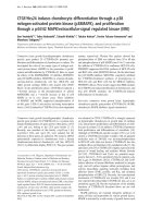
Elution profiles (Fig. 1) show that the antiviral activity
eluted mostly as a single peak, around an M
r
of 43 000 for
TrIFN-c (Fig. 1A), 50 000 for LeIFN-c (1B), and 34 000
for nonglycosylated rIFN-c. The scheme with TrIFN-c
(Fig. 1A) was however, more complex; in ELISA, a single
peak eluted at around 43 000, while the antiviral assay
detected two peaks, one at 43 000 and slightly above, and
one around 17–19 000. This second peak was most prob-
ably due to the presence of IFN-d in the crude uterine flush,
which had previously been shown to be monomeric, with an
M
r
around 19 000 [25]. This peak was not detected by
ELISA.
Unexpectedly, for leucocytic IFN-c,andtoalesserextent
for rIFN-c, the maximum ELISA score was delayed by one
and two fractions with regard to antiviral activity. One
possibility is that our ELISA is more specific for shortened
IFN-c molecules (see below).
TrIFN-c therefore appears to be essentially, if not
entirely, dimeric, similar to natural LeIFN-c and unglycos-
ylated recombinant IFN-c.
Monomers of TrIFN-c are glycosylated and have
shorter polypeptide chains
Both TrIFN-c and LeIFN-c were metabolically radio-
labelled with [
35
S]Met, then immunoprecipitated with rabbit
antiserum as described in Materials and methods. The
Fig. 1. Sephadex G-75 gel filtration profiles of three preparations of
crude porcine IFN-c. (A) TrIFN-c derived from uterine flushes of a
day-15 pregnant gilt. (B) crude natural LeIFN-c.(C)recombinant
IFN-c (E.coli).IneachfractionIFN-c concentration (d) was meas-
ured by ELISA, and antiviral activity (h) was determined by antiviral
assay on MDBK cells. Molecular weight standards and void volume
(V
0
) are indicated by arrows, and the black rectangle designates the
elution area of expected IFN-c monomers.
Ó FEBS 2002 Structure of trophoblastic interferon-c (Eur. J. Biochem. 269) 2775
monomers were analysed by denaturing SDS/PAGE
(Fig. 2). The results were clearly contrasted: in the immu-
noprecipitate from leukocytes, LeIFN-c consisted of four
major bands (lane 1: M
r
24 800; 22 000; 19 800; 17 500), the
M
r
24 800 band being slightly more pronounced. These four
bands resolved into two bands on deglycosylation (lane 2:
16 000 and 14 000). As for TrIFN-c, only two main bands
were visible at 22 500 and 18 000 (lane 3), which yielded one
main band with an M
r
of 14 400 after N-glycosidase F
treatment (lane 4), suggesting a single major polypeptide
chain, but macroheterogeneity at the two potential glyco-
sylation sites present on the IFN-c polypeptide core. They
could differ in the rate and site of glycosylation, considering
the 22 500-Da band as bi-glycosylated and the 18 000-Da
band as monoglycosylated (Fig. 2). The deglycosylated
14 400-Da band may correspond to the truncation of about
20 amino acids in the embryonic IFN-c molecule, as the
expected mass of full-length porcine IFN-c polypeptide is
around 16 780 Da.
In order to check if a full-length TrIFN-c form could be
found in the trophoblast cells, which would be indicative of
extracellular degradation, the same immunoprecipitation
was performed on the conceptus cell lysate in parallel with
the supernatant (Fig. 3). SDS/PAGE revealed only one
major band in the cell lysate, at an apparent M
r
of 23 000–
24 000 (lane 2), which was slightly higher than the largest
of the major monomers found in the supernatant (lane 1).
Because of the scarcity of intracellular material, it was not
possible to analyse the effect of N-Glycosydase F on this
band, which casts a doubt on its glycosylation status.
However, no equivalent of the largest LeIFN-c species
(24 800) was found. Furthermore, the larger amount of
TrIFN-c found, when compared to the previous experi-
ment (Fig. 2), revealed that the 22 500 and 18 000 bands
were the major but not the only components of TrIFN-c;
two minor bands at M
r
24 000 and 20 500 were also
visible. These band most probably correspond to the
diglycosylated and monoglycosylated forms of the minor
polypeptide of M
r
16 000 obtained after N-glycosidase F
treatment (lane 3).
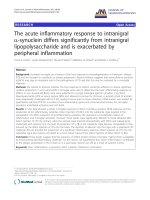
TrIFN-c has an intact N-terminus, and a truncated
C-terminus
Immunoaffinity-purified trophoblastic IFN-c, obtained
from pregnant uterine flushes, was electrophoresed in
SDS/PAGE, after treatment (or mock treatment) with
N-glycosydase F (Fig. 4A). Again, two major bands were
found at M
r
22 500 and 18 000 (lane 1), and upon
deglycosylation, one major band at M
r
14 400 was seen.
But unlike TrIFN-c collected in the supernatant of cultured
conceptuses, a minor deglycosylated band was obtained at
M
r
12 000 (lane 2). The two major TrIFN-c polypeptides
yielded no residue by Edman microsequencing, a result
compatible with a blocked pyroglutamate N-terminus (the
expected mature sequence is Q-A-P-F-F-K-E-I-T-I-L-K-).
Immunopurified TrIFN-c was then treated with pyroglu-
Fig. 2. SDS/PAGE profiles of [
35
S]Met metabolically labelled native
TrIFN-c and LeTrIFN-c. Lane 1, control LeTrIFN-c.Lane2,
N-glycosidase F treated LeIFN-c.Lane3,controlTrIFN-c.Lane4,
N-glycosidase F-treated TrIFN-c.
Fig. 3. SDS/PAGE profiles of [
35
S]Met-labelled TrIFN-c after immu-
noprecipitation by rabbit anti-(porcine IFN-c)Ig.Lane 1, glycosylated
conceptus IFN secreted in the supernatant. Lane 2, intracellular
TrIFN-c.Lane3,conceptussecretedIFN-c treated with N-Glycosi-
dase F.
Fig. 4. Mass determination of deglycosylated TrIFN-c species. (A)
SDS/PAGE profiles of native TrIFN-c obtained in flushings of Day-15
pregnant uterus, control (lane 1) and N-glycosidase F treated (lane 2).
(B) Mass spectrum obtained by MALDI-TOF of the M
r
14 400
polypeptide. (C) Mass spectrum of the M
r
12 000 polypeptide.
2776 A. Cencic
ˇ
et al. (Eur. J. Biochem. 269) Ó FEBS 2002
tamate aminopeptidase, and re-submitted to the microseq-
uencing process. In the largest band, the A-P-F-F-K
sequence appeared with a moderate yield, thus confirming
that the native N-terminus was intact.
In a second step, the mass analysis of the two deglycos-
ylated bands was performed by MALDI-TOF. The 14 400-
Da species yielded a main peak at (M + H)
+
14 742.0 Da
(Fig. 4B). This measured mass is compatible with a
nonglycosylated polypeptide starting with an N-terminal
pyroglutamate and ending at C-terminal L126. Indeed such
a peptide has a theoretical sequence mass (average) of
14 712.0 Da, to which 17 Da must be substracted for
N-terminal pyroglutamate, and 2 Da must be added for two
N-glycosidase F-induced N fi D transitions, and 48 Da
added for oxidation of three residues (probably the three
M), respectively. The calculated (M + H)
+
obtained is
then 14 746.0 Da, a value which differs by 4 Da from the
observed mass.
The MALDI-MS analysis of the minor peak with an M
r
of 12 000 yielded an observed (M + H)
+
of 12 635.0 Da
(Fig. 4C). This is compatible with a deglycosylated poly-
peptide with R
107
as C-terminus. Indeed such a 1–107
polypeptide with N-terminal pyroglutamate, three oxidized
residues and two Asn/Asp transitions gives a calculated
(M + H)
+
of 12 635.5, that is a 0.5-Da difference with the
measured value. The second peak of the MALDI spectrum
was measured at 12 762.4 Da (D
mass
¼ 127.4 Da), a mass
compatible with a peptide cleaved behind R107.
Therefore, it is most probable that TrIFN-c is mostly
composed of a polypeptide in which the C-terminus is
cleaved after L126 (a lack of 17 residues), and of a minor
polypeptide which is further cleaved, that is after R107 (a
lack of 36 residues).
TrIFN-c N-glycans contain no sialic acid, and have
limited heterogeneity
The tryptic peptide analysis of the four main bands obtained
in PAGE were performed. We chose to point to data
obtained for the M
r
22 500 species. Figure 5 shows the
complete sequence of pig IFN-c [26] with its theoretical
trypsin cleavage sites, the peptides found upon MALDI
analysis (underlined), and the deduced C-termini of each
14.74 and 12.63 kDa species (arrows). The coverage of the
molecule was rather high as peptide analysis amounted to
87.3% of the sequence Q1-L126. Table 1 shows the
comparison between theoretical and measured masses of
tryptic peptides as provided by MALDI. Three main
conclusions could be drawn: (a) on the peptide 1–6, the
Fig. 5. Complete amino-acid sequence of mature porcine IFN-c [26].
Being 143 residues long, it includes two glycosylatable Asn at positions
16 and 83 (in grey). Expected trypsin cleavages are marked by slashes,
and peptides analysed by MALDI-TOF are underlined. The two
arrows point to the inferred C-termini of each 14.74 and 12.63 kDa
species.
Table 1. MALDI-TOF tryptic peptide analysis of the M
r
22 500 TrIFN-c species.
Peptide
start
Peptide
end Sequence
Theoretical
(M + H)+
Measured
(M + H) +
Dmass
(meas – theor) Remarks
1 6 QAPFFK 737.400 720.372 )17.028 N-term. pyroglut.
7 12 EITILK 716.455 716.452 )0.003
13 34 DYF…ILK 2397.198 4540.260
4394.224
4250.802
2143.062
1997.026
1853.604
Glycosylation
Glycosylation
Glycosylation
44 55 IIQSQIVSFYFK 1472.814 1472.847 0.033
56 61 FFEIFK 830.444 830.459 0.015
62 68 DNQAIQR 844.427 844.452 0.025
69 74 SMDVIK 692.364 692.348
708.351
)0.016
15.987 Oxidized Met
75 80 QDMFQR 824.372 824.395
840.379
0.023
16.007 Oxidized Met
81 88 FLNGSSGK 809.416 2805.871
2951.970
1996.455
2142.554
Glycosylation
Glycosylation
98 107 IPVDNLQIQR 1195.679 1195.762 0.083
89 94 LNDFEK 765.377 765.383 0.006
109 115 AISELIK 773.476 773.480 0.004
116 123 VMNDLSPR 931.466 931.502
947.480
0.036
16.014 Oxidized Met
Ó FEBS 2002 Structure of trophoblastic interferon-c (Eur. J. Biochem. 269) 2777
presence of N-terminal pyroglutamate is confirmed. (b) On
peptides 69–74, 75–80 and 116–123, the three Met residues
are oxidized. (c) On peptides 13–34 and 81–88, a mass excess
of 2143 Da is the most probable signature of the same
glycan conjugate. This mass is compatible with a sugar
moiety made of three fucoses plus six N-acetylglucosamines
plus three hexoses (monoisotopic mass ¼ 2142.80 Da).
Table 1 and Fig. 6 show that other peaks differed from the
major one by 146 Da, corresponding to the mass of fucose.
Therefore, a microheterogeneity exists with at least three
glycoform variants at each site, depending on the fucose
content (one, two or three). Whether this is the real situation
on the native molecule, or a consequence of laser-induced
cleavage of fucose in the course of MALDI analysis is not
known.
The same analysis performed on the band at M
r
18 500
(data not shown) indicated that the same glycan motif was
present on the tryptic peptide 13–34, but absent on the
peptide 81–88, for which the measured value was 809.42 Da
[theoretical (M + H)
+
value is 809.41 Da]. We can there-
fore conclude that if the main deglycosylated peptide is
indeed 14.74 kDa, then the two main species of natural
TrIFN-c found in uterine flushings have molecular masses
of 19.03 kDa and 16.88 kDa, corresponding to diglycosyl-
ated and monoglycosylated isoforms, respectively, the latter
isoform being glycosylated on N16. As expected, the
correspondance between the measured masses and observed
M
r
in SDS/PAGE is quite good for nonglycosylated
proteins, but not for glycosylated ones, as the latter have
lowered electrophoretic mobility.
Specific antiVSV activity of TrIFN-c is reduced
Table 2 shows results concerning the antiviral activity of
TrIFN-c, in comparison with LeIFN-c and two species of
recombinant IFN-c, including the glycosylated rGIFN-c
produced in transfected RK13 cells [18]. The specific activity
of TrIFN-c on MDBK cells challenged with VSV was
1–5 · 10
5
UÆmg
)1
of IFN-c (ELISA reactive), i.e. approxi-
mately 10 times lower than that of its ÔadultÕ equivalent
(LeIFN-c), and 20–50 times less than the two recombinant
forms.
TrIFN-c has an antiproliferative activity (APA)
Immunoaffinity-purified IFN-c from uterine flushes did
exert an APA on different cells. We first checked on pig
swine testis cells that possible residual IFN-d was not a
Fig. 7. Compared antiproliferative effect of TrIFN-c and other porcine
IFN-c on several cell lines. Dilutions 1–6 represent serial threefold
dilutions of purified IFNs, all of them being adjusted before assay to
1 lgÆmL
)1
(A) ST cells treated with TrIFN-c in the absence (hatched
bars) or presence (black bars) of rabbit antiserum 652 to type I IFN.
(B,C,D): EL cells, TBA cells and MDBK cells, respectively, treated
with serial dilutions of TrIFN-c (black bars), rGIFN-c produced in
RK13 cells (grey bars) and rIFN-c expressed in E. coli (white bars).
Values are means of four replicate assays per dilution, and the errors
bars give the positive value of the SEM.
Fig. 6. MALDI-TOF analysis of tryptic peptide 13–34 from the 22 500
IFN-c species. The area shown is an enlargement of the total mass
spectrum. The main peak at an (M + H)
+
of 4540.26 is compatible
with a N-glycosylation on N16 having the proposed structure drawn
above the peak, including three fucose residues. Two other peaks on
the left, with Dmass of 146.03 and 146.42 Da with each other, are
compatible with masses of the peptide 13–34 with 2 and 1 fucose,
respectively. (monoisotopic mass of fucose: 146.04). The two peaks
marked with an asterisk represent the Na adducts of the two main
peaks. The unmarked peak could not be identified. Schematic structure
of the glycan conjugate was inferred by analogy with data obtained for
human IFN-c [27].N-acetylglucosamine (j);hexose(d); fucose ( fi ).
Table 2. Specific antiviral activity of TrIFN-c by comparison with other natural and recombinant IFN-c.
Cell line
IFN-c origin
LeIFN-c rGIFN-c rIFN-c TrIFN-c
Specific activity MDBK 1–5 · 10
5
1–5 · 10
6
2–3.5 · 10
6
5–10 · 10
6
(UÆmg
)1
IFN-c)
2778 A. Cencic
ˇ
et al. (Eur. J. Biochem. 269) Ó FEBS 2002
significant effector of any APA by comparing the effect of
dilutions from 300 ngÆmL
)1
to 1.2 ngÆmL
)1
in the absence
or presence of antiserum to porcine type IFN (Fig. 7A),
known to neutralize IFN-d [8]. Other cells were tested for
their proliferation in the presence of TrIFN-c,andtwo
purified recombinant proteins, one glycosylated (rGIFN-c
produced in eucaryotic cells), the other free of carbohydrate
chains (rIFN-c produced in E. coli). Figure 7B–D shows
that, with cell-related differences, trophoblastic IFN-c
exerted the same (in MDBK cells) or even more pronounced
APA (in endometrial cells and trophoblast cell line TBA)
than its recombinant counterparts. On pig EL and TBA
cells, TrIFN-c was the most active on cell growth inhibition,
especially in the first four dilutions, that is in the range of
300–11 ngÆmL
)1
. On these same cell lines, recombinant
E. coli-derived IFN-c was found the least active, which
suggests that the glycosylation status is important for cell
growth inhibition.
DISCUSSION
Embryonic TrIFN-c is the only IFN-c secreted by a
nonlymphoid tissue. It is also a unique case among all IFN
species, because it is intensely induced under physiological
conditions (at the time of trophoblast implantation).
TrIFN-c is secreted in substantial amount, simultaneously
with IFN-d, in a polarized manner, by the trophectoderm.
The precise structure and function of this embryonic,
epithelial IFN-c has not been clarified to date. In this work,
we have demonstrated that structural, biochemical and
biological differences exist between TrIFN-c and LeIFN-c.
As shown by gel filtration chromatography, TrIFN-c is
accessible to the uterine lumen in the form of relatively
heterogeneous glycosylated dimer with an apparent M
r
of
43 000. A shift towards a lower M
r
was noted for TrIFN-c,
when compared to LeIFN-c, which eluted as a major
heterogeneous peak at a M
r
between 50 000 and 60 000. On
the other hand, rIFN-c exhibits no macroheterogeneity, as
it elutes as one homogeneous peak at around 34 000, a size
compatible with the correct predicted size of a biologically
active dimeric protein. We can therefore conclude that
functional embryonic IFN-c (TrIFN-c), like LeIFN-c,isa
dimer. The weak antiviral activity found in fractions
corresponding to monomers is certainly that of IFN-d,
with an M
r
around 19 000, which is also present in uterine
flushes [25].
As revealed by the electrophoretic profiles of
35
S-labelled
TrIFN-c and LeIFN-c immunoprecipitates, TrIFN-c
monomers differ from the LeIFN-c in terms of their
polypeptide length and glycosylation pattern. Electropho-
retic profile of TrIFN-c exhibits two major bands that are
equimolar, with an apparent M
r
values of 22 500 and
18 000, thus suggesting that dimers are composed of equal
proportions of mono glycosylated and biglycosylated
monomers. The two glycoforms resolve into a major
M
r
14 000 band upon enzymatic deglycosylation with
N-glycosidase F. The fact that TrIFN-c secreted in the
supernatant of conceptus in culture presents with the same
truncation as TrIFN-c collected in uterine fluids suggests
that the cleavage of natural TrIFN-c is not due to
endometrial peptidases. The pig trophectoderm has been
shown to express various proteases, among which plasmi-
nogen activator and different matrix metalloproteinases,
which could, directly or by activation of protease cascades,
lead to the observed cleavage of TrIFN-c [27,28]. In
addition, in the flushed fluids, where TrIFN-c is supposed to
be present in its bioavailable form, a minor polypeptide
variant was observed after treatment with N-glycosidase F,
with an apparent M
r
of 12,500, corresponding to a still more
cleaved polypeptide.
The MALDI-TOF resolution of deglycosylated TrIFN-c
monomers, obtained from the uterine flush, confirmed
results obtained by SDS/PAGE electrophoresis. The major
form of TrIFN-c molecule is a polypeptide with a mass of
14 741 Da and a minor one with a mass of 12 634 Da. As
confirmed by MALDI analysis, the two polypeptides found
in TrIFN-c are truncated at the C-terminus. The major
polypeptide lacks 17 C-terminal amino acids, as compared
to the full length sequence, and a minor one is further
truncated by 36 residues. Porcine LeIFN-c and TrIFN-c
monomers are glycosylated, unlike human IFN-c,where
nonglycosylated forms have also been found in crude
preparations [29]. In the TrIFN-c molecule, little variability
in the glycan structure are observed. Only three variants in
glycan composition were found at both N-glycosylation
sites, which differ only by the number of bound
L
-fucose
molecules. Surprisingly, TrIFN-c glycans terminate with
N-acetylglucosamine and not with sialic acid like for human
IFN-c. Indeed, post-translational modifications, including
glycosylation, are strongly dependant upon the type and
physiological status of producing cells, and may signifi-
cantly influence the characteristics of a glycoprotein
[16,17,29]. From this point of view, no direct comparison
has been possible with the glycan structure and heterogen-
eity of porcine LeIFN-c, for which low amounts obtained in
phytohaemagglutinin-activated pig PBL did not allow the
same mass spectroscopy analysis.
As a consequence of the C-terminal truncation, the native
TrIFN-c lacks seven basic residues, in particular the R-K-
R-K-R cluster (residues 127–131). It is therefore expected to
be less positively charged than LeIFN-c or rIFN-c,which
comprise full-length molecules. Indeed, unlike the two other
IFNs, TrIFN-c, when analyzed by chromatofocusing, did
not yield a readable pI, as it did not bind to a Mono-P
column. In addition, attempts at binding TrIFN-c onto
CM-cellulose columns at neutral pH were unsuccessful
(data not shown). Although the calculated pI is 10.66 for the
full length IFN-c molecule and 9.66 for the 1–126 polypep-
tide, TrIFN-c behaves as a molecule without measurable net
charge.
Concerning biological activities, we found divergent
results for antiviral and APA. The data shown in Table 2
suggest that TrIFN-c is much less antiviral than LeIFN-c
and rIFN-c, as far as VSV challenge is concerned. It is
possible however, that the relative values for TrIFN-c
specific activity are underestimated, if it happened that the
specificity of our ELISA test was slightly or significantly
better for truncated molecules. In any case, the C-terminal
truncation of TrIFN-c is most probably not the only reason
for the reduced antiviral activity of TrIFN-c on MDBK
cells (Table 2), such as that previously described for
HuIFN-c [14]. The specific glycan composition that we
found for TrIFN-c might also contribute to this reduced
antiviral activity.
On the other hand, we observed that TrIFN-c exhibits an
APA on homologous (ST, TBA, EL) and heterologous
Ó FEBS 2002 Structure of trophoblastic interferon-c (Eur. J. Biochem. 269) 2779
(MDBK) cells, that does not significantly differ from intact
nonglycosylated rIFN-c and intact glycosylated porcine
rGIFN-c (Fig. 7). Moreover, the APA of TrIFN-c on EL
and TBA cells was even higher as compared to the intact
porcine IFN-c. It seems that, especially in homologous cell
lines, an intact IFN-c C-terminus is not essential for its
biological function, as was shown for human IFN-c [30].
Our results also confirm previous data, namely that IFN-c
antiviral and APAs can be dissociated [31–33].
Our results shed some light on the specific structure and
properties of this atypical porcine trophoblastic IFN-c,
produced by a polarized epithelium. It is probable that the
structural and chemical characteristics of TrIFN-c affects its
bioavailability and biological effect(s) on the maternal
uterus. In particular, this shortened version of IFN-c,
lacking a well known ECM-binding sequence and with very
weak net charge, could be more prone than full-length IFN-
c to cross the endometrial epithelium, and to reach cellular
targets located in the uterine mucosa (e.g. lymphoid or
endothelial cells). It is possible that the very particular
context
9
in which this embryonic IFN-c is produced, namely
between two opposite epithelia, has favoured the selection
of a functionally adapted molecule, which differs from adult
lymphoid IFN-c more by its bioavailability in this particular
context than by its biological activity.
ACKNOWLEDGEMENTS
We would like to thank Christiane De Vaureix for her technical help.
This work was supported by grants from the Slovenian Scientific
Foundation and from the French Ministry of Foreign Affairs.
REFERENCES
1. De Maeyer, E. & De Maeyer-Guignard, J. (1988) Interferons and
Other Regulatory Cytokines.JohnWiley&sons,NewYork.
2. Ijzermans, J.N.M. & Marquet, R.L. (1989) Interferon-gamma: a
Review. Immunobiol. 179, 456–473.
3. Baron, S., Tyring, S.K., Fleischmann Jr, R.W., Coppenhaver,
D.H., Niesel, D.W., Klimpel, G.R., Stanton, J. & Hughes, T.K.
(1991) The interferons, Mechanisms of action and clinical appli-
cations. J. Am. Med Assoc. 266, 1375–1383.
4. Degre, M. (1996) Interferons and other cytokines in bacterial
infections. J. Interferon Cytokine Res. 16, 417–426.
5. Cross, J.C. & Roberts, R.M. (1989) Porcine conceptuses secrete an
interferon during the pre-attachment period of early pregnancy.
Biol. Reprod. 40, 1109–1118.
6. La Bonnardie
`
re, C., Martinat-Botte
´
,F.,Terqui,M.,Lefe
`
vre,
F., Zouari, K., Martal, J. & Bazer, F.W. (1991) Production of
two species of interferon by Large White and Meishan pig con-
ceptuses during the peri-attachment period. J. Reprod. Fertil. 91,
469–478.
7. Lefe
`
vre, F., Martinat-Botte
´
, F., Guillomot, M., Zouari, K.,
Charley, B. & La Bonnardie
`
re, C. (1990) Interferon-gamma gene
and protein are spontaneously expressed by the porcine tropho-
ectoderm early in gestation. Eur. J. Immunol. 20, 2485–2490.
8. Lefe
`
vre, F. & Boulay, V. (1993) A novel and atypical type one
interferon gene expressed by trophoblast during early pregnancy.
J. Biol. Chem. 268, 19760–19768.
9. Arakawa, T., Hsu, Y.R., Chang, D., Stebbing, N. & Altrock, B.
(1986) Structure and activity of glycosylated human interferon-c.
J. Interferon Res. 6, 687–695.
10. Ealick, S.E., Cook, W.J., Kumar, S.V., Carson, M., Nagabhushan,
T.L., Trotta, P.P. & Bugg, C.E. (1991) Three-dimensional structure
of recombinant human interferon-c. Science 252, 689–701.
11. Denesyuk, A.I., Zavyalov, V.P. & Korpela, T. (1994) Common
structural patterns of cytokine outer surfaces. Bioch. Biophy. Res.
Comm. 201, 1396–1400.
12. Dorner, F., Scriba, M. & Weil, R. (1973) Interferon: evidence for
its glycoprotein nature. Proc. Natl Acad. Sci. USA 70, 1981–1985.
13. Sareneva, T., Pirhonen, J., Cantell, K., Kalkkinen, N. & Julkunen,
I. (1994) Role of N-glycosylation in the synthesis, dimerization
and secretion of human interferon-c. Biochem. J. 303, 1–10.
14. Arakawa, T., Narachi, M.A., Hsu, Y.R., Everett, R.R., Lai, P.H.
& Fish, E.N. (1989) The effect of C-terminal processing on the
activity of human interferon-c. Drug Des. Del. 4, 217–225.
15. Honda, S., Asano, T., Kajio, T., Nakagawa, S., Ikeyama, S. &
Ichimori, Y. (1987) Differential purification by immunoaffinity
chromatography of two carboxy-terminal portion-deleted deriv-
atives of recombinant human interferon-c from Escherichia coli.
J. Interferon Res. 7, 145–154.
16. James, D.C., Goldman, M.H., Hoare, M., Jenkins, N., Oliver,
R.W.A., Green, B.N. & Freedman, R.B. (1996) Posttranslational
processing of recombinant human IFN-c in animal expression
systems. Protein Sci. 5, 331–340.
17. Hooker, A. & James, D. (1998) The glycosylation heterogeneity of
recombinant human IFN-c. J. Interferon Cytokine Res. 18,287–
295.
18. Cencic, A., Lefe
`
vre, F., Koren, S. & La Bonnardie
`
re, C. (1999)
Tetracycline-controlled expression of glycosylated porcine inter-
feron-c in mammalian cells. Anim. Biotechnol. 10, 63–79.
19. Cencic, A. (1995) Porcine immune interferon. M.Phil. Thesis.
Biotechnical faculty, University of Ljubljana.
20. Vandenbroeck, K., Willems, L., Billiau, A., Opdenakker, G. &
Huybrechts, R. (1994) Glycoform heterogeneity of porcine inter-
feron-c expressed in Sf9 cells. Lymphok. Cytok. Res. 13, 253–258.
21. La Bonnardie
`
re, C. & Laude, H. (1981) High interferon titer in
newborn pig intestine during experimentally induced viral
enteritis. Infect. Immun. 32, 28–31.
22. McClurkin, A.W. & Norman, J.O. (1966) Studies on Transmis-
sible gastroenteritis of swine II. selected characteristics of a cyto-
pathogenic virus common to five isolates from transmissible
gastroenteritis. Can. J. Comp. Med. Vet. Sci. 30, 190–198.
23. Laemmli, U.K. (1970) Cleavage of structural proteins during
theassemblyoftheheadofthebacteriophageT4.Nature 227,
680–685.
24. Messer, M., Griffiths, M., Rismiller, P.D. & Shaw, D.C. (1997)
Lactose synthesis in a monotreme, the echidna (Tachyglossus
aculeatus): isolation and amino acid sequence of echidna alpha-
lactalbumin. Comp.Biochem.Physiol.188B, 403–410.
25. Niu, P.D., Lefe
`
vre, F., Me
`
ge, D. & La Bonnardie
`
re, C. (1995)
Atypical porcine type I interferon. Biochemical and biological
characterization of the recombinant protein expressed in insect
cells. Eur. J. Biochem. 230, 200–206.
26. Dijkmans, R., Vandenbroeck, K., Beuken, E. & Billiau, A. (1990)
Sequence of the porcine interferon-gamma (IFN-gamma) gene.
Nucleic Acids Res. 25, 4259.
27. Geisert, R.D. & Yelich, J.V. (1997) Regulation of conceptus
development and attachment in pigs. J. Reprod Fertil. 52
(supplement), 133–149.
28. Menino, A.R., Hogan, A., Schultz, G.A., Novak, S., Dixon, W. &
Foxcroft, G.H. (1997) Expression of proteinases and proteinase
inhibitors during embryo-uterine contact in the pig. Dev. Genet.
21, 68–74.
29. Mortz, E., Sareneva, T., Julkunen, I. & Roepstorff, P. (1996) Does
matrix-assisted laser desorption/ionization mass spectrometry
allow analysis of carbohydrate heterogeneity in glycoproteins? A
study of natural human interferon-c. J. Mass Spectr. 31, 1109–
1118.
30. Luk, S.K., Jay, E. & Jay, F.T. (1990) Structure-function analysis
of the human interferon-gamma. The COOH terminus is not
essential for functional activity. J. Biol. Chem. 265, 13314–13319.
2780 A. Cencic
ˇ
et al. (Eur. J. Biochem. 269) Ó FEBS 2002
31. Carter, W.A., Swartz, H. & Gillespie, D.H. (1985) Independent
evolution of antiviral and growth-modulating activities of inter-
feron. J. Biol. Resp. Mod. 4, 447–459.
32. Goldberg, M., Belkowski, L.S. & Bloom, B.R. (1989) Regulation
of macrophage growth and antiviral activity by interferon-gamma.
J. Cell Biol. 109, 1331–1340.
33. Caruso, A., Tiberio, L., De Rango, C., Bonfanti, C., Flamminio,
G.,Gribaudo,G.,Monti,E.,Viani,E.,Manca,N.,Garotta,G.,
Landolfo, S. & Turano, A. (1993) A monoclonal antibody to the
NH2-terminal segment of human IFN-c selectively interferes with
the antiproliferative activity of the lymphokine. J. Immunol. 150,
1029–1035.
Ó FEBS 2002 Structure of trophoblastic interferon-c (Eur. J. Biochem. 269) 2781