Báo cáo Y học: Expression of uncoupling protein-3 in subsarcolemmal and intermyofibrillar mitochondria of various mouse muscle types and its modulation by fasting docx
Bạn đang xem bản rút gọn của tài liệu. Xem và tải ngay bản đầy đủ của tài liệu tại đây (186.23 KB, 7 trang )
Expression of uncoupling protein-3 in subsarcolemmal
and intermyofibrillar mitochondria of various mouse muscle
types and its modulation by fasting
Maria Jimenez, Cedric Yvon, Lorenz Lehr, Bertrand Le
´
ger, Patrick Keller, Aaron Russell, Franc¸oise Kuhne,
Pierre Flandin, Jean-Paul Giacobino and Patrick Muzzin
Department of Medical Biochemistry, Faculty of Medicine, University of Geneva, Switzerland
Uncoupling protein-3 (UCP3) is a mitochondrial inner-
membrane protein abundantly expressed in rodent and
human skeletal muscle which may be involved in energy
dissipation. Many studies have been performed on the
metabolic regulation of UCP3 mRNA level, but little is
known about UCP3 expression at the protein level. Two
populations of mitochondria have been described in skeletal
muscle, subsarcolemmal (SS) and intermyofibrillar (IMF),
which differ in their intracellular localization and possibly
also their metabolic role. To examine if UCP3 is differen-
tially expressed in these two populations and in different
mouse muscle types, we developed a new protocol for
isolation of SS and IMF mitochondria and carefully valid-
ated a new UCP3 antibody. The data show that the density
of UCP3 is higher in the mitochondria of glycolytic muscles
(tibialis anterior and gastrocnemius) than in those of oxi-
dative muscle (soleus). They also show that SS mitochondria
contain more UCP3 per mg of protein than IMF mito-
chondria. Taken together, these results suggest that oxida-
tive muscle and the mitochondria most closely associated
with myofibrils are most efficient at producing ATP. We
then determined the effect of a 24-h fast, which greatly
increases UCP3 mRNA (16.4-fold) in muscle, on UCP3
protein expression in gastrocnemius mitochondria. We
found that fasting moderately increases (1.5-fold) or does
not change UCP3 protein in gastrocnemius SS or IMF
mitochondria, respectively. These results show that modu-
lation of UCP3 expression at the mRNA level does not
necessarily result in similar changes at the protein level and
indicate that UCP3 density in SS and IMF mitochondria can
be differently affected by metabolic changes.
Keywords: fasting; intermyofibrillar mitochondria; muscle
type; subsacorlemmal; uncoupling protein-3 (UCP3).
The first uncoupling protein described, uncoupling protein-1
(UCP1), is an inner-mitochondrial membrane protein,
which, by dissipating the mitochondrial proton gradient
driven by the respiratory chain, uncouples oxidation from
phosphorylation and therefore produces heat instead of
ATP. UCP1 was found to be exclusively expressed in brown
adipose tissue (for review see [1]).
The novel UCP3, discovered in 1997, is abundantly
expressed in rodent and human skeletal muscle. Its high
sequence homology with UCP1 suggested that it had similar
uncoupling activities [2]. In fact, using heterologous yeast
and mammalian cell expression systems, UCP3 was shown
to decrease the mitochondrial membrane potential, as
measured by uptake of potential sensitive fluorescent dyes
(reviewed in [3–5]). Also recent data obtained using muscle
mitochondria of UCP3 knockout (UCP3KO) mice [6,7] and
of transgenic mice overexpressing UCP3 in their skeletal
muscle [8] confirmed the uncoupling activity of UCP3. A
more recent study clearly established that UCP3 is, like
UCP1, a H
+
transporter sensitive to nucleotides and fatty
acids [9].
Many studies have been performed on the metabolic
regulation of brown adipose tissue and muscle UCP3
mRNA expression in both rodents and humans (for review
see [3–5]). Very few of the control mechanisms of UCP3
observed in muscle at the mRNA level have so far been
studied at the protein level. The reasons for this are the
difficulty in obtaining specific antibodies and validation
tests and also good Western blot conditions.
Skeletal muscle mitochondria consist of two distinct
subfractions, the subsarcolemmal (SS) and intermyofibrillar
(IMF) mitochondria, located beneath the sarcolemma and
between the myofibrils, respectively. These two mitochond-
rial populations possess different characteristics, such as
higher cardiolipin content and more elevated state-3 respir-
ation rate in IMF mitochondria [10].
In this study, we validated an antibody to UCP3 using a
fully controlled Western blot technique and present a
comparative study in mice of UCP3 protein expression in
SS and IMF mitochondria of various muscle types. We also
examined UCP3 protein expression in the two mitochon-
dria populations of fed and fasted mouse gastrocnemius
muscle.
Correspondence to P. Muzzin, De
´
partement de Biochimie Me
´
dicale,
Centre Me
´
dical Universitaire, 1 rue Michel Servet,
CH-1211 Gene
`
ve 4 Switzerland.
Fax: 41 22 702 5502, Tel.: 41 22 702 5492,
E-mail:
Abbreviations: UCP, uncoupling protein; SS, subsarcolemmal;
IMF, intermyofibrillar; COX, cytochrome oxidase; UCP3KO,
UCP3 knockout.
(Received 17 January 2002, revised 12 April 2002,
accepted 23 April 2002)
Eur. J. Biochem. 269, 2878–2884 (2002) Ó FEBS 2002 doi:10.1046/j.1432-1033.2002.02953.x
MATERIALS AND METHODS
Bio-Rad Protein Assay and nonfat dry milk were purchased
from Bio-Rad Laboratories (Hercules, CA, USA). ECL kit
Hyperfilm ECL and Coomassie blue (PhastGelÒ BlueR)
were obtained from Amersham International Biotech
(Amersham, Bucks, UK), the antibody to human UCP3
C-terminus (CabrX) from Research Diagnostics, Inc (San
Antonio, LA, USA) and the monoclonal antibody to the
cytochrome oxidase (COX) subunit IV were from Molecu-
lar Probes (Eugene, OR, USA). Goat anti-rabbit and anti-
mouse immunoglobulins were purchased from Santa Cruz
Biotechnology, Inc (Santa Cruz, CA, USA). Trizol reagent
was from Life Technologies (Basel, Switzerland). The
protease inhibitor cocktail was purchased from Sigma (St
Louis, MO, USA).
Animals
Three-month-old female C57BL/6J mice fed ad libitum a
standard laboratory chow and maintained under a 12-h
light/dark cycle at 23 °C were used. All animals were caged
individually during the experimental periods. Mice were
either fed ad libitum orfastedforaperiodof24hwithfree
access to water. The animals were killed by cervical
dislocation, and tibialis anterior, gastrocnemius and soleus
muscles were carefully dissected and kept on ice. In fasted
animals and the respective controls, one gastrocnemius
muscle was used for the mitochondria preparation and the
other for RNA isolation. All experiments were performed in
accordance with the Office Ve
´
te
´
rinaire de Gene
`
ve author-
ization covering animal experiments.
Preparation of muscle mitochondria
SS and IMF mitochondria were prepared from skeletal
muscle by the following procedure. Muscle (50–250 mg)
was minced with scissors in 5 mL ice-cold homogenization
buffer containing 100 m
M
sucrose, 180 m
M
KCl, 10 m
M
EDTA, 5 m
M
MgCl
2
,1m
M
ATP, 50 m
M
Tris/HCl,
pH 7.4, and 0.06% protease inhibitor cocktail. They were
then homogenized using a Teflon pestle in an ice-cold glass
Potter–Elvehjem homogenizer (clearance 0.37 mm, 10 up
and down strokes, 1800 r.p.m.; clearance 0.12 mm, 2 up and
down strokes, 1800 r.p.m.). The homogenate was centri-
fuged at 1600 g for 10 min at 4 °C. The pellet was kept at
4 °C for the extraction of IMF mitochondria. The super-
natant was filtered through two layers of surgical gauze and
then centrifuged at 9200 g for 10 min at 4 °C. The resulting
SS mitochondria pellet was suspended in the appropriate
volume of distilled water. Being located just beneath the
sarcolemmal membrane, the SS mitochondria are easier to
extract from the muscle by homogenization than IMF
mitochondria. In a first attempt to prepare IMF mitochon-
dria, we used the original technique of Krieger et al. [11],
which involves digestion by the Nagarse protease. The
latter, even when tightly controlled, was found by Western
blot analysis to degrade UCP3. Therefore we adopted
another technique in which we broke down myofibrils by
strong mechanical disruption with a tight-fitting homoge-
nizer. The 1600 g pellet was resuspended in 4 mL ice-cold
homogenization buffer containing 100 m
M
KCl, 1 m
M
EDTA, 5 m
M
MgSO
4
,1m
M
ATP, 50 m
M
Tris/HCl,
pH 7.4, and 0.06% protease inhibitor cocktail using a
Teflon pestle in an ice-cold glass Elvehjem homogenizer
(clearance 0.12 mm for 3 min, 1800 rpm). The resulting
homogenate was centrifuged at 1600 g for 10 min at 4 °C.
After filtration through two layers of surgical gauze, the
supernatant was centrifuged at 15 000 g for 45 min at 4 °C,
and the resulting IMF mitochondrial pellet was resuspended
in an appropriate volume of distilled water. Mitochondrial
protein concentrations were determined as described by
Bradford [12] using the Bio-Rad Protein Assay, with BSA as
a standard. Isolated mitochondria were stored at )20 °Cas
15-lg mitochondrial protein aliquots.
Western blotting
Purified muscle mitochondria (15 lg) were dried under
vacuum and resuspended in 10 lL loading buffer contain-
ing 50% glycerol, 5% SDS, 2.5% bromophenol blue and
0.5
M
Tris/HCl, pH 6.8. The samples were electrophoresed
on a 12% polyacrylamide/0.1% SDS gel, and transferred to
a poly(vinylidene difluoride) membrane by electroblotting
transfer with a buffer containing 10% methanol, 25 m
M
Tris/HCl, pH 6.8, and 190 m
M
glycine. The transfer was
performed for 15 h at 30 V, 70 mA and 4 °C. After blotting,
the gel was stained with 0.1% Coomassie blue to check
transfer efficiency. No band was visible below 50 kDa on
the gel. The membrane was blocked with a NaCl/P
i
buffer
containing 0.1% Tween and 2% nonfat dry milk. This same
buffer was used for all subsequent hybridizations. UCP3
protein was detected using CabrX at a concentration of
1 lgÆmL
)1
. The membrane was washed twice in NaCl/P
i
containing 0.1% Tween and hybridized with a 1 : 1000
diluted goat anti-rabbit peroxidase-labeled secondary anti-
body. The signals were detected by chemiluminescence
using a standard ECL kit and developed Hyperfilm ECL
film. They were quantified by scanning photodensitometry
using ImageQuant Software 3.3 (Molecular Dynamics,
Sunnyvale, CA, USA). COX protein was detected as above
using a 1 : 1000 diluted monoclonal antibody specific for
COX subunit IV and a 1 : 1000 diluted goat anti-mouse
peroxidase-labeled secondary antibody. To compare the
UCP3 signals, linear standard curves were constructed using
increasing concentrations of the human and mouse UCP3
recombinant proteins provided by Dr Michele Chiesi at
Novartis (Basel, Switzerland) and Stratagene (La Jolla, CA,
USA), respectively. The specificity of the antibody to UCP3
was tested using UCP3KO muscle mitochondria, which
were described by Gong et al. [7] and generously provided
by Dr Mary Ellen Harper (University of Ottawa, Ottawa,
Ontario, Canada). The replication-defective recombinant
adenoviral vector that contains the human UCP3 cDNA
under the transcriptional control of the cytomegalovirus
promoter was constructed as previously described [13].
Real-time quantitative RT-PCR
Total muscle RNA was isolated using the Trizol reagent
technique according to the manufacturer’s instructions.
Oligo-dT first-strand cDNA was synthesized from 2 lg
total RNA using Superscript II reverse transcriptase.
Real-time PCR was performed using a Lightcycler rapid
thermal cycler system and designated software (Roche
Diagnostics Ltd, Rotkreuz, Switzerland) according to the
Ó FEBS 2002 UCP3 protein quantitation (Eur. J. Biochem. 269) 2879
manufacturer’s instructions. Reactions were performed in a
20-lL reaction mixture containing 50 ngÆlL
)1
of first-
strand cDNA, 0.5 l
M
primers and 2.4 m
M
MgCl
2
.Nucleo-
tides, Taq DNA polymerase, and buffer were included in the
Lightcycler-DNA Master SYB Green I mix (Roche Diag-
nostics). The PCR protocol consisted of 2 min of denatur-
ing at 95 °C, followed by 30 cycles with 95 °Cdenaturing
for 1 s, 56 °C annealing for 5 s and 72 °C extension for
16 s. The fluorescent product was detected at the end of the
72 °C extension period. To confirm the amplification
specificity, the PCR product was subjected to a melting
curve analysis and then agarose gel electrophoresis. A linear
standard curve was constructed using known concentra-
tions of a mouse UCP3 plasmid. The 10-fold serial dilutions
ranged between 3.0 ngÆlL
)1
and 3.0 pgÆlL
)1
. The concen-
trations of the experimental samples were calculated by
comparison with the standard curves. Background fluores-
cence was removed by setting a noise band. The number of
cycles at which the best-fit line through the log-linear
portion of each amplified curve intersected the noise band
was inversely proportional to the log copy number [14]. The
samples were also normalized against b-actin using the same
conditions as described above.
Northern blot analysis
Total RNA from gastrocnemius muscle was isolated using
the Trizol reagent technique. Total RNA (20 lg) was
separated on a 1.2% agarose/formaldehyde gel and trans-
ferred to nylon membrane as described by Boss et al.[16].
To detect UCP3 mRNA, we used a probe derived from a
full-length rat UCP3 cDNA [15]. The probe was labeled
by random priming with [a-
32
P]dCTP (Amersham).
Hybridization and washing were carried out as previously
reported [16]. Blots were exposed to Hyperfilm ECL films
(Amersham) at )80 °C with intensifying screens. The
signals on the autoradiograms were quantified by scanning
photodensitometry using ImageQuant Software version
3.3. Hybridization of the blots with a [c-
32
P]ATP-labeled
synthetic oligonucleotide specific for the 18S rRNA
subunit was used to correct for differences in the amounts
of RNA loaded on to the gel. Student’s unpaired t test
was used to determine statistical significance.
RESULTS
Validation of antibodies to UCP3
Table 1 shows a list of antibodies that have been used for
the Western blot analysis of UCP3 protein expression in
rodents. The studies performed on UCP3KO or transgenic
mice overexpressing UCP3 in their skeletal muscle provide
convincing validation of the antibodies used. The Lilly
antibody to mouse and rat UCP3 [6] and the Chemicon
antibody to human UCP3 (AB3046) [7] were found to react
specifically with mouse UCP3, as the signal observed in the
wild-type animal was found to be abolished in UCP3KO
mouse muscle. The a-Diagnostic antibody to human UCP3
(UCP32-A) was found to cross-react specifically with
human UCP3 as validated in a UCP3 transgenic mouse
model [8]. In the other studies in Table 1, in which the
possible modulations of UCP3 at the protein level were
analysed, validated Lilly, Chemicon and a-Diagnostic
antibodies were used [17–21]. Pedraza et al.[22]usedan
antibody to human UCP3 from a-Diagnostic (UCP31-A)
which was not validated in UCP3 transgenic mice but using
human UCP3 transfected cells.
In this study, we used a new antibody to human UCP3
C-terminus from Research Diagnostics (CabrX) and devel-
oped a Western-blot technique that optimizes protein
transfer. We obtained a 34-kDa UCP3 signal that was
validated using a UCP3KO mouse model, adenovirus
human UCP3-transfected cells and recombinant UCP3.
In our hands, the CabrX antibody showed higher
sensitivity than the other commercial antibodies listed in
Table 1.
Table 1. Western blot analysis of rodent muscle mitochondria with UCP3 antibodies. TM3, 3rd transmembrane domain; TM4, 4th transmembrane
domain; h, human.
Reference Antibody name (supplier) Transgenic animals Transfected cells
Species
(muscle mitochondria)
Vidal-Puig et al. [6] Peptide sequence between
TM3 and TM4 of mouse
and rat UCP3 (Lilly)
UCP3KO – Mouse muscle
Gong et al. [7] C-Terminus of human UCP3,
AB3046 (Chemicon)
UCP3KO – Mouse muscle
Cadenas et al. [17] C-Terminus of human UCP3,
AB3046 (Chemicon)
– hUCP3/HEK293 Rat muscle (starvation)
Zhou et al. [19] C-Terminus of human UCP3,
AB3046 (Chemicon)
– – Rat muscle (exercise, hypoxia,
AMPkinase activation)
Clapham et al. [8] C-Terminus of human UCP3,
UCP32-A (a-Diagnostic)
hUCP3Tg – Mouse muscle
Sivitz et al. [18] C-Terminus of human UCP3,
UCP32-A (a-Diagnostic)
– – Rat muscle (fasting, leptin)
Jucker et al. [20,21] C-Terminus of human UCP3,
UCP32-A (a-Diagnostic)
– – Rat muscle (fasting, T3)
Pedraza et al. [22] Peptide sequence between
TM2 and TM3 of human
UCP3, UCP31-A (a-Diagnostic)
– hUCP3/HEK293 Mouse muscle (lactation)
2880 M. Jimenez et al.(Eur. J. Biochem. 269) Ó FEBS 2002
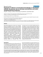
Muscle mitochondria prepared from UCP3KO mice [7]
were compared with wild-type mitochondria. As shown in
Fig. 1A lanes 1 and 2, the strong signal observed in wild-
type mouse mitochondria was absent from those of
UCP3KO mice. Figure 1A (lane 3) shows that the CabrX
antibody reacts with the mouse recombinant protein. The
size of the signal is higher than 34 kDa because of the
presence of a His
6
tag in the mouse recombinant protein.
C
2
C
12
cells, which do not express UCP3, were infected
with an adenovirus containing the human UCP3 gene. As
shown in Fig. 1B, no signal was observed in the wild-type
C
2
C
12
cells whereas the human UCP3 was detected, at the
expected size of 34 kDa, in the infected cells and in a sample
of the human UCP3 recombinant protein.
Taken together these results demonstrate that the 34-kDa
signal observed in our Western blots is due to a specific
interaction between the CabrX antibody and mouse or
human UCP3.
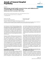
Figure 2A shows that the UCP3 signal of human
recombinant protein interacting with the antibody to
human UCP3 increases linearly as a function of increasing
amounts of the protein over the relatively large range
5–30 ng. Figure 2B shows data from a similar experiment
performed with mouse recombinant protein. Representative
signals obtained at different concentrations of recombinant
protein are shown under the figures. The times of exposure
werethesameforFig2AandB.
The reproducibility of the Western blot quantification
was analyzed using mouse gastrocnemius and tibialis muscle
mitochondria. The mean variation between quadruplicates
for four different samples was 27 ± 7% and 20 ± 6% for
UCP3 and COX, respectively. It should be stressed that
larger and unpredictable variations were observed when
values obtained with a given sample on two different gels
were compared. Therefore we only compared values
obtained on the same gel for all our subsequent quantitative
studies.
Preparation of SS and IMF mitochondria
We developed a technique using selective conditions of
mechanical disruption to prepare SS and IMF mitochon-
dria. As shown in Table 2, the quantity of IMF mitochon-
dria recovered from 1 g gastrocnemius muscle was 1.7-fold
higher than SS mitochondria. The specific and total levels of
COX protein were not significantly different in IMF and SS
mitochondria. The yield in mitochondria, which was
determined by comparing the level of COX protein in the
sum of the two mitochondria populations with that in the
homogenate, was 88%.
Expression of UCP3 protein in SS and IMF mitochondria
of various muscles
Figure 3A illustrates the distribution of UCP3 in SS and
IMF mitochondria obtained from different types of mouse
muscle, i.e. tibialis anterior (two-thirds fast oxidative
glycolytic, one-third glycolytic), gastrocnemius (one-third
slow oxidative, one-third fast oxidative glycolytic, one-third
fast glycolytic) and soleus (90% slow oxidative). It can be
seen in Fig. 3A that the UCP3 protein levels in SS
mitochondria (expressed as arbitrary units per mg mitoch-
ondrial protein) are higher in the tibialis anterior and
gastrocnemius than in the soleus (1.4-fold and 1.7-fold,
respectively). The levels of UCP3 in IMF mitochondria are
also higher in the tibialis anterior and gastrocnemius
muscles than in the soleus muscle (2.2-fold and 1.8-fold,
respectively). UCP3 is expressed at a significantly lower level
in IMF than SS mitochondria in the three types of muscle
(by 37%, 58% and 46% in tibialis anterior, gastrocnemius
and soleus muscle, respectively). No difference was observed
in the level of COX per mg of mitochondrial protein in the
three muscle types and in IMF vs. SS mitochondria, except
Fig. 2. Increase in the UCP3 signal as a function of increasing amounts
of (A) human recombinant protein and (B) mouse recombinant protein.
Representative signals are shown under the figures.
Fig. 1. Western blot analysis. (A) Western-blot signals obtained with
20 lg mitochondria isolated from wild-type (lane 1) or UCP3KO (lane
2) mouse gastrocnemius. Lane 3, 20 ng mouse recombinant UCP3. (B)
20 lg homogenate from C
2
C
12
wild-type myoblasts (lane 1) and C
2
C
12
myoblasts infected with adenoviruses containing the human UCP3
gene (lane 2). Lane 3, 40 ng human recombinant UCP3. The immu-
noblots were hybridized with CabrX (Anti-hUCP3) antibody. They
were also hybridized with antibodies to COX (Anti-COX) and pro-
hibitin. Representative signals are shown under the figures.
Table 2. Recovery of SS and IMF mitochondria from gastrocnemius
muscle. The results are expressed as means ± SEM from the number
of experiments in parentheses.
SS IMF
Protein yield
(mg protein per g muscle)
1.9 ± 0.2 (4) 3.2 ± 0.2 (3)
a
COX protein specific level
(arbitrary units per mg protein)
131 ± 12 (4) 86 ± 26 (3)
COX protein total level
(arbitrary units per g muscle)
239 ± 14 (4) 265 ± 69 (3)
a
P < 0.01 in IMF vs. SS mitochondria.
Ó FEBS 2002 UCP3 protein quantitation (Eur. J. Biochem. 269) 2881
for the gastrocnemius, where the level of COX is lower in
IMF than SS mitochondria by 43% (Fig. 3B). As shown in
Fig. 3C, the UCP3/COX ratio in SS mitochondria is higher
in the gastrocnemius than in the soleus (1.5-fold) and in
IMF mitochondria in the tibialis anterior and in the
gastrocnemius than in the soleus (2.0-fold and 1.9-fold,
respectively). In the gastrocnemius and soleus muscles, the
UCP3/COX ratio is 37% and 41% lower, respectively, in
IMF than SS mitochondria. This indicates that the compo-
sitions of the two mitochondrial populations are different.
UCP3 mRNA was determined by quantitative RT-PCR
in the same muscles to allow a comparison between the
respective UCP3 mRNA and protein levels. As shown in
Fig. 4, UCP3 mRNA levels were higher in the tibialis
anterior and gastrocnemius muscles than in the soleus
muscle (1.7-fold and 2.1-fold, respectively). Therefore the
relative amounts of UCP3 protein and mRNA in the three
muscles are comparable.
Effect of fasting on UCP3 protein expression
We and others have shown that fasting induces upregula-
tion of UCP3 mRNA expression in skeletal muscle of mice
and rats (for review see [5]). To study further the regulation
of UCP3 in fasting, we measured the protein levels in SS and
IMF mitochondria of gastrocnemius muscle in 24 h-fasted
mice. Figure 5 illustrates the effects of 24-h fasting on the
expression of muscle UCP3 and COX per mg of protein in
both mitochondria populations. After a 24-h fast, the UCP3
protein level was increased 1.5-fold (P < 0.01) in SS
mitochondria, whereas it was unaffected in IMF mitochon-
dria. COX protein level was found to be unchanged by
fasting. In five animals from each group of mice, we also
Fig. 3. UCP3 protein levels (A) and COX protein levels (B) in mouse
tibialis anterior (TA), gastrocnemius (Gn) and soleus (So) muscle SS
(empty columns) and IMF (shaded columns) mitochondria (20 lg). (A)
The immunoblots were hybridized with CabrX antibody. The signals
were quantified by scanning photodensitometry and are presented as
means ± SEM of absolute values, n ¼ 4–6. **P <0.02 and
***P < 0.001 vs. SS mitochondria; #P < 0.05 vs. tibialis anterior
values; °P <0.05 and°°°P < 0.005 vs. gastrocnemius values. (B)
Same as in (A) except that the immunoblots were hybridized with a
COX antibody. ***P < 0.005 vs. SS mitochondria. (C) UCP3 values
normalized using the corresponding COX values. *P <0.05 and
***P < 0.005 vs. SS mitochondria; #P < 0.05 vs. tibialis anterior
values; °°°P < 0.005 vs. gastrocnemius values.
Fig. 4. UCP3 mRNA levels in mouse tibialis anterior (TA), gastroc-
nemius (Gn) and soleus (So) muscle. The results, obtained by real-time
quantitative RT-PCR as described in Materials and Methods, are
presented as means ± SEM of values normalized using actin.
*P < 0.05 vs. gastrocnemius values.
2882 M. Jimenez et al.(Eur. J. Biochem. 269) Ó FEBS 2002
determined the UCP3 mRNA levels in gastrocnemius
muscle. We observed that fasting induced a 16.4-fold
(P < 0.001) increase in UCP3 mRNA expression, whereas
it augmented the total amount of UCP3 protein by 1.5-fold
in the gastrocnemius from the same animals.
DISCUSSION
This study first validates a Western blot technique for the
detection and quantitation of UCP3 using a specific and
sensitive antibody raised against 14 amino acids located at
the C-terminus of human UCP3 protein (CabrX). The use
of both mitochondria from knockout mouse gastrocnemius
and homogenates of C
2
C
12
myoblasts infected with an
adenovirus encoding for human UCP3 allowed clear
validation of our Western blot signal. Good cross-reactivity
of the CabrX antibody with mouse recombinant UCP3 was
also demonstrated. The signals can be quantitated and
compared with a reasonable degree of accuracy, but only if
obtained on the same gel.
The density of UCP3 protein (i.e. the intensity of the
signal expressed per mg of proteins) was similar in the SS
mitochondria of the two glycolytic muscles, tibialis anterior
and gastrocnemius, and higher than that in the oxidative
muscle soleus. The same was observed with IMF mito-
chondria. It is noteworthy that identical results were
obtained in this study when the levels of UCP3 mRNA
were measured in the tibialis anterior, gastrocnemius and
soleus muscles. Hesselink et al. [23], who studied the
distribution of UCP3 protein in various human muscle
types by immunofluorescence, showed a higher expression
of UCP3 protein in glycolytic than oxidative fibers of vastus
lateralis muscle. Our results show that these findings cannot
be extended to rodent muscles. If indeed the UCP3 protein
was more abundantly expressed in glycolytic than oxidative
muscles, its density would be higher in the tibialis anterior
than in the gastrocnemius.
The density of COX showed a tendency to be lower in
IMF than SS mitochondria, the difference being significant
only in the gastrocnemius muscle. In rats, muscle IMF
mitochondria have been shown to display higher state-III
respiration [10,11] and slightly higher COX and lower
succinate dehydrogenase activities [10] than SS mitochon-
dria. The results of our study, showing that the COX
protein level tended to be lower in IMF than SS mitochon-
dria is not in total agreement with these results.
When the density of UCP3 was compared in SS and IMF
mitochondria, it was found to be higher in SS mitochondria
of all three muscle types studied.
Our UCP3 data show that (a) the muscle type that relies
most on oxidative phosphorylation for ATP synthesis, i.e.
the soleus muscle, contains less UCP3 per mg of mitoch-
ondrial protein than glycolytic muscle types and (b) in all
muscle types the mitochondria most closely associated with
the myofibrills, i.e. the IMF mitochondria, have a lower
UCP3 density.
If UCP3 is an uncoupling protein, the oxidative muscle
(soleus) and the IMF mitochondria (which should be most
involved in muscle contraction) would be less prone to
uncoupling and therefore more efficient at producing ATP.
In a recent paper on pig muscle SS and IMF mitochondria,
Lombardi et al. [24] showed that IMF mitochondria had a
higher capacity for ATP production than SS. The possible
role of the lower level of UCP3 in this difference would be
interesting to study.
Most studies on regulation of UCP3 expression have
investigated changes in UCP3 mRNA levels. Fasting has
repeatedly been shown to dramatically increase muscle
UCP3 mRNA in rats and mice [5]. This is surprising
because UCP3, which has been shown to exhibit uncoupling
activity, would be expected to be turned off in muscle under
conditions that dictate energy sparing such as starvation. In
this study, a 24-h fasting period, which was shown to
increase gastrocnemius muscle UCP3 mRNA level 16.4-
fold, was found to induce an increase of 1.5-fold in the
UCP3 protein level of gastrocnemius muscle from the same
animal. Consistent with these observations, Cadenas et al.
[17] and Sivitz et al. [18] reported increases in rat UCP3
protein induced by fasting that were less than half those of
UCP3 mRNA level in experiments carried out in parallel.
These results are in agreement with studies reporting no
change in mitochondrial proton conductance [17] and
energy coupling [21] in muscle of starved rats. Thus,
UCP3 protein level rather than mRNA expression would be
appear to be an indication of UCP3 activity in rodent
muscle. The observation of a marked increase in UCP3
Fig. 5. Effect of a 24-h fast on (A) UCP3 protein levels and (B) COX protein levels in mouse gastrocnemius SS (empty columns) and IMF (shaded
columns) mitochondria (20 lg). The methods for the detection and quantitation of UCP3 and COX are described in the legend of Fig. 3. Data are
presented as means ± SEM, n ¼ 9–11. (A) ***P < 0.005 vs. SS mitochondria; ##P < 0.01 vs. respective mitochondrial population in muscle of
fed mice. (B) **P <0.01and*P < 0.05 vs. SS mitochondria.
Ó FEBS 2002 UCP3 protein quantitation (Eur. J. Biochem. 269) 2883
mRNA associated with only a minor change at the protein
level during fasting suggests that UCP3 expression is
regulated at the post-transcriptional level and therefore that
modulation of UCP3 expression at the mRNA level does
not necessarily result in similar changes at the protein level.
We can speculate that the increase in UCP3 mRNA
produces an RNA pool ready to be rapidly translated after
the initiation of refeeding. This point will be addressed in
future studies. The present findings put into perspective the
so-called Ôfasting paradoxÕ, suggesting that UCP3 activity
might not be modified in the muscle during fasting.
Furthermore our data show that the two mitochondria
populations are differently affected by metabolic changes.
Further studies on the regulation of UCP3 protein expres-
sion may have important implications for the understanding
of its physiological role.
In conclusion, in this study, using a carefully validated
antibody to UCP3 and a new protocol to prepare SS and
IMF mitochondria, UCP3 was found at higher density in
mitochondria of glycolytic muscle than in those of oxidative
muscle. Our data also indicate that SS mitochondria contain
more UCP3 than IMF mitochondria, raising the possibility
that these organelles have different capacities for oxidative
ATP production and a moderate increase in UCP3 protein
content in SS mitochondria of fasted mice.
ACKNOWLEDGEMENTS
This work was supported by grants from the Swiss National Science
Foundation no. 31-53707.98 and 31-54306.98. We are indebted to the
Office Fe
´
de
´
ral du Sport Macolin, the Fonds Euge
`
ne Rapin, the
Fondation du Centenaire de la socie
´
te
´
Suisse d’Assurances ge
´
ne
´
rales sur
la vie humaine pour la sante
´
publique et les recherches me
´
dicales and
the Roche Research Foundation.
REFERENCES
1. Cannon, B. & Nedergaard, J. (1985) The biochemistry of an
inefficient tissue: brown adipose tissue. Essays Biochem. 20,
110–164.
2. Boss, O., Samec, S., Paoloni-Giacobino, A., Rossier, C., Dulloo, A.,
Seydoux, J., Muzzin, P. & Giacobino, J.P. (1997) Uncoupling
protein-3: a new member of the mitochondrial carrier family with
tissue-specific expression. FEBS Lett. 408, 39–42.
3. Boss, O., Muzzin, P. & Giacobino, J.P. (1998) The uncoupling
proteins, a review. Eur. J. Endocrinol. 139, 1–9.
4. Muzzin, P., Boss, O. & Giacobino, J.P. (1999) Uncoupling protein
3: its possible biological role and mode of regulation in rodents
and humans. J. Bioenerg. Biomembr. 31, 467–473.
5. Boss, O., Hagen, T. & Lowell, B.B. (2000) Uncoupling proteins 2
and 3: potential regulators of mitochondrial energy metabolism.
Diabetes. 49, 143–156.
6. Vidal-Puig, A.J., Grujic, D., Zhang, C.Y., Hagen, T., Boss, O.,
Ido, Y., Szczepanik, A., Wade, J., Mootha, V., Cortright, R.,
Muoio, D.M. & Lowell, B.B. (2000) Energy metabolism in
uncoupling protein 3 gene knockout mice. J. Biol. Chem. 275,
16258–16266.
7. Gong, D.W., Monemdjou, S., Gavrilova, O., Leon, L.R., Marcus-
Samuels,B.,Chou,C.J.,Everett,C.,Kozak,L.P.,Li,C.,Deng,C.,
Harper, M.E. & Reitman, M.L. (2000) Lack of obesity and nor-
mal response to fasting and thyroid hormone in mice lacking
uncoupling protein-3. J. Biol. Chem. 275, 16251–16257.
8. Clapham, J.C., Arch., J.R., Chapman, H., Haynes, A., Lister, C.,
Moore,G.B.,Piercy,V.,Carter,S.A.,Lehner,I.,Smith,S.A.,
Beeley, L.J., Godden, R.J., Herrity, N., Skehel, M., Changani,
K.K.,Hockings,P.D.,Reid,D.G.,Squires,S.M.,Hatcher,J.,
Trail, B., Latcham, J., Rastan, S., Harper, A.J., Cadenas, S.,
Buckingham, J.A., Brand, M.D. & Abuin, A. (2000) Mice over-
expressing human uncoupling protein-3 in skeletal muscle are
hyperphagic and lean. Nature (London) 406, 415–418.
9. Echtay,K.S.,Winkler, E.,Frischmuth, K.&Klingenberg, M.(2001)
Uncoupling proteins 2 and 3 are highly active H(+) transporters
and highly nucleotide sensitive when activated by coenzyme Q
(ubiquinone). Proc. Natl Acad. Sci. USA 98, 1416–1421.
10. Cogswell, A.M., Stevens, R.J. & Hood, D.A. (1993) Properties of
skeletal muscle mitochondria isolated from subsarcolemmal and
intermyofibrillar regions. Am. J. Physiol. 264, C383–C389.
11. Krieger, D.A., Tate, C.A., McMillin-Wood, J. & Booth, F.W.
(1980) Populations of rat skeletal muscle mitochondria after
exercise and immobilization. J. Appl. Physiol. 48, 23–28.
12. Bradford, M.M. (1976) A rapid and sensitive method for the
quantitation of microgram quantities of protein utilizing the
principle of protein-dye binding. Anal. Biochem. 72, 248–254.
13. Muzzin, P., Eisensmith, R.C., Copeland, K.C. & Woo, S.L. (1996)
Correction of obesity and diabetes in genetically obese mice by
leptin gene therapy. Proc. Natl Acad. Sci. USA 93, 14804–14808.
14. Higuchi, R., Fockler, C., Dollinger, G. & Watson, R. (1993)
Kinetic PCR analysis: real-time monitoring of DNA amplification
reactions. Biotechnology (NY) 11, 1026–1030.
15. Boss, O., Samec, S., Desplanches, D., Mayet, M.H., Seydoux, J.,
Muzzin, P. & Giacobino, J.P. (1998) Effect of endurance training
on mRNA expression of uncoupling proteins 1, 2, and 3 in the rat.
FASEB J. 12, 335–339.
16. Boss, O., Samec, S., Kuhne, F., Bijlenga, P., Assimacopoulos-
Jeannet, F., Seydoux, J., Giacobino, J.P. & Muzzin, P. (1998)
Uncoupling protein-3 expression in rodent skeletal muscle is
modulated by food intake but not by changes in environmental
temperature. J. Biol. Chem. 273,5–8.
17. Cadenas, S., Buckingham, J.A., Samec, S., Seydoux, J., Din, N.,
Dulloo, A.G. & Brand, M.D. (1999) UCP2 and UCP3 rise in
starved rat skeletal muscle but mitochondrial proton conductance
is unchanged. FEBS Lett. 462, 257–260.
18. Sivitz,W.I.,Fink,B.D.&Donohoue,P.A.(1999)Fastingand
leptin modulate adipose and muscle uncoupling protein: divergent
effects between messenger ribonucleic acid and protein expression.
Endocrinology. 140, 1511–1519.
19. Zhou, M., Lin, B.Z., Coughlin, S., Vallega, G. & Pilch, P.F. (2000)
UCP-3 expression in skeletal muscle: effects of exercise, hypoxia,
and AMP-activated protein kinase. Am. J. Physiol. Endocrinol.
Metab. 279, E622–E629.
20. Jucker, B., Dufour, S., Ren, J., Cao, X., Previs, S., Underhill, B.,
Cadman, K. & Shulman, G. (2000) Assessment of mitochondrial
energy coupling in vivo by
13
C/
31
PNMR.Proc. Natl Acad. Sci.
USA 97, 6880–6884.
21. Jucker,B.,Ren,J.,Dufour,S.,Cao,X.,Previs,S.,Cadman,K.&
Shulman, G. (2000)
13
C/
31
P NMR assessment of mitochondrial
energy coupling in skeletal muscle of awake fed and fasted rats.
J. Biol. Chem. 275, 39279–39286.
22. Pedraza, N., Solanes, G., Carmona, M.C., Iglesias, R., Vinas, O.,
Mampel, T., Vazquez, M., Giralt, M. & Villarroya, F. (2000)
Impaired expression of the uncoupling protein-3 gene in skeletal
muscle during lactation: fibrates and troglitazone reverse lacta-
tion-induced downregulation of the uncoupling protein-3 gene.
Diabetes 49, 1224–1230.
23. Hesselink, M.K.C., Keizer, H.A., Borghouts, L.B., Schaart, G.,
Kornips, C.F.P., Slieker, L.J., Slopp, K.W., Saris, W.H.M. &
Schrauwen, P. (2001) Protein expression of UCP3 differs between
type 1, type 2a, and type 2b fibers. FASEB J. 15, 1071–1073.
24. Lombardi, A., Damon, M., Vincent, A., Goglia, F. & Herpin, P.
(2000) Characterisation of oxidative phosphorylation in skeletal
muscle mitochondria subpopulations in pig: a study using top-
down elasticity analysis. FEBS Lett. 475, 84–88.
2884 M. Jimenez et al.(Eur. J. Biochem. 269) Ó FEBS 2002