Báo cáo Y học: The structure of the O-chain of the lipopolysaccharide of a prototypal diarrheagenic strain of Hafnia alvei that has characteristics of a new species under the genus Escherichia pot
Bạn đang xem bản rút gọn của tài liệu. Xem và tải ngay bản đầy đủ của tài liệu tại đây (464.09 KB, 7 trang )
The structure of the O-chain of the lipopolysaccharide of a prototypal
diarrheagenic strain of
Hafnia alvei
that has characteristics of a new
species under the genus
Escherichia
Reine Eserstam
1
, Thushari P. Rajaguru
1,2
, Per-Erik Jansson
1
, Andrej Weintraub
3
and M. John Albert
4
1
Clinical Research Center, Analytical unit, Karolinska Institute, Huddinge Hospital, Huddinge, Sweden;
2
Department of Chemistry,
University of Peradeniya, Peradeniya, Sri Lanka;
3
Karolinska Institute, Department of Microbiology, Pathology and Immunology,
Division of Clinical Bacteriology, Huddinge University Hospital, Sweden;
4
Department of Microbiology, Faculty of Medicine, Kuwait
University, Safat, Kuwait
The structure of the O-polysaccharide of the lipopolysac-
charide from a diarrheal strain isolated in Bangladesh
was studied with sugar, and methylation analysis, NMR
spectroscopy, mass spectrometry and partial acid hydrolysis.
The strain was first designated as Hafnia alvei, but later
found to be a possible new species in the genus Escherichia.
Two different polysaccharides were detected, a major and a
minor one. The structure of the major polysaccharide is gi-
ven below, while the structure of the minor one was not
investigated. The structure of the repeating unit was estab-
lished as
→6)-β-
D
-Galf-(1→3)-β-
D
-GalpNAc-(1→3)-β-
D
-Galp-(1→
α-NeuAc
↑
6
2
The structure does not resemble any of the previously
investigated lipopolysaccharide O-chains from Escherichia
coli or H. alvei, but could fit in either group based on types of
sugar residues and acidity.
Phenotypic microbiological studies cannot definitely
assign it to either species of the two genera. Genetic
hybridization studies indicate that the Bangladeshi isolates
may require a new species designation under the genus
Escherichia.
Keywords: lipopolysaccharide; Escherichia; Hafnia alvei;
diarrhea; neuraminic acid.
Hafnia alvei is a Gram negative bacterium and a member of
the family Enterobacteriaceae. There are reports of associ-
ation of H. alvei with diarrhoea in Canada [1] and Finland
[2], but the mechanism of diarrhoea caused by this organism
in these locations remains unknown [3]. However, some
isolates of a bacterium typed as H. alvei from patients with
diarrhoea in Bangladesh produced diarrhoea in rabbits by
attaching and effacing (AE) lesions in the intestinal mucosa
that are characteristic of the lesions produced by entero-
pathogenic Escherichia coli [4]. Like enteropathogenic
E. coli,theseH. alvei isolates possess a homologous patho-
genicity island in the chromosome locus for enterocyte
effacement (LEE), which is responsible for producing
attaching and effacing lesions [5]. LEE encodes a type III
secretory system [6]. Secretion of the virulence factors leads
to effacement of the microvillus structure and reorganiza-
tion of the actin cytoskeleton to form a pedestal-like
structure, the attaching and effacing lesion [7]. AE lesion
formation is critical in mediating diarrhoea production in
the host, but its exact role in disease is not known. Recent
results from conventional biochemical analyses, testing of
susceptibility to cephalothin, lysis by a Hafnia-specific
phage, and amplification of the outer membrane protein
gene phoE with species-specific primers support the identi-
fication of these isolates as members of the genus Escheri-
chia rather than Hafnia alvei [8]. We studied the structure of
the O-chain of the lipopolysaccharide of one them.
MATERIALS AND METHODS
Bacterium, cultivation and isolation
of lipopolysaccharide and O-specific polysaccharide
The Hafnia alvei, strain number 10457, was from the culture
collection of the International Centre for Diarrhoeal
Disease Research, Bangladesh (ICDDR, B), Dhaka. This
strain was isolated from a patient with diarrhoea and was
positive for the AE property [15]. The bacterium was grown
in TY-medium, and the lipopolysaccharide isolated by
centrifugation and extraction of bacterial cells with hot
aqueous phenol [16]. The polysaccharide was analysed as
Correspondence to P E. Jansson, Karolinska Institute,
Clinical Research Center, Novum, Huddinge University Hospital,
S-141 86 Huddinge, Sweden.
Fax: + 46 8 58583820, Tel.: + 46 8 58583821,
E-mail:
Abbreviations: AE, attaching and effacing; Hex, hexose; DEPT, dis-
torsionless enhanced polarization transfer; HMBC, heteronuclear
multiple-bond correlation; HSQC, heteronuclear single-quantum
coherence; LEE, locus for enterocyte effacement; TMS, trimethylsilyl.
(Received 19 March 2002, revised 6 May 2002,
accepted 21 May 2002)
Eur. J. Biochem. 269, 3289–3295 (2002) Ó FEBS 2002 doi:10.1046/j.1432-1033.2002.03009.x
the lipopolysaccharide, or degraded with 0.1
M
acetic acid/
sodium acetate, pH 4.2, for 4 h at 100 °Ctogivethe
O-polysaccharide. The O-polysaccharide was isolated by
gel-permeation chromatography on a column (70 · 2.6 cm)
of Sephadex G-50 using 0.05
M
pyridinium acetate, pH 4.5,
as eluent and monitoring with a differential refractometer.
Sugar analysis
Hydrolysis was performed with 2
M
trifluoroacetic acid
(120 °C, 2 h), and monosaccharides identified by GLC as
their alditol acetates. Sugars were analysed on a Hewlett–
Packard 5880 GLC instrument on a DB-5 fused-silica
capillary column and a temperature gradient of 160 °C
(1 min) to 250 °Cat3°CÆmin
)1
. The absolute configura-
tions were determined by GLC of acetylated glycosides
with (+)-2-butanol, as described previously, but with the
modification that acetates were used [9–11]. Neuraminic
acid methyl glycoside methyl ester was analyzed as the
trimethylsilyl (TMS)-derivative and authentic reference.
A colorimetric test for Kdo using thiobarbituric acid was
also made [17].
Methylation analysis
Methylation was carried out with methyl iodide in dimethyl
sulfoxide in the presence of sodium methylsulfinylmethanide
[18]. The methylated polysaccharide was purified using Sep–
Pak C18 cartridges. Hydrolysis was performed as described
for sugar analysis; partially methylated monosaccharides
were converted into alditol acetates and analyzed by GLC
and GLC-MS on a Hewlett Packard 5890 chromatograph
equipped with a NERMAG R10–10 L mass spectrometer,
using the above conditions. Identification was made using
reference data.
NMR spectroscopy
1
H- and
13
C-NMR spectra were recorded with a JEOL
GSX-270 or JEOL JNM ECP500 instruments or solutions
in
2
H
2
Oat70or85°C. Chemical shifts are reported with
internal acetone (d
H
2.25, d
C
31.00) as reference. Mixing
times of 30–160 ms were used in TOCSY experiments, and
for NOESY 100 and 300 ms.
MALDI mass spectrometry
MALDI mass spectrometry in the positive mode was run on
a Finnigan Lasermat instrument using dihydroxybenzoic
acid acid as matrix. Between 10 and 20 scans were
accumulated and added. The neuraminic-acid-free polysac-
charide was treated with 0.01
M
acetic acid for 1 h at 65 °C
and then neutralized with dilute sodium hydroxide solution.
RESULTS AND DISCUSSION
Hafnia alvei strain number 10457 was grown in tryptone-
yeast (TY) medium and harvested by centrifugation.
Extraction with hot phenol/water (1 : 1, v/v) gave a lipo-
polysaccharide in the aqueous phase, which was recovered
and freeze dried. Ultracentrifugation of the lipopolysaccha-
ride gave a pellet and an upper phase, the latter containing
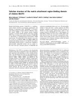
most of the material. SDS/PAGE of the two materials
(Fig. 1) in the upper phase and the pellet showed identical
patterns and it was therefore concluded that the same
polysaccharide was present. A hydrolysate of the upper
phase, analyzed as alditol acetates, revealed as
D
-glucose,
D
-galactose,
D
-galactosamine,
L
-glycero-
D
-manno-heptose,
and
D
-glucosamine in the proportions 6 : 65 : 19 : 6 : 3.
The relatively high proportion of heptose may be the result
of short chains. It is not a component in the polysaccharide
as demonstrated in the MS analysis (see below). The
absolute configurations of the sugars were established by
GLC analysis of the acetylated (+)-2-butyl glycosides
[9–11]. Methanolysis of the sample and analysis by GLC-
MS gave, in addition to the sugars mentioned, neuraminic
acid. The pellet showed essentially the same compounds.
To verify that the material was an O-polysaccharide or
exclude the possibility, the content of Kdo was checked with
the thiobarbiturate method. It showed that the content of
Kdo in the crude material, the pellet, and the supernatant
was 11, 15, and 12 lgÆmg
)1
, respectively, corresponding to
one Kdo per 70 sugars or two per 35, i.e a significant
amount of Kdo.
Treatment of the upper phase with acetic acid buffer of
pH 4.2 followed by gel chromatography on a Sephadex
G-50 column gave a major O-polysaccharide peak at the
void volume (O-polysaccharide) and a minor peak just after.
The material in the major peak was devoid of neuraminic
acid. The second minor peak, which was included in the
column and had four signals in the anomeric region of the
1
H-NMR spectrum at d 5.07, 4.92, 4.80, and 4.49, clearly
different from the major compound. The presence of two
different polysaccharides in Hafnia has not been observed
before. It was not clear whether it was an lipopolysaccharide
or a capsular polysaccharide and the fraction was not
further investigated. The proportion of the minor polysac-
charide was indicated by the size of the minor peaks in the
1
H-NMR spectrum (Fig. 2), especially as the peak near
d 5.0 was separate enough to be able to make a quantitative
estimation, approximately 5%.
In the methylation analysis of the O-polysaccharide,
derivatives corresponding to 6-substituted galactofuranose,
3-substituted galactopyranose and 3-substituted galactosa-
Fig. 1. SDS/PAGE of the upper phase (1) and the pellet (2) obtained on
ultracentrifugation of the Hafnia alvei lipopolysaccharide. For com-
parison lipopolysaccharide from a smooth (Shigella flexneri,3)anda
rough bacterium (Salmonella typhimurium Ra, 4) was run simulta-
neously.
3290 R. Eserstam et al. (Eur. J. Biochem. 269) Ó FEBS 2002
mine were detected, thus indicating a linear polysaccharide
consisting of repeats with three sugar residues. This was
corroborated by the
1
H-NMR spectrum, which showed
signals for anomeric protons at d 5.11 (J small), 4.76
(J 7.7 Hz) and 4.46 (J 7.7 Hz), for ring protons, and for an
N-acetyl methyl group at d 2.05. The first chemical shift
should belong to a furanoside as these normally have small
J-values, the second and third signal have typical J-values
for b-linked sugars with galacto-pyranose configuration.
The absence of NeuAc was evident as no methylene-
deoxyresonances could be detected. The
13
C-NMR spec-
trum showed signals inter alia for anomeric carbons at d
109.9, 104.1 and 103.6. The first value is very high and
characteristic of a b-furanosidic sugar. A distorsionless
enhanced polarization transfer (DEPT) spectrum revealed
that the substituted hydroxymethyl group, C-6 of the
galactofuranose, is located at d 71.8, thus among those of
secondary carbons. The
1
H- and
13
C-NMR spectra were
assigned with 2D NMR spectra including COSY, TOCSY,
NOESY, HSQC, and HMBC. Overlap in the spin systems
were in some cases a problem, but could be overcome with a
combination of the spectra. Residues and spin systems are
denoted A–C in Table 1. Indeed, the A residue was the
furanoside as evident from the correlation in the HSQC-
spectrum between d 5.11/109.9. The possibility to trace
signals beyond that H-2 was limited, but two of the signals
in the
13
C-NMR spectrum at 82–84 p.p.m. could be shown
to derive from C-2 and C-4 in A, and to correlate to proton
signals at approximately d 4.07. B could be assigned to the
GalNAc residue as evident from the chemical shift of C-2
signal, which appeared at d 52.2, typical for C-N signals. A
downfield shift of the signal for C-3 signal to d 78.7
corroborated the linkage position. Residue C gave in the
TOCSY spectrum three correlations, up to H-4, which had
aresonanceatd 4.16, from the high value it was confirmed
that it was galactose. In addition, the couplings of H-4 (1D/
2D) were small, indicative of the galacto configuration. In
the
13
C-NMR spectrum, it was evident that two signals were
present at d 75.6, the second being assigned to C-5 in residue
C, close to the value in the monomer. The sequence of
Fig. 2. The
1
H-NMR spectrum of the Hafnia
alvei lipopolysaccharide in D
2
O.
Table 1.
1
H- and
13
C-NMR chemical shifts (d, p.p.m.) for different H. alvei polysaccharides. The N-acetyl group in the GalNAC residue appears at d
2.05/22.7/176.1 in the O-polysaccharide and in the GalNAc and NeuAc residues at d 2.05/22.8–22.9/175.8 in the lipopolysaccharide.
Sugar residue 1 2 3 4 5 6 7 8 9
H. alvei O-polysaccharide
fi 6)-b-
D
-Galf-(1 fi (A) 5.11 4.09 4.06 4.06 4.02 3.75, 4.04
109.9 82.1 77.6 83.7 70.2 71.8
fi 3)-b-
D
-GalpNAc-(1 fi (B) 4.76 4.05 3.82 4.02 3.71 3.77
103.6 52.2 78.7 70.2 75.6 61.6
fi 3)-b-
D
-Galp-(1 fi (C) 4.46 3.62 3.76 4.16 3.71 3.77
104.1 70.7 82.6 69.3 75.6 61.6
Native H. alvei lipopolysaccharide
fi 6)-b-
D
-Galf-(1 fi (A) 5.11 4.09 4.05 4.04 4.00 3.89, 3.89
109.9 82.8 77.8 83.8 70.6 72.1
fi 3)-b-
D
-GalpNAc-(1 fi (B) 4.73 4.04 3.83 4.07 3.70 3.75, 3.75
103.7 52.4 78.8 68.5 75.6 61.7
fi 3,6)-b-
D
-Galp-(1 fi (C) 4.43 3.65 3.72 4.19 3.73 3.62, 3.92
104.2 70.6 82.8 69.0 73.4 64.1
a-NeuAc-(1 fi (D) – – 2.74, 1.68 3.74 3.84 3.70 3.78 4.03
174.2 101.2 41.0 69.0 52.6 73.8 69.0 72.4 63.4
Ó FEBS 2002 New species under the genus Escherichia (Eur. J. Biochem. 269) 3291
sugars was indicated by the following H/C correlations in
the HMBC spectrum, d 5.11/78.7 (A H-1/B C-3), d 4.73/82.6
(B H-1/C C-3), and d 4.44/71.8 (C H-1/A C-6). The
disaccharide elements A–B, B–C, and C-A were thus present
to make up the chain as
→6)-β-
D
-Galf-(1→3)-β-
D
-GalpNAc-(1→3)-β-
D
-Galp-(1→
A
B
C
The next step was to analyze the native lipopolysaccha-
ride. Methylation analysis of the native lipopolysaccharide
gave major GLC peaks corresponding to 6-substituted
galactofuranose, 3,6-disubstituted galactose, and 3-substi-
tuted 2-acetamido-2-deoxy-
D
-galactose. In addition, smaller
peaks corresponding to the minor component polysaccha-
ride were observed. The repeating unit of the polysaccharide
thus contains a terminal NeuAc, and the above mentioned
residues. A comparison to the methylation analysis data on
the O-polysaccharide, indicates that the terminal NeuAc
should be substituting the galactose residue in the
6-position.
For the full characterization of the lipopolysaccharide,
with NeuAc still present, an NMR sample was prepared
from the native lipopolysaccharide. The spectrum had
broadened lines but were surprisingly good with resolved
couplings (Fig. 2). The
1
H-NMR spectrum of the lipopoly-
saccharide showed signals for three anomeric protons at
d 5.11 (J small), 4.73 (J 7.7 Hz), 4.43 (J 7.7 Hz), thus close
to those observed for the O-polysaccharide. In the high field
region signals for a methylene group, assigned to CH
2
in
NeuAc were observed at d 2.75 and 1.68, the large difference
establishing the presence of an axial carboxyl group and an
a-linkage in the NeuAc residue. Signals for N-acetyl groups
deriving from NeuAc and GalNAc were present at d 2.05. In
the
13
C-NMR spectrum, the corresponding signals were
present inter alia at d 109.9, 104.2, 103.7, and 101.2 for
anomeric carbons and at d 41.0 and 22.8–22.9 for methylene
and methyl groups, respectively. The spectrum resembled
that of the O-polysaccharide, but some changes were
evident. The signals at d73.8, 69.0, 63.4, and 52.6 were
higher than the others and were subsequently assigned to
the NeuAc residue.
Analysis of both of the
1
H- and the
13
C-NMR spectra
using 1D and 2D techniques gave the data shown in
Table 1, where the residues are referred to as A–D, D being
the additional NeuAc residue. Most of the signals could be
unambiguously assigned. The
13
C-NMR spectrum showed
28 signals of the possible 30. The assignment was made
essentially as described for the O-polysaccharide and by
comparison with the spectra of the O-polysaccharide.
Figures 3–6 show the COSY, TOCSY, HSQC and HMBC
spectra, respectively.
From the large glycosylation shifts of signals from C-6 in
A, C-3 and C-6 in B,andC-3inC, the linkage positions
were verified. The absence of any glycosylation shift in D
further indicated that it was terminal. The chemical shift
displacement of the C-6 signal in C to d 64.1, is small,
typical for substitution with ketosides. An HMBC experi-
ment showed the following inter-residue correlations from
anomeric protons to linkage carbons: A H-1/B C-3 (5.11/
78.8), B H-1/C C-3 (4.73/82.8) corroborating elements A–B
and B–C. From the NOE spectrum the following inter-
residue correlations between H-1 and protons on linkage
carbons were observed: A H-1/B H-3 (5.11/3.82), demon-
strating the element A–B. Correlations 4.73/3.72 and 4.43/
3.90 are in accord with elements B–C and C–A but
ambiguous due to signal overlap. From the combined data,
however, the following structure can be postulated for the
repeating unit
→6)-β-
D
-Galf-(1→3)-β-
D
-GalpNAc-(1→3)-β-
D
-Galp-(1→
α-NeuAc
↑
6
2
A
B
C
D
Fig. 3. The COSY spectrum the of the Hafnia
alvei lipopolysaccharide showing the anomeric
and the ring proton region.
3292 R. Eserstam et al. (Eur. J. Biochem. 269) Ó FEBS 2002
MALDI-MS of the O-polysaccharide
The O-polysaccharide, i.e. the desialylated polysaccharide
chain, was also characterized by MALDI-MS of the
fragmented chain, anticipated to be facile to cleave with
acid, as furanosides were present. Thus, the O-polysaccha-
ride was treated with aqueous 0.01
M
acetic acid and the
samples were withdrawn at different times. Good spectra
were obtained after approximately 1 h. The spectra showed
three series of ions, all sodiated, the first at m/z 1626, 2149,
2677, 3204, 3730, and 4254, the second at m/z 1825, 2352,
2881, and 3401, and the third at m/z 1946, 2475, 3001, 3528,
and 4054. All series are spaced with approximately
527 atomic mass units (amu), corresponding to the molecu-
lar mass of the repeating unit containing two hexoses and
one acetamidohexose. The first series corresponds to a
multiple of the repeating unit, thought to be derived from
the expected hydrolysis of the furanosidic linkage, thus
(HexNAc-Hex-Hexf)
n
. The second series contains a mul-
tiple of the repeat plus an additional acetamidohexose
(203 amu) and the third contains a multiple of the repeat
plus two additional hexose residues (324 amu). This implies
that not only the furanosidic linkage is acid-labile, but also
that of the b-D-GalNAc residue. Thus, assuming that the
linkage second most easily hydrolyzed is that of the
acetamidohexose, the ions in the second and the third series
correspond to the formulas (HexNAc-Hex-Hex)
n
-HexNAc
and Hex-Hex-(HexNAc-Hex-Hex)
n
and the sequence is
further established.
The initial phenotypic characterization of the strain 10457
with a commercial identification system, API-20 E identified
the strain as Hafnia alvei [4]. Additional phenotypic
characterization and partial 16S rRNA sequencing of a
set of isolates identified them not as typical Hafnia alvei, but
Fig. 5. HSQC spectrum of the Hafnia alvei
lipopolysaccharide showing the anomeric and
the ring proton/carbon region and including
high resolution
1
H- and
13
C-NMR spectra.
Fig. 4. TOCSY spectra of the Hafnia alvei
lipopolysaccharide showing the correlations
deriving from the anomeric protons. Mixing
times were from 30 to 160 ms.
Ó FEBS 2002 New species under the genus Escherichia (Eur. J. Biochem. 269) 3293
unique isolates [12]. Further phenotypic characterization
suggested that these isolates are neither Hafnia alvei nor
Escherichia coli, but closely related to the genus Escherichia
[8]. DNA hybridization studies suggested that these isolates
deserve a new species name under the genus Escherichia
(J. Albert, Kuwait University, Safat, Kuwait, personal
communication). The unique structure of the O-chain of
one of these isolates further confirms this conclusion.
The lipopolysaccharide structures of both H. alvei and
E. coli are known [13,14]. More than 20 strains from
H. alvei have been investigated and both amino sugars and
acidic sugars are frequent. Furanosidic sugars are also
observed. Neuraminic acid has been found once but only as
an internal residue. Of the more than 60 E. coli strains that
have been investigated, many contain hexoses and hexosa-
mines, as well as acid functions. Also here, neuraminic acid
has been found only internally. Furanosidic galactose is
present, but not common. As a whole, the structural
features of the investigated strain cannot be related to any
particular structure among those studied of Hafnia and
Escherichia. The terminal neuraminic acid is, however, an
interesting feature, normally associated with glycoproteins
and glycolipids. The biological properties of the novel
lipopolysaccharide of this strain remain to be elucidated.
ACKNOWLEDGEMENTS
The authors thank Mrs G. Alvelius for help in mass spectrometry and
Mrs M. So
¨
rensson for microbiology work. This work was supported
by grants from the Swedish Natural Science Research Council, the
Swedish Medical Research Council (No. B95-16X-11227-01 A), and
the International Science Programs, Uppsala University, Sweden. Mrs
Farrah Vesali is thanked for some preliminary experiments.
REFERENCES
1. Ratnam, S. (1991) Etiologic role of Hafnia alvei in human
diarrheal illness. J. Clin. Microbiol. 59, 4744–4745.
2. Ridell, J., Siitonen, A., Paulin, L., Mattila, L., Korkeala, H. &
Albert, M.J. (1994) Hafnia alvei in stool specimens from patients
with diarrhoea and healthy controls. J. Clin. Microbiol. 32, 2335–
2337.
3. Ismaili, A., Bourke, B., de-Azavedo, J.C., Ratnam, S., Karmali,
M.A. & Sherman, P.M. (1996) Heterogeneity in phenotypic and
genotypic characteristics among strains of Hafnia alvei. J. Clin.
Microbiol. 34, 2973–2979.
4. Albert,M.J.,Alam,K.,Islam,M.,Montanaro,J.,Rahman,H.,
Haider,K.,Hossain,M.A.,Kibriya,A.K.M.G.&Tzipori,S.
(1991) Hafnia alvei: a probable diarrheal pathogen of humans.
Infect. Immun. 59, 1507–1513.
5. McDaniel, T.K., Jarvis, K.G., Donnenberg, M.S. & Kaper, J.B.
(1995) A genetic locus of enterocyte effacement conserved among
diverse enterobacterial pathogens. Proc. Natl Acad. Sci. 92, 1664–
1668.
6.Jarvis,K.G.,Giron,J.A.,Jerse,A.E.,McDaniel,T.K.,
Donnenberg, M.S. & Kaper, J.B. (1995) Enteropathogenic
Escherichia coli contains a putative type III secretion system
necessary for the export of proteins involved in attaching and
effacing lesion formation. Proc.NatlAcad.Sci.USA92, 7996–
8000.
7.Rosenshine,I.,Ruschkowski,S.,Stein,M.,Reinscheid,D.J.,
Mills, S.D. & Finlay, B.B. (1996) A pathogenic bacterium triggers
epithelial signals to form a functional bacterial receptor that
mediates actin pseudopod formation. EMBO J. 15, 2613–2624.
8. Janda, J.M., Abbott, S.L. & Albert, M.J. (1999) Prototypal diar-
rheagenic strains of Hafnia alvei are actually members of the genus
Escherichia. J. Clin. Microbiol. 37, 2399–2401.
9. Gerwig, G.J., Kamerling, J.P. & Vliegenthart, J.F.G. (1978)
Determination of the D and L configuration of neutral mono-
saccharides by high resolution capillary glc. Carbohydr. Res. 62,
349–357.
10. Gerwig, G.J., Kamerling, J.P. & Vliegenthart, J.F.G. (1979)
Determination of the absolute configuration of monosaccharides
in complex carbohydrates by capillary glc. Carbohydr. Res. 77,
1–7.
11. Leontein, K., Lindberg, B. & Lo
¨
nngren, J. (1978) Assignment of
the absolute configuration by glc of their acetylated glycosides
formed from chiral alcohols. Carbohydr. Res. 62, 159–162.
12. Ridell, J., Siitonen, A., Paulin, L., Lindroos, O., Korkeala, H. &
Albert, M. (1995) Characterization of Hafnia alvei with bio-
chemical test, RAPD-PCR and partial sequencing of the 16S
rRNA gene. J. Clin. Microbiol. 33, 2372–2239.
Fig. 6. The full HMBC spectrum of the Hafnia
alvei lipopolysaccharide.
3294 R. Eserstam et al. (Eur. J. Biochem. 269) Ó FEBS 2002
13. Knirel, Y.A. & Kochetkov, N.K. (1994) The structure of lipo-
polysaccharides of gram-negative bacteria. III. The structure
of O-antigens. Biochemistry 59, 1325–1383.
14. Jansson, P E. (1999) The chemistry of O-polysaccharide chains in
bacterial lipopolysaccharides. EndotoxininHealthandDisease
(Brade, H., Opal, S.M., Vogel, S.N. & Morrison, D.C., eds), pp.
155–178. Marcel Dekker, New York.
15. Albert, M.J., Faruque, S.M., Ansaruzzaman, M., Islam, M.M.,
Haider, K., Alam, K., Kabir, I. & Robins-Browne, R. (1992)
Sharing of virulence-associated properties at the phenotypic and
genetic levels between enteropathogenic Escherichia coli and
Hafnia alvei. J. Med. Microbiol. 37, 310–314.
16. Westphal, O. & Jann, K. (1965) Bacterial lipopolysaccharides.
Extraction with phenol-water and further applications of the
procedure. Methods Carbohydr. Chem. 5, 83–91.
17. Karkhanis, Y.D., Zeltner, J.Y., Jackson, J.J. & Carlo. D.J. (1978)
A new and improved microassay to determine 2-keto-3-deoxy-
octonate in lipopolysaccharide of Gram-negative bacteria. Anal.
Biochem. 85, 595–601.
18. Jansson, P E., Kenne, L., Liedgren, H., Lindberg, B. & Lo
¨
nng-
ren, J. (1976) A practical guide to the methylation analysis of
carbohydrates. In Chemical Communications. pp. 1–75. University
of Stockholm, Stockholm.
Ó FEBS 2002 New species under the genus Escherichia (Eur. J. Biochem. 269) 3295