Characterization of rat and mouse acidic milk oligosaccharides based on hydrophilic interaction chromatography coupled with electrospray tandem mass spectrometry
Bạn đang xem bản rút gọn của tài liệu. Xem và tải ngay bản đầy đủ của tài liệu tại đây (3.03 MB, 10 trang )
Carbohydrate Polymers 259 (2021) 117734
Contents lists available at ScienceDirect
Carbohydrate Polymers
journal homepage: www.elsevier.com/locate/carbpol
Characterization of rat and mouse acidic milk oligosaccharides based on
hydrophilic interaction chromatography coupled with electrospray tandem
mass spectrometry
Jiaqi Li a, b, 1, Maorong Jiang c, 1, JiaoRui Zhou d, Junjie Ding a, Zhimou Guo a, b, Ming Li d,
Fei Ding c, Wengang Chai e, Jingyu Yan a, b, *, Xinmiao Liang a, b, *
a
Dalian Institute of Chemical Physics, Chinese Academy of Sciences, Key Laboratory of Separation Science for Analytical Chemistry, Dalian, 116023, China
University of Chinese Academy of Sciences, Beijing, 100049, China
c
Key Laboratory of Neuroregeneration of Jiangsu and Ministry of Education, Co-Innovation Center of Neuroregeneration, Nantong University, Nantong, Jiangsu,
226001, China
d
Department of Microecology, College of Basic Medical Science, Dalian Medical University, Dalian, 116044, China
e
Glycosciences Laboratory, Faculty of Medicine, Imperial College London, Hammersmith Campus, London, W12 0NN, United Kingdom
b
A R T I C L E I N F O
A B S T R A C T
Keywords:
Milk oligosaccharides
Rat and mouse
Structural characterization
Electrospray mass spectrometry
Oligosaccharides are one of the most important components in mammalian milk. Milk oligosaccharides can
promote colonization of gut microbiota and protect newborns from infections. The diversity and structures of
MOs differ among mammalian species. MOs in human and farm animals have been well-documented. However,
the knowledge on MOs in rat and mouse have been very limited even though they are the most-widely used
models for studies of human physiology and disease. Herein, we use a high-sensitivity online solid-phase
extraction and HILIC coupled with electrospray tandem mass spectrometry to analyze the acidic MOs in rat
and mouse. Among the fifteen MOs identified, twelve were reported for the first time in rat and mouse together
with two novel sulphated oligosaccharides. The complete list of acidic oligosaccharides present in rat and mouse
milk is the baseline information of these animals and should contribute to biological/biomedical studies using
rats and mice as models.
1. Introduction
Breast milk is the primary source of nutrition for the mammals and
plays pivotal roles for their growth and development (Ballard &
Morrow, 2013; Victora et al., 2016). In humans, oligosaccharides are
one of the most abundant components in milk in addition to proteins and
fats (Bode, 2012; Kunz, Rudloff, Baier, Klein, & Strobel, 2000). They are
involved in numerous functions such as balancing infant’s gut micro
biota as prebiotic (Bode, 2012; Marcobal et al., 2010), antiadhesive
antimicrobials (Bode, 2012; Craft, Thomas, & Townsend, 2019; Lin
et al., 2017), immune system modulators (Comstock et al., 2017;
Newburg, 2009; Zuurveld et al., 2020) and nutrients for brain
development (Charbonneau et al., 2016; Wang et al., 2019).
In recent years, there have been an increasing number of reports
describing the contents, diversities and differences of oligosaccharides
from different mammalian milk (Fukuda et al., 2010; Kumar & Deepak,
2019; Mineguchi et al., 2018; Tao, Ochonicky, German, Donovan, &
Lebrilla, 2010; Verruck, Santana, de Olivera Müller, & Prudencio,
2018). The major difference has been found in milk between human and
non-human mammals, e.g. bovine, ovine, chimpanzee, and other farm
and nonfarm mammals (Urashima, Saito, Nakamura, & Messer, 2001).
Compared to the human milk, non-human mammalian milk contains
much less oligosaccharides, in which sialylated milk oligosaccharides
(SMOs) are the major components (Albrecht, Lane, Marino, Al Busadah,
Abbreviations: MOs, milk oligosaccharides; SMOs, sialylated milk oligosaccharides; SPE, solid-phase extraction; HILIC, hydrophilic interaction chromatography;
ESI-MS, electrospray mass spectrometry; CID, collision-induced dissociation; PBS, phosphate-buffered saline; ACN, acetonitrile; TIC, total ion chromatogram; Glc,
glucose; Gal, galactose; GlcNAc, N-acetylglucosamine; Neu5Ac, N-acetylneuraminic acid; Su, sulphate; SL, sialyl lactose; SLN, sialyllactosamine; DSL, disialylated
lactose; LST, sialyl-lacto-N-tetraosese; LNTri, lacto-N-trisaccharide; R, C3H8O3; NH4FA, ammonium formate.
* Corresponding authors.
E-mail addresses: (J. Yan), (X. Liang).
1
Jiaqi Li and Maorong Jiang contributed equally to this work.
/>Received 7 November 2020; Received in revised form 5 January 2021; Accepted 26 January 2021
Available online 2 February 2021
0144-8617/© 2021 The Authors. Published by Elsevier Ltd. This is an open access article under the CC BY license ( />
J. Li et al.
Carbohydrate Polymers 259 (2021) 117734
& Carrington, 2014). Due to the much lower content of milk oligosac
charides in non-human mammals than that in human and interference
from the large amount of lactose, the detection and analysis of SMOs
have not been straightforward. Various methods have been developed to
overcome these problems (Mineguchi et al., 2018; Monti, Cattaneo,
Orlandi, & Curadi, 2015). We have recently established an online
solid-phase extraction with hydrophilic interaction chromatography
(HILIC) followed by negative-ion electrospray mass spectrometry
(ESI-MS) method for profiling SMOs in the human and other mammalian
milk (Yan et al., 2018). It showed great promise for detection and
sequence determination of acidic oligosaccharides in the milk, espe
cially for the low content acidic oligosaccharides in the non-human
mammalian milk.
Among the non-human mammals, rats produce much less milk. This
poses considerable difficulty for the study of rat milk oligosaccharides
and there have been very limited reports on the oligosaccharides in rats.
However, rats share 90 % of the genome with humans (Dvorak et al.,
2004) and have been a prevalent model in biomedical research. Almost
all disease-related genes in human we currently know of have equivalent
ones within the rat genome, and this makes rat a suitable research tool
for human disease (Jacob & Kwitek, 2002). Well-established strains of
rats are currently used extensively in study of many human diseases. The
rat has allowed us to build up an incredible wealth of knowledge about
basic biology and complex physiological interactions, and has served as
a model of human disease and learning, much of which has been
translated to greater knowledge about humans (Serikawa et al., 2014),
and used to answer many research questions (Melina, 2010).
Scientists can now breed genetically-altered transgenic rats or mice,
carrying genes similar to those that cause human diseases. Likewise,
selected genes can be turned off or made inactive, creating “knockout”
rats or mice which can be used to evaluate the effects of cancer-causing
chemicals (carcinogens) and assess drug safety (Corpet & Pierre, 2005;
Vlaming et al., 2006). Rats and mice are very useful research animals
also because their anatomy, physiology, genetics, and all basic biology
and biochemistry are well-understood, making the changes of their be
haviours and characteristics readily identifiable during specific in
vestigations (Gosling, 2001).
Apart from directly affecting the survival and development of the
newborns, rat milk has other important biological functions (Briffa et al.,
2017; Dvorak et al., 2004; Egelrud, Helander, & Olivecrona, 1970;
Kariakin & Alekseev, 1991; Meng et al., 2013). However, rat milk
compositions, particularly the milk oligosaccharides, have not been
well-documented. Rat milk oligosaccharides were reported half a cen
tury ago. Due to the difficulty in collection of sufficient amounts of milk
only three acidic oligosaccharides have been described so far: 3’-sia
lyllactose (3’-SL), 6’-sulphated lactose (6’-Su-L) and 6’-sulphate-3’-sia
lyllactose (6’-Su-3’-SL) (Carubelli, Ryan, Trucco, & Caputto, 1961; Choi
& Carubelli, 1968; Kuhn, 1972; Naccarato, Ray, & Wells, 1975).
In the present work, we aim to carry out a comprehensive study on
the acidic oligosaccharides in rat and mouse milk, by detection, profiling
and sequencing of acidic oligosaccharides. For profiling, the online dual
functional HPLC coupled with ESI-MS (Yan et al., 2018) is used, in which
the SPE is for removal of the dominant lactose and enrichment of the
acidic oligosaccharides, while the subsequent HILIC is for their detailed
separation. Collision-induced dissociation tandem ESI-MS (ESI-
CID-MS/MS) is then used for sequence (Chai et al., 2006) and sialic acid
α2-3/α2-6 linkage analysis (Wheeler & Harvey, 2000). The complete list
of acidic oligosaccharides presents in the milk of rats and mice resulted
from this study can be considered as one of background information of
these animals and should be useful to future biological and biomedical
studies using rats and mice as models.
2. Materials and methods
2.1. Reagents and materials
HPLC-grade ACN was obtained from Merck (Darmstadt, Germany).
Ammonium formate and formic acid were from J&K Scientific (Beijing,
China). All other reagents used in this work were of analytical grade or
higher. Water was purified by a Milli-Q water purification system
(Billerica, USA). Rat milk sample was obtained from mature breast rats
in Experimental Animal Center of Nantong University (Nantong, China).
Mouse mammary glands tissue extracts were prepared at Dalian Medical
University (Dalian, China).
2.2. Preparation of milk oligosaccharides
Two lactating healthy rats were selected for breast milk collection
three times a day with the help of manual squeezing over a period of one
week. The 1 mL rat milk sample collected was stored at − 40 ◦ C before
lyophilization. The freeze-dried milk powder was then dissolved in
water at a concentration of 30 mg/mL. The resulting concentrated milk
was centrifuged at 8000 rpm for 10 min at 4 ◦ C. After the removal of the
top lipid layer, two volumes of ethanol were added to the mixture, and
the mixture was stored at 4 ◦ C for 6 h. The mixture was then centrifuged
at 8000 rpm for 10 min at 4 ◦ C. The supernatant contains the oligo
saccharides and was used for analysis.
As mouse milk was difficult to collect directly from lactating mouse
by squeesing, mammary tissue was used for extraction of milk oligo
saccharides. After sacrifice, the entire mammary gland of maternal
mouse was gently peeled off with a scalpel, and then immersed in
phosphate-buffered saline (PBS) until the white milk was extracted
completely. After the removal of the top lipid layer by centrifugation at
8000 rpm for 10 min at 4 ◦ C, two volumes of ethanol were added into the
200 μL supernatant to obtain an ethanol/water mixture and centrifuged
at 8000 rpm. The supernatant was dried and redissolved in 50 μL 50 %
ACN/H2O solution. The mixture was then centrifuged again, and the
supernatant was used for further analysis.
2.3. Online SPE-HILIC and ESI-CID-MS/MS
Online SPE-HILIC-ESI-MS/MS was carried out according to the pre
vious report (Yan et al., 2018). The analysis platform was established by
using an Ultimate 3000 UHPLC system (Thermo-Fisher Scientific, Milan,
Italy) followed an SCIEX X500B QTOF (AB Sciex, Foster city, CA USA) or
an Agilent Q-TOF mass spectrometer (Agilent Technologies 6450 UHD).
The UHPLC is consisted of a column compartment, an autosampler, a
10-port valve and a dual gradient pump system. After injection the
sialylated oligosaccharides in milk sample pass through “Click TE-GSH”
column (5 μm, 2.1 × 50 mm), and separated by XAmide column (5 μm,
2.1 × 150 mm, Acchrom, Beijing, China) with a flow rate of 0.2 mL/min
and the following mobile phase: solvent A, ACN; solvent B, NH4FA (100
mM, pH 3.2); solvent C, H2O. Gradient in “Click TE-GSH” column was
0− 10 min, A/B (80/20); 10− 30 min A/B (80/20) to A/B (40/60);
30.1–45 min, A/B (80/20). Gradient in XAmide column was 0− 6 min,
A/B/C (80/10/10); 6− 36 min, A/B/C (80/10/10) to A/B/C (50/40/10);
36.1–45 min (80/10/10). Both MS and MS/MS spectra were acquired in
the negative-ion mode with an acquisition rate of 1 s per spectrum over a
mass range of m/z 300–2000 (for MS) and m/z 100–2000 (for MS/MS).
The ion source gas 1 was set at 45 psi, gas 2 at 50 psi, and source tem
perature at 450 ◦ C. detection using IDA survey. Precursor-ion selection
was carried out automatically by the data system based on ion abun
dance and dynamic background subtractions. Seven precursors were
selected from each MS spectrum and collision energy of − 65 V ± 20 V
was used for collision-induced dissociation (CID). When using the Agi
lent Q-TOF mass spectrometer, the drying gas temperature was at 350 ◦ C
with a flow rate of 8.0 L/min. The capillary was set at 3500 V and
fragmentor 175 V. The skimmer voltage was at ‒ 65 V. Both MS and
2
J. Li et al.
Carbohydrate Polymers 259 (2021) 117734
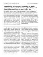
Fig. 1. Profiles of acidic oligosaccharides from rat milk. (a) Total ion chromatogram, (b) Extracted single-ion chromatograms.
MS/MS spectra were acquired in the negative-ion mode with an acqui
sition rate of 1 s per spectrum. Precursor-ion selection was made auto
matically by the data system based on ion abundance. Three precursors
were selected from each MS spectrum to carry out product-ion scanning.
Collision energy of 40 V was used for CID.
(and α-Neu5Gc in the case of non-human mammals). We here use
negative-ion ESI-MS for detection and composition analysis of the acidic
oligosaccharides as the native reducing sugars and ESI-CID-MS/MS for
subsequent sequencing. For the low quantity of milk oligosaccharides in
rats and mice, their reduction by chemical methods can eliminate
possible HPLC chromatographic peak splitting due to the separation of
α/β anomers and therefore increase ion signals. Reducing terminal
derivatization may also improve HPLC detection by UV or fluorescence.
However, after reduction or reducing-terminal tagging the fragmenta
tion patterns also change completely (Zhang et al., 2013), and the
unique features established for sequence assignment (Chai et al., 2006)
and sialic acid linkage determination (Wheeler & Harvey, 2000) are lost,
and therefore reducing sugars without derivatization are used for
negative-ion LC–MS.
3. Results and discussion
As acidic oligosaccharides are the major components of oligosac
charides in animal milk, we focused on the analysis of acidic oligosac
charides using the method developed for profiling sialylated
oligosaccharides (Yan et al., 2018). Based on the retention mechanism of
different oligosaccharides on the SPE and HILIC column, we considered
that the online SPE-HILIC method developed for sialylated oligosac
charides could also be applicable to sulphated ones. ESI-MS was used for
detection and the compositions of mammalian milk oligosaccharides can
be readily derived from the deprotonated molecules [M− H]− as their
biosynthetic pathways and the common backbone structures have been
well established. Almost all human milk oligosaccharides contain a
lactose unit (Galβ1-4Glc) at their reducing end, while N-acetyllactos
amine (Galβ1-4GlcNAc) can also be found in non-human mammalian
milk. The disaccharide cores can be extended by type 1
(-Galβ1-3GlcNAcβ1-) and type 2 (-Galβ1-4GlcNAcβ1-) chains as linear or
branched sequences. These are often terminated by a few α-mono
saccharide residues including α-Gal, α-GalNAc, α-Fuc, and α-Neu5Ac
3.1. Profiling of acidic oligosaccharides in rat milk by SPE-HILIC-MS
After removal of lactose and the possible neutral oligosaccharide
components by the SPE “Click TE-GSH” column, sialylated and sulph
ated oligosaccharides were eluted out and separated by the HILIC amide
column. In the total ion chromatogram (TIC) shown in Fig. 1a, three
major components (#3, #5 and #13) were obtained. The [M− H]− of
peaks #3 and #5 are identical at m/z 632.2, with the composition of
Hex2Neu5Ac1 (H2S1), and these two can be tentatively assigned as the
two isomeric sialylated lactose (SL) widely found in mammalian milk as
3
J. Li et al.
Carbohydrate Polymers 259 (2021) 117734
Table 1
Acidic milk oligosaccharides identified in rat and mouse by LC-ESI-MS/MS.
Peak
Noa
RTb
1
[M-H]−
Compositionc
Short
namee
Structured
Found
Calc’d
4.1
421.04
421.07
H2Su1
Gal(6Su)β1-4Glc
2
7.5
673.22
673.24
H1N1S1
3
4
5
6
7
7.7
9.2
9.2
10.5
10.8
632.21
673.22
632.21
794.25
835.27
632.21
673.24
632.21
794.27
835.29
8
11.9
835.27
9
12.6
10
Relative
content (%)f
Humang
Bovineg
References
Rat
Mouse
6’-Su-Lac
3.45
0.06
–
–
Neu5Acα2-3Galβ1-4GlcNAc
3’-SLN
0.08
0.02
–
+
H2S1
H1N1S1
H2S1
H3S1
H2N1S1
Neu5Acα2-3Galβ1-4Glc
Neu5Acα2-6Galβ1-4GlcNAc
Neu5Acα2-6Galβ1-4Glc
Neu5Acα2-3Galβ1-3Galβ1-4Glc
Neu5Acα2-3GlcNAcβ1-3Galβ1-4Glc
100
0.41
45.6
0.65
0.22
81.4
0.61
100
0.18
1.47
+
+
+
+
–
+
+
+
+
–
835.29
H2N1S1
Neu5Acα2-6(GlcNAcβ1-3)Galβ1-4Glc
0.01
0.04
+
–
997.33
997.34
H3N1S1
0.16
0.10
+
–
Chai et al. (2006)
13.6
997.33
997.34
H3N1S1
LSTc
1.97
11.2
+
+
Chai et al. (2006)
11
12
15.5
18.8
923.30
753.19
923.31
753.20
H2S2
H1N1S1Su1
Neu5Acα2-6(Galβ1-3)GlcNAcβ1-3
Galβ1-4Glc
Neu5Acα2-6Galβ1-4GlcNAcβ1-3
Galβ1-4Glc
Neu5Acα2-8Neu5Acα2-3Galβ1-4Glc
Neu5Acα2-3Gal(6Su)β1-4GlcNAc
3’-SL
6’-SLN
6’-SL
3’’S-β3’-GL
3’-S-LNTriII
6’-S-LNTriII
LSTb
Barba & Caputto
(1965)
Albrecht et al.
(2014)
Chai et al. (2006)
Chai et al. (2006)
Chai et al. (2006)
Yan et al. (2018)
Albrecht et al.
(2014)
Yan et al. (2018)
0.22
0.02
0.04
0.08
–
–
+
–
(Taufik, 2012)
–
13
20.0
712.16
712.17
H2S1Su1
Neu5Acα2-3Gal(6Su)β1-4Glc
44.0
0.01
–
–
14
24.6
1085.34
1085.36
H3S2
0.06
n.d.
–
–
15
26.6
1077.30
1077.30
H3N1S1Su1
Neu5Acα2-3Galβ1-3(Neu5Acα2-6)
Galβ1-4Glc
Neu5Acα2-6Galβ1-4GlcNAcβ1-3 Gal
(6Su)β1-4Glc
DSL
3’-S-6’-SuLN
3’-S-6’-SuL
DSβ3’-GL
Su-6’-LSTc
0.06
0.01
–
–
Choi & Carubelli
(1968)
Albrecht et al.
(2014).
–
a
b
c
d
e
f
g
HPLC peak numbers.
Retention time (in min).
H, Hex; N, HexNAc; S, Neu5NAc, Su, SO3H.
Proposed structure based on MS/MS and comparison with literature data.
Trivial name is given based on MS/MS analysis and comparison with literature data. S, Sialylated; Su, Sulphated.
Relative intensity to the most intense ion as 100 %, n.d.: not detected (relative content below 0.01 %).
+, present; -, not present.
the main components. The broad peak at 20 min, #13, with a [M− H]− at
m/z 712.2, an increase of 80 Da m/z 632.2, was deduced as the sulph
ated SL with a composition of H2S1Su1 (Su: sulphate) previously found
in rat milk (Choi & Carubelli, 1968; Sturman, Lin, Higuchi, & Fellman,
1985).
Additional minor components can be found by extracted ion chro
matograms (EICs) using different m/z values observed during MS
scanning (Fig. 1b). EIC of m/z 421.0 showed a single peak (Peak #1)
which was considered as the sulphated lactose H2Su1 (Barba & Caputto,
1965; Choi & Carubelli, 1968). EIC of m/z 673.2 exhibited two peaks,
#2 and #4, and from the composition of H1N1S1 (N: HexNAc) these can
be considered as the sialyllactosamine (SLN) isomers. The peak split of
both #2 and #4 indicated that a GlcNAc is at the reducing end as the
separation of the α and β anomers of HexNAc tends to be more promi
nent. Peaks #6–#10 were all identified as mono-sialylated oligosac
charides (Fig. 1b and Table 1) while #11–#15 each contain two acidic
groups either di-sialylated (#11 and #14) or mono-sialylated and
mono-sulphated (#12, #13 and #15). Clearly sulphate is similar to sialic
acid to have stronger electrostatic interaction with the amide stationary
phase and increased retention time. The largest oligosaccharides found
are pentasaccharides but there was no fucose detected in any of the rat
milk oligosaccharides. Apart from SLN (#2 and #4) and SL (#3 and #5)
discussed above, two more well resolved isomeric pairs were detected:
#7/#8 (H2N1S1), and #9/#10 (H3N1S1).
of the isomeric structures.
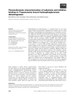
SL with α2-3 or α2-6 linkages (peaks #3 and #5, respectively) were
identified by their different fragmentations. Consistent to literature data
(Chai et al., 2006), characteristic fragments C2 (m/z 470), 0,2A2 (m/z
410) and 0,2A2-CO2 (m/z 306) in the spectrum of #5 (Fig. 2b) indicated a
Neu5Ac α2-6-linked lactose (6’-SL), whereas, the unique fragments 2,
4
A3-CO2 (m/z 468) and B2-CO2 (m/z 408) identified a Neu5
Acα2-3-linked lactose (3’-SL). 3’-SL and 6’-SL are most common acidic
oligosaccharides in mammalian milk. In human, the content of 6’-SL is
usually higher than that of 3’-SL, but in non-human mammals, 3’-SL is
often of higher concentration than 6’-SL. The presence of 6’-SL in rat
milk has not been previously reported and this was likely due to the low
abundance of 6’-SL and insufficient resolving power during oligosac
charide separation. Here, we identified both 3’-SL and 6’-SL in similar
concentrations as those found in other non-human mammals. A pair of
sialylated N-acetyllactosamine isomers, 6’-SLN (peak #4) and 3’-SLN
(peak #2), were also found in rat milk. Similar characteristic fragment
ions to those of 6’-SL and 3’-SL were observed. Again, the 2-6 linkage
specific fragment 0,4A2-CO2 (m/z 306) (Wheeler & Harvey, 2000) was
only present in the spectrum of 6’-SLN (Fig. 2d) but not in the 2-3 linked
3’-SLN (Fig. 2c), and therefore the isomers could be readily
differentiated.
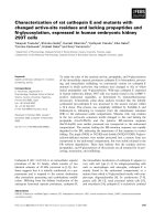
Only one peak (#6) was found to have the composition of H3S1.
Three possible structures including Neu5Acα2-3Galβ1-3 Galβ1-4Glc,
Neu5Acα2-3(Galβ1-6)Galβ1-4Glc and Galβ1-3(Neu5Acα2-6)Galβ1-4Glc
have been reported in non-human mammalian milk(Urashima et al.,
2001). Apart from the 2-3/2-6 linkage of the Neu5Ac, the position of the
extra Gal is the main point of assignment. Although a branched Gal can
produce fragment ion B1 at m/z 161, a decarboxylated B2 ion (B2-CO2) at
m/z 408 suggested the Neu5Ac linked to a Gal (Fig. 3c). The C3 ion at
m/z 632 further identified a Neu5Ac-Gal-Gal- sequence. The D-ion m/z
3.2. Sequence determination of monosialylated oligosaccharide by ESICID-MS/MS
Different fragmentation patterns in negative ion ESI-CID-MS/MS
(Chai et al., 2006) was then used to determine the sequence and par
tial linkages of the detected milk oligosaccharides and to differentiation
4
J. Li et al.
Carbohydrate Polymers 259 (2021) 117734
Fig. 2. ESI-CID-MS/MS spectra of sialyllactose and sialyl-N-acetyllactosamine. (a) 3’-SL, (b) 6’-SL, (c) 3’-SLN, (d) 6’-SLN. Structures are shown to indicate the
proposed fragmentation.
161 produced by the Gal is typical for a 3-linked residue. Finally, the
lack of Neu5Acα2-6 specific fragment m/z 306 indicated a α2-3-linked
sialic acid. Therefore 3”S-β3’-GL with the sequence of Neu5
Acα2-3Galβ1-3Galβ1-4Glc (Table 1) can be tentatively proposed.
Two peaks, #7 and #8, were found with the composition of H2N1S1
([M− H]− at m/z 835). Although the spectral signals of peak #8 is very
weak (Fig. 1b), from the product-ion spectra the isomeric pair can still be
assigned based on some important ions observed. The branched
sequence of #8 is apparent from the C1 at m/z 202 and B1α at m/z 290
(Fig. 3b). Further glycosidic cleavage at B2 (m/z 655) and its desialy
lated ion B2-S (m/z 364) identified the branching point at the Gal as the
tetrasaccharide structure 6’-S-LNTri-II, GlcNAcβ1-3(Neu5Acα2-6)
Galβ1-4Glc, which was found previously in other reports (Albrecht et al.,
2014; Yan et al., 2018) (Table 1). The linear sequence of #7 can be
deduced by the B1 at m/z 290 and B2 at m/z 493. The double glycosidic
D-type ion D1-2 at m/z 202 indicated the internal GlcNAc 3-linked to the
Gal, and therefore 3’-S-LNTri-II (Neu5Acα2-3GlcNAcβ1-3Galβ1-4Glc)
can be proposed (Table 1).
Peaks #9 and #10 can be readily assigned as LSTb and LSTc
(Table 1), respectively, by comparison of the product-ion spectra
(Fig. 3d and e) with literature data (Chai et al., 2006), and by the
fragment ions obtained. In the spectrum of #9, the full set of sequence
ions B1α (m/z 290), C1 (m/z 179), C2 (m/z 673) and C3 (m/z 835) defined
the sequence, and 0,4A2-CO2 indicated the Neu5Ac2-6 linkage and D1-2
(m/z 493) suggested a 3-linked GlcNAc. Peak #10 was similarly assigned
as LSTc (Table 1). The assignment was confirmed by comparison with
both retention times and product-ion spectra of standard LSTb and LSTc
(Figs. S-1 and S-2).
The nine monosialylated oligosaccharides described above are
common acidic oligosaccharides in mammalian milk.
3.3. Sequence determination of disialyated and sulphated
oligosaccharides by ESI-CID-MS/MS
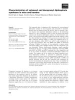
The oligosaccharides in peak #11 and #14 are both disialylated.
Peak #11 has a composition of H2S2 and is considered as disialylated
lactose (DSL). As shown in Fig. 4a, the Neu5Ac-Neu5Ac- sequence can
unambiguously assigned by B1 m/z 290 and B2 m/z 581, the latter
accompanied by a decaboxylated ion m/z 537 with an α2-8 linkage. Y1 at
m/z 632 can further confirm this sequence. The lack of Neu5Acα2-6
specific ion at m/z 597 (306 + 291), equivalent to m/z 306 in the case of
monosialylated oligosaccharides (see above for discussion), highly
indicated an α2-3 linkage between the Neu5Ac and Gal. The linkage
between the two sialic acid residues was tentatively assigned as α2-8 as
those found in bovine milk (Veh et al., 1981) and buffalo colostrum
(Aparna & Salimath, 1995). Therefore, peak #11 can be identified as
Neu5Acα2-8Neu5Acα2-3Galβ1-4Glc.
The disialylated oligosaccharide in peak #14 contains an additional
hexose (Fig. 4b). The absence of the characteristic fragments m/z 581
and 537 for Neu5Acα2-8Neu5Ac- (as shown in Fig. 4a for DSL) and the
presence of the mono-desialylated ion m/z 794 indicate the two sialic
acids at different positions. A weak ion at m/z 161 from a double
glycosidic D-type cleavage indicated a 3-linked Gal in the penta
saccharide. Although the product-ion spectrum was very weak and
insufficient fragment ions to give a full assignment, a sequence of
Neu5Acα2-3Galβ1-3(α2-6Neu5Ac)Galβ1-4Glc (Fig. 4b) can be specu
lated which was previously named as DSβ3’-GL. These two disialylated
oligosaccharides have been found in the milk of domestic animals
(Albrecht et al., 2014).
The remaining four oligosaccharides are all sulfated. Peak #1 was
identified as the 6’-sulfated lactose (Table 1) as the sulfate on the Gal is
apparent by the presence of strong ion pair of B1 m/z 241 and C1 m/z 259
5
J. Li et al.
Carbohydrate Polymers 259 (2021) 117734
Fig. 3. ESI-CID-MS/MS spectra of sialylated oligosaccharides. (a) 3′ -S-LNTri-II, (b) 6′ -S-LNTri-II, (c) 3′ ′ -S-β3′ -GL, (d) LSTb, (e) LSTc. Structures are shown to indicate
the proposed fragmentation.
6
J. Li et al.
Carbohydrate Polymers 259 (2021) 117734
signals in addition to the facile loss of the sulphate, it is difficult to have a
definitive assignment from the mass spectral fragmentation but the
likely sulphation at the 6-position of the Gal is assumed and sulphated
LSTc, Neu5Ac2-6Gal1-4GlcNAc1-3Gal(6Su)1-4Glc, is proposed.
Although a phosphate group is also of 80 Da and phosphorylated
oligosaccharides have been found in animal milk (Urashima et al.,
2001), #12 and #15 were assigned as sulphated. This is because sul
phation has been identified in rat milk oligosaccharides (#1 and #13)
(Choi & Carubelli, 1968; Sturman et al., 1985) and it is unlikely sul
phation and phosphorylation can occur in the milk of the same animals.
It has been recently recognized that human milk oligosaccharides
play important roles in shaping up the infant’s intestinal microbiota
composition and in serving as soluble decoy receptors preventing
pathogen attachment to infant mucosal surfaces and lower the risk for
viral, bacterial and protozoan parasite infections. Although milk oligo
saccharides in general do not have nutritional value, early work spec
ulated for the possible nutritional importance of sulphate in
oligosaccharides present in rat milk. Sulphate is not considered as an
essential nutrient in mature mammals but it could be a nutrient in the
neonate. In an experiment using 35S, sulphated SL was found to be
hydrolysed in the gut of rat neonates, and the sulphur absorbed as
inorganic sulphate (Sturman et al., 1985). The presence of this may
permit the simultaneous delivery of two essential nutrients, sulphate
and calcium, in early life, avoiding the precipitation of insoluble calcium
sulphate in the milk (Sturman et al., 1985). However, in human infants
the function of milk oligosaccharides is primarily protective rather than
nutritional (Newburg, 2000).
3.4. Comparison of acidic oligosaccharides in rat, mouse and human milk
Analysis of oligosaccharides in mouse milk is more challenging due
to the very small amount of mouse milk available and the difficulty for
collection directly from lactating mouse. A single mouse mammary tis
sue was used for extraction of milk oligosaccharides. Fourteen acidic
oligosaccharides were similarly detected (Table 1) but DSβ3’-GL (#14)
was not found. For comparison, Fig. 6 shows the two acidic oligosac
charide profiles from rat and mouse milk. To make the low abundant
peaks more visible different magnifying factors were applied (please
note the different colours representing different magnifying factors).
There is an apparent difference in relative abundances of oligosaccha
rides in rat and mouse milk (Table 1). In rat, 3’-SL is most abundant,
whereas in mouse it is 6’-SL. The content of sulphated oligosaccharides
in rat milk was much higher than those in mouse milk. Apart from 6’-SL
and 3’-SL, sulphated 3’-SL is the most abundant with a relative intensity
of 44.0 %, but it is less than 0.01 % in mouse milk. In mouse milk, LSTc is
the third most abundant one.
The 15 acidic oligosaccharides detected in rat and mouse milk can be
compared with the 30 sialylated oligosaccharides in human milk iden
tified in a previous study (Yan et al., 2018). As shown in Table 1, seven
oligosaccharides are common in both rat and human and these include
3’-SL, 6’-SL, 6’-SLN, 3’’-β3’-GL, 6’-S-LNTri-II, LSTb and LSTc. The other
eight oligosaccharides are absent in human milk. Compared with do
mestic animals (such as cow, goat and sheep), rat and mouse share more
common oligosaccharides with human. The acidic oligosaccharides in
mouse milk are more similar to human milk due to the higher contents of
6’-SL and LSTc.
Fig. 4. ESI-CID-MS/MS spectra of disialylated oligosaccharides. (a) DSL, (b)
DSβ3’-GL. Structures are shown to indicate the proposed fragmentation.
(Fig. 5a). Peak #13 was the 3’-sialyl-6’-sulfated lactose (3’-S-6’-Su-L,
Fig. 5b and Table 1). B1 at m/z 290 and lack of 0,4A2-CO2 at m/z 306
indicated a 3-linked Neu5Ac. Y1 at m/z 421 is indicative of the sulfate on
the lactose moiety. Extensive decarboxylation and desulphation made it
impossible to assign exactly the position of the sulphate group. The two
sulfated oligosaccharides have been reported previously, and the sul
phate group was identified by elemental composition and the Gal-6-Oposition assigned by methylation analysis (Barba & Caputto, 1965;
Choi & Carubelli, 1968; Michael et al., 2013).
Peak #12 can be readily assigned by comparison with the spectrum
of peak #13 (3’-S-6’-Su-L (Fig. 5b). The reducing terminal disaccharide
N-acetyllactosamine rather than lactose is apparent from their compo
sitions (H1N1S1Su1and H2S1Su1, respectively) and the Y1 ion at m/z
462 (compared with Y1 at m/z 421 in the spectrum of #13, Fig. 5b).
Although very weak signal due to the extremely low content (0.06 %,
Table 1) NeuAcα2-3Galβ(6Su)1-4GlcNAc.
With the composition of H3N1S1Su1, peak #15 was predicted to be
either the sulphated LSTb or LSTc which are present in rat milk (peak #9
and #10, Table 1). As LSTc is more abundant (1.97 %) compared with
LSTb (0.16 %), sulphated LSTc was the most possible structure. As
shown in Fig. 5d, the glycosidic ions C1 at m/z 308 and B2 at m/z 452
clearly identified the sialic acid at the non-reducing end while the sul
phate is not at this Gal. The 0,2A3-h at m/z 554 also indicated the absence
of sulphate on the GlcNAc. Therefore, the sulphate at the lactose site
could be assigned. Due to very low concentration and extremely weak
4. Conclusions
In this work we carried out a comprehensive analysis of oligosac
charides using 1 mL of rat milk or 1 mouse gland tissue. We detected and
identified 15 acidic oligosaccharides and these include 9 mono
sialylated, 2 disialylated, 1 monosulphated, and 3 both monosulphated
and monosialylated. Among these, 12 are reported here for the first time
in rat milk and 2 are novel structures. As some of oligosaccharides are in
very low concentrations this precludes fully sequence assignment. The
7
J. Li et al.
Carbohydrate Polymers 259 (2021) 117734
Fig. 5. ESI-CID-MS/MS spectra of sulphate and sialylated oligosaccharides. (a) 6’-L-O-sulphate, (b) 3’-SL-6’-O-sulphate, (c) 3’-SLN-6’-O-sulphate, (d) LSTc-6’-Osulphate. Structures are shown to indicate the proposed fragmentation.
Fig. 6. Overlay extracted single-ion chromatograms of oligosaccharides in (a) rat milk, (b) mouse milk. Peaks 4,6,7, 9 and 11 were magnified by a factor of 20, peaks
2, 8, 12, 14 and 15 were magnified by a factor of 200 in rat milk. Peaks 4, 6, 7, 9 and 11 were magnified by a factor of 40, peaks 2, 8, 13, 14 and 15 were magnified by
a factor of 1000 in mouse milk. Legends: yellow circle, galactose; purple diamond, N-acetylneuraminic acid; blue square, N-acetylglucosamine; and blue cir
cle, glucose.
8
J. Li et al.
Carbohydrate Polymers 259 (2021) 117734
sulphation is likely at the 6-O-position of the Gal at the reducing side.
When this position is occupied by sialic acid sulphation does not seem to
take place.
Egelrud, T., Helander, H., & Olivecrona, T. (1970). Gastric digestion and uptake of milk
fat in the suckling rat. Acta Physiologica Scandinavica, 13.
Fukuda, K., Yamamoto, A., Ganzorig, K., Khuukhenbaatar, J., Senda, A., Saito, T., et al.
(2010). Chemical characterization of the oligosaccharides in Bactrian camel
(Camelus bactrianus) milk and colostrum. Journal of Dairy Science, 93, 5572–5587.
Gosling, S. D. (2001). From mice to men: What can we learn about personality from
animal research? Psychological Bulletin, 127, 45.
Jacob, H. J., & Kwitek, A. E. (2002). Rat genetics: Attaching physiology and
pharmacology to the genome. Nature Reviews Genetics, 3, 33–42.
Kariakin, M., & Alekseev, N. (1991). The characteristics of the motor activity of rat pups
during the milk ejection reflex. Fiziologicheskii zhurnal SSSR imeni IM Sechenova, 77,
83–88.
Kuhn, N. (1972). The lactose and neuraminlactose content of rat milk and mammary
tissue. Biochemical Journal, 130, 177–180.
Kumar, K., & Deepak, D. (2019). Structural characterization of novel milk
oligosaccharide Aurose from cow colostrum. Journal of Molecular Structure, 1176,
394–401.
Kunz, C., Rudloff, S., Baier, W., Klein, N., & Strobel, S. (2000). Oligosaccharides in
human milk: Structural, functional, and metabolic aspects. Annual Review of
Nutrition, 20, 699–722.
Lin, A. E., Autran, C. A., Szyszka, A., Escajadillo, T., Huang, M., Godula, K., et al. (2017).
Human milk oligosaccharides inhibit growth of group B Streptococcus. Journal of
Biological Chemistry, 292, 11243–11249.
Marcobal, A., Barboza, M., Froehlich, J. W., Block, D. E., German, J. B., Lebrilla, C. B.,
et al. (2010). Consumption of human milk oligosaccharides by gut-related microbes.
Journal of Agricultural and Food Chemistry, 58, 5334–5340.
Melina, R. (2010). Why do medical researchers use mice. Life’s little mysteries.
LiveScience.
Meng, L., Forouhar, F., Thieker, D., Gao, Z., Ramiah, A., Moniz, H., et al. (2013).
Enzymatic basis for N-glycan sialylation structure of rat α2, 6-sialyltransferase
(st6Gal1) reveals conserved and unique features for glycan sialylation. Journal of
Biological Chemistry, 288, 34680–34698.
Michael, L. P., Shin-Yi, Y., Chu-Wen, C., Ming-Yi, H., Lotten, T., & Keiichiro, S. (2013).
KSGal6ST generates galactose-6-O-sulfate in high endothelial venules but does not
contribute to L-selectin-dependent lymphocyte homing. Glycobiology, 23, 381–394.
Mineguchi, Y., Miyoshi, M., Taufik, E., Kawamura, A., Asakawa, T., Suzuki, I., et al.
(2018). Chemical characterization of the milk oligosaccharides of some Artiodactyla
species including giraffe (Giraffa camelopardalis), sitatunga (Tragelaphus spekii),
deer (Cervus nippon yesoensis) and water buffalo (Bubalus bubalis). Glycoconjugate
Journal, 35, 561–574.
Monti, L., Cattaneo, T. M. P., Orlandi, M., & Curadi, M. C. (2015). Capillary
electrophoresis of sialylated oligosaccharides in milk from different species. Journal
of Chromatography A, 1409, 288–291.
Naccarato, W., Ray, R., & Wells, W. (1975). Characterization and tissue distribution of 6O-beta-D-galactopyranosyl myo-inositol in the rat. Journal of Biological Chemistry,
250, 1872–1876.
Newburg, D. S. (2000). Oligosaccharides in human milk and bacterial colonization.
Journal of Pediatric Gastroenterology and Nutrition, 30, S8–S17.
Newburg, D. S. (2009). Neonatal protection by an innate immune system of human milk
consisting of oligosaccharides and glycans. Journal of Animal Science, 87, 26–34.
Serikawa, T., Mashimo, T., Kuramoto, T., Voigt, B., Ohno, Y., & Sasa, M. (2014).
Advances on genetic rat models of epilepsy. Experimental Animals, 2015(64), 1–7.
Sturman, J. A., Lin, Y. Y., Higuchi, T., & Fellman, J. (1985). N-acetylneuramin lactose
sulfate: A newly identified nutrient in milk. Pediatric Research, 19, 216–219.
Tao, N., Ochonicky, K. L., German, J. B., Donovan, S. M., & Lebrilla, C. B. (2010).
Structural determination and daily variations of porcine milk oligosaccharides.
Journal of Agricultural and Food Chemistry, 58, 4653–4659.
Urashima, T., Saito, T., Nakamura, T., & Messer, M. (2001). Oligosaccharides of milk and
colostrum in non-human mammals. Glycoconjugate Journal, 18, 357–371.
Veh, R. W., Michalski, J.-C., Corfield, A. P., Sander-Wewer, M., Gies, D., & Schauer, R.
(1981). New chromatographic system for the rapid analysis and preparation of
colostrum sialyloligosaccharides. Journal of Chromatography A, 212, 313–322.
Verruck, S., Santana, F., de Olivera Müller, C., & Prudencio, E. S. (2018). Thermal and
water sorption properties of Bifidobacterium BB-12 microcapsules obtained from
goat’s milk and prebiotics. LWT-Food Science and Technology, 98, 314321.
Victora, C. G., Bahl, R., Barros, A. J., Franỗa, G. V., Horton, S., Krasevec, J., et al. (2016).
Breastfeeding in the 21st century: Epidemiology, mechanisms, and lifelong effect.
Lancet, 387, 475–490.
Vlaming, M., Mohrmann, K., Wagenaar, E., de Waart, D. R., Elferink, R. O., Lagas, J. S.,
et al. (2006). Carcinogen and anticancer drug transport by Mrp2 in vivo: Studies
using Mrp2 (Abcc2) knockout mice. Journal of Pharmacology and Experimental
Therapeutics, 318, 319–327.
Wang, H. X., Chen, Y., Haque, Z., de Veer, M., Egan, G., & Wang, B. (2019). Sialylated
milk oligosaccharides alter neurotransmitters and brain metabolites in piglets: An in
vivo magnetic resonance spectroscopic (MRS) study. Nutritional Neuroscience, 1–11.
Wheeler, S. F., & Harvey, D. J. (2000). Negative ion mass spectrometry of sialylated
carbohydrates: Discrimination of N-acetylneuraminic acid linkages by MALDI-TOF
and ESI-TOF mass spectrometry. Analytical Chemistry, 72, 5027–5039.
Yan, J., Ding, J., Jin, G., Yu, D., Yu, L., Long, Z., et al. (2018). Profiling of sialylated
oligosaccharides in mammalian milk using online solid phase extraction-hydrophilic
interaction chromatography coupled with negative-ion electrospray mass
spectrometry. Analytical Chemistry, 90, 3174–3182.
CRediT authorship contribution statement
Jiaqi Li: Data curation, Formal analysis, Writing - original draft.
Maorong Jiang: Methodology. JiaoRui Zhou: Data curation. Junjie
Ding: Data curation. Zhimou Guo: Conceptualization, Project admin
istration. Ming Li: Funding acquisition. Fei Ding: Methodology. Wen
gang Chai: Funding acquisition, Writing - review & editing. Jingyu
Yan: Funding acquisition, Writing - review & editing. Xinmiao Liang:
Funding acquisition, Project administration.
Declaration of Competing Interest
The authors report no declarations of interest.
Acknowledgments
The work is supported in part by the National Natural Science
Foundation of China (21934005, 22074143, and 31900920), and by the
March of Dimes Prematurity Research Center grant (22-FY18-821) and
the Wellcome Trust Biomedical Resource grant (WT 218304/Z/19/Z).
Appendix A. Supplementary data
Supplementary material related to this article can be found, in the
online version, at doi: />References
Albrecht, S., Lane, J. A., Marino, K., Al Busadah, K. A., Carrington, S. D., et al. (2014).
A comparative study of free oligosaccharides in the milk of domestic animals. British
Journal of Nutrition, 111, 1313–1328.
Aparna, H. S., & Salimath, P. V. (1995). Disialyl lactose from buffalo colostrum: Isolation
and characterization. Carbohydrate Research, 268, 313–318.
Ballard, O., & Morrow, A. L. (2013). Human milk composition: Nutrients and bioactive
factors. Pediatric Clinics of North America, 60, 49–74.
Barba, H. S., & Caputto, R. (1965). Isolation and identification of a lactose sulphate ester
from rat mammary gland. Biochimica et Biophysica Acta (BBA)-Mucoproteins and
Mucopolysaccharides, 101, 367–369.
Bode, L. (2012). Human milk oligosaccharides: Every baby needs a sugar mama.
Glycobiology, 22, 1147–1162.
Briffa, J. F., O’Dowd, R., Moritz, K. M., Romano, T., Jedwab, L. R., McAinch, A. J., et al.
(2017). Uteroplacental insufficiency reduces rat plasma leptin concentrations and
alters placental leptin transporters: Ameliorated with enhanced milk intake and
nutrition. The Journal of Physiology, 595, 3389–3407.
Carubelli, R., Ryan, L. C., Trucco, R. E., & Caputto, R. (1961). Neuramin-lactose sulfate, a
new compound isolated from the mammary gland of rats. Journal of Biological
Chemistry, 236, 2381–2388.
Chai, W., Piskarev, V. E., Mulloy, B., Liu, Y., Evans, P. G., Osborn, H. M., et al. (2006).
Analysis of chain and blood group type and branching pattern of sialylated
oligosaccharides by negative ion electrospray tandem mass spectrometry. Analytical
Chemistry, 78, 1581–1592.
Charbonneau, M. R., O’Donnell, D., Blanton, L. V., Totten, S. M., Davis, J. C.,
Barratt, M. J., et al. (2016). Sialylated milk oligosaccharides promote microbiotadependent growth in models of infant undernutrition. Cell, 164, 859–871.
Choi, H. U., & Carubelli, R. (1968). Neuramine-lactose, neuramine-lactose sulfate, and
lactose sulfate from rat mammary glands. Isolation, purification, and permethylation
studies. Biochemistry, 7, 4423–4430.
Comstock, S. S., Li, M., Wang, M., Monaco, M. H., Kuhlenschmidt, T. B.,
Kuhlenschmidt, M. S., et al. (2017). Dietary human milk oligosaccharides but not
prebiotic oligosaccharides increase circulating natural killer cell and mesenteric
lymph node memory t cell populations in noninfected and rotavirus-infected
neonatal piglets. Journal of Nutrition, 147, 1041–1047.
Corpet, D. E., & Pierre, F. (2005). How good are rodent models of carcinogenesis in
predicting efficacy in humans? A systematic review and meta-analysis of colon
chemoprevention in rats, mice and men. European Journal of Cancer, 41, 1911–1922.
Craft, K. M., Thomas, H. C., & Townsend, S. D. (2019). Sialylated variants of lacto-Ntetraose exhibit antimicrobial activity against Group B Streptococcus. Organic &
Biomolecular Chemistry, 17, 1893–1900.
Dvorak, B., Halpern, M. D., Holubec, H., Dvorakova, K., Dominguez, J. A.,
Williams, C. S., et al. (2004). Rat milk decreases necrotizing enterocolitis in a rat
model. Protecting infants through human milk (pp. 471–473).
9
J. Li et al.
Carbohydrate Polymers 259 (2021) 117734
Zhang, H., Zhang, S., Tao, G., Zhang, Y., Mulloy, B., Zhan, X., et al. (2013). Typing of
blood-group antigens on neutral oligosaccharides by negative-ion electrospray
ionization tandem mass spectrometry. Analytical Chemistry, 85, 5940–5949.
Zuurveld, M., van Witzenburg, N. P., Garssen, J., Folkerts, G., Stahl, B., van’t Land, B.,
et al. (2020). Immunomodulation by human milk oligosaccharides: The potential
role in prevention of allergic diseases. Frontiers in Immunology, 11.
Taufik, E., Fukuda, K., Senda, A., Saito, T., Williams, C., & Tilden, C. (2012). Structural
characterization of neutral and acidic oligosaccharides in the milks of strepsirrhine
primates: greater galago, aye-aye, Coquerel’s sifaka and mongoose lemur.
Glycoconjugate Journal, 29(2–3), 119–134.
10