Magnetic microspheres based on pectin coated by chitosan towards smart drug release
Bạn đang xem bản rút gọn của tài liệu. Xem và tải ngay bản đầy đủ của tài liệu tại đây (4.72 MB, 10 trang )
Carbohydrate Polymers 265 (2021) 118013
Contents lists available at ScienceDirect
Carbohydrate Polymers
journal homepage: www.elsevier.com/locate/carbpol
Magnetic microspheres based on pectin coated by chitosan towards smart
drug release
Thalia S.A. Lemos , Jaqueline F. de Souza , Andr´e R. Fajardo *
Laborat´
orio de Tecnologia e Desenvolvimento de Comp´
ositos e Materiais Polim´ericos (LaCoPol), Universidade Federal de Pelotas (UFPel), Campus Cap˜
ao do Le˜
ao s/n,
96010-900, Pelotas, RS, Brazil
A R T I C L E I N F O
A B S T R A C T
Keywords:
Magnetic
Biopolymers
Microspheres
Smart materials
Stimuli-responsive system
Drug delivery
This study reports the preparation of microspheres of pectin and magnetite nanoparticles coated by chitosan to
encapsulate and deliver drugs. Magnetic-pectin microspheres were obtained by ionotropic gelation followed by
polyelectrolyte complexation with chitosan. Characterization data show that magnetite changes the physico
chemical and morphological properties of the microspheres compared to the non-magnetic samples. Using
metamizole (Mtz) as a drug model, the magnetic microspheres showed appreciable encapsulation efficiency (85
%). Release experiments performed in simulated gastric (pH 1.2) and intestinal (pH 6.8) fluids suggested that the
release process is pH-dependent. At pH 6.8, the Mtz release is favored achieving 75 % after 12 h. The application
of an external magnetic field increased the release to 91 % at pH 6.8, indicating that the release also is magneticdependent. The results suggest that the magnetic microspheres based on pectin/chitosan biopolymers show the
potential to be used as a multi-responsive drug delivery system.
1. Introduction
The first examples of drug delivery systems (DDS) based on polymers
were reported almost five decades ago and have since attracted the
attention of several researcher fields (Wong et al., 2018). In summary,
this success is attributable to the many advantages offered by these
delivery systems as compared to free-drug formulations. Some attributes
of polymeric DDS include the ability to maintain drug concentration
within a desirable range, increase drug bioavailability, a decrease of side
effects and administration doses, and increase of patient compliance to
the treatment (Gunter et al., 2018; Wong et al., 2018). Overall, these
features allowed enhancing the efficiency of several drugs and medica
ment treatments for various diseases and conditions (Jafari et al., 2020;
Li et al., 2020).
Nowadays, the main challenges related to the development of more
efficient polymeric DDS are related to the improvement of drug encap
sulation efficiency and release (Patra et al., 2018). Specifically, drug
release is a critical stage since it is related to the success of the DDS. The
release of a drug (or other bioactive compounds) from a polymeric
system can occur continuously or cyclically over a long period or it can
be triggered by an external stimulus (Karimi et al., 2016). This last
mechanism has gained importance as an efficient strategy to overcome
two potential shortcomings related to the releasing process: (i) the
inability to deliver the loaded drug and (ii) burst release effects (Pham
et al., 2020). In recent years, researchers have developed polymeric DDS
able to control their release mechanism according to changes on
different environmental parameters (such as pH condition, temperature,
ionic strength, light incidence, and electric and magnetic fields) (Raza
et al., 2019; Thevenot et al., 2013). Although these parameters can be
modulated under the physiological environment, in which the DDS is
administrated, some of them can be invasive and cause undesired effects
(Senapati et al., 2018). In light of this, some authors claim that the use of
DDS endowed with stimuli-responsive magnetic properties is a prom
ising alternative to overcome the aforementioned limitations (Frachini
& Petri, 2019; Price et al., 2018). The efficiency of these responsive
systems can be ascribed to the use of external magnetic fields, which
enable controlling the DDS actuation remotely. According to Farah
(2016), the main advantage of magnetic-responsive DDS is the reduction
in the dose and side effects of the drug. Additionally, therapeutic re
sponses in target organs can be achieved by a small fraction of the free
drug due to the improvement of the drug bioavailability. The magnetic
response is typically obtained by focusing an extracorporeal magnetic,
which is less invasive than other responsive systems (Mura et al., 2013).
Iron oxides such as Fe3O4 (magnetite) and γ-Fe2O3 (maghemite) have
* Corresponding author.
E-mail address: (A.R. Fajardo).
/>Received 29 January 2021; Received in revised form 27 February 2021; Accepted 26 March 2021
Available online 2 April 2021
0144-8617/© 2021 Elsevier Ltd. This article is made available under the Elsevier license ( />
T.S.A. Lemos et al.
Carbohydrate Polymers 265 (2021) 118013
been predominantly used to induce magnetic properties in polymeric
DDS because of their biocompatibility and low toxicity properties
(Ghazanfari et al., 2016). Moreover, the affinity of these oxides with
water allows the interaction of the same with different biological spe
cies. Consequently, the incorporation of these oxides into natural ma
terials like polysaccharides may result in smart drug delivery systems.
The use of polysaccharides is preferred by several investigators devoted
to preparing magnetic DDS since they enable a good dispersion and
stabilization of the iron oxide particles (Chang et al., 2011). Of course,
the use of polysaccharides in the preparation of DDS is also stimulated
owing to their interesting properties such as biocompatibility, biode
gradability, non-toxicity, renewability, low-cost, and processability (Oh
et al., 2009). Among the polysaccharides suitable to this application,
pectin, a natural polymer component of all plant cell walls has been
poorly explored. Pectin (Pec) is a complex polysaccharide, predomi
nantly linear, consisting mainly of methoxy esterified α(1→4)-linked
D-galacturonic acid units that according to their esterification degree
can form gels (Lara-Espinoza et al., 2018). Capel et al. (2006) demon
strate that Pec with a low esterification degree undergoes ionotropic
crosslinking in the presence of Ca2+ ions resulting in a stable hydrogel.
This gel-forming ability of Pec can also be useful to form polyelectrolyte
complexes with polycationic species, like chitosan, a well-known chitin
derivative. Chitosan (Cs), a linear copolymer polysaccharide consisting
of β(1→4)-linked D-glucosamine and N-acetyl-D-glucosamine units
widely used in pharmaceutical and biomedical applications owing to its
biological properties (Younes & Rinaudo, 2015). The protonable amino
groups of Cs can interact strongly with the carboxylate-rich structure of
Pec resulting in a polyelectrolyte complex (Rampino et al., 2016).
Earlier studies demonstrated that the stability of Pec/Cs complexes can
be modified by changing external conditions like pH and temperature,
which allows ranking these materials as potential DDS with sensitive
properties (Maciel et al., 2015; Sigaeva et al., 2020).
Herein, we prepared microspheres consisting of pectin and magne
tite nanoparticles, which were coated by a chitosan layer, and hypoth
esize that they can be used as a multi-responsive DDS. The magnetic
microspheres were loaded with metamizole (Mtz), which is a pyrazolone
derivative commonly used to treat various pain conditions (e.g., post
operative pain, colic pain, cancer pain, and migraine) in humans and
veterinary practices (Jasiecka et al., 2014). A series of experiments were
performed to investigate the behavior and mechanism associated with
the Mtz release under different simulated physiological conditions
(gastric and intestinal fluids) and with and without the presence of an
external magnetic field.
2. Materials and methods
2.1. Materials
Orange (Citrus sinensis) peels were obtained from the student
restaurant at Universidade Federal de Pelotas (Pelotas, RS, Brazil).
Pectin (Pec) was isolated from orange peels and fully desesterified as
reported by Lessa et al. (2017). Chitosan (Cs, Mv of 87,000 g/mol and 85
% deacetylated) was purchased from Golden-Shell Biochemical
(Yuhuan, China). Magnetite nanopowder (iron (II,III) oxide, 97 % of
purity, 50− 100 nm particle size, and magnetization saturation of
91 emu g− 1) was purchased from Sigma-Aldrich (St. Louis, MO, USA).
Metamizole sodium salt (Mtz, 351.36 g mol− 1) was purchased from
Sanofi Aventis Pharma (Bombain, India). Calcium chloride (CaCl2) was
purchased from Synth (Diadema, SP, Brazil). All other chemicals were of
analytical grade and were utilized without further purification.
2.2. Preparation of the magnetic microspheres
Magnetic Pec@Cs microspheres were prepared using a two-step
process adapting a methodology described by Rashidzadeh et al.
(2020). Scheme 1 outlines the microspheres preparation processes.
Firstly, Pec was completely solubilized in distilled water at a concen
tration of 3 wt-% and magnetite nanoparticles (1 wt-% related to the Pec
dry weight) were added. The system was homogenized using an ultra
sonic bath (42 kHz for 15 min at 30 ◦ C) and transferred to a syringe
equipped with a needle (inner diameter of 1 mm). Next, the Pec/mag
netite solution was dropped (speed 1 ml min− 1) into CaCl2 solution (10
wt-%, 20 mL), which was kept under mild orbital stirring (~100 rpm) at
room temperature. The as-formed microspheres were left to maturate in
CaCl2 solution for 15 min. After that, the microspheres were recovered
by filtration and thoroughly washed with distilled water to remove the
excess of Ca2+ ions. No release of magnetite was observed during this
step.
In the sequence, the Pec/magnetite microspheres were put in contact
with a Cs solution (1 wt-%, acetic acid solution 1.5 v/v-%, pH 3) under
low stirring (~100 rpm) for 2 h at room temperature. Lastly, the mi
crospheres coated by Cs were recovered and washed with distilled water
and oven-dried (35 ◦ C, 24 h). The prepared microspheres were denoted
as mag-Pec@Cs, respectively. For comparative and characterization
Scheme 1. The experimental approach used to prepare magnetic-pectin microspheres coated by chitosan.
2
T.S.A. Lemos et al.
Carbohydrate Polymers 265 (2021) 118013
purposes, microspheres without magnetite (denoted as Pec@Cs) and
without the Cs coating (denoted as mag-Pec) were also prepared using
similar procedures.
2.4. Characterization
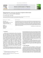
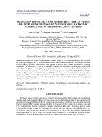
Photographs of the as-prepared Pec@Cs and mag-Pec@Cs micro
spheres (wet state) were taken with a digital camera (Fig. 1a and b).
Furthermore, photographs of mag-Pec@Cs microspheres immersed in
the aqueous medium were taken in the absence and presence of an
external magnet (neodymium permanent magnets, NdFeB, 20 × 10 mm,
grade N52) (Fig. 1c and d). The average size of the prepared micro
spheres (wet state) was measured using a calibrated digital Vernier
caliper micrometer (resolution 0.01 mm). For each microsphere type,
the average size was calculated from the data measured from 50 samples
chosen randomly. Data are expressed as mean ± standard error of the
mean.
The prepared microspheres were characterized by Fourier Trans
formed Infra-Red (FTIR) spectroscopy, X-ray Diffraction (XRD), Ther
mogravimetric Analysis (TGA), and Scanning Electron Microscopy
(SEM). Before the FTIR, XRD, and TG analyses the as-prepared micro
spheres (wet state) were crushed using a mortar and then oven-dried
(50 ◦ C for 48 h). The powdered samples were sieved before use. FTIR
spectra were recorded in a Shimadzu (model Affinity) spectrometer
(Japan) operating in the region from 4000–400 cm− 1 with a resolution
of 4 cm− 1 and 64 scan acquisitions. The samples were blended with KBr
and pressed into discs before FTIR analysis. XRD diffraction patterns
were obtained on a Siemens (model D500) diffractometer (Germany)
using Cu-Kα radiation (λ ≈ 1.54 Å), at a tube voltage of 40 kV, and tube
current of 30 mA. TGA analysis was performed with a Shimadzu (model
DTG60) analyzer (Japan) under an N2(g) atmosphere. SEM images were
recorded using a JEOL (model JSM-6610LV) microscope (USA). Before
SEM visualization, the samples were swelled in distilled water, frozen in
N2(l), freeze-dried (-55 ◦ C for 48 h) and sputter-coated with gold.
The liquid uptake capacity was evaluated by swelling experiments
2.3. Drug encapsulation
The preparation of Mtz loaded-microspheres was made using the
same process described in the previous section with minor modifica
tions. Herein, Mtz (1 mg) was added to the Pec or Pec/magnetite solu
tions before their dripping in the CaCl2 solution. It is important to
mention that the amount of Mtz (1 mg) was selected from previous ex
periments. Two sets of Mtz loaded-microspheres were prepared;
Pec@Cs/Mtz and mag-Pec@Cs/Mtz, respectively. The Mtz content
encapsulated within the microspheres was determined using a UV–vis
spectrometer (Perkin-Elmer, model Lambda 24, USA). For this, the Mtzloaded microspheres (1 g) were completely crushed and soaked in PBS
(0.01 mol L− 1, pH 7.4) for 24 h under stirring. The obtained solutions
were centrifuged (5000 rpm for 15 min) and the supernatants were
analyzed by UV–vis spectrometry at λ =271 nm. The Mtz content was
estimated using a previously built calibration curve (R2 > 0.999). From
these data, the encapsulation efficiency (EE%) and drug loading (DL%)
were calculated per Eq. (1) and (2). All samples were analyzed in
triplicate.
EE% =
[amount of Mtz within the analyzed microspheres]
x 100
[amount of Mtz initally added to the microspheres]
(1)
DL% =
[amount of Mtz in the microspheres]
x 100
[amount of microspheres]
(2)
Fig. 1. Digital photographs of the as-prepared (a) Pec@Cs and (b) mag-Pec@Cs microspheres. The mag-Pec@Cs microspheres immersed in aqueous medium (c) in
the absence and (d) presence of an external magnet.
3
T.S.A. Lemos et al.
Carbohydrate Polymers 265 (2021) 118013
gelation between carboxylate groups of pectin and Ca2+ ions (Kim et al.,
2017). Next, the pectin-based microspheres were allowed to interact
with chitosan, a polycationic polysaccharide, resulting in the coat of the
surface of the microspheres. Herein, the residual carboxylate groups of
pectin interact electrostatically with the amino protonated groups of
chitosan. The Pec@Cs microspheres exhibited a colorless nature and
spherical geometry (Fig. 1a). Although the introduction of magnetite did
not affect the geometry of the microspheres, the mag-Pec@Cs showed a
dark color characteristic of the magnetic nanoparticles embedded into
the polymer matrix (Fig. 1b). Photographs taken from the prepared
mag-Pec@Cs microspheres in aqueous media (Fig. 1c) show that they
moved toward an external magnetic field (Fig. 1d) indicating a suc
cessful magnetization behavior.
Table 1 compares the average size and pHPZC data estimated for
different prepared microspheres samples. As observed, the presence of
magnetite in the microsphere formulation decreased their average size
as compared to the bare sample (Pec@Cs). Probably, magnetite nano
particles interact with functional groups distributed along the pectin
chains (hydroxyl and carboxyl groups) increasing the crosslinking den
sity within the magnetic microspheres, and thus average size decreases
(Kondaveeti et al., 2016). Also, the microspheres coated by the chitosan
layer (mag-Pec@Cs and Pec@Cs) exhibited a higher average size sug
gesting the successful deposition of this polysaccharide on the surface of
the pectin-based microspheres. This is a typical result reported by other
studies that use chitosan as a coating agent for different particulate
systems (Frank et al., 2020). Overall, the experimental approach used
here to prepare microspheres (coated or not) seems to be efficient to
obtain microspheres with certain regularity of size and shape. It is
important to mention that despite the above-discussed features, the
average size calculated for these different microspheres systems are
statistically similar.
The point of zero charge (PZC) is the pH of the suspension at which
the net charge on the surface of the microspheres is zero (i.e., [H+] ≈
[OH− ]). Generally, the pHPZC value is of great importance since it gives
information on pH ranges where the surface of the microsphere is
positively or negatively charged (Allouss et al., 2019). Also, this
parameter can be useful to investigate the surface charge density of the
prepared microspheres. According to the data presented in Table 1,
mag-Pec exhibits a negatively charged surface at pH conditions higher
than 2.83, owing to the carboxylate groups of pectin. Thus, at pH 3
(experimental condition) the surface of these microspheres is ready to
interact electrostatically with the cationic chains of chitosan. Indeed, the
chitosan-coated microspheres (mag-Pec@Cs and Pec@Cs) exhibited
higher pHPZC values, confirming the coating process. Due to the chitosan
layer, the pH range where the surface of the microspheres is negatively
charged is shortened. Additionally, the pHPZC estimation suggests that
magnetite does not affect the surface charge of the prepared micro
spheres, probably because it remains embedded within the pectin core.
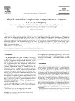
SEM images recorded from the mag-Pec, mag-Pec@Cs, and Pec@Cs
microspheres were used to investigate their morphology and micro
structure. As shown in Fig. 2, all microsphere samples exhibited a
spherical-like shape with different levels of roughness and cracks. Ac
cording to Jeddi & Mahkam (2019), the cracks appear due to the drying
process and can be ascribed to the high volume of water inside the
polymer matrices. The SEM images of mag-Pec (Fig. 2a and b) show that
this sample has a more uniform and compact surface, which strengthens
the suggestion that magnetite increased the crosslinking density of the
performed in simulated gastric fluid (SGF, pH 1.2) and simulated in
testinal fluid (SIF, pH 6.8) (Pereira et al., 2013). For this, dry micro
spheres (50 mg) were put into vials filled with 50 ml of the swelling
medium at room temperature and slow stirring. At predetermined in
tervals, the microspheres were collected, the excess of liquid on their
surfaces was carefully removed, and then, they were weighed again. The
swelling ratio at each time interval was calculated per Eq. (3):
Swelling(%) =
[ws − wd ]
x 100
wd
(3)
where ws is the weight of samples after swelling at a predetermined
interval and wd is the weight at dry state. The swelling experiments were
performed in triplicate.
The point of zero charge (PZC), a parameter that describes the con
dition when the electrical charge density on the bead surface is zero, was
estimated from the difference between the initial and final pHs of the
immersion solution (Kosmulski, 2020). Briefly, 200 mg of microspheres
were placed into vials containing NaCl solution (50 mL, 0.1 mol L− 1)
with different pHs (from 2 to 12). The pH was adjusted with HCl or
NaOH solution (0.1 mol L− 1) using a Hannah (model HI2211) pH Meter
(Brazil). The vials were kept under low orbital stirring for 24 h to reach
equilibrium. Thus, the microspheres were withdrawn from each vial and
the final pH (pHf) of the solutions was measured immediately. The dif
ference between the initial (pH0) and final pHs (ΔpH = pH0 – pHf) was
plotted against pH0. The pH where the ΔpH is equal to zero was ascribed
as pHPZC.
2.5. In vitro release experiments
The Mtz release behavior from the prepared microspheres was
assessed through in vitro experiments using two different media; SGF
(pH 1.2) and SIF (pH 6.8) both without the presence of enzymes (Pereira
et al., 2013). A certain amount of the Mtz-loaded microspheres (200 mg)
were placed into vials filled with 50 ml of the release medium (SGF or
SIF), which were kept at 37 ± 1 ◦ C with mild orbital stirring (50 rpm)
over the whole experiment (12 h duration). At predetermined time in
tervals, stirring was stopped and aliquots (3 mL) were withdrawn,
centrifuged (5000 rpm for 5 min), and spectrophotometrically analyzed
at λ =271 nm. An equivalent volume of fresh release medium was
refilled in the system immediately to keep the total volume constant.
The cumulative release percentages after each time interval were
calculated per Eq. (4). Again, all procedures were done in triplicate.
Cumulative release (%) =
[amount of Mtz released at time t]
x 100
[amount of Mtz loaded in microspheres]
(4)
To verify the effect of an external magnetic field (EMF) on the Mtz
release behavior similar in vitro experiments were carried. However, a
permanent cylindrical neodymium permanent magnet (NdFeB,
20 × 10 mm, grade N52) was positioned on the top of the vial containing
the microspheres and the release medium (externally), while another
identical magnet was placed at the bottom. Again, SGF and SIF were
used as releasing media. At predetermined time intervals, aliquots were
withdrawn from each vial and the amount of Mtz released was estimated
by UV–vis measurements (at λ =271 nm). The cumulative release was
calculated per Eq. (4).
3. Results and discussion
Table 1
Average size and pHPZC values estimated for different microspheres samples.
3.1. Characterization of the prepared magnetic microspheres
The dripping approach used to prepare the Pec@Cs microspheres
(with and without magnetite) resulted in spherical-like materials as
demonstrated in Fig. 1. Microspheres were instantaneously formed after
the dripping of pectin solution into CaCl2 solution due to the ionotropic
Microspheres
Average size (mm)a
pHpzc
mag-Pec
mag-Pec@Cs
Pec@Cs
3.05 ± 0.14
3.28 ± 0.38
3.69 ± 0.36
2.83 ± 0.06
5.73 ± 0.10
5.70 ± 0.17
a
4
The average size was calculated from wet microspheres.
T.S.A. Lemos et al.
Carbohydrate Polymers 265 (2021) 118013
Fig. 2. Images obtained by SEM from dried (a,b) mag-Pec, (c,d) mag-Pec@Cs and (e,f) Pec@Cs microspheres.
the Ca2+ affects the electrostatic environment around the functional
groups of pectin causing changes in the intensity of multiple bands
compared to the spectrum of raw pectin. For example, the band ascribed
to O–H stretching is sharpened and its center is moved to 3422 cm− 1,
– O stretching is shifted to 1630
while the band ascribed to asymmetric C–
− 1
cm . Similar results concerning this kind of microspheres were re
ported in the literature (Assifaoui et al., 2010; Lessa et al., 2017). Also,
the appearance of a new band at 554 cm− 1 can be associated with the
Fe–O bond, indicating the successful entrapment of magnetite nano
particles on the pectin-based microspheres (Marin et al., 2018). FTIR
spectrum of raw chitosan exhibited a broad band centered at 3402 cm− 1
due to O–H and N–H stretching (hydroxyl and amine groups) and
bands at 2901 cm− 1, 1638 cm− 1, 1570 cm− 1, and 1235 cm− 1 corre
– O stretching (amide I),
sponding to C–H stretching (CH3 groups), C–
N–H bending (amide II), and C–N stretching (amide III) (Brugnerotto
et al., 2001). Bands at 1163 cm− 1 and 1082 cm− 1 are due to C–C and
C–O stretching related to the saccharide structure of chitosan (Gonza
lez-Pabon et al., 2019). After the coating of the mag-Pec microspheres
with chitosan, some discrepancies were noticed in the spectrum ob
tained for mag-Pec@Cs. The bands associated with the carboxyl groups
of pectin were shifted to 1628 cm− 1, while the bands corresponding to
amino groups of chitosan were reduced in intensity and shifted to
1552 cm− 1, respectively. The shifting of these bands to lower wave
number regions is caused by the electrostatic interaction among the
pectin matrix. Besides, a denser polymer matrix retains a smaller volume
of water, which may explain the lower cracking on its surface. At higher
magnification (Fig. 2b) it can be observed that the mag-Pec microsphere
has a highly rough and irregular surface, with polyhedric particles of
variable sizes. In contrast, SEM images of the mag-Pec@Cs and Pec@Cs
(Fig. 2c–f) revealed that the chitosan coating increased the cracks on the
surface of the microspheres, while it reduced the surface roughness.
Similar reports are done by other authors that have utilized chitosan as a
coating agent for microspheres (Finotelli et al., 2010; Rashidzadeh et al.,
2020). Comparing mag-Pec@Cs and Pec@Cs, their morphologies are
quite similar indicating that magnetite nanoparticles embedded on the
pectin core exert a negligible effect on microspheres surfaces.
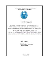
FTIR spectroscopy was used to evaluate the microsphere formation
and chitosan-coating process. All obtained spectra are shown in Fig. 3a.
The spectrum of raw pectin exhibited a broad band centered at
3418 cm− 1 due to O–H stretching (hydroxyl groups) and other char
acteristic bands at 2930 cm− 1, 1642 cm− 1, and 1421 cm− 1 ascribed to
–O
C–H stretching (CHx groups) and asymmetric and symmetric C–
stretching (carboxyl groups) (Lessa et al., 2017). The bands at
1157 cm− 1, 1100 cm− 1, and 1035 cm− 1 are due to C–O–C stretching
(glycosidic bond, ring) and C–C/C–O stretching (Demir et al., 2020).
After the mag-Pec formation, the bands associated with the hydroxyl
and carboxyl groups of pectin were shifted to different wavenumber due
to the bind of such groups to Ca2+ ions (Lessa et al., 2017). Moreover,
5
T.S.A. Lemos et al.
Carbohydrate Polymers 265 (2021) 118013
nanoparticles, with following corresponding indices (220), (311), (400),
(511), and (440) (JCPDS number #19-0629) (Dar & Shivashankar,
2014). The presence of these diffraction peaks confirms the entrapment
of magnetite into the microspheres without changing its structure (Xiao
et al., 2011). Besides, the absence of new diffraction peaks compared to
the bare microspheres (Pec@Cs) suggests the magnetite nanoparticles
did not affect the polymer matrix ordering.
TGA/DTG analysis was performed to evaluate the thermal behavior
of the prepared microspheres and results are shown in Fig. 4a and b. TGA
curve of raw pectin exhibited two weight loss stages, where the first
(between 30 and 125 ◦ C) caused a weight loss of 15 % due to the
evaporation of water. The second stage (between 195 and 290 ◦ C, with a
maximum at 241 ◦ C) is due to the thermal depolymerization of the
pectin backbone resulted in a weight loss of 43 % (Lessa et al., 2017). At
500 ◦ C, the residual weight of pectin was around 42 %. Similarly, raw
chitosan exhibited two main weight loss stages. The first weight loss
around of 10 % (between 30 and 130 ◦ C) was due to the evaporation of
adsorbed water, while the second weight loss stage (between 230 and
400 ◦ C, with a maximum at 303 ◦ C) was attributed to the thermal
decomposition and deacetylation of chitosan backbone (Nam et al.,
2010). For chitosan, the residual weight at 500 ◦ C was around 43 %. For
the microspheres (Pec@Cs and mag-Pec@Cs), TGA curves were quite
similar; however, some discrepancies can be noticed. In summary, both
curves exhibited three main weight loss stages. In the first stages (be
tween 30 and 120 ◦ C), Pec@Cs lost around 17 % of weight, while
mag-Pec@Cs around 21 % due to the water evaporation. This data re
veals that the entrapment of magnetite into the pectin matrix increased
the water content into the magnetic microsphere compared to the bare
sample. It is worthy to point out that both microspheres samples were
thoroughly dried under identical conditions (up to constant weight)
before TGA analysis. Moreover, comparable finds were also reported by
Jeddi & Mahkam (2019). The second and third stages were observed
between 210 and 350 ◦ C and are attributed to the thermal decomposi
tion of each polysaccharide. For Pec@Cs, the maximum temperatures
for pectin and chitosan decomposition were found to be at 257 ◦ C and
303 ◦ C and the total weight loss was around 25 %. For mag-Pec@Cs, the
maximum temperatures were found to be 251 ◦ C and 302 ◦ C, while the
weight loss was around 29 %. This result suggests the presence of
magnetite has a slightly negative effect on the thermal stability of the
mag-Pec@Cs microspheres. Additionally, at 500 ◦ C it was found that the
residual weight of Pec@Cs was higher than that observed for
mag-Pec@Cs. Probably, the magnetite nanoparticles catalyzed the
thermal decomposition of pectin/chitosan chains explaining the ob
tained results. Indeed, some papers have described the ability of metal
oxides like to Fe3O4 to accelerate the thermal decomposition of poly
saccharides (Jurikova et al., 2012; Ziegler-Borowska et al., 2016).
The liquid uptake is an essential property of hydrophilic materials
and paramount for functional DDS. Herein, the liquid uptake capacity of
the prepared microspheres was evaluated by swelling experiments per
formed in SGF (pH 1.2) and SIF (pH 6.8). The swelling curves built for
Pec@Cs and mag-Pec@Cs are shown in Fig. 5a and b. Both microspheres
swelled quickly in SGF achieving high swelling rates before 30 min. For
Pec@Cs, the swelling rate seems to slow down after 20− 25 min and,
then, the equilibrium is achieved close to 60 min. Next, the swelling
tends to level off until the end of the experiment. The maximum swelling
rate calculated for this sample in SGF was around 233 %. Conversely,
mag-Pec@Cs exhibited a slightly faster initial swelling achieving the
equilibrium sooner than the bare microspheres (ca. 30 min). For these
microspheres, the maximum swelling rate was around 275 %. In general
lines, Pec@Cs and mag-Pec@Cs showed a high swelling performance,
which can be explained by the acidic condition of SGF that affects the
charge of the different functional groups. Under this pH condition, the
amino groups in chitosan and carboxyl groups in pectin are both pro
tonated. As a result, the electrostatic interaction between pectin and
chitosan decreases, as well as the pectin-Ca2+ interactions (Lofgren
et al., 2002). Simultaneously, the repulsive forces among the protonated
Fig. 3. (a) FTIR spectra recorded from raw pectin and chitosan and prepared
microspheres (mag-Pec, mag-Pec@Cs, and Pec@Cs). (b) XRD patterns of raw
pectin and chitosan and prepared microspheres (Pec@Cs and mag-Pec@Cs).
–COO− groups of pectin and –NH+
3 groups of chitosan. The absence of
new bands strengthens the suggestion that only electrostatic interactions
occur between the polysaccharides. Similar results were reported to
authors that used chitosan to coat microspheres based on alginate, a
carboxyl-rich polysaccharide (Jeddi & Mahkam, 2019; Rashidzadeh
et al., 2020). It is important to note that the band associated with the
magnetite is still observed in the mag-Pec@Cs spectrum. Finally, as
shown in Fig. 3a, the spectrum of the Pec@Cs microspheres showed to
be similar to mag-Pec@Cs indicating that the presence of magnetite does
not affect the electrostatic interaction between pectin and chitosan.
Fig. 3b shows the XRD patterns obtained for raw pectin and chitosan
and Pec@Cs and mag-Pec@Cs microspheres. As observed, the XRD
pattern of pectin exhibited some diffraction peaks at 2θ ≈ 12.7◦ , 20.5◦ ,
26.2◦ , and 30.1◦ indicating that this polysaccharide has some crystal
linity (Kumar & Chauhan, 2010). Probably, crystalline regions are
formed as a result of intra and intermolecular hydrogen bonds among
the pectin chains. For chitosan, it was observed a typical broad
diffraction peak at 2θ ≈ 20.3◦ indicating its semi-crystalline nature
(Lessa et al., 2018). The XRD pattern obtained for the Pec@Cs micro
spheres did not exhibit any diffraction peak indicating the prevalence of
amorphous structure. Indeed, the electrostatic interaction between the
pectin-Ca2+ ions and pectin-chitosan disrupts the crystalline regions in
the raw polysaccharides, explaining the amorphous nature of Pec@Cs
microspheres. In contrast, the XRD pattern of mag-Pec@Cs microspheres
exhibited diffraction peaks at 2θ ≈ 30.2◦ , 35.7◦ , 43.3◦ , 57.2◦ , and 62.7◦ ,
which correspond to the typical reflection planes of cubic Fe3O4
6
T.S.A. Lemos et al.
Carbohydrate Polymers 265 (2021) 118013
Fig. 4. (a) TGA and (b) DTG curves obtained for raw pectin and chitosan and prepared microspheres (mag-Pec, mag-Pec@Cs, and Pec@Cs).
Overall, a lower crosslinked density favors the water uptake process
(Bueno et al., 2013). Moreover, such impairment caused by magnetite in
the ionotropic crosslinking can also explain the lower thermal stability
of mag-Pec@Cs, as observed from TGA/DTG analysis.
In SIF (pH 6.8), the liquid uptake capacity of both microspheres was
noticeably lower than in SGF, as shown in Fig. 5b. This trend highlights
that the prepared microspheres are exceedingly sensitive to pH varia
tions. Under neutral pH, the mag-Pec@Cs microspheres showed again a
faster swelling profile compared to the Pec@Cs. However, at this pH
condition, the swelling equilibrium was achieved faster than in acidic
conditions (before 10 min). The maximum swelling rate was calculated
to be 68 % and 180 % for Pec@Cs and mag-Pec@Cs, respectively. At pH
6.8, the carboxyl groups in pectin and amino groups in chitosan are
deprotonated, which increases the interaction between pectin chains
and Ca2+ ions. At the same time, the electrostatic interactions between
pectin and chitosan decrease. However, the chitosan coat probably re
mains on the surface of microspheres since hydrogen bonds can be
formed between the polysaccharides. Furthermore, this suggestion is
strengthened by the low solubility of chitosan in neutral and alkaline pH
conditions (Nie et al., 2016). As demonstrated by these swelling ex
periments, the pH-sensitive properties of Pec@Cs and mag-Pec@Cs can
be attractive to trigger and control the release of encapsulated bioactive
compounds like drugs, for example.
3.2. Release experiments
In vitro experiments were conducted to investigate the release ability
of the prepared microspheres using Mtz a model drug. Earlier to the
release experiments, the encapsulation efficiency (EE%) and drug
loading (DL%) were estimated. For Pec@Cs/Mtz, EE% and DL% were
calculated to be 85 ± 1 % and 0.14 ± 0.02 %, while mag-Pec@Cs/Mtz
showed EE% and DL% equal to 88 ± 2 % and 0.15 ± 0.04 %, respec
tively. From a statistical viewpoint, the results concerning both micro
sphere samples are similar. However, it can be mentioned that both
microspheres showed EE% values higher than 85 %, indicating a mini
mal loss of Mtz during the encapsulation process.
Fig. 6a and b show the release profile of Mtz from Pec@Cs/Mtz magPec@Cs/Mtz in SGF (pH 1.2) and SIF (pH 7.4) at 37 ◦ C. Moreover,
additional release experiments were performed with the loaded mag
netic microspheres using an external magnetic field (EMF) to evaluate its
effect on the Mtz release. In SGF, the drug release occurred quickly
during the first hour of the experiment for all tested samples, mainly for
the microspheres exposed to EMF. Next, the release process slows down
and remained constant until the end of the experiment. After 12 h, the
percentages of Mtz released from Pec@Cs/Mtz, mag-Pec@Cs/Mtz, and
mag-Pec@Cs/Mtz (with EMF) in SGF were calculated to be around 18 %,
21 %, and 26 %, respectively. These results seem to be inconsistent with
Fig. 5. Swelling profile of Pec@Cs and mag-Pec@Cs microspheres in (a) SGF
(pH 1.2) and (b) SIF (pH 6.8) at 37 ◦ C.
amino groups in chitosan increase. Thus, the polymer matrix expands
allowing that a high amount of liquid moves inward the microspheres. It
is important to inform that the hydrophilic nature of both poly
saccharides enhances the liquid uptake capacity of the prepared mi
crospheres. The data depicted in Fig. 5a also reveals that mag-Pec@Cs
microspheres have a higher liquid uptake capacity than Pec@Cs. In
practical terms, the addition of 1 wt-% of magnetite allowed to increase
the maximum swelling by 42 %. Probably, the presence of magnetite
nanoparticles impaired the ionotropic crosslinking of pectin chains by
Ca2+ ions reducing the crosslinking density within the microspheres.
7
T.S.A. Lemos et al.
Carbohydrate Polymers 265 (2021) 118013
Fig. 6. In vitro Mtz release profile from Pec@Cs/Mtz and mag-Pec@Cs/Mtz microspheres in (a) SGF (pH 1.2) and (b) SIF (pH 6.8) at 37 ◦ C. For mag-Pec@Cs/Mtz the
release experiments were performed in the absence and presence of an external magnetic field (EMF).
experiment without EMF. In summary, the release experiments indicate
that both microspheres (Pec@Cs/Mtz, mag-Pec@Cs/Mtz) are sensitive
to pH, while mag-Pec@Cs/Mtz is simultaneously sensitive to EMF.
To gain insights about the release process and mechanism, all results
shown in Fig. 6 were fitted by different mathematical models of drug
release. Herein, Higuchi, Korsmeyer-Peppas, and Weibull models were
utilized. The Higuchi model (Eq. (5)) is often used for the assessment of
drug release from polymeric matrices via diffusion-controlled processes
(Mircioiu et al., 2019). Korsmeyer-Peppas is a semi-empirical model (Eq.
(6)) generally used to analyze drug release when the mechanism is not
well known or multiple mechanisms are involved (Korsmeyer et al.,
1983). Moreover, this model enables only fitting the data related to the
first 60 % of drug release. Finally, Weibull is an empirical model (Eq.
(7)) frequently used to analyze the drug release from micro and nano
particles in different experimental conditions (Ignacio et al., 2017).
the swelling data that demonstrated that under acidic conditions both
Pec@Cs/Mtz and mag-Pec@Cs/Mtz microspheres exhibit high liquid
uptake capacities. To explain these results, it should be noticed that the
Mtz molecule contains negatively charged groups that can interact with
the chitosan coat that under acidic conditions is positively charged (due
to its protonated amino groups). Similar results were reported by Bhise
et al. (2008) and Sun et al. (2010) that designed DDS based on chitosan
for sustained release of anionic drugs such as naproxen and enoxaparin.
According to the authors, the interactions between cationic chitosan and
the anionic drugs form stable systems from which the drugs are released
over a more prolonged time interval. These finds corroborate the high
values of log P calculated for Mtz under these release conditions (log
P ≥ 3.05). It is important to mention that the cationic nature of the
chitosan coat under acidic conditions also can be ranked as an additional
advantage since it is responsible for mucoadhesion via ionic interaction
with the mucus of the gastric system (Shafabakhsh et al., 2020).
Results depicted in Fig. 6a also reveal that the presence of an EMF
increases the Mtz release rate from mag-Pec@Cs and promotes a gain of
5 % in the cumulative amount released after 12 h compared to the
conventional release (i.e., without EMF). This find confirms that magPec@Cs show magnetic-responsible behavior. As explained by Rashid
zadeh et al. (2020), the magnetic nanoparticles embedded into the mi
crosphere’s matrix are agitated and moved under the influence of EMF,
which leads to the relaxing of polymer chains. Thus, this relaxation
phenomenon may have led to mechanical deformation and subsequent
tensile stresses, resulting in an enhancement in the amount of drug
released (Paulino et al., 2012; Rashidzadeh et al., 2020). Additionally,
under EMF the magnetic nanoparticles are aligned within the micro
spheres decreasing the barrier effect against the drug release process
(Marin et al., 2018).
The Mtz release from the Pec@Cs/Mtz and mag-Pec@Cs/Mtz in SIF
showed a similar profile compared to SGF media (Fig. 6b). Overall, the
drug was released quickly at the beginning of the experiment, and, then,
the release process slows down as time goes on. However, after 12 h the
amount of Mtz released from the microspheres is markedly higher than
that estimated in SGF. Herein, the percentages of Mtz released from
Pec@Cs/Mtz, mag-Pec@Cs/Mtz, and mag-Pec@Cs/Mtz (with EMF)
after 12 h were calculated to be around 71 %, 75 %, and 91 %,
respectively. These results can be explained by the absence of charges in
the chitosan layer under neutral conditions (i.e., absence of interaction
with Mtz molecules). Besides, at pH 6.8 the carboxylic groups in pectin
are deprotonated increasing the negatively repulsive forces with the
anionic Mtz, thus, favoring the release. Hence, the calculated values of
log P (≤ 1.04) were noticeably lower than those calculated for Mtz in
SGF. Furthermore, under EMF the drug release process was enhanced
again. The Mtz release increased by 16 % after 12 h compared to the
Mt = kH t0.5
(5)
Mt/M = kKP tn
(6)
∞
Mt/M = 1 − e−
∞
atb
(7)
Herein, Mt refers to the amount of cumulative drug released at each time
(t), M∞ is the amount of cumulative drug release at infinite time, kH and
kKP are the Higuchi and Korsmeyer-Peppas constants, and n is the release
exponent associated with the drug release mechanism. Furthermore, in
Eq. (7), the parameters a and b are the "scale" and "shape" factors in the
Weibull distribution (Ignacio et al., 2017). The fitting parameters ob
tained from the mathematical models are summarized in Table 2.
Analyzing the coefficients of determination (R2) given in Table 2, it is
observed that the highest R2 values were obtained for the Weibull
model, indicating that this model adjusts well to the experimental data.
Indeed, the Weibull model had the best fit for all tested samples and
conditions. In this context, the parameter b ("shape" factor) can be used
as an indicator of the mechanism of transport for the drug through the
polymeric matrix. Generally, a value of b < 0.75 denotes Fickian diffu
sion, while a value in the range 0.75 < b < 1.0 denotes a combined
mechanism (Fickian diffusion and swelling-controlled transport). Values
of b > 1 are associated with a complex transport/release mechanism (i.
e., a combination of different mechanisms such as erosion, diffusion, and
swelling) (Mircioiu et al., 2019). From Table 2, it is noticed that Mtz
release from Pec@Cs and mag-Pec@Cs microspheres change according
to the release media. In SGF, the release mechanism is guided by Fickian
diffusion, while in SIF it changes to a combined mechanism (Fickian
diffusion and swelling-controlled transport). Curiously, the presence of
an EMF does not affect the release mechanism of mag-Pec@Cs. It means
8
T.S.A. Lemos et al.
Carbohydrate Polymers 265 (2021) 118013
Release media
media suggesting that the microspheres prepared in this study also
exhibit a magnetic-sensitivity property. Based on our finds, the magnetic
microspheres can be considered potential candidates for drug delivery
applications, particularly in colon-localized delivery or in cancer ther
apy (tumor inhibition).
Parameter
SGF (pH
1.2)
SIF (pH
6.8)
CRediT authorship contribution statement
kH
R2
kKP
n
R2
a
b
Td
R2
kH
R2
kKP
n
R2
a
b
Td
R2
kH
R2
kKP
n
R2
a
b
Td
R2
0.021
0.738
0.546
0.383
0.849
0.910
0.511
0.687
0.993
0.020
0.748
0.619
0.251
0.917
1.014
0.453
0.558
0.989
0.044
0.432
0.731
0.256
0.637
2.060
0.496
0.712
0.995
0.090
0.587
0.618
0.410
0.812
1.584
0.910
0.942
0.988
0.081
0.784
0.625
0.282
0.952
1.647
0.886
0.744
0.986
0.189
0.399
0.721
0.310
0.709
2.855
0.970
0.989
0.990
Table 2
Fitting parameters obtained from the mathematical models of Higuchi,
Korsmeyer-Peppas and Weibull to the experimental data of Mtz release from
prepared microspheres in SGF and SIF at 37 ◦ C.
Microspheres
Model
Higuchi
Pec@Cs/Mtz
KorsmeyerPeppas
Weibull
Higuchi
mag-Pec@Cs/Mtz
KorsmeyerPeppas
Weibull
Higuchi
mag-Pec@Cs/Mtz
(with EMF)
KorsmeyerPeppas
Weibull
Thalia S.A. Lemos: Methodology, Formal analysis, Investigation,
Writing - original draft. Jaqueline F. de Souza: Methodology, Formal
´ R. Fajardo: Supervision, Project admin
analysis, Investigation. Andre
istration, Writing - review & editing.
Declaration of Competing Interest
The authors report no declarations of interest.
Acknowledgments
The authors are thankful to CNPq (Process 404744/2018-4) for
financial support. A.R.F. also thanks CNPq for his PQ fellowship (Process
303872/2019-5). This study was financed in part by the Coordenaỗ
ao de
Aperfeiỗoamento de Pessoal de Nớvel Superior, Brazil (CAPES/Proap),
Finance Code 001.
References
Allouss, D., Essamlali, Y., Amadine, O., Chakir, A., & Zahouily, M. (2019). Response
surface methodology for optimization of methylene blue adsorption onto
carboxymethyl cellulose-based hydrogel beads: adsorption kinetics, isotherm,
thermodynamics and reusability studies. RSC Advances, 9(65), 37858–37869.
/>Assifaoui, A., Loupiac, C., Chambin, O., & Cayot, P. (2010). Structure of calcium and zinc
pectinate films investigated by FTIR spectroscopy. Carbohydrate Research, 345(7),
929–933. />Barboza, F. M., Machado, W. M., Olchanheski, L. R., de Paula, J. P., Zawadzki, S. F.,
Fernandes, D., & Farago, P. V. (2014). PCL/PHBV microparticles as innovative
carriers for oral controlled release of manidipine dihydrochloride. Scientific World
Journal. />Bhise, K. S., Dhumal, R. S., Paradkar, A. R., & Kadam, S. S. (2008). Effect of drying
methods on swelling, erosion and drug release from chitosan-naproxen sodium
complexes. AAPS PharmSciTech, 9(1), 1–12. />Brugnerotto, J., Lizardi, J., Goycoolea, F. M., Arguelles-Monal, W., Desbrieres, J., &
Rinaudo, M. (2001). An infrared investigation in relation with chitin and chitosan
characterization. Polymer, 42(8), 3569–3580. />(00)00713-8
Bueno, V. B., Siqueira, D. F., Bentini, P. R., & Catalani, L. H. (2013). Synthesis and
swelling behavior of xanthan-based hydrogels. Carbohydrate Polymers, 92(2),
1091–1099. />Capel, F., Nicolai, T., Durand, D., Boulenguer, P., & Langendorff, V. (2006). Calcium and
acid induced gelation of (amidated) low methoxyl pectin. Food Hydrocolloids, 20(6),
901–907. />Chang, P. R., Yu, J. G., Ma, X. F., & Anderson, D. P. (2011). Polysaccharides as stabilizers
for the synthesis of magnetic nanoparticles. Carbohydrate Polymers, 83(2), 640–644.
/>Dar, M. I., & Shivashankar, S. A. (2014). Single crystalline magnetite, maghemite, and
hematite nanoparticles with rich coercivity. RSC Advances, 4(8), 4105–4113.
/>Demarchi, C. A., Debrassi, A., Buzzi, F. D., Correa, R., Cechinel, V., Rodrigues, C. A., &
Greneche, J. M. (2014). A magnetic nanogel based on O-carboxymethylchitosan for
antitumor drug delivery: Synthesis, characterization and in vitro drug release. Soft
Matter, 10(19), 3441–3450. />Demir, D., Ceylan, S., Gokturk, D., & Bolgen, N. (2020). Extraction of pectin from albedo
of lemon peels for preparation of tissue engineering scaffolds. Polymer Bulletin.
/>Farah, F. H. (2016). Magnetic microspheres: A novel drug delivery system. Journal of
Analytical & Pharmaceutical Research, 3(5). />japlr.2016.03.00067, 00067.
Finotelli, P. V., Da Silva, D., Sola-Penna, M., Rossi, A. M., Farina, M., Andrade, L. R.,
Takeuchi, A. Y., & Rocha-Leao, M. H. (2010). Microcapsules of alginate/chitosan
containing magnetic nanoparticles for controlled release of insulin. Colloids and
Surfaces B-Biointerfaces, 81(1), 206–211. />colsurfb.2010.07.008
Frachini, E. C. G., & Petri, D. F. S. (2019). Magneto-responsive hydrogels: Preparation,
characterization, biotechnological and environmental applications. Journal of the
Brazilian Chemical Society, 30(10), 2010–2028. />
that the electrostatic interactions between the polysaccharides exert a
higher effect on the drug release process than the presence of an EMF.
This find corroborates other similar studies focused on the use of mag
netic polymeric systems as DDS (Demarchi et al., 2014; Uva et al., 2015).
From the Weibull model, the values of a ("scale" factor) can be used to
calculate the Td parameter [a = (Td)b], which corresponding to the time
required to release 63.2 % of the encapsulated drug (Barboza et al.,
2014). The values of Td obtained reveal that the presence of EMF
allowed an accelerated Mtz release from mag-Pec@Cs in both tested
media.
4. Conclusions
Here, we succeed in preparing magnetic microspheres based on
pectin/magnetite coated by chitosan. The microspheres were obtained
by ionotropic gelation of pectin with Ca2+ ions followed by poly
electrolyte complexation of a chitosan coating. According to FTIR and
XRD, the magnetite nanoparticles were entrapped into the micro
spheres. SEM and TGA/DTG analyses showed that magnetite caused
changes in the morphology and thermal stability of the microspheres.
Moreover, these characterization analyses confirmed the coating of the
pectin/magnetite microspheres by chitosan. Swelling experiments per
formed in simulated gastric fluid (SGF, pH 1.2) and simulated intestinal
fluid (SIF, pH 6.8) revealed that the presence of magnetite enhances the
liquid uptake capacity of the microspheres in both media. Additionally,
metamizole (Mtz) was efficiently encapsulated into the magnetic mi
crospheres, and in vitro release experiments were performed in SGF and
SIF. The results showed that the release process can be adjusted by
varying the pH of the medium and it is favored in SIF. Weibull model
better fitted the release data, indicating that the release mechanism is
guided by Fickian diffusion in SGF, while in the SIF medium it changes
to a combined mechanism (Fickian diffusion and swelling-controlled
transport). Release experiments in the presence of an external mag
netic field (EMF) showed that the drug release is boosted in both release
9
T.S.A. Lemos et al.
Carbohydrate Polymers 265 (2021) 118013
Frank, L. A., Onzi, G. R., Morawski, A. S., Pohlmann, A. R., Guterres, S. S., & Contri, R. V.
(2020). Chitosan as a coating material for nanoparticles intended for biomedical
applications. Reactive & Functional Polymers, 147, 104–459. />10.1016/j.reactfunctpolym.2019.104459
Ghazanfari, M. R., Kashefi, M., Shams, S. F., & Jaafari, M. R. (2016). Perspective of Fe3O4
nanoparticles role in biomedical applications. Biochemistry Research International,
2016. />Gonzalez-Pabon, M. J., Figueredo, F., Martinez-Casillas, D. C., & Corton, E. (2019).
Characterization of a new composite membrane for point of need paper-based microscale microbial fuel cell analytical devices. PloS One, 14(9). />10.1371/journal.pone.0222538
Gunter, E. A., Markov, P. A., Melekhin, A. K., Belozerov, V. S., Martinson, E. A.,
Litvinets, S. G., & Popov, S. V. (2018). Preparation and release characteristics of
mesalazine loaded calcium pectin-silica gel beads based on callus cultures pectins for
colon-targeted drug delivery. International Journal of Biological Macromolecules, 120,
2225–2233. />Ignacio, M., Chubynsky, M. V., & Slater, G. W. (2017). Interpreting the Weibull fitting
parameters for diffusion-controlled release data. Physica A-Statistical Mechanics and
Its Applications, 486, 486–496. />Jafari, M., Sriram, V., Xu, Z. Y., Harris, G. M., & Lee, J. Y. (2020). Fucoidan-doxorubicin
nanoparticles targeting P-selectin for effective breast cancer therapy. Carbohydrate
Polymers, 249, 116–837. />Jasiecka, A., Maslanka, T., & Jaroszewski, J. J. (2014). Pharmacological characteristics
of metamizole. Polish Journal of Veterinary Sciences, 17(1), 207–214. />10.2478/pjvs-2014-0030
Jeddi, M. K., & Mahkam, M. (2019). Magnetic nano carboxymethyl cellulose-alginate/
chitosan hydrogel beads as biodegradable devices for controlled drug delivery.
International Journal of Biological Macromolecules, 135, 829–838. />10.1016/j.ijbiomac.2019.05.210
Jurikova, A., Csach, K., Miskuf, J., Koneracka, M., Zavisova, V., Kubovcikova, M., &
Kopcansky, P. (2012). Thermal analysis of magnetic nanoparticles modified with
dextran. Acta Physica Polonica A, 121(5-6). />APhysPolA.121.1296, 1296-129.
Karimi, M., Ghasemi, A., Zangabad, P. S., Rahighi, R., Basri, S. M. M., Mirshekari, H.,
Amiri, M., Pishabad, Z. S., Aslani, A., Bozorgomid, M., Ghosh, D., Beyzavi, A.,
Vaseghi, A., Aref, A. R., Haghani, L., Bahrami, S., & Hamblin, M. R. (2016). Smart
micro/nanoparticles in stimulus-responsive drug/gene delivery systems. Chemical
Society Reviews, 45(5), 1457–1501. />Kim, C., Park, K. S., Kim, J., Jeong, S. G., & Lee, C. S. (2017). Microfluidic synthesis of
monodisperse pectin hydrogel microspheres based on in situ gelation and settling
collection. Journal of Chemical Technology & Biotechnology, 92(1), 201–209. https://
doi.org/10.1002/jctb.4991
Kondaveeti, S., Cornejo, D. R., & Petri, D. F. S. (2016). Alginate/magnetite hybrid beads
for magnetically stimulated release of dopamine. Colloids and Surfaces BBiointerfaces, 138, 94–101. />Korsmeyer, R. W., Gurny, R., Doelker, E., Buri, P., & Peppas, N. A. (1983). Mechanisms of
solute release from porous hydrophilic polymers. International Journal of
Pharmaceutics, 15(1), 25–35. />Kosmulski, M. (2020). The pH dependent surface charging and points of zero charge.
VIII. Update. Advances in Colloid and Interface Science, 275. />j.cis.2019.102064
Kumar, A., & Chauhan, G. S. (2010). Extraction and characterization of pectin from apple
pomace and its evaluation as lipase (steapsin) inhibitor. Carbohydrate Polymers, 82
(2), 454–459. />Lara-Espinoza, C., Carvajal-Millan, E., Balandran-Quintana, R., Lopez-Franco, Y., &
Rascon-Chu, A. (2018). Pectin and pectin-based composite materials: Beyond food
texture. Molecules, 23(4). />Lessa, E. F., Gularte, M. S., Garcia, E. S., & Fajardo, A. R. (2017). Orange waste: A
valuable carbohydrate source for the development of beads with enhanced
adsorption properties for cationic dyes. Carbohydrate Polymers, 157, 660–668.
/>Lessa, E. F., Nunes, M. L., & Fajardo, A. R. (2018). Chitosan/waste coffee-grounds
composite: An efficient and eco-friendly adsorbent for removal of pharmaceutical
contaminants from water. Carbohydrate Polymers, 189, 257–266. />10.1016/j.carbpol.2018.02.018
Li, B. W., Yuan, Z. F., He, Y. W., Hung, H. C., & Jiang, S. Y. (2020). Zwitterionic
nanoconjugate enables safe and efficient lymphatic drug delivery. Nano Letters, 20
(6), 4693–4699. />Lofgren, C., Walkenstrom, P., & Hermansson, A. M. (2002). Microstructure and
rheological behavior of pure and mixed pectin gels. Biomacromolecules, 3(6),
1144–1153. />Maciel, V. B. V., Yoshida, C. M. P., & Franco, T. T. (2015). Chitosan/pectin
polyelectrolyte complex as a pH indicator. Carbohydrate Polymers, 132, 537–545.
/>Marin, T., Montoya, P., Arnache, O., Pinal, R., & Calderon, J. (2018). Development of
magnetite nanoparticles/gelatin composite films for triggering drug release by an
external magnetic field. Materials & Design, 152, 78–87. />matdes.2018.04.073
Mircioiu, C., Voicu, V., Anuta, V., Tudose, A., Celia, C., Paolino, D., Fresta, M.,
Sandulovici, R., & Mircioiu, I. (2019). Mathematical modeling of release kinetics
from supramolecular drug delivery systems. Pharmaceutics, 11(3). />10.3390/pharmaceutics11030140
Mura, S., Nicolas, J., & Couvreur, P. (2013). Stimuli-responsive nanocarriers for drug
delivery. Nature Materials, 12, 991–1003. />Nam, Y. S., Park, W. H., Ihm, D., & Hudson, S. M. (2010). Effect of the degree of
deacetylation on the thermal decomposition of chitin and chitosan nanofibers.
Carbohydrate Polymers, 80(1), 291–295. />carbpol.2009.11.030
Nie, J. Y., Wang, Z. K., & Hu, Q. L. (2016). Difference between chitosan hydrogels via
alkaline and acidic solvent systems. Scientific Reports, 6. />srep36053
Oh, J. K., Lee, D. I., & Park, J. M. (2009). Biopolymer-based microgels/nanogels for drug
delivery applications. Progress in Polymer Science, 34(12), 1261–1282. https://doi.
org/10.1016/j.progpolymsci.2009.08.001
Patra, J. K., Das, G., Fraceto, L. F., Campos, E. V. R., Rodriguez-Torres, M. D. P., AcostaTorres, L. S., & Shin, H. S. (2018). Nano based drug delivery systems: recent
developments and future prospects. Journal of Nanobiotechnology, 16. https://doi.
org/10.1186/s12951-018-0392-8
Paulino, A. T., Pereira, A. G. B., Fajardo, A. R., Erickson, K., Kipper, M. J., Muniz, E. C., &
Tambourgi, E. B. (2012). Natural polymer-based magnetic hydrogels: Potential
vectors for remote-controlled drug release. Carbohydrate Polymers, 90(3),
1216–1225. />Pereira, A. G. B., Fajardo, A. R., Nocchi, S., Nakamura, C. V., Rubira, A. F., & Muniz, E. C.
(2013). Starch-based microspheres for sustained-release of curcumin: Preparation
and cytotoxic effect on tumor cells. Carbohydrate Polymers, 98(1), 711–720. https://
doi.org/10.1016/j.carbpol.2013.06.013
Pham, S. H., Choi, Y., & Choi, J. (2020). Stimuli-responsive nanomaterials for application
in antitumor therapy and drug delivery. Pharmaceutics, 12(7). />10.3390/pharmaceutics12070630
Price, P. M., Mahmoud, W. E., Al-Ghamdi, A. A., & Bronstein, L. M. (2018). Magnetic
drug delivery: Where the field is going. Frontiers in Chemistry, 6. />10.3389/fchem.2018.00619
Rampino, A., Borgogna, M., Bellich, B., Blasi, P., Virgilio, F., & Cesaro, A. (2016).
Chitosan-pectin hybrid nanoparticles prepared by coating and blending techniques.
European Journal of Pharmaceutical Sciences, 84, 37–45. />ejps.2016.01.004
Rashidzadeh, B., Shokri, E., Mahdavinia, G. R., Moradi, R., Mohamadi-Aghdam, S., &
Abdi, S. (2020). Preparation and characterization of antibacterialmagnetic-/pHsensitive alginate/Ag/Fe3O4 hydrogel beads for controlled drug release. International
Journal of Biological Macromolecules, 154, 134–141. />ijbiomac.2020.03.028
Raza, A., Rasheed, T., Nabeel, F., Hayat, U., Bilal, M., & Iqbal, H. M. N. (2019).
Endogenous and exogenous stimuli-responsive drug delivery systems for
programmed site-specific release. Molecules, 24(6). />molecules24061117
Senapati, S., Mahanta, A. K., Kumar, S., & Maiti, P. (2018). Controlled drug delivery
vehicles for cancer treatment and their performance. Signal Transduction and Targeted
Therapy, 3. />Shafabakhsh, R., Youse, B., Asemi, Z., Nikfar, B., Mansournia, M. A., & Hallajzadeh, J.
(2020). Chitosan: A compound for drug delivery system in gastric cancer-a review.
Carbohydrate Polymers, 242. />Sigaeva, N. N., Vil’danova, R. R., Sultanbaev, A. V., & Ivanov, S. P. (2020). Synthesis and
properties of chitosan- and pectin-based hydrogels. Colloid Journal, 82(3), 311–323.
/>Sun, W., Mao, S. R., Wang, Y. J., Junyaprasert, V. B., Zhang, T. T., Na, L. D., & Wang, J.
(2010). Bioadhesion and oral absorption of enoxaparin nanocomplexes. International
Journal of Pharmaceutics, 386(1-2), 275–281. />ijpharm.2009.11.025
Thevenot, J., Oliveira, H., Sandre, O., & Lecommandoux, S. (2013). Magnetic responsive
polymer composite materials. Chemical Society Reviews, 42(17), 7099–7116. https://
doi.org/10.1039/c3cs60058k
Uva, M., Mencuccini, L., Atrei, A., Innocenti, C., Fantechi, E., Sangregorio, C., &
Barbucci, R. (2015). On the mechanism of drug release from polysaccharide
hydrogels cross-linked with magnetite nanoparticles by applying alternating
magnetic fields: The case of DOXO delivery. Gels, 1(1), 24–43. />10.3390/gels1010024
Wong, C. Y., Al-Salami, H., & Dass, C. R. (2018). Microparticles, microcapsules and
microspheres: A review of recent developments and prospects for oral delivery of
insulin. International Journal of Pharmaceutics, 537(1-2), 223–244. />10.1016/j.ijpharm.2017.12.036
Xiao, B., Wan, Y., Zhao, M. Q., Liu, Y. Q., & Zhang, S. M. (2011). Preparation and
characterization of antimicrobial chitosan-N-arginine with different degrees of
substitution. Carbohydrate Polymers, 83(1), 144–150. />carbpol.2010.07.032
Younes, I., & Rinaudo, M. (2015). Chitin and chitosan preparation from marine sources.
Structure, properties and applications. Marine Drugs, 13(3), 1133. />10.3390/md13031133
Ziegler-Borowska, M., Chelminiak, D., Kaczmarek, H., & Kaczmarek-Kedziera, A. (2016).
Effect of side substituents on thermal stability of the modified chitosan and its
nanocomposites with magnetite. Journal of Thermal Analysis and Calorimetry, 124(3),
1267–1280. />
10