Strategies to load therapeutics into polysaccharide-based nanogels with a focus on microfluidics: A review
Bạn đang xem bản rút gọn của tài liệu. Xem và tải ngay bản đầy đủ của tài liệu tại đây (2.4 MB, 17 trang )
Carbohydrate Polymers 266 (2021) 118119
Contents lists available at ScienceDirect
Carbohydrate Polymers
journal homepage: www.elsevier.com/locate/carbpol
Review
Strategies to load therapeutics into polysaccharide-based nanogels with a
focus on microfluidics: A review
N. Zoratto a, E. Montanari b, *, M. Viola a, J. Wang a, T. Coviello a, C. Di Meo a, *, P. Matricardi a
a
b
Department of Drug Chemistry and Technologies, Sapienza University of Rome, 00185 Roma, Italy
Institute of Pharmaceutical Sciences, Department of Chemistry and Applied Biosciences, ETH Zurich, 8093 Zurich, Switzerland
A R T I C L E I N F O
A B S T R A C T
Keywords:
Nanogels
Polysaccharides
Drug loading methods
Microfluidics
Nanogels-based vaccines
Nowadays nanoparticles are increasingly investigated for the targeted and controlled delivery of therapeutics, as
suggested by the high number of research articles (2400 in 2000 vs 8500 in 2020). Among them, almost 2%
investigated nanogels in 2020. Nanogels or nanohydrogels (NGs) are nanoparticles formed by a swollen threedimensional network of synthetic polymers or natural macromolecules such as polysaccharides. NGs represent
a highly versatile nanocarrier, able to deliver a number of therapeutics. Currently, NGs are undergoing clinical
trials for the delivery of anti-cancer vaccines. Herein, the strategies to load low molecular weight drugs, (poly)
peptides and genetic material into polysaccharide NGs as well as to formulate NGs-based vaccines are summa
rized, with a focus on the microfluidics approach.
1. Introduction
In 1999 Alexander V. Kabanov and Serguei V. Vinogradov intro
duced the term NanoGel™ referring to an innovative nano drug delivery
system formed by a hydrophilic polymer network of cross-linked poly
ethyleneimine and carbonyldiimidazole-activated polyethylene glycol
(PEG), using an emulsification/solvent evaporation technique (Vinog
radov et al., 1999). This chemically cross-linked NG was used to deliver
antisense oligonucleotides (Kabanov & Vinogradov, 2009). However,
already few years before, Junzo Sunamoto and Kazunari Akyioshi
described the phenomenon of the physical cross-linking (self-assembly)
of cholesterol (Ch)-modified polysaccharides, such as pullulan (Pul),
mannan (Man) and hyaluronic acid (HA), which resulted in the forma
tion of hydrogels with a nano-scale size (Akiyoshi et al., 1993; Lee &
Akiyoshi, 2004; Nakai et al., 2012; Yamane et al., 2009).
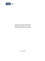
NGs are nano-sized three-dimensional networks (Fig. 1) able to
absorb a large amount of water and to easily swell and de-swell in
aqueous media.
NGs are generally soft, hydrophilic, biocompatible and represent a
highly versatile nano-system able to deliver a variety of bioactive
Abbreviations: AA, asiatic acid; Alg, alginate; Alg-CHO, aldehyde-functionalized alginate; Alg-PDEA, alginate-poly(2-(diethylamino)ethyl methacrylate); ALN,
alendronate; AmPs, antimicrobial peptides; APCs, antigen-presenting cells; BoHc/A, botulinum type-A neurotoxin subunit antigen Hc; BSA, bovine serum albumin;
CDDP, cisplatin-based HA nanocomplexes; CDs, cyclodextrins; CMD-SS-LCA, carboxymethyl dextran-lithocholic acid; Cs, chitosan; CSLNs, cationic solid lipid
nanoparticles; DA, desoxycholic acid; DCs, dendritic cells; DD, deacetylation degree; DEAE, diethyl amino ethyl amine; DEX, dexamethasone; Dex, dextran; DHA, 1,4dihydroxyanthraquinone; DOX, doxorubicin; DSB, di-strylbenzene derivative; dsDNA, double-stranded DNA; E.E., encapsulation efficiency percentage; FA, folic acid;
FNC, flash nanocomplexation; Gel, gellan; Gel-Ch, gellan-cholesterol; Gel-Rfv, gellan-riboflavin; GSH, glutathione; HA, hyaluronic acid; HA-AT, thiolated alkyl
derivative of hyaluronic acid; HA-APBA, hyaluronan‑boronic acid; HA-Ch, hyaluronan-cholesterol; HA-Rfv, hyaluronan-riboflavin; HBsAg, surface protein of Hep
atitis B virus; HCPT, hydroxycamptothecin; IDA, iminodiacetic acid; MA, malonic acid; Man, mannan; MIC, minimum inhibitory concentration; miRNA, microRNA;
MIVM, multi-inlet vortex mixer; mRNA, messenger RNA; MW, molecular weight; NGs, nanogels; OVA, ovalbumin; PDI, polydispersity index; pDNA, plasmid DNA;
PAA, poly(acrylicacid); PEI, polyethylenimine; PEG, polyethylene glycol; PIR, piroxicam; PPZ, perphenazine; PTX, paclitaxel; Pul, pullulan; Pul-Ch, pullulancholesterol; pβ-CD, poly-β-cyclodextrin; RA, retinoic acid; RGD, Arg-Gly-Asp; rHBsAg, recombinant hepatitis B surface antigen; siRNA, short interfering RNA; SODB1,
superoxide dismutase; SpAcDEX, spermine-modified acetalated dextran; ssDNA, single-strended DNA; TA, tannic acid; TOPSi, thermally oxidized porous silicon
particles; TPP, pentasodium triphosphate; TT, tetanus taxoid.
* Corresponding authors.
E-mail addresses: (N. Zoratto), (E. Montanari), (M. Viola), ju.
(J. Wang), (T. Coviello), (C. Di Meo),
(P. Matricardi).
/>Received 11 February 2021; Received in revised form 4 April 2021; Accepted 15 April 2021
Available online 28 April 2021
0144-8617/© 2021 The Authors.
Published by Elsevier Ltd.
This is an
( />
open
access
article
under
the
CC
BY-NC-ND
license
N. Zoratto et al.
Carbohydrate Polymers 266 (2021) 118119
swelling and de-swelling nature in response to external stimuli such as
solvent composition, light, temperature, pH, pressure, magnetic and
electric fields, NGs have attracted attention as functional smart mate
rials for biotechnological and biomedical applications (Eckmann et al.,
2014; Zha et al., 2011). NGs can be prepared from natural (i.e., poly
saccharides, polypeptides) and/or synthetic polymers (i.e., poly lacticco-glycolic acid, PEG, polyglycolic acid, polycaprolactone, poly(Nisopropylacrylamid), poly(methylmethacrylate), poly(acrylicacid), pol
yacrilamide, poly(N-vinyl-pyrrolidone) and depending on the kind of
network linkages, NGs are classified into two groups: physically or
´ & Etrych, 2018). Herein, only
chemically cross-linked NGs (Kousalova
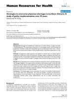
polysaccharide NGs are described. Polysaccharides are biopolymers
consisting of chains of monosaccharide or disaccharide units joined by
glycosidic bonds (Fig. 2) (Coviello et al., 2007). Polysaccharides are
usually non-toxic, biocompatible and biodegradable (Mizrahy & Peer,
2012). Both hydrophilic and hydrophobic therapeutics have been
entrapped into polysaccharide NGs with a significant enhancement of
both the drug bioavailability and pharmacological activity (Kabanov &
Vinogradov, 2009; Vinogradov, 2010). Herein, the strategies to load
molecular or macromolecular therapeutics into NGs and to formulate
polysaccharide-based vaccines are reported, with a focus on
microfluidics.
Microfluidics is emerging as a promising strategy dealing with the
Fig. 1. Schematic representation of a hydrogel, microgel and NG.
molecules such as hydrophobic as well as hydrophilic drugs, (poly)
peptides and genetic material (Choi et al., 2009; Ganguly et al., 2014;
Montanari et al., 2013; Montanari et al., 2018). Indeed, the porosity of
the NGs network provides a reservoir for loading molecular and
macromolecular therapeutics as well as protecting them from the envi
ronmental degradation. Furthermore, because of their inherent rapid
Fig. 2. Average structures and/or repeating units of the various reported polysaccharides: A) Pul; B) Man; C) HA; D) Cs; E) Alg; F) Gel; G) Dex; H) β-1,3-D-glucan;
I) heparin.
2
N. Zoratto et al.
Carbohydrate Polymers 266 (2021) 118119
manipulation of small volumes of fluids (from pico-to-nanoliter) inside a
miniaturized device, with a millisecond mixing time and a real-time
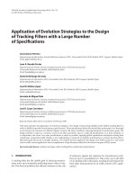
monitoring. Microfluidic devices are made of a number of materials,
such as silicon, glass borosilicate and polydimethylsiloxane which are
patterned into micrometer-sized channels, whilst syringe pumps provide
the driving force for the fluid flow in the microchannels (Fig. 3). Fluid
manipulation occurs by an active or passive control in the microfluidic
device. Active control means that external forces (e.g., magnetic or
electric fields, heat) are responsible for the flow movement, whilst in the
passive control the fluidic movement is governed by channel geometries
and/or by liquid flow rates. In a microfluidic device, the nucleation and
growth stages of nanoparticles can be spaced from the position where
the solution mixing takes place, leading to a precise control in the par
ticle size and morphology, hence to a polydispersity index (PDI)
reduction (Hung & Lee, 2007; Ma et al., 2017). Furthermore, the particle
size can be finely tuned by modifying the flow rate and ratio of the
phases. All these features make microfluidics a cost-effective, repro
ducible and scalable technology (Shrimal et al., 2020). The drug loading
into NGs is usually achieved by polymer emulsification or by exploiting
other approaches such as solvent extraction, solvent diffusion, solvent
evaporation or coacervation within the microfluidic device. In all these
conditions, drug-loaded NGs are formed in a single step, improving both
the NGs drug loading efficiency and the ability to release drugs in a
controlled fashion (Chiesa et al., 2018). However, some shortcomings
still need to be overcome: for example, organic solvents should be
avoided since they may have poor biocompatibility and may affect the
activity of the encapsulated molecules. Moreover, the drug-loaded NGs
production should be further optimized in terms of fabrication process
and drug delivery efficacy (Ma et al., 2020). The next sections describe
the strategies that can be adopted in the formulation of drug-loaded
NGs, with a focus on the microfluidic approach.
or physical cross-linking. In this respect, T. Thambi et al. loaded the
poorly water-soluble anticancer drug doxorubicin (DOX) into carbox
ymethyl Dex-lithocholic acid-based NGs (CMD-SS-LCA). An organic so
lution of DOX was added to the aqueous polymer solution, forming an
oil-in-water emulsion followed by a dialysis against water that led to
the pure drug-loaded NGs formation (Thambi et al., 2014). R. Guo et al.
prepared both chemically and physically cross-linked Alg-poly(2(diethylamino)ethyl methacrylate) (PDEA) NGs for the delivery of
hydroxycamptothecin (HCPT). At neutral pH, HCPT exhibits the
lactone-ring-opened structure which is water soluble. Therefore, a HCPT
aqueous solution (at pH = 8) was firstly added to a mixture of Alg-DEA,
followed by the chemical polymerization of DEA monomers, initiated by
K2S2O8, and the physical crosslinking of Alg chains by CaCO3. As the
chemical polymerization proceeded, the lower pH led to the formation
of the HCPT into its water-insoluble lactone, thus allowing the drug
entrapment in the hydrophobic core of the Alg-PDEA NGs (Guo et al.,
2007).
Inorganic compounds were also loaded into polysaccharide NGs. M.
C. Coll Ferrer and colleagues synthetized NGs based on a lysozyme core
and a Dex shell in which AgNO3 was loaded by the autoclaving process.
The high temperature allowed the reduction of Ag+ to Ag0, in a process
in which lysozyme contributed to the in situ reduction and stabilization
of Ag/NGs. The amount of embedded Ag increased with the increase of
lysozyme content. Unfortunately, the loss or retention of the lysozyme
activity was not shown after the NGs formation (Coll Ferrer et al., 2014).
The in bulk loading methods might take long incubation time (i.e.,
overnight) (Pedrosa et al., 2014; Thambi et al., 2014) and might require
the use of organic solvents (Bertoni et al., 2018; Stefanello et al., 2017;
Thambi et al., 2014). Moreover, drug encapsulation is often achieved by
a two-step procedure: NGs are firstly synthetized and then the payload is
loaded (Pedrosa et al., 2014; Stefanello et al., 2017). Furthermore, the
sterilization process represents another critical issue. In order to over
come some of these disadvantages, the autoclaving process was exploi
ted (Manzi et al., 2017). The aqueous suspension of the amphiphilic
hyaluronan-riboflavin (HA-Rfv) polymer was added to the drug film and
then autoclaved to form sterile and drug loaded NGs. Piroxicam (PIR),
dexamethasone (DEX) and PTX were efficiently loaded into HA-Rfv NGs
by exploiting this approach (Manzi et al., 2017). In other works, auto
claving was used to achieve drug-loaded Gel-Ch (Musazzi et al., 2018),
Gel-riboflavin (Gel-Rfv) (Musazzi et al., 2018) and HA-Rfv NGs (Di Meo
et al., 2015), which were loaded with a number of hydrophobic mole
cules in a single step, confirming the versatility of this method. However,
the autoclaving process cannot be used for encapsulating thermosensitive drugs (Montanari et al., 2019). Moreover, the molecular
weight (MW) of the polysaccharide may decrease after autoclaving, thus
producing new chemical species. Taking into account the limits of the
autoclaving approach and considering that all the described strategies
may lead to high batch variability (i.e., large size distribution and high
polydispersity) and to the formulation of low yield of nano-systems, a
robust procedure for a scalable production of NGs is still under
investigation.
2. Loading of low molecular weight drugs into NGs
2.1. Physical loading by hydrophobic forces
A number of poorly water-soluble drugs have been loaded into NGs,
offering the advantage to enhance their apparent water solubility
(Table IA). For example, the chemical functionalization of the poly
saccharide chains with hydrophobic moieties allows the formation of
amphiphilic derivatives able to self-assemble into NGs with internal
hydrophobic residues, which can host hydrophobic drugs. Typically, the
increase of NGs hydrophobicity enables higher loading capability, as
well as longer sustained release profiles (Bewersdorff et al., 2019). One
strategy for loading poorly-water soluble drugs into NGs is the incuba
tion of the preformed nanoparticle suspension with a concentrated
organic solution of the bioactive molecule (Pedrosa et al., 2014; Stefa
nello et al., 2017). Hydrophobic molecules can also be loaded mean
while the NGs are formed. This can be achieved by adding the bioactive
molecules in the polymer solution before the gelation process that refers
to the formation of a polymeric three-dimensional network by chemical
Fig. 3. An example of a microfluidic setup for the preparation of drug-loaded NGs.
3
N. Zoratto et al.
Carbohydrate Polymers 266 (2021) 118119
Table I
Summary of the physical A) and chemical B) loading strategies employed in the preparation of polysaccharide-based NGs loaded with low molecular weight drugs.
A
Physical loading
Class of therapeutics
Starting material
Loading driving
forces
Loading strategy
Advantages/Disadvantages
References
Low-molecular
weight
hydrophobic drugs
Amphiphilic polysaccharides
Hydrophobic
forces
NGs incubation with the drug
solution
- Long incubation time
- Organic solvents may be
required
- Drug encapsulation
achieved by a two step
procedure
- Low efficiency
- Organic solvents may be
required
- Pedrosa et al.,
2014
- Stefanello et al.,
2017
- Montanari et al.,
2019
- Yang et al., 2011
- Sterile and drug-loaded NGs
formed in a single-step
procedure
- High reproducibility
- Incompatible with thermosensitive drugs
- Change of the polymer Mw
- High reproducibility
- Control over the size, PDI
and compactness of NGs
- Coll Ferrer et al.,
2014
- Manzi et al., 2017
- Musazzi et al.,
2018
- Di Meo et al.,
2015
- Majedi et al.,
2013
- Majedi et al.,
2014
- Wannasarit et al.,
2019
- Kłodzi´
nska et al.,
2019
- Liu et al., 2015
- Bongiovì et al.,
2020
- Gref et al., 2006
Addition of the drug solution to the
polymer suspension, followed by
NGs formation
Autoclaving process
Microfluidics/Millifluidics
Low-molecular
weight
hydrophobic drugs
CDs/polysaccharide mixtures or
polysaccharides containing
chelating moieties
Complexation or
coordination
NGs incubation with the drug
solution
CDs incubation with the drug,
followed by NGs formation
Low-molecular
weight drugs
Charged polysaccharides
Electrostatic
interactions
Incubation of the chelating polymer
with the drug, followed by NGs
formation
Addition of drug to the polymer
solution, followed by NGs
formation
- Long incubation time
- Drug encapsulation
achieved by a two/three step
procedure
- Long incubation time
- Drug encapsulation
achieved by a two step
procedure
- Drug loaded NGs formed in a
single step procedure
- Low versatility
- Typically good E.E.%
- Possible interference with
the NGs formation
NGs incubation with the drug
- Possible low stability in
human fluids
Autoclaving process
- Sterile and drug-loaded NGs
formed in a single-step
procedure
- High reproducibility
- Incompatible with thermosensitive drugs
- Change of the polymer Mw
- High reproducibility
- Control over the size, PDI
and compactness of NGs
Microfluidics/Millifluidics
Thambi et al.,
2014
- Gref et al., 2006
- Thiele et al., 2011
- Ohta et al., 2016
- Deacon et al.,
2015
Rossi et al., 2017
- Rajaonarivony
et al., 1993
- Curcio et al.,
2015
- Yang et al., 2011
- Zhang et al., 2006
- Schmitt et al.,
2010
- Curcio et al.,
2015
- Montanari et al.,
2018
- Moradikhah
et al., 2020
- Dong &
Hadinoto, 2017
(continued on next page)
4
N. Zoratto et al.
Carbohydrate Polymers 266 (2021) 118119
Table 1 (continued)
B
Chemical loading
Stimuli responsiveness
Ligand
Starting material
Loading strategy
References
pH-responsive NGs
DOX
Aldehyde-functionalised Alg
Dihydrazide-modified HA
HA‑boronate
Heparin
Cystamine-modified HA
Cystamine-modified Pul
Schiff base condensation
Hydrazone linkage
Cyclic boronic ester formation
Amide bond formation
Disulphide linkage
Disulphide linkage
-
Redox-responsive NGs
Tannic acid
Aminated-RA and aminated-FA
DOX
Protoporphyrin IX
Among the investigated approaches, microfluidics appears to offer a
number of advantages: I) the possibility to finely tune the size of the
nanoparticles and, hence, the nanoparticle compactness by modifying
the polymer concentration and the flow ratio of the dispersed and
continuous phase; II) the significant reduction of the PDI; III) the high
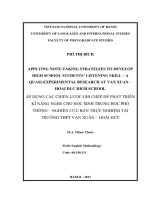
reproducibility; IV) the lack of user talent variability. F. S. Majedi et al.,
prepared PTX-loaded hydrophobically modified Cs-based NGs by using a
flow focusing microfluidic device (Fig. 4A) (Majedi et al., 2013). The Tshape microfluidic device was provided with two inlets for aqueous
buffer (pH = 9), to achieve two water streams at the flow-focusing Tjunction, and one inlet for the mixture of palmitoyl-Cs and PTX at acidic
pH (pH 5.5). In the microfluidic device, the pH increase induces the
simultaneous deprotonation of the Cs hydrophobic side chains and the
Cs amine groups, leading to self-aggregation, thus the NGs formation.
The mixing time was in the millisecond scale; the flow ratio of Cs (pH =
5.5) and aqueous buffer (pH = 9.0) streams was changed from 0.03 to
0.2. By controlling the flow ratio, it was possible to finely tune the size,
the surface charge and the density of the NGs. Compared to the con
ventional mixing method, this approach allowed the formation of more
stable NGs with high encapsulation efficiency percentage (E.E., up to
95% and 60% for microfluidic-formed and bulk mixing-formed NGs,
respectively) and a remarkably lower PDI (PDI < 0.2 for the
microfluidic-formed and PDI > 0.6 for the bulk mixing-formed NGs).
Moreover, the E.E. of NGs increased by increasing the functionalization
degree of the hydrophobically modified Cs, thanks to the hydrophobic
nature of the drug. Furthermore, by reducing the mixing times in the
microfluidic device, higher E.E. were obtained, since more hydrophobic
moieties could interact with the PTX molecules during the NGs forma
tion (Majedi et al., 2013; Majedi et al., 2014). Similarly, drug-loaded
hydrophobically modified Dex NGs were obtained by grafting poly
(lauryl methacrylate-co-methacrylic acid) onto acetylated Dex and were
prepared by nanoprecipitation in a glass-capillary microfluidic device,
as shown in Fig. 4B. Specifically, an ethanolic solution containing the
polymer and asiatic acid (AA, a pentacyclic triterpenoid with anticancer
activity) with a flow rate of 2 mL/h was used as inner phase, whilst an
Pei et al., 2018
Yin et al., 2018
Montanari et al., 2016
Tran et al., 2012
Yin et al., 2018
Xia et al., 2017
aqueous solution at pH 7.4 with a flow rate of 20 mL/h was selected as
an outer phase. NGs formation and loading occurred in a single step
when the polymer solution was quickly mixed with the outer fluid. The
resulting NGs, exhibited a quite low PDI value (0.16) and a high E.E. (~
80%) (Wannasarit et al., 2019). The same nanoprecipitation method was
also exploited by S. Kłodzinska et al. for the preparation of octenyl
succinic anhydride-modified HA NGs loaded with azithromycin. The
polymer solution was injected into the outer streams of a three-inlet
microfluidic chip at a flow rate of 5.4 mL/min, whilst the azi
thromycin acidic solution was injected into the centre stream of the
device at a flow rate of 1.2 mL/min, yielding a combined flow of 12.1
mL/min. By optimizing the working parameters, the highest azi
´ ska et al., 2019). Other hydrophobic
thromycin E.E. was 45% (Kłodzin
drugs, such as imatinib and a mixture of PTX and sorafenib were
encapsulated into HA-based NGs and hybrid porous silicon-acetylated
Dex NGs, respectively, via microfluidic, obtaining E.E. of almost 50%
(Bongiovì et al., 2020; Liu et al., 2015).
2.2. Physical loading by electrostatic interactions
The basic principle of electrostatic forces is that oppositely charged
polymer derivatives and bioactive molecules give rise to strong in
teractions in aqueous phase. By using this approach, the loading of the
guest molecules can occur during the NGs formation (Table IA). E.
Montanari et al., loaded the highly hydrophilic drug gentamicin, into
self-assembled HA-Ch-based NGs, by exploiting the electrostatic in
teractions between the positively charged antibiotic molecules and the
negatively charged polymer chains, at a suitable pH value. Although
gentamicin is highly hydrophilic, E.E. ~ 36% and good sustained-release
were achieved (Montanari et al., 2018). A number of antibiotics have a
net positive charge under physiological pH and, hence, negatively
charged polysaccharides may represent suitable materials for their de
livery (Deacon et al., 2015; Rossi et al., 2017). Tobramycin, an amino
glycoside antibiotic, was encapsulated in physically crosslinked Alg/Csbased NGs by J. Deacon et al. Since tobramycin and Alg can strongly
Fig. 4. Schematic illustration of A) PTX loaded HMCS, reprinted with permission from (Majedi et al., 2013). Copyright (2013) The Royal Society of Chemistry; and B)
AA loaded ADMAP NGs preparation via microfluidics, reprinted with permission from (Wannasarit et al., 2019). Copyright (2019) John Wiley & Sons.
5
N. Zoratto et al.
Carbohydrate Polymers 266 (2021) 118119
interact via electrostatic interactions, tobramycin-loaded NGs were
prepared by mixing the drug with the Alg solution, followed by the
addition of Cs, with the aim to form self-assembled polyelectrolytes NGs.
The binding energy of tobramycin with Alg was investigated by
isothermal titration calorimetry demonstrating that the association be
tween the drug and the polymer was enthalpically driven (ΔH = −
40.33 kcal/mol), with a resulting free energy (ΔG) of − 7.98 kcal/mol
(Deacon et al., 2015).
The anticancer drug DOX was physically entrapped into Alg-based
NGs during the ionotropic gelation process by M. Rajaonarivony et al.
In fact, a solution of calcium chloride was added to Alg solutions con
taining various concentrations of DOX, followed by the addition of a
solution of poly-lysine. The electrostatic interactions between the cal
cium ions and the oligopolyguluronic sequences of Alg led to the for
mation of the so called “egg-box structure”, as evidenced by the presence
of polymer aggregates. The further addition of the poly-lysine solution
resulted in the formation of a polyelectrolyte complex thanks to its
interaction with the mannuronic residues of the Alg chains, trans
forming the Alg‑calcium aggregates in small and well-defined NGs. The
loading efficiency values were in the range of 93–97% (Rajaonarivony
et al., 1993). DOX can exhibit both hydrophobic moieties and ionizable
groups: in fact, DOX is positively charged at physiological pH (pKa 8.6)
whilst in its deprotonated form it is hydrophobic. Consequently,
depending on the pH of the formulation, as well as on the physicochemical properties of NGs, DOX might be encapsulated via electro
static or hydrophobic forces (Yang et al., 2011).
The physical entrapment by electrostatic interactions is usually
simple and leads to relatively high E.E. (Curcio et al., 2015; Schmitt
et al., 2010; Zhang et al., 2006). However, this approach might suffer of
some limitations: the physical entrapment into preformed NGs may
result in an initial burst release of the cargo since part of the drug
molecules might be absorbed onto the NGs surface and, on the other
hand, the simultaneous incubation of the drug molecules with the
polymer chains may interfere with the NGs formation (Vrignaud et al.,
2011). Microfluidics was exploited for loading alendronate (ALN) into
Cs/pentasodium triphosphate (TPP) NGs, by a hydrodynamic flow
focusing method in a cross-junction microfluidic device. Specifically, a
solution of Cs/ALN (pH 6.5) at a flow rate of 1 μL/min and two TPP
solutions (pH = 3) at a flow rate of 5, 7 or 10 μL/min were injected in the
core flow and lateral flows of the microfluidic device, respectively. At
these pH values, the zwitterionic ALN interacted with the positively
charged Cs, forming NGs with a narrow size distribution (Moradikhah
et al., 2020). Also millifluidics represents a synthetic platform for the
continuous preparation of NGs with tuneable sizes, lower susceptibility
to particle fouling, and higher production throughput (Dong & Hadi
noto, 2017). Millifluidics allows the use of a larger amount of fluids than
microfluidics as well as the fluid manipulation in larger channels (~ 1
mm). As a result, millifluidic chips are usually easier and cheaper to
manufacture than the microfluidic ones (Lohse et al., 2013). A direct
comparison between the millifluidic and the bulk mixing approaches for
the formation of drug-loaded polysaccharide NGs was reported by B.
Dong et al. which employed the antipsychotic perphenazine (PPZ) and
Dex sulphate. PPZ and DXT solutions were separately injected into a
millifluidic reactor containing a Y-junction connector, in order to pro
mote the mixing between the two phases. The driving force for the NGs
formation was the electrostatic interaction between the positively
charged PPZ and the negatively charged Dex. Although the two ap
proaches exhibited similar trends in terms of particle sizes, pH depen
dence, zeta potential values and stability data, some remarkable
differences were reported. In fact, NGs produced via millifluidic showed
a smaller size distribution and higher PPZ E.E. values than those found in
the samples prepared in bulk (87 ± 11 nm vs 73 ± 40 nm for the particle
size, whilst 85% vs 64% for the E.E.) (Dong & Hadinoto, 2017).
2.3. Loading by complexation or coordination
The drug encapsulation into polysaccharide NGs can be also ach
ieved through the formation of an inclusion complex between the drug
and the nanocarrier (Table IA). Drug entrapment via complexation or
coordination offers the advantage to avoid the use of surfactants or
organic solvents. In this respect, polysaccharides should be properly
modified with molecules able to complex the drug as, for example, cy
clodextrins (CDs). The covalent bonds of CDs to the polysaccharide
backbone may allow the CDs to: I) retain their ability to form inclusion
complexes between poor water-soluble drugs and the hydrophobic
cavity of CDs, without decreasing the hydrophilicity of the overall
structure and; II) enable NGs to entrap certain drugs and to release them
in a controlled fashion (Moya-Ortega et al., 2012; Yuan et al., 2013).
R. Gref et al., prepared self-assembled NGs based on hydrophobically
modified Dex (MD) and poly-β-cyclodextrin (pβ-CD). Two different
drugs, benzophenone and tamixifen, were loaded into NGs. Benzophe
none was incorporated following two strategies: by the formation of an
inclusion complex of the drug with the pβ-CD before the mixing with MD
or by loading the drug directly within the NGs (Fig. 5 A and B), whilst
tamoxifen was incorporated by exploiting only the first strategy. Both
drugs were selected thanks to their ability to form inclusion complexes
with β-CD. The hydrophobic cavity of β-CD fulfils two requirements: the
capability to form complexes with the hydrophobic moieties of MD,
leading to stable self-assemblies via ‘lock and key’ mechanism, and the
possibility to entrap lipophilic drugs. NMR spectra of benzophenone-pβCD solutions showed the shift of the ortho, para and metha protons of
the benzophenone, suggesting the formation on an inclusion complex
between the drug and the pβ-CD (Gref et al., 2006). C. Thiele et al.
developed self-assembled NGs based on negative oxidized starch chains
and positive CD derivative molecules. 1,4-dihydroxyanthraquinone
(DHA) was loaded into oxidized starch- β-CD NGs through the forma
tion of an inclusion complex with the hydrophobic cavity of the β-CD.
The drug loading increased with the increasing of the particle sizes of
NGs, up to a maximum value of 86% (Thiele et al., 2011). The drug
loading by coordination was reported by S. Ohta et al. Cisplatin (CDDP)incorporated HA nanocomplexes were prepared by using a chelating
ligand-metal coordination cross-linking reaction. HA was previously
chemically modified with two chelating moieties, namely iminodiacetic
acid (IDA) and malonic acid (MA). Then, CDDP was loaded by mixing
HA-IDA or HA-MA derivatives with CDDP, followed by heating. In this
way, spherical and CDDP loaded NGs were formed in a single step. The
ligand-conjugated HA was possibly cross-linked via bridging of ligands
by CDDP or via the hydrophobic forces of CDDP with the coordinated
ligands that lose their hydrophilicity through coordination (Ohta et al.,
2016). To the best of our knowledge, the microfluidics approach was
never employed, for loading low molecular weight drugs into poly
saccharide NGs by complexation or coordination.
2.4. Chemical loading by smart linkages
The conjugation of drugs to polysaccharide NGs via chemical bonds
leads to higher drug stability; however, it is not feasible with every kind
of molecule and it is usually more time and cost consuming. Further
more, the drug degradation may occur once hard conditions are
required. Last, but not least, the drug should be linked to NGs with co
valent linkages which can be cleaved in vivo (and possibly in situ) in
order to perform its therapeutic activity. In this respect, a number of
linkages responsive to a wide range of stimuli (i.e., pH, light, tempera
ture, enzymatic or redox reactions) were investigated in the last decades
(Wang et al., 2019).
Among the pH-responsive linkages, those based on imines or boronic
esters have been studied to load drugs into polysaccharide NGs
(Table IB). Imine bonds can be hydrolysed under very slightly acidic
conditions (pH ~ 6.8) which are, for example, typical of solid tumours.
In a work of M. Pei et al. Alg was oxidized with sodium periodate into
6
N. Zoratto et al.
Carbohydrate Polymers 266 (2021) 118119
Fig. 5. Schematic representation of NGs formation from MD and a cross-linked pβ-CD, redrawn from R. Gref et al., 2006. The drug was incorporated into the nanoassemblies by A) the formation of an inclusion complex of the drug with pβ-CD before the mixing with MD and B) by the drug loading within the preformed NGs.
aldehyde-functionalized Alg (Alg-CHO) before the conjugation with
DOX (E.E., 37%), via direct Schiff base reaction. Such work highlighted a
reasonable loading efficacy, responsiveness, and physiological stability
of the nano-formulations (Pei et al., 2018). Boronic acids bind to diols,
forming cyclic boronic esters which are pH-responsive, being cleavable
under acidic conditions. In fact, B − O bonds show different hydrolytic
stability when involving tricoordinated boron atoms (at low pH, easily
hydrolyzable) or the quaternarized ones (at neutral or basic pH, more
stable against hydrolysis) (Springsteen & Wang, 2002). Diols show a
number of different structures, including sugars and catechols and
typically, the affinity for boronates of sugar diols is markedly lower than
that of aromatic diols (Gennari et al., 2017). Such pH-responsiveness has
been exploited by E. Montanari and co-workers to develop HA‑boronic
acid (HA-APBA)-based NGs loaded with the poly-catechol tannic acid
(TA). TA worked both as a drug and as a bi-functional cross-linker, for
the NGs formation. HA-APBA spontaneously reacted with TA at neutral
pH, yielding NGs with a size that decreases with decreasing HA MW (e.
g., 200 nm for 4.4 × 10⁴ g/mol, 400 nm for 7.37 × 10⁵ g/mol). The
boronate esters made NGs stable at physiological pH, but their hydro
lysis in an acidic environment (pH = 5) led to swelling/solubilization,
potentially allowing TA release in endosomal compartments (Montanari
et al., 2016) (Fig. 6). A similar approach was also explored by F. Abdi
and co-workers (Abdi et al., 2020).
Boronic esters also show a redox responsive behaviour. In fact, C − B
bonds can be easily cleaved by oxidants (i.e., hydrogen peroxide),
possibly working as a tool to release payloads under oxidative conditions
(e.g., sites of inflammation) (de Gracia Lux et al., 2012). Redox
responsive linkages can be also degraded by glutathione (GSH). Since in
tumour cells GSH concentration can reach values four time higher than
those in normal cells (Bansal & Simon, 2018), GSH-responsive nano
particles were engineered to improve the delivery and the release of
therapeutics into cancer cells (Alejo et al., 2019). In this respect, the
most studied linkage is the disulphide bridge which is cleaved by GSH
especially in cells, leading to the intracellular drug release. T. Yin et al.
coupled adryamicin/DOX to a disulphide-hydrazine-functionalized HA
(E.E., 75%), forming dual responsive (redox and pH) NGs (Yin et al.,
2018). J. Xia et al. exploited the 4-dimethylami-nopyridine activation of
Pul followed by cystamine functionalization and protoporphyrin IX
photosensitizer conjugation through amide bond (Xia et al., 2017). A
novel microfluidic approach was proposed by T.H. Tran et al. with the
Fig. 6. Schematic representation of pH-responsive HA‑boronic acid-based NGs, chemically cross-linked with TA through reversible boronate esters. Reprinted with
permission from (Montanari et al., 2016). Copyright (2016) John Wiley & Sons.
7
N. Zoratto et al.
Carbohydrate Polymers 266 (2021) 118119
partially folded or misfolded proteins typically expose hydrophobic
domains on their surface which might cause irreversible protein ag
gregation; molecular chaperones reversibly bind the protein hydro
phobic regions, thus preventing misfolding and/or aggregation and
preserving the protein activity (Eichner et al., 2011). In this respect, NGs
formed by amphiphilic polysaccharides, such as Pul-Ch, were exten
sively investigated (Nomura et al., 2003; Takahashi et al., 2011). In
teractions between NGs and proteins arise from complex mechanisms,
which may be predominantly electrostatic, hydrophobic, as well as
being complemented by Van der Waals forces (Salmaso & Caliceti,
2013). These forces can be optimized in order to accommodate specific
proteins, by modifying the NGs structure and the external medium
during the loading. For example, NGs based on amphiphilic poly
saccharides, are able to encapsulate proteins, predominantly through
the orientation of the hydrophobic residues on the protein surface to
wards the hydrophobic moieties within the NGs (Akiyama et al., 2007).
Typically, a higher number of protein hydrophobic residues show rather
strong forces with hydrophobized polysaccharide-based NGs, leading to
both, high E.E. values and stability of the nano-formulations (Takahashi
et al., 2011). Moreover, the extent of the protein interactions with NGs,
also depends on the size and MW of the protein. K. Akiyoshi and col
leagues demonstrated that the loading of small proteins, like insulin,
increased with an increase of the hydrophobic moieties linked to the
polysaccharide chains, whilst larger proteins, like bovine serum albumin
(BSA) showed a different behaviour (Akiyoshi et al., 1998).
(Poly)peptides can be physically or chemically entrapped into NGs:
in fact, the loading of (poly)peptides mainly occurs through either
passive diffusion into the NGs (already formed) or through in-situ
crosslinking of the NGs in the presence of the protein molecules
(Table II).
aim to develop heparin-based NGs delivering retinoic acid (RA) and folic
acid (FA). RA and FA were coupled via acid cleavable bonds to NGs. The
modified heparin was previously synthesized in a solvent-resistant lab
on-a-chip microreactor, mixing heparin, FA and ethyl dimethylamino
propyl carbodiimide in formamide and RA in dimethyl formamide, at
different flow rates to modulate the coupling ratio of RA to heparin. The
used organic phase ratios were 1:1 (v/v). Successively, the modified
heparin was able to self-assemble in aqueous medium into NGs in which
RA and FA were covalently loaded (E.E., 94% and 40% for RA and FA,
respectively) (Tran et al., 2012). This approach allowed to overcome the
solubility issues related to the bulk reactions, but it did not avoid the use
of organic solvents.
3. Strategies to load (poly)peptides into NGs
Cytokines, growth factors and antibodies are examples of biologi
cally active proteins which represent a promising class of macromolec
ular therapeutics of the last decades (Desai & Brightling, 2009; Martino
et al., 2015; Scott et al., 2012). However, proteins are often unstable and
quickly degrade in the human body, because of the activity of enzymes
(e.g., proteases), the side-products of cell metabolism (i.e., radicals) or
acidic pH conditions (Lecker et al., 2006; Uzman et al., 2000). It is
therefore necessary to find strategies which allow protecting the struc
ture, controlling the release and localising the delivery of proteins in the
human body, thus guaranteeing the effectiveness of the therapy with less
side effects (Arnfast et al., 2014). This might be achieved by using
nanocarriers, which can improve the biological half-life of proteins as
well as their effectiveness in situ (Ray et al., 2017; Solaro et al., 2010).
However, the protein encapsulation into nanocarriers represents a
crucial step which should avoid the aggregation of the macromolecules,
hence, the loss of protein activity. Self-assembled Pul-Ch NGs show a
peculiar ability, the so called ‘artificial chaperone activity’, that offers a
number of advantages, like the prevention of protein aggregation and
precipitation during the entrapment step (Hashimoto et al., 2018). In
fact, in living systems, molecular chaperons regulate the protein folding:
3.1. Physical loading
T. G. Van Thienen and collaborators prepared protein-loaded Dex
NGs by using liposomal vesicles as reactors (Van Thienen et al., 2007).
Table II
Summary of the physical A) and chemical B) loading strategies employed in the preparation of poly(peptides)-loaded NGs.
A
Physical loading
Class of
therapeutics
Starting material
Loading driving forces
Loading strategy
References
Poly(peptides)
Pul-Ch
Dex-derivative
Hydrophobic forces
Van der Waals forces
Alg
Van der Waals forces
NGs incubation with the cargo
Protein addition to the polymer solution/suspension,
followed by NGs formation
Microfluidics
Octenyl succinic anhydridemodified HA
Cs
Both hydrophobic forces and
electrostatic interactions
Electrostatic interactions
Microfluidics
- Akiyoshi et al., 1998
- Van Thienen et al.,
2007
- Bazban-Shotorbani
et al., 2016
- Water et al., 2015
Flash nanocomplexation
- He et al., 2017
B
Chemical loading
Stimuli
responsiveness
Ligand
Starting material
Loading strategy
References
Redox-responsive
Synthetic antigenic peptides
Cationic Dex containing methacylamidedisulphide linker
Cationic Dex containing methacylamidedisulphide linker
Anionic methacrylated Dex
Disulphide conjugation after NGs
formation
Disulphide conjugation after NGs
formation
Disulphide conjugation after NGs
formation
Disulphide conjugation before NGs
formation
Schiff base condensation after NGs
formation
Schiff base condensation
before NGs formation
- Kordalivand et al.,
2019
- Li et al., 2016
S-acethylthioacetate OVA
pH-responsive
RNase A modified with Traut's
reagent
Cysteinylated exendin-4
(Pyridyldithio)-propionate Cs
Bovine haemoglobin
Aldeide-functionalized Dex
OVA
Oxidazed Alg
8
- Kordalivand et al.,
2018
- Ahn et al., 2013
- Wei et al., 2017
- Zhang et al., 2017
N. Zoratto et al.
Carbohydrate Polymers 266 (2021) 118119
Same NGs were coated with a lipid layer (coated-NGs or naked-NGs,
respectively) and the effect of the cross-link density of the NGs
network was investigated by following the release of BSA and lysozyme.
The cross-link density had a clear effect both on BSA and lysozyme
release; in fact, higher cross-link density leads to a slower release of the
two proteins. Moreover, compared to naked-NGs, coated-NGs released
BSA more slowly. In contrast, the release of lysozyme from coated- and
naked-NGs occurred similarly. Furthermore, lysozyme was released
faster than BSA from NGs, independently from the cross-link density.
This may be ascribed to the different protein size, being lysozyme much
smaller than BSA (14.7 kDa and 66.7 kDa for lysozyme and BSA,
respectively). The encapsulated lysozyme retained 75% of its biological
activity after the loading process. Pore size and density of the NGs
network are important parameters which should be finely controlled,
both for an efficient loading and for a sustained release of (poly)pep
tides. In this respect, S. Bazban-Shotorbani and co-workers used a crossjunction flow focusing microfluidic chip for developing Alg-based NGs
with controlled pore sizes, dimensions and density (Fig. 7A) (BazbanShotorbani et al., 2016; Hasani-Sadrabadi et al., 2012). Specifically, Alg
solution was used as inner phase at a flow rate of 0.5 μL/min, whilst a
CaCl2 solution was injected into the two lateral streams at the flow rate
in the range of 24.0–2.8 μL/min. The relationship between the “on-chip”
time of mixing and the average pore size was studied: the increase in the
flow ratio led to an increase in the pore size and dimensions of the NGs
and to a decrease of their density. This approach was then employed for
studying both the loading and release of a model protein, BSA. There
was a direct relationship between pore size of NGs and the release rate.
NGs formed by bulk mixing showed the fastest release rate of BSA,
probably due to the lack of control in the NGs structure, which leads to
the formation of pores with the largest average sizes, whereas lower
release profiles were obtained by decreasing the flow rate of the NGs
formation, and hence the pore size (Bazban-Shotorbani et al., 2016).
Although microfluidics seems to finely tune the BSA release from NGs,
no data regarding the BSA biological activity retention are reported,
after its encapsulation into Alg-based NGs. Another important param
eter that should be considered is the NGs tortuosity, which is the path
that molecular or macromolecular therapeutics should cross to diffuse
throughout the network (Saltzman, 2001). Tortuosity can also be tuned
with a microfluidic apparatus, by changing the polymeric content in the
chip: high polymer concentration increases tortuosity leading to a
slower release rate (Bazban-Shotorbani et al., 2016). The microfluidicbased system was also used by J.J. Water and collaborators to formu
late self-assembled Novicidin-loaded octenyl succinic anhydride-HA
NGs (Water et al., 2015). Novicidin belongs to the group of
antimicrobial peptides (AmPs). AmPs are an emergent class of antimi
crobial agents, consisting of 10–50 amino acids, typically having overall
positive charge and amphiphilic three-dimensional structure (Zasloff,
2002). In this work, the aqueous polymer solution was injected into the
lateral streams of a three-channelled microfluidic platform, whilst the
novicidin solution was injected in the central one. The polymernovicidin ratio was fixed at 9:1. The study evidenced that the flow
rate was the main determinant for both ζ-potential and E.E. of NGs: in
fact, by increasing the flow rates an increase in ζ-potential values was
observed, whereas the E.E. was inversely related to the flow rate. By
contrast, the mean hydrodynamic diameter of NGs was not significantly
affected by any parameter in the microfluidic chip, suggesting that the
flow rate manly has an impact on the internal structure and organization
of NGs, without affecting the sizes (Water et al., 2015). Moreover, to
assess whether novicidin encapsulation into NGs leads to lower anti
microbial activity, a standard minimum inhibitory concentration (MIC)
test was performed, demonstrating no reduction of the antimicrobial
activity against S. aureus (Water et al., 2015).
Another novel approach that offers a high degree of control over
particle size and distribution is the flash nanocomplexation (FNC)
(Santos et al., 2016). FNC is a technique that allows the continuous and
scalable production of uniform polyelectrolyte nanocomplexes, thanks
to the kinetically controlled and rapid mixing of aqueous polycation and
polyanion streams, which collide in the jet mixer (Lee et al., 2019).
Despite the bulk mixing or pipetting procedures, which are widely used
in laboratory-scale preparations, but often lead to low reproducibility of
the samples, FNC allows preparing highly reproducible nanostructures
in a continuous flow operation process, which is amenable to the scaleup production (Santos et al., 2016). In this respect, Z. He and coworkers, developed insulin-entrapped Cs/tripolyphosphate (Cs/TPP)based NGs (He et al., 2017). After adding Cs, TPP, insulin and water into
the four inlets of a multi-inlet vortex mixer (MIVM) device (Fig. 7B),
three essential parameters were controlled: flow rate, Cs/TPP/insulin
mass ratio and pH. In fact, the average size of NGs decreased from 115 to
45 nm as the flow rate increased from 1 to 25 mL/min; the loading
content of insulin increased with the Cs/TPP ratio and it was strongly
dependent by the final pH of the mixture in the MIVM chamber,
reaching E.E. of ~90% at pH 6.5. On the other hand, Cs/ TPP NGs
prepared by drop-wise addition and bulk mixing, exhibited larger size
and PDI, lower E.E. (62 and 42% for drop-wise addition and bulk mix
ing, respectively) and released the double amount of insulin within the
first 2 h, compared to NGs prepared by the FNC method.
Fig. 7. Schematic representation of A) microfluidic-based system, reprinted with permission from (Bazban-Shotorbani et al., 2016). Copyright (2016) American
Chemical Society; and B) flash nanocomplexation, for producing (poly)peptides-loaded NGs, reprinted with permission from (He et al., 2017). Copyright
(2017) Elsevier.
9
N. Zoratto et al.
Carbohydrate Polymers 266 (2021) 118119
3.2. Chemical loading by smart linkages
intracellular localization and release. In 2018, the first nanoformulation
(Onpattro), based on ionizable lipids delivering siRNA, was approved by
the Food and Drug Administration for the treatment of polyneuropathies
(Akinc et al., 2019; Kulkarni et al., 2018). Two years later PfizerBioNtech and Moderna exploited a similar technology for formulating
the anti-Covid 19 vaccines, delivering mRNA encoding genetic variants
of the SARS-CoV-2 spike protein (Nature Nanotechnology, 2020; Shin
et al., 2020). Despite lipid nanoparticles, also polysaccharide-based NGs
have been investigated for loading and delivering several RNA- and
DNA-based materials (Cevher et al., 2012; Khan et al., 2012; Kumari &
Badwaik, 2019; Raemdonck et al., 2013), as reported in this chapter.
Strategies for loading (poly)peptides into NGs, by exploiting covalent
linkages have been also investigated, with the aim to achieve more
stable nano-systems capable to release the cargo in situ, in a responsive
fashion. In this respect, N. Kordalivand et al. developed NGs loaded with
antigenic peptides via disulphide bonds (Kordalivand et al., 2018). A
number of synthetic peptides, with the MW of ~2.5 × 10³ g/mol were
synthesized via fluorenylmethyloxycarbonyl solid phase approach, with
the aim to introduce CTL and CD4+ T-helper epitopes, for the induction
of T-cell response. The employed polysaccharide was a methacrylatederivatized Dex which was functionalized with a methacrylamidedisulphide linker. NGs were obtained by inverse mini-emulsion tech
nique, photo-polymerized and, finally, the cys-ending peptides were
covalently conjugated to NGs. This procedure allowed to obtain a redoxresponsive nano-system with high peptide content (E.E., 86–96%) and
an average size of ~200 nm. A similar strategy was adopted by D. Li
et al. that linked the functionalized S-acethylthioacetate ovalbumin
(OVA) to Dex-methacrylate-based NGs, after a deacetylation step (Li
et al., 2016). N. Kordalivand et al. linked RNase A (1.4 × 10⁴ g/mol)
through disulphide bonds to Dex-methacrylate-based NGs, with the aim
to trigger the protein release under reductive conditions (Kordalivand
et al., 2018). The nano-complex was modified in situ with the Traut's
reagent, in order to obtain responsive covalent linkages. A yeast RNA
digestion assay showed that 86% of the RNase A biological activity was
retained after conjugation, whilst the RNase A E.E. was 72%. Even the
peptide Exendin 4 was conjugated via responsive disulphide bonds to Csbased NGs, by S. Ahn et al. (Ahn et al., 2013). After the reaction, NGs
exhibited an average size of 100 nm, whilst the conjugated Exendin 4
retained its starting biological activity, which was assessed by a glucoseinduced insulin secretion study carried out on INS-1 pancreatic β-cells.
X. Wei et al. linked bovine haemoglobin (6.4 × 10⁴ g/mol) to Dex-based
NGs by exploiting the imine bond, via a Schiff-base reaction, which was
carried out in three steps (Wei et al., 2017). Dex was modified with a
succinic-dopamine moiety and subjected to spontaneous self-assembly
under acidic conditions. NGs were then oxidized with sodium period
ate to obtain both crosslinking and ring-opening formation of aldehyde
moieties, available for the subsequent haemoglobin conjugation through
Schiff-base reaction. NGs showed dimensions of approximately 350 nm
that were reduced to 260 nm after the haemoglobin conjugation (E.E.,
34%). Authors claimed the oxygen affinity of loaded haemoglobin was
higher than that of free haemoglobin; however, data regarding the
retention of the biological activity of the protein were not reported, after
loading.
A similar strategy was explored by C. Zhang et al., that developed
pH-responsive NGs by ionic crosslinking of two types of functionalized
Alg (Zhang et al., 2017). The first played the targeting role, bearing an
aminophenyl-α-D-mannopyranoside moiety (MAN-Alg), the second
worked as a drug-carrier being conjugated to the model OVA protein via
iminic bond (OVA-Alg), through the oxidation step, followed by a Schiffbase reaction. NGs showed an average size of 310 nm and an E.E. of
51%.
4.1. Physical loading
Genetic material was loaded into NGs (Table IIIA) mainly using two
encapsulation strategies (Barclay et al., 2019). The first is named ‘presynthetic loading’ and is based on the mixing of nucleic acids with the
polymer chains during the NGs formation (Fig. 8 A), whilst the second is
defined as ‘post-synthetic loading’ and refers to the nucleic acid
adsorption on the already formed NGs (Fig. 8 B).
4.1.1. Pre-synthetic loading:
The pre-synthetic loading allows the one-step preparation of geneloaded NGs and usually ensures good E.E. and protection of the
nucleic acids from degradation. (Kandil & Merkel, 2019; Vauthier et al.,
2013). Typically, Cs is widely used for engineering gene material-based
polysaccharide NGs (Lee et al., 2009; Wang et al., 2017) thanks to its
polycationic nature that allows to establish electrostatic interactions
with the negatively charged nucleic acids. H.D. Han et al. entrapped
siRNA into Arg-Gly-Asp (RGD) peptide modified Cs via ionic gelation.
RGD peptide was previously conjugated with Cs by thiolation reaction
and then TPP and siRNA were mixed with the RGD-Cs polymer solution.
siRNA/RGD-Ch NGs were spontaneously formed under stirring at 25 ◦ C.
The NGs size was around 200 nm and the presence both of RGD and
siRNA in NGs was confirmed by fluorescence microscopy, using FITClabeled RGD (green) and Alexa555-labeled siRNA. Unfortunately, the
E.E. of siRNA in the formulation was not reported (Han et al., 2010). A
similar strategy was employed by C. He et al. who modified Cs with
methyl iodide, mannose and cysteine forming the mannose-modified
trimethyl Cs-cysteine (MTC) derivative. Subsequently, siRNA and TPP
were dissolved in water and added dropwise to the MTC solution under
stirring at 37 ◦ C for 30 min, with the aim to allow the NGs formation via
ionic gelation. The NGs size was around 150 nm and the nanosystem was
tested in vivo via oral administration. Unfortunately, even in this work
the E.E. of siRNA in the formulation was not reported (He et al., 2013).
The MW and deacetylation degree (DD) of Cs might influence the gene
encapsulation capacity and the transfection efficiency of NGs, in relation
to the number of available cationic moieties. In this respect, E. Lallana
and co-workers formulated Cs/HA NGs loaded with mRNA or siRNA and
studied the effects of parameters, such as the Cs MW and DD on the E.E.
and on the transfection efficiency. mRNA- and siRNA-loaded Cs/HA NGs
were produced with a two-step process, consisting of an initial RNA/Cs
complexation, followed by the addition to HA. NGs with a size between
200 and 300 nm were obtained. The different Cs MW and DD did not
affect the ability of NGs to entrap mRNA or siRNA: in fact, both RNAs
were quantitatively entrapped (E.E. >95%) into NGs. Moreover, they
did not even affect the ability of NGs in protecting both the loaded siRNA
and mRNA. On the other hand, the molecular size of the payload
affected the NGs size, with siRNA providing smaller NGs than mRNA.
Furthermore, siRNA was more easily released from NGs than mRNA and
better mRNA transfection was observed with larger MW Cs, whereas no
clear influence of Cs MW was seen on siRNA activity. (Lallana et al.,
2017). Although its polyanionic nature, HA has been investigated as a
material for gene delivery, thanks to its ability to target specific re
ceptors (e.g., CD44) (Lee et al., 2007). J.S. Park and co-workers prepared
HA-shielded polyethylenimine (PEI)/pDNA NGs in HEPES-buffered
4. Loading of genetic material into NGs
Gene transfer refers to the insertion of one or multiple foreign genes
or genetic sequences in a specific and identified cell population, by using
a selected gene delivery system (Doudna, 2020; Remaut et al., 2007).
Messenger RNA (mRNA), short interfering RNA (siRNA), microRNA
(miRNA), plasmid DNA (pDNA), single-stranded DNA (ssDNA), doublestranded DNA (dsDNA) can be introduced in the human body with the
aim to treat a number of diseases (Cullis, 2015; Friedmann & Roblin,
1972; Hao et al., 2017; Verma & Weitzman, 2005). However, unpro
tected RNA- and DNA-based materials are quickly degraded in the body
fluids, therefore nanoparticles play a fundamental role in shielding the
cargo from degradation and in offering control over its biodistribution,
10
N. Zoratto et al.
Carbohydrate Polymers 266 (2021) 118119
Table III
Summary of the physical A) and chemical B) loading strategies employed in the preparation of gene-loaded polysaccharide NGs.
A
Physical loading
Class of therapeutics
Starting material
Loading driving
forces
Loading strategy
References
mRNA, siRNA, miRNA, pDNA, ssDNA, ds-DNA
Cs or Cs derivatives
Electrostatic
interactions
- Han et al., 2010
- He et al., 2013;
- Lallana et al., 2017
HA derivatives or HA/PEI
mixtures
Electrostatic
interactions
Cationic Pul derivatives
Electrostatic
interactions
Addition of genetic material to the polymer suspension or
solution, followed by NGs formation
Addition of genetic material to the polymer solution, followed
by NGs formation
Microfluidics
Addition of genetic material to the polymer solution, followed
by NGs formation
Microfluidics
Mixing of genetic material with the polymer solution,
followed by NGs formation
Adsorption of genetic material on the NGs shell
Cationic Dex derivatives
Electrostatic
interactions
NGs incubation with genetic material
- Ho et al., 2009
- Park et al., 2016
- Agnello et al., 2017
- Wang et al., 2014
- San Juan et al., 2009
- Chen et al., 2017
- Zhao et al., 2015;
- Hu et al., 2019; Liu
et al., 2019;
- Zink et al., 2019
B
Chemical loading
Stimuli responsiveness
Ligand
Starting material
Loading strategy
References
Redox-responsive NGs
Thiolated siRNA
Reducible HA derivative
Disulphide conjugation before NGs formation
Disulphide-linked poly-siRNA
Thiolated glycol Cs
Disulphide conjugation before NGs formation
- Park et al., 2013
- Lee, Kong, et al., 2014
- Lee, Lee, et al., 2014
solutions. Firstly, the polycationic PEI was mixed with the polyanionic
pDNA forming complexes which were then coated with HA, producing
NGs. PEI:HA ratio ranged from 1:1 to 1:10 leading to an increase of the
NGs size from 70 to 150 nm, respectively. Unfortunately, the E.E. of PEI/
pDNA complexes in NGs was not reported (Park et al., 2016). Also, Pul
has been exploited for formulating NGs for gene delivery. For example,
FA- and PEI-modified Pul derivatives were used to encapsulate pDNA
and siRNA, leading to P-PEI/pDNA and P-PEI-FA/pDNA NGs (Wang
et al., 2014).
Microfluidics has been used for developing RNA- or DNA-loaded
nanomaterials with the aim to make the formulations scalable and to
allow the simultaneous evaluation of several transfection conditions
(Giupponi et al., 2018; Kim et al., 2011; Leung et al., 2012). Y.P. Ho et al.
prepared Cs/pDNA nanocomplexes by controlling the rapid mixing of
labeled-pDNA with Cs in a microfluidic T-junction device at the flow
rate of 12 or 20 nL s− 1. The formation kinetics of the nanocomplexes was
confirmed by quantum-dot-mediated FRET, even though the NGs size
and DNA loading were not reported (Ho et al., 2009). S. Agnello and
colleagues developed HA-EDA-C18 NGs by using a hydrophilic splitand-recombine micromixer containing 12 mixing stages (Agnello
et al., 2017). Different solutions were pumped in different microfluidic
channels, leading to the formation of NGs by modulating their selfassembly into a micromixer chip. Four different flow ratios, equal to
0.05, 0.1, 0.25, and 0.5 (expressed as the ratio between the flow of the
polymer dispersion and the external phase) were employed by keeping
constant the flow rate of the polymer dispersion at 100 μL/min and
varying the flow rate of the external phase between 2000 and 200 μL/
min. The particle size increased from 150 to 450 nm according to the
increase of the flow ratio from 0.05 to 0.5, which was ascribed to the
controlled nanoprecipitation of amphiphilic HA-EDA-C18 due to the
diffusion of the polymer into the saline external phase and its nucleation.
The group employed a similar HA-based NGs for the complexation of
siRNA (Palumbo et al., 2015).
already formed NGs and DNA or RNA (Park et al., 2016). However, it
should be taken into account that such approach might lead to several
disadvantages: I) lower stability of the drug in the NGs network and,
hence, in the body fluid; II) possible initial burst release of the cargo; III)
less protection of the DNAs or RNAs by NGs. For the post-synthetic
loading, the polycationic Cs-based NGs have been extensively exploi
ted (Bao et al., 2011; Edson et al., 2018; Yeo et al., 2010). Polycationic
Pul derivatives were also synthetised by using, for instance, diethyl
amino ethyl amine (DEAE) (San Juan et al., 2007; San Juan et al., 2009)
or PEI (Mao et al., 2010; Rekha & Sharma, 2011; Ambattu & Rekha,
2015). These Pul derivatives might show high cationic charge density,
contributing to enhance the condensation ability of nucleic acids and to
improve the gene loading efficiency. L. Chen et al. reported the Pulbased amphiphilic bifunctional polymer for the co-delivery of both Dox
and pDNA for cancer therapy (Chen et al., 2017). In this study, Pul was
grafted to desoxycholic acid (DA) and PEI. DA was selected to form a
hydrophobic core for the encapsulation of DOX, whilst positively
charged PEI, located on the NGs hydrophilic shell, allowed the encap
sulation of the negatively charged pDNA with high loading efficiency
value, even though the exact E.E. was not reported. The NGs size was
around 160 nm.
Cationic amino acids, such as arginine and histidine as well as PEI,
were grafted to Dex for achieving NGs-based gene delivery systems (Hu
et al., 2019; Liu et al., 2019; Zhao et al., 2015; Zink et al., 2019).
Typically, cationic materials are essential for forming stable nanocomplexes with DNA or RNA, even though they might display higher
cytotoxicity than neutral or negatively charged materials, due to the
possible mitochondrial and lysosomal damages as well as to the strong
interactions that can establish with the plasma membranes, leading to
their disruption (Fră
ohlich, 2012). This is why, long-term therapies based
on these formulations are still not feasible, due to an insufficient
compliance. However, single or double injections of these materials did
not report highly relevant side effects, so far (Feldman et al., 2019;
Mulligan et al., 2020).
4.1.2. Post-synthetic loading
Another approach for the gene loading into polysaccharide NGs, is
the formation of nanocomplexes via electrostatic interactions between
11
N. Zoratto et al.
Carbohydrate Polymers 266 (2021) 118119
Fig. 8. Strategies for gene encapsulation: A) pre-synthetic loading and B) post-synthetic loading.
4.2. Chemical loading by smart linkages
our knowledge, works in which gene-based materials were covalently
loaded into polysaccharide NGs via microfluidics were not found.
It is worth noting that even if NGs have been investigated for
delivering DNAs and RNAs, so far the most efficient non-viral delivery
vector is based on ionizable or cationic lipid nanoparticles (Buck et al.,
2019; Jayaraman et al., 2012; Kulkarni et al., 2018). Therefore, further
studies are necessary for improving the ability of NGs to maximize the
potency of DNA- or RNA-based therapeutics.
The use of covalent crosslinking was recently investigated for
loading genes into polysaccharide NGs (Table IIIB). Covalent linkages
between NGs and the genetic material should allow higher stability and
protection of the cargo, however two requirements are necessary: I)
DNAs and RNAs should not be damaged during the crosslinking reaction
that might require hard conditions; II) the linkage between DNAs or
RNAs and NGs should be cleaved in the way that the cargo is released
without any structural modification, in the appropriate intracellular
compartment. A common strategy is, for example, the formation of
redox responsive linkages (i.e., disulphide bridge), which allow
exploiting the redox processes that normally occur in the intracellular
environments. This approach was followed by K. Park and co-workers
for the preparation of HA-siRNA conjugates through chemical cross
linking (Park et al., 2013). The thiolated siRNA was covalently coupled
to the positive HA derivative that was previously functionalized with a
bifunctional linker, achieving an E. E. of 75%. Then, a tight complex was
obtained by adding the polycationic linear-PEI that allowed the forma
tion of NGs with an average size of 250 nm. The linkage responsiveness
was confirmed by gel electrophoresis, using tris(2-carboxyethyl)
phosphine as a reducing agent. The resulting NGs appeared to show
higher in vitro gene silencing efficiency than the non-cleavable HA-based
NGs (60–70% vs 30–40%, respectively). A similar strategy was
employed by M.Y. Lee et al. who conjugated thiolated siRNA to
disulphide-linker-functionalized HA (65% E.E.), followed by their
complexation with lipoprotein-based cationic solid lipid nanoparticles
(CSLNs). In this way, NGs with the average size of ~300 nm were ach
ieved and were capable of an efficient liver-specific transfection and
gene silencing (Lee, Kong, et al., 2014). Another approach was proposed
by S.J. Lee et al. (Lee, Lee, et al., 2014). The system was based on sense
and antisense strand couples of a thiol-modified siRNA annealed
together and then linked to form a disulphide-linked poly-siRNA chain
with thiol extremities under basic conditions. These poly-siRNA chains
were then assembled and cross-linked to thiolated glycol Cs, exhibiting
up to 78% gene silencing. The modification on the 5′ end-thiolated
siRNA did not affect the RNA activity (Lee et al., 2010). To the best of
5. Strategies to formulate vaccine-based NGs
Recently, NGs have been engineered to stimulate or suppress im
mune responses or to enhance antigen delivery in the prevention or
treatment of infections (Cordeiro et al., 2015), cancer (Muraoka et al.,
2014), allergies (Ferreira et al., 2013) and autoimmune diseases (Feng
et al., 2019). Among them, Pul-Ch NGs have been evaluating in clinical
´ndez-Adame et al., 2019). The in
trials as anti-cancer vaccines (Herna
terest in polysaccharide NGs relies on their ability to deliver and to
protect antigens in vivo (Han et al., 2018) as well as to mimic the
composition of natural pathogens (Cordeiro et al., 2015; Neamtu et al.,
2017). In this respect, in vitro studies have shown that NGs may activate
the humoral immunity (Dacoba et al., 2019), i.e. maturation of dendritic
cells (DCs, a class of antigen-presenting cells), behaving as synthetic
adjuvants (Dobrovolskaia & McNeil, 2007). The NGs interaction with
antigen-presenting cells (APCs) mainly depends on the polymer MW,
size, shape, surface charge, hydrophobicity/hydrophilicity ratio of NGs
´ndez-Adame et al., 2019). Such properties also affect the
(Herna
entrapment efficiency and the release of the NGs cargos. So far, NGsbased vaccines have been mainly formulated by using synthetic pep
tides and full-length proteins containing one or several epitopes of a
pathogen protein that might be recognized by B and T cells as well as by
using genes (DNA or RNA) encoding a protein (Ferreira et al., 2013).
Positively charged NGs-based vaccines have been widely exploited as
they show high uptake by DCs and can be prepared without using
organic solvents (which may alter the antigen immunogenicity). Spe
cifically, Cs NGs have been investigated for intranasal vaccine design,
thanks to the easy NGs formation via ionic gelation in aqueous
12
N. Zoratto et al.
Carbohydrate Polymers 266 (2021) 118119
environments and the intrinsic positive charges that facilitate the
interaction with the negatively charged mucins on the mucosal surfaces.
Physically crosslinked Cs NGs were used for the delivery of the recom
binant hepatitis B surface antigen (rHBsAg) to induce immunization
against hepatitis B infection (Prego et al., 2010). The formation and the
encapsulation of the rHBsAg was obtained by dissolving the antigens in
the TPP solution and adding it to a Cs solution. Free amino groups of Cs
were able to interact with the negatively charged antigen molecules. The
encapsulation of tetanus taxoid (TT) into Cs/TPP NGs was also investi
gated, leading to high and long-lasting IgG immune responses, after
mice nasal administration (Vila et al., 2002). The same Cs/TPP system
and loading strategy were also used to encapsulate the enzymatic extract
of Streptococcus equi (S. equi), the recombinant disulphide isomerase
protein (recNcPDI) of Neospora caninum and the superoxide dismutase
(SODB1) of Leishmania with the aim to induce vaccination against
S. equi, Neospora caninum and Leishmaniosis infections, respectively
(Danesh-Bahreini et al., 2011; Debache et al., 2011; Figueiredo et al.,
2012). Cs/TPP NGs were decorated with the mannosylate derivative of
HA (HA-Man) by A. Gennari and co-workers with the aim to obtain a
synergic DCs targeting: HA and mannose interact with CD44 receptors
and mannose-binding lectins (typical DC pattern recognition receptors),
respectively. The use of low MW Cs enabled a better exposure of HAMan followed by a significant increase in NGs uptake, suggesting that
the interactions with mannose-binding receptors require a correct ligand
presentation (Gennari et al., 2016).
Other polysaccharides, such as Dex (Kordalivand et al., 2019; Li
et al., 2016), Man and beta glucans (Jin et al., 2018) were used as car
riers for vaccines, thanks to their presence in the cell walls of a wide
range of pathogens, i.e. bacteria, yeast. Such property enables their
recognition by APCs and, hence, working as integrated adjuvant (Cor
deiro et al., 2015).
Cationic Pul-Ch NGs were loaded with botulinum type-A neurotoxin
subunit antigen Hc (BoHc/A) and were evaluated as effective vehicles
for adjuvant-free intranasal vaccines in mice. NGs loading was achieved
by incubating NGs suspension with vaccine antigen at 1:1 M ratio.
Cationic NGs were administered intranasally to mice and they were
retained in the nasal mucosa thanks to the interactions with the anionic
epithelial cell layer, whilst their cargo was taken up by DCs after its
release. The immunized mice showed high concentration of botulinic
neurotoxin/A specific IgA and IgG antibodies and they survived without
any clinical sign when infected with C. botulinum-producing neurotoxin
(BONT/A) intraperitoneally or intranasally, indicating that the NGsbased vaccine nanoformulation induced both systemic and mucosal
protective immunity (Nochi et al., 2010). The same Pul-Ch or cationic
Pul-Ch NGs were also exploited for the design of several anticancervaccines, most of which have been investigating in phase I or I + II
clinical trials (Kawabata et al., 2007; Kitano et al., 2006; Kyogoku et al.,
2016; Shimizu et al., 2008). In all these works, the antigen loading
within Pul-Ch NGs was obtained by mixing NGs with the cargos, thus
leading to the formation of hydrophobic forces between the Ch moieties
of the NGs and the hydrophobic domains of the antigen molecules.
Several studies were focused on the use of Cs NGs to encapsulate
DNA or RNA for both parental and mucosal vaccination (Bivas-Benita
et al., 2004; Cambridge et al., 2013; Khatri et al., 2008). Positive Cs NGs
loaded with pDNA encoding the surface protein of Hepatitis B virus
(HBsAg) were prepared by K. Khatri et al. NGs were obtained via ionic
gelation between Cs and pDNA/TPP mixture, showing E.E. of 96%. Fe
male BALB/c mice were vaccinated intranasally with Cs/pDNA NGs,
resulting in both systemic and mucosal humoral immune responses.
Specifically, Cs-based NGs induced a 9-fold increase of the anti-HBsAg
IgG compared to the conventional alum-adsorbed vaccine, suggesting
the adjuvant ability of Cs NGs (Khatri et al., 2008). Similarly, M. BivasBenita et al., prepared Cs NGs loaded with pDNA encoding different
epitopes of Mycobacterium tuberculosis by coacervation. Authors showed
that Cs NGs protected the payload from nuclease degradation, induced
the maturation of DCs and increased the IFN-γ secretion from T-cells
after pulmonary mucosal immunization (Bivas-Benita et al., 2004). The
same coacervation approach was used by C. D. Cambridge et al. with the
aim to encapsulate pDNA encoding for the major outer membrane
protein (MOMP) of Chlamydia trachomatis, into CS NGs. After parental
vaccination, the MOMP gene transcript was expressed locally and sys
temically in mice tissues (Cambridge et al., 2013). Other cationic
polysaccharide derivatives, i.e quaternized β-glucan or spermine-Man
were investigated for developing genetic material-loaded NGs as vac
cines (Ruan et al., 2014; Tahara & Akiyoshi, 2015; Wang et al., 2012).
So far, only few works describe the use of microfluidics for the
preparation of NGs-based vaccines. F. Fontana et al. used nano
precipitation in a glass-capillary device with a co-flow geometry, in
order to prepare the initial two layers of the nanovaccine, based on the
spermine-modified acetalated Dex (SpAcDEX) and on the thermally
oxidized porous silicon particles (TOPSi). Specifically, TOPSi NGs were
suspended into an ethanol solution of SpAcDEX and their mixture was
used as the inner phase at the flow rate of 2 mL/h, whilst 1% polyvinyl
alcohol was selected as the outer phase at the flow rate of 40 mL/h (the
used volume ratios were 1:20 for the inner and outer phases, respec
tively). NGs showed an average size of ~240 nm and a low PDI value
(0.08). Such NGs were then conjugated to a model antigen (Trp2) to
form TOPSi@SpAcDEX@Trp2 NGs or coated with vesicles derived from
cancer cells (CCM), to form TOPSi@SpAcDEX@CCM NGs. After the Trp2
conjugation, both NGs average size and PDI increased up to ~400 nm
and 0.29, respectively. The obtained nanosuspension was reported to
show immunostimulant properties in human cells, promoting the
expression of co-stimulatory signals and the secretion of pro-inflam
matory cytokines (Fontana et al., 2017).
6. Conclusions
Since their discovery, NGs have become an important nanocarrier for
delivering a wide range of molecular and macromolecular therapeutics.
Since there is not a single universal method that allow the achievement
of suitable drug-loaded NGs formulations, a number of strategies have
been explored for loading NGs with biologically active compounds, ac
cording to both the drug and NGs physico-chemical properties. Con
ventional bulk approaches such as ultrasonic, dialysis, spray drying and
high-pressure homogenization methods, rely on the breakdown of bulk
products (top-down approach) or the nanoprecipitation/self-assembling
of monomers (bottom-up approach), (Hamdallah et al., 2020; Zhang
et al., 2019), allowing the loading of a number of therapeutics into NGs
(i.e., low MW drugs, (poly)peptides, gene-based materials). However,
the bulk NGs production might be significantly affected by high batch
variability, poor-reproducibility and lack of control over the experi
mental variables. Moreover, both the synthesis and sterilization steps of
drug-loaded NGs are far from making these formulations scalable at the
industrial level, also due to the high production costs. This is why, the
NGs clinical translation is still facing several challenges, even though
some NGs formulations already reached the clinical trials as anti-cancer
vaccines. In this respect, the autoclaving process showed several ad
vantages: sterile and drug-loaded NGs (both with low molecular weight
hydrophobic and hydrophilic drugs) were produced in a single step (i.e.,
a sterile cycle at 121 ◦ C, for 20 min) and with high reproducibility (de
Rugeriis et al., 2013). Autoclaving might enhance the hydrophobic
forces and electrostatic interactions between the drug and the polymer
chains, meanwhile NGs are formed possibly thanks to the reduction of
the polymer MW. The mechanism with which the autoclaving process
forms drug-loaded NGs needs of further investigation, although this
approach is not suitable for thermo-sensitive drugs. On the other hand,
microfluidics might represent a promising approach, possibly improving
the scalability of nano-formulations (Tang et al., 2020). In fact, micro
fluidics shows the advantage to make the production of drug-loaded NGs
faster and more reproducible than the bulk methods. Although micro
fluidics started affecting the NGs design and production, this technology
still needs to address several issues: I) the solvent and high-temperature
13
N. Zoratto et al.
Carbohydrate Polymers 266 (2021) 118119
incompatibility for the microfluidic devices; II) the high costs and
complexities in their fabrication; III) the difficulty to prepare large
sample amounts (i.e., grams or kilograms) (Valencia et al., 2012).
Therefore, the improvement of the drug-loaded NGs production is still
required, with the aim to accelerate the NGs clinical translation.
Bivas-Benita, M., Van Meijgaarden, K. E., Franken, K. L. M. C., Junginger, H. E.,
Borchard, G., Ottenhoff, T. H. M., & Geluk, A. (2004). Pulmonary delivery of
chitosan-DNA nanoparticles enhances the immunogenicity of a DNA vaccine
encoding HLA-A*0201-restricted T-cell epitopes of Mycobacterium tuberculosis.
Vaccine, 22, 1609–1615.
Bongiovì, F., Fiorica, C., Palumbo, F. S., Pitarresi, G., & Giammona, G. (2020).
Hyaluronic acid based nanohydrogels fabricated by microfluidics for the potential
targeted release of Imatinib: Characterization and preliminary evaluation of the
antiangiogenic effect. International Journal of Pharmaceutics, 573, 118851.
Buck, J., Grossen, P., Cullis, P. R., Huwyler, J., & Witzigmann, D. (2019). Lipid-based
DNA therapeutics: Hallmarks of non-viral gene delivery. ACS Nano, 13, 3754–3782.
Cambridge, C. D., Singh, S. R., Waffo, A. B., Fairley, S. J., & Dennis, V. A. (2013).
Formulation, characterization, and expression of a recombinant MOMP Chlamydia
trachomatis DNA vaccine encapsulated in chitosan nanoparticles. International
Journal of Nanomedicine, 8, 1759.
Cevher, E., Demir, A., & Sefik, E. (2012). Gene delivery systems: Recent Progress in viral
and non-viral therapy. In Recent Advances in Novel Drug Carrier Systems (pp.
437–470).
Chen, L., Ji, F., Bao, Y., Xia, J., Guo, L., Wang, J., & Li, Y. (2017). Biocompatible cationic
pullulan-g-desoxycholic acid-g-PEI micelles used to co-deliver drug and gene for
cancer therapy. Materials Science and Engineering: C, 70, 418–429.
Chiesa, E., Dorati, R., Pisani, S., Conti, B., Bergamini, G., Modena, T., & Genta, I. (2018).
The microfluidic technique and the manufacturing of polysaccharide nanoparticles.
Pharmaceutics, 10, 267.
Choi, K. Y., Min, K. H., Na, J. H., Choi, K., Kim, K., Park, J. H., Kwon, I. C., & Jeong, S. Y.
(2009). Self-assembled hyaluronic acid nanoparticles as a potential drug carrier for
cancer therapy: Synthesis, characterization, and in vivo biodistribution. Journal of
Materials Chemistry, 19, 4102–4107.
Coll Ferrer, M. C., Dastgheyb, S., Hickok, N. J., Eckmann, D. M., & Composto, R. J.
(2014). Designing nanogel carriers for antibacterial applications. Acta Biomaterialia,
10, 2105–2111.
Cordeiro, A. S., Alonso, M. J., & de la Fuente, M. (2015). Nanoengineering of vaccines
using natural polysaccharides. Biotechnology Advances, 33, 1279–1293.
Coviello, T., Matricardi, P., Marianecci, C., & Alhaique, F. (2007). Polysaccharide
hydrogels for modified release formulations. Journal of Controlled Release, 119, 5–24.
Cullis, P. (2015). Personalised medicine revolution (pp. 1–161). Vancouver Barkeley:
Greystone Books.
Curcio, M., Blanco-Fern´
andez, B., Costoya, A., Concheiro, A., Puoci, F., & AlvarezLorenzo, C. (2015). Glucose cryoprotectant affects glutathione-responsive antitumor
drug release from polysaccharide nanoparticles. European Journal of Pharmaceutics
and Biopharmaceutics, 93, 281–292.
Dacoba, T. G., Omange, R. W., Li, H., Crescente-Campo, J., Luo, M., & Alonso, M. J.
(2019). Polysaccharide nanoparticles can efficiently modulate the immune response
against an HIV peptide antigen. ACS Nano, 13, 4947–4959.
Danesh-Bahreini, M. A., Shokri, J., Samiei, A., Kamali-Sarvestani, E., Barzegar-Jalali, M.,
& Mohammadi-Samani, S. (2011). Nanovaccine for leishmaniasis: Preparation of
chitosan nanoparticles containing Leishmania superoxide dismutase and evaluation
of its immunogenicity in BALB/c mice. International Journal of Nanomedicine, 6, 835.
de Gracia Lux, C., Joshi-Barr, S., Nguyen, T., Mahmoud, E., Schopf, E., Fomina, N., &
Almutairi, A. (2012). Biocompatible polymeric nanoparticles degrade and release
cargo in response to biologically relevant levels of hydrogen peroxide. Journal of the
American Chemical Society, 134, 15758–15764.
de Rugeriis Maria Cristina, Montanari Elita, di Meo Chiara, Matricardi Pietro (2013).
Method for preparing nanohydrogels, WO2014/199318.
Deacon, J., Abdelghany, S. M., Quinn, D. J., Schmid, D., Megaw, J., Donnelly, R. F.,
Jones, D. S., Kissenpfennig, A., Elborn, J. S., Gilmore, B. F., Taggart, C. C., &
Scott, C. J. (2015). Antimicrobial efficacy of tobramycin polymeric nanoparticles for
Pseudomonas aeruginosa infections in cystic fibrosis: Formulation, characterisation
and functionalisation with dornase alfa (DNase). Journal of Controlled Release, 198,
55–61.
Debache, K., Kropf, C., Schütz, C. A., Harwood, L. J., Kă
auper, P., Monney, T., Rossi, N.,
Laue, C., Mccullough, K. C., & Hemphill, A. (2011). Vaccination of mice with
chitosan nanogel-associated recombinant NcPDI against challenge infection with
Neospora caninum tachyzoites. Parasite Immunology, 33, 81–94.
Desai, D., & Brightling, C. (2009). Cytokine and anti-cytokine therapy in asthma: Ready
for the clinic? Clinical and Experimental Immunology, 158, 10–19.
Di Meo, C., Montanari, E., Manzi, L., Villani, C., Coviello, T., & Matricardi, P. (2015).
Highly versatile nanohydrogel platform based on riboflavin-polysaccharide
derivatives useful in the development of intrinsically fluorescent and cytocompatible
drug carriers. Carbohydate Polymers, 115, 502–509.
Dobrovolskaia, M. A., & McNeil, S. E. (2007). Immunological properties of engineered
nanomaterials. Nature Nanotechnology, 2, 469–478.
Dong, B., & Hadinoto, K. (2017). Direct comparison between millifluidic and bulk-mixing
platform in the synthesis of amorphous drug-polysaccharide nanoparticle complex.
International Journal of Pharmaceutics, 523, 42–51.
Doudna, J. A. (2020). The promise and challenge of therapeutic genome editing. Nature,
578, 229–236.
Eckmann, D. M., Composto, R. J., Tsourkas, A., & Muzykantov, V. R. (2014). Nanogel
carrier design for targeted drug delivery. Journal of Materials Chemistry B, 2,
8085–8097.
Edson, J. A., Ingato, D., Wu, S., Lee, B., & Kwon, Y. J. (2018). Aqueous-soluble, acidtransforming chitosan for efficient and stimuli-responsive gene silencing.
Biomacromolecules, 19, 1508–1516.
Eichner, T., Kalverda, A. P., Thompson, G. S., Homans, S. W., & Radford, S. E. (2011).
Conformational conversion during amyloid formation at atomic resolution.
Molecular Cell, 41, 161–172.
Funding sources
The authors acknowledge the financial support from Sapienza Uni
versity of Rome, Italy (“Finanziamenti di Ateneo per la Ricerca Scien
tifica Grant. N. RM120172AE742B3B). N.Z. was supported by the
SAPIENZA fellowship (A.R. n. 2580 del 23/12/2019). The authors thank
Michael Burger and Zhi Luo for proofreading of the manuscript.
Author contribution
N.Z.: literature searches and review, manuscript writing, figure
design; M.V., W.J.: literature searches and review; T.C., P.M., C.D.M:
manuscript revision and funding; E.M.: literature searches and review,
manuscript writing and revision.
Declaration of competing interest
None.
References
Abdi, F., Michel, R., Poirot, R., Dakir, M., Sancey, L., Ravaine, V., & Auz´
ely-Velty, R.
(2020). Dynamic covalent chemistry enables reconfigurable all-polysaccharide
nanogels. Macromolecular Rapid Communications, 41, Article 2000213.
Agnello, S., Bongiovì, F., Fiorica, C., Pitarresi, G., Palumbo, F. S., Di Bella, M. A., &
Giammona, G. (2017). Microfluidic fabrication of physically assembled nanogels and
micrometric fibers by using a hyaluronic acid derivative. Macromololecular Materials
and Engineering, 302, 1700265.
Ahn, S., Lee, I. H., Lee, E., Kim, H., Kim, Y. C., & Jon, S. (2013). Oral delivery of an antidiabetic peptide drug via conjugation and complexation with low molecular weight
chitosan. Journal of Controlled Release, 170, 226–232.
Akinc, A., Maier, M. A., Manoharan, M., Fitzgerald, K., Jayaraman, M., Barros, S., …
Cullis, P. R. (2019). The Onpattro story and the clinical translation of nanomedicines
containing nucleic acid-based drugs. Nature Nanotechnology, 14, 1084–1087.
Akiyama, E., Morimoto, N., Kujawa, P., Ozawa, Y., Winnik, F. M., & Akiyoshi, K. (2007).
Self-assembled nanogels of cholesteryl-modified polysaccharides: Effect of the
polysaccharide structure on their association characteristics in the dilute and
semidilute regimes. Biomacromolecules, 8, 2366–2373.
Akiyoshi, K., Deguchi, S., Moriguchi, N., Yamaguchi, S., & Sunamoto, J. (1993). Selfaggregates of hydrophobized polysaccharides in water. Formation and
characteristics of nanoparticles. Macromolecules, 26, 3062–3068.
Akiyoshi, K., Kobayashi, S., Shichibe, S., Mix, D., Baudys, M., Wan Kim, S., &
Sunamoto, J. (1998). Self-assembled hydrogel nanoparticle of cholesterol-bearing
pullulan as a carrier of protein drugs: Complexation and stabilization of insulin.
Journal of Controlled Release, 54, 313–320.
Alejo, T., Uson, L., & Arruebo, M. (2019). Reversible stimuli-responsive nanomaterials
with on-off switching ability for biomedical applications. Journal of Controlled
Release, 314, 162–176.
Ambattu, L. A., & Rekha, M. R. (2015). Collagen synthesis promoting pullulan-PEIascorbic acid conjugate as an efficient anti-cancer gene delivery vector. Carbohydrate
Polymers, 126, 52–61.
Arnfast, L., Madsen, C. G., Jorgensen, L., & Baldursdottir, S. (2014). Design and
processing of nanogels as delivery systems for peptides and proteins. Therapeutic
Delivery, 5, 691–708.
Bansal, A., & Simon, M. C. (2018). Glutathione metabolism in cancer progression and
treatment resistance. Journal of Cell Biology, 217, 2291–2298.
Bao, H., Pan, Y., Ping, Y., Sahoo, N. G., Wu, T., Li, L., Li, J., & Gan, L. H. (2011). Chitosanfunctionalized graphene oxide as a nanocarrier for drug and gene delivery. Small, 7,
1569–1578.
Barclay, T. G., Day, C. M., Petrovsky, N., & Garg, S. (2019). Review of polysaccharide
particle-based functional drug delivery. Carbohydrate Polymers, 221, 94–112.
Bazban-Shotorbani, S., Dashtimoghadam, E., Karkhaneh, A., Hasani-Sadrabadi, M. M., &
Jacob, K. I. (2016). Microfluidic directed synthesis of alginate nanogels with tunable
pore size for efficient protein delivery. Langmuir, 32, 4996–5003.
Bertoni, S., Liu, Z., Correia, A., Martins, J. P., Rahikkala, A., Fontana, F., … Santos, H. A.
(2018). pH and reactive oxygen species-sequential responsive nano-in-micro
composite for targeted therapy of inflammatory bowel disease. Advanced Functional
Materials, 28, 1806175.
Bewersdorff, T., Gruber, A., Eravci, M., Dumbani, M., Klinger, D., & Haase, A. (2019).
Amphiphilic nanogels: Influence of surface hydrophobicity on protein corona,
biocompatibility and cellular uptake. International Journal of Nanomedicine, 14, 7861.
14
N. Zoratto et al.
Carbohydrate Polymers 266 (2021) 118119
Feldman, R. A., Fuhr, R., Smolenov, I., Ribeiro, A. M., Panther, L., Watson, M., …
Ciaramella, G. (2019). mRNA vaccines against H10N8 and H7N9 influenza viruses of
pandemic potential are immunogenic and well tolerated in healthy adults in phase 1
randomized clinical trials. Vaccine, 37, 3326–3334.
Feng, X., Xu, W., Li, Z., Song, W., Ding, J., & Chen, X. (2019). Immunomodulatory
nanosystems. Advanced Science, 6, 1900101.
Ferreira, S. A., Gama, F. M., & Vilanova, M. (2013). Polymeric nanogels as vaccine
delivery systems. Nanomedicine: Nanotechnology, Biology and Medicine, 9, 159–173.
Figueiredo, L., Cadete, A., Gonỗalves, L. M. D., Corvo, M. L., & Almeida, A. J. (2012).
Intranasal immunisation of mice against Streptococcus equi using positively charged
nanoparticulate carrier systems. Vaccine, 30, 6551–6558.
Fontana, F., Shahbazi, M. A., Liu, D., Zhang, H., Mă
akilă
a, E., Salonen, J., … Santos, H. A.
(2017). Multistaged nanovaccines based on porous silicon@acetalated dextran@
cancer cell membrane for cancer immunotherapy. Advanced Materials, 29, 1603239.
Friedmann, T., & Roblin, R. (1972). Gene therapy for human genetic disease? Science,
175, 949955.
Fră
ohlich, E. (2012). The role of surface charge in cellular uptake and cytotoxicity of
medical nanoparticles. International Journal of Nanomedicine, 7, 5577.
Ganguly, K., Chaturvedi, K., More, U. A., Nadagouda, M. N., & Aminabhavi, T. M. (2014).
Polysaccharide-based micro/nanohydrogels for delivering macromolecular
therapeutics. Journal of Controlled Release, 193, 162–173.
Gennari, A., Gujral, C., Hohn, E., Lallana, E., Cellesi, F., & Tirelli, N. (2017). Revisiting
boronate/diol complexation as a double stimulus-responsive bioconjugation.
Bioconjugate Chemistry, 28, 1391–1402.
Gennari, A., Pelliccia, M., Donno, R., Kimber, I., & Tirelli, N. (2016). Mannosylation
allows for synergic (CD44/C-type lectin) uptake of hyaluronic acid nanoparticles in
dendritic cells, but only upon correct ligand presentation. Advanced Healthcare
Materials, 5, 966–976.
Giupponi, E., Visone, R., Occhetta, P., Colombo, F., Rasponi, M., & Candiani, G. (2018).
Development of a microfluidic platform for high-throughput screening of non-viral
gene delivery vectors. Biotechnology and Bioengineering, 115, 775–784.
Gref, R., Amiel, C., Molinard, K., Daoud-Mahammed, S., S´
ebille, B., Gillet, B.,
Beloeil, J. C., Ringard, C., Rosilio, V., Poupaert, J., & Couvreur, P. (2006). New selfassembled nanogels based on host-guest interactions: Characterization and drug
loading. Journal of Controlled Release, 111, 316–324.
Guo, R., Zhang, L., Jiang, Z., Cao, Y., Ding, Y., & Jiang, X. (2007). Synthesis of alginic
acid-poly[2-(diethylamino)ethyl methacrylate] monodispersed nanoparticles by a
polymer-monomer pair reaction system. Biomacromolecules, 8, 843–850.
Hamdallah, S. I., Zoqlam, R., Erfle, P., Blyth, M., Alkilany, A. M., Dietzel, A., & Qi, S.
(2020). Microfluidics for pharmaceutical nanoparticle fabrication: The truth and the
myth. International Journal of Pharmaceutics, 584, 119408.
Han, H. D., Mangala, L. S., Lee, J. W., Shahzad, M. M. K., Kim, H. S., Shen, D., Nam, E. J.,
Mora, E. M., Stone, R. L., Lu, C., Lee, S. J., Roh, J. W., Nick, A. M., LopezBerestein, G., & Sood, A. K. (2010). Targeted gene silencing using RGD-labeled
chitosan nanoparticles. Clinical Cancer Research, 16, 3910–3922.
Han, J., Zhao, D., Li, D., Wang, X., Jin, Z., & Zhao, K. (2018). Polymer based
nanomaterials and applications for vaccines and drugs. Polymers, 10, 31.
Hao, Y., Kauffman, K. J., & Anderson, D. G. (2017). Delivery technologies for genome
editing. Nature Reviews Drug Discovery, 16, 387.
Hasani-Sadrabadi, M. M., Majedi, F. S., Vandersarl, J. J., Dashtimoghadam, E.,
Ghaffarian, S. R., Bertsch, A., Moaddel, H., & Renaud, P. (2012). Morphological
tuning of polymeric nanoparticles via microfluidic platform for fuel cell applications.
Journal of the American Chemical Society, 134, 18904–18907.
Hashimoto, Y., Sada-Atsu Mukai, S., Sasaki, Y., & Akiyoshi, K. (2018). Nanogel tectonics
for tissue engineering: Protein delivery systems with nanogel chaperones. Advanced
Healthcare Materials, 7, 1800729.
He, C., Yin, L., Tang, C., & Yin, C. (2013). Multifunctional polymeric nanoparticles for
oral delivery of TNF-α siRNA to macrophages. Biomaterials, 31, 2843–2854.
He, Z., Santos, J. L., Tian, H., Huang, H., Hu, Y., Liu, L., Leong, K. W., Chen, Y., &
Mao, H. Q. (2017). Scalable fabrication of size-controlled chitosan nanoparticles for
oral delivery of insulin. Biomaterials, 130, 28–41.
Hern´
andez-Adame, L., Angulo, C., García-Silva, I., Palestino, G., & Rosales-Mendoza, S.
(2019). An overview of nanogel-based vaccines. Expert Review of Vaccines, 18,
951–968.
Ho, Y. P., Chen, H. H., Leong, K. W., & Wang, T. H. (2009). The convergence of quantumdot-mediated fluorescence resonance energy transfer and microfluidics for
monitoring DNA polyplex self-assembly in real time. Nanotechnology, 20, Article
095103.
Hu, Y., Wang, H., Song, H., Young, M., Fan, Y., Xu, F. J., Qu, X., Lei, X., Liu, Y., &
Cheng, G. (2019). Peptide-grafted dextran vectors for efficient and high-loading gene
delivery. Biomaterials Science, 7, 1543–1553.
Hung, L. H., & Lee, A. P. (2007). Microfluidic devices for the synthesis of nanoparticles
and biomaterials. Journal of Medical and Biological Engineering, 27, 1.
Jayaraman, M., Ansell, S. M., Mui, B. L., Tam, Y. K., Chen, J., Du, X., … Hope, M. J.
(2012). Maximizing the potency of siRNA lipid nanoparticles for hepatic gene
silencing in vivo. Angewandte Chemie, 124, 8657–8661.
Jin, J. W., Tang, S. Q., Rong, M. Z., & Zhang, M. Q. (2018). Synergistic effect of dual
targeting vaccine adjuvant with aminated β-glucan and CpG-oligodeoxynucleotides
for both humoral and cellular immune responses. Acta Biomaterialia, 78, 211–223.
Kabanov, A. V., & Vinogradov, S. V. (2009). Nanogels as pharmaceutical carriers: Finite
networks of infinite capabilities. Angewandte Chemie International Edition, 48,
5418–5429.
Kandil, R., & Merkel, O. M. (2019). Recent progress of polymeric nanogels for gene
delivery. Current Opinion in Colloid & Interface Science, 39, 11–23.
Kawabata, R., Wada, H., Isobe, M., Saika, T., Sato, S., Uenaka, A., Miyata, H., Yasuda, T.,
Doki, Y., Noguchi, Y., Kumon, H., Tsuji, K., Iwatsuki, K., Shiku, H., Ritter, G.,
Murphy, R., Hoffman, E., Old, L. J., Monden, M., & Nakayama, E. (2007). Antibody
response against NY-ESO-1 in CHP-NY-ESO-1 vaccinated patients. International
Journal of Cancer, 120, 2178–2184.
Khan, W., Hosseinkhani, H., Ickowicz, D., Hong, P.-D., Yu, D.-S., & Domb, A. J. (2012).
Polysaccharide gene transfection agents. Acta Biomaterialia, 8, 4224–4232.
Khatri, K., Goyal, A. K., Gupta, P. N., Mishra, N., & Vyas, S. P. (2008). Plasmid DNA
loaded chitosan nanoparticles for nasal mucosal immunization against hepatitis B.
International Journal of Pharmaceutics, 354, 235–241.
Kim, J., Hwang, I., Britain, D., Chung, T. D., Sun, Y., & Kim, D. H. (2011). Microfluidic
approaches for gene delivery and gene therapy. Lab on a Chip, 11, 3941–3948.
Kitano, S., Kageyama, S., Nagata, Y., Miyahara, Y., Hiasa, A., Naota, H., Okumura, S.,
Imai, H., Shiraishi, T., Masuya, M., Nishikawa, M., Sunamoto, J., Akiyoshi, K.,
Kanematsu, T., Scott, A. M., Murphy, R., Hoffman, E. W., Old, L. J., & Shiku, H.
(2006). HER2-specific T-cell immune responses in patients vaccinated with
truncated HER2 protein complexed with nanogels of cholesteryl pullulan. Clinical
Cancer Research, 12, 7397–7405.
Kłodzi´
nska, S. N., Wan, F., Jumaa, H., Sternberg, C., Rades, T., & Nielsen, H. M. (2019).
Utilizing nanoparticles for improving anti-biofilm effects of azithromycin: A head-tohead comparison of modified hyaluronic acid nanogels and coated poly (lactic-coglycolic acid) nanoparticles. Journal of Colloid and Interface Science, 555, 595–606.
Kordalivand, N., Li, D., Beztsinna, N., Sastre Torano, J., Mastrobattista, E., van
Nostrum, C. F., Hennink, W. E., & Vermonden, T. (2018). Polyethyleneimine coated
nanogels for the intracellular delivery of RNase A for cancer therapy. Chemical
Engineering Journal, 340, 32–41.
Kordalivand, N., Tondini, E., Lau, C. Y. J., Vermonden, T., Mastrobattista, E.,
Hennink, W. E., Ossendorp, F., & Van Nostrum, C. F. (2019). Cationic synthetic long
peptides-loaded nanogels: An efficient therapeutic vaccine formulation for induction
of T-cell responses. Journal of Controlled Release, 315, 114–125.
Kousalov´
a, J., & Etrych, T. (2018). Polymeric nanogels as drug delivery systems.
Physiological Research, 67, S305–S317.
Kulkarni, J. A., Darjuan, M. M., Mercer, J. E., Chen, S., van der Meel, R., Thewalt, J. L., …
Cullis, P. R. (2018). On the formation and morphology of lipid nanoparticles
containing ionizable cationic lipids and siRNA. ACS Nano, 12, 4787–4795.
Kumari, L., & Badwaik, H. R. (2019). Polysaccharide-based nanogels for drug and gene
delivery, polysaccharide carriers for drug delivery (pp. 497–557). Woodhead
Publishing.
Kyogoku, N., Ikeda, H., Tsuchikawa, T., Abiko, T., Fujiwara, A., Maki, T., Yamamura, Y.,
Ichinokawa, M., Tanaka, K., Imai, N., Miyahara, Y., Kageyama, S., Shiku, H., &
Hirano, S. (2016). Time-dependent transition of the immunoglobulin g subclass and
immunoglobulin E response in cancer patients vaccinated with cholesteryl pullulanmelanoma antigen gene-a4 nanogel. Oncology Letters, 12, 4493–4504.
Lallana, E., Rios De La Rosa, J. M., Tirella, A., Pelliccia, M., Gennari, A., Stratford, I. J., …
Tirelli, N. (2017). Chitosan/hyaluronic acid nanoparticles: Rational design revisited
for RNA delivery. Molecular Pharmaceutics, 14, 2422–2436.
Lecker, S. H., Goldberg, A. L., & Mitch, W. E. (2006). Protein degradation by the
ubiquitin-proteasome pathway in normal and disease states. Journal of the American
Society of Nephrology, 17, 1807–1819.
Lee, H., Mok, H., Lee, S., Oh, Y. K., & Park, T. G. (2007). Target-specific intracellular
delivery of siRNA using degradable hyaluronic acid nanogels. Journal of Controlled
Release, 119, 245–252.
Lee, I., & Akiyoshi, K. (2004). Single molecular mechanics of a cholesterol-bearing
pullulan nanogel at the hydrophobic interfaces. Biomaterials, 25, 2911–2918.
Lee, J. I., Kim, H. S., & Yoo, H. S. (2009). DNA nanogels composed of chitosan and
Pluronic with thermo-sensitive and photo-crosslinking properties. International
Journal of Pharmaceutics, 373, 93–99.
Lee, M. Y., Kong, W. H., Jung, H. S., & Hahn, S. K. (2014). Hyaluronic acid-siRNA
conjugates complexed with cationic solid lipid nanoparticles for target specific gene
silencing. RSC Advances, 4, 19338–19344.
Lee, S. J., Lee, A., Hwang, S. R., Park, J. S., Jang, J., Huh, M. S., Jo, D. G., Yoon, S. Y.,
Byun, Y., Kim, S. H., Kwon, I. C., Youn, I., & Kim, K. (2014). TNF-α gene silencing
using polymerized siRNA/thiolated glycol chitosan nanoparticles for rheumatoid
arthritis. Molecular Therapy, 22, 397–408.
Lee, S. Y., Huh, M. S., Lee, S., Lee, S. J., Chung, H., Park, J. H., … Kwon, I. C. (2010).
Stability and cellular uptake of polymerized siRNA (poly-siRNA)/polyethylenimine
(PEI) complexes for efficient gene silencing. Journal of Controlled Release, 141,
339–346.
Lee, V. E., Scott, D. M., Prud’homme, R. K., & Priestley, R. D. (2019). Flash nanoprecipitation and-complexation to produce polymer colloids. Polymer Colloids, 9, 61.
Leung, A. K. K., Hafez, I. M., Baoukina, S., Belliveau, N. M., Zhigaltsev, I. V.,
Afshinmanesh, E., … Cullis, P. R. (2012). Lipid nanoparticles containing siRNA
synthesized by microfluidic mixing exhibit an electron-dense nanostructured core.
The Journal of Physical Chemistry C, 116, 18440–18450.
Li, D., Sun, F., Bourajjaj, M., Chen, Y., Pieters, E. H., Chen, J., Van Den Dikkenberg, J. B.,
Lou, B., Camps, M. G. M., Ossendorp, F., Hennink, W. E., Vermonden, T., & Van
Nostrum, C. F. (2016). Strong: In vivo antitumor responses induced by an antigen
immobilized in nanogels via reducible bonds. Nanoscale, 8, 1959219604.
Liu, D., Zhang, H., Mă
akilă
a, E., Fan, J., Herranz-Blanco, B., Wang, C. F., Rosa, R.,
Ribeiro, A. J., Salonen, J., Hirvonen, J., & Santos, H. A. (2015). Microfluidic assisted
one-step fabrication of porous silicon@acetalated dextran nanocomposites for
precisely controlled combination chemotherapy. Biomaterials, 39, 249–259.
Liu, Y., Zhao, N., & Xu, F. J. (2019). PH-responsive degradable dextran-quantum dot
nanohybrids for enhanced gene delivery. ACS Applied Materials & Interfaces, 11,
34707–34716.
Lohse, S. E., Eller, J. R., Sivapalan, S. T., Plews, M. R., & Murphy, C. J. (2013). A simple
millifluidic benchtop reactor system for the high-throughput synthesis and
15
N. Zoratto et al.
Carbohydrate Polymers 266 (2021) 118119
functionalization of gold nanoparticles with different sizes and shapes. ACS Nano, 7,
4135–4150.
Ma, J., Lee, S. M. Y., Yi, C., & Li, C. W. (2017). Controllable synthesis of functional
nanoparticles by microfluidic platforms for biomedical applications-a review. Lab on
a Chip, 17, 209–226.
Ma, Q., Cao, J., Gao, Y., Han, S., Liang, Y., Zhang, T., … Sun, Y. (2020). Microfluidicmediated nano-drug delivery systems: From fundamentals to fabrication for
advanced therapeutic applications. Nanoscale, 12, 15512–15527.
Majedi, F. S., Hasani-Sadrabadi, M. M., Hojjati Emami, S., Shokrgozar, M. A.,
Vandersarl, J. J., Dashtimoghadam, E., Bertsch, A., & Renaud, P. (2013).
Microfluidic assisted self-assembly of chitosan based nanoparticles as drug delivery
agents. Lab on a Chip, 13, 204–207.
Majedi, F. S., Hasani-Sadrabadi, M. M., Vandersarl, J. J., Mokarram, N., HojjatiEmami, S., Dashtimoghadam, E., Bonakdar, S., Shokrgozar, M. A., Bertsch, A., &
Renaud, P. (2014). On-chip fabrication of paclitaxel-loaded chitosan nanoparticles
for cancer therapeutics. Advanced Functional Materials, 24, 432–441.
Manzi, G., Zoratto, N., Matano, S., Sabia, R., Villani, C., Coviello, T., Matricardi, P., & Di
Meo, C. (2017). “Click” hyaluronan based nanohydrogels as multifunctionalizable
carriers for hydrophobic drugs. Carbohydrate Polymers, 174, 706–715.
Mao, S., Sun, W., & Kissel, T. (2010). Chitosan-based formulations for delivery of DNA
and siRNA. Advanced Drug Delivery Reviews, 62, 12–27.
Martino, M. M., Briquez, P. S., Maruyama, K., & Hubbell, J. A. (2015). Extracellular
matrix-inspired growth factor delivery systems for bone regeneration. Advanced Drug
Delivery Reviews, 94, 41–52.
Mizrahy, S., & Peer, D. (2012). Polysaccharides as building blocks for nanotherapeutics.
Chemical Society Reviews, 41, 2623–2640.
Montanari, E., Capece, S., Di Meo, C., Meringolo, M., Coviello, T., Agostinelli, E., &
Matricardi, P. (2013). Hyaluronic acid nanohydrogels as a useful tool for BSAO
immobilization in the treatment of melanoma cancer cells. Macromolecular
Bioscience, 13, 1185–1194.
Montanari, E., Di Meo, C., Coviello, T., Gueguen, V., Pavon-Djavid, G., & Matricardi, P.
(2019). Intracellular delivery of natural antioxidants via hyaluronan nanohydrogels.
Pharmaceutics, 11, 532.
Montanari, E., Gennari, A., Pelliccia, M., Gourmel, C., Lallana, E., Matricardi, P., …
Tirelli, N. (2016). Hyaluronan/tannic acid nanoparticles via catechol/boronate
complexation as a smart antibacterial system. Macromolecular Bioscience, 16,
1815–1823.
Montanari, E., Oates, A., Di Meo, C., Meade, J., Cerrone, R., Francioso, A., …
Matricardi, P. (2018). Hyaluronan-based nanohydrogels for targeting intracellular
S. Aureus in human keratinocytes. Advanced Healthcare Materials, 7, 1701483.
Moradikhah, F., Doosti-Telgerd, M., Shabani, I., Soheili, S., Dolatyar, B., & Seyedjafari, E.
(2020). Microfluidic fabrication of alendronate-loaded chitosan nanoparticles for
enhanced osteogenic differentiation of stem cells. Life Sciences, 254, 117768.
Moya-Ortega, M. D., Alvarez-Lorenzo, C., Concheiro, A., & Loftsson, T. (2012).
Cyclodextrin-based nanogels for pharmaceutical and biomedical applications.
International Journal of Pharmaceutics, 428, 152–163.
Mulligan, M. J., Lyke, K. E., Kitchin, N., Absalon, J., Gurtman, A., Lockhart, S., …
Jansen, K. U. (2020). Phase I/II study of COVID-19 RNA vaccine BNT162b1 in
adults. Nature, 586, 589–593.
Muraoka, D., Harada, N., Hayashi, T., Tahara, Y., Momose, F., Sawada, S., Mukai, S.,
Akiyoshi, K., & Shiku, H. (2014). Nanogel-based immunologically stealth vaccine
targets macrophages in the medulla of lymph node and induces potent antitumor
immunity. ACS Nano, 8, 9209–9218.
Musazzi, U. M., Cencetti, C., Franz´e, S., Zoratto, N., Di Meo, C., Procacci, P.,
Matricardi, P., & Cilurzo, F. (2018). Gellan nanohydrogels: Novel nanodelivery
systems for cutaneous administration of piroxicam. Molecular Pharmaceutics, 15,
1028–1036.
Nakai, T., Hirakura, T., Sakurai, Y., Shimoboji, T., Ishigai, M., & Akiyoshi, K. (2012).
Injectable hydrogel for sustained protein release by salt-induced association of
hyaluronic acid nanogel. Macromolecular Bioscience, 12, 475–483.
Nanomedicine and the COVID-19 vaccines. Nature Nanotechnology, 15(2020), 963.
Neamtu, I., Rusu, A. G., Diaconu, A., Nita, L. E., & Chiriac, A. P. (2017). Basic concepts
and recent advances in nanogels as carriers for medical applications. Drug Delivery,
24, 539–557.
Nochi, T., Yuki, Y., Takahashi, H., Sawada, S. I., Mejima, M., Kohda, T., Harada, N.,
Kong, I. G., Sato, A., Kataoka, N., Tokuhara, D., Kurokawa, S., Takahashi, Y.,
Tsukada, H., Kozaki, S., Akiyoshi, K., & Kiyono, H. (2010). Nanogel antigenic
protein-delivery system for adjuvant-free intranasal vaccines. Nature Materials, 9,
572–578.
Nomura, Y., Ikeda, M., Yamaguchi, N., Aoyama, Y., & Akiyoshi, K. (2003). Protein
refolding assisted by self-assembled nanogels as novel artificial molecular
chaperone. FEBS Letters, 553, 271–276.
Ohta, S., Hiramoto, S., Amano, Y., Sato, M., Suzuki, Y., Shinohara, M., … Ito, T. (2016).
Production of cisplatin-incorporating hyaluronan nanogels via chelating ligandmetal coordination. Bioconjugate Chemistry, 27, 504–508.
Palumbo, F. S., Bavuso Volpe, A., Bongiovì, F., Pitarresi, G., & Giammona, G. (2015).
A new hyaluronic acid derivative obtained from atom transfer radical
polymerization as a siRNA vector for CD44 receptor tumor targeting. Macromolecular
Bioscience, 15, 1605–1615.
Park, J. S., Yi, S. W., Kim, H. J., & Park, K. H. (2016). Receptor-mediated gene delivery
into human mesenchymal stem cells using hyaluronic acid-shielded
polyethylenimine/pDNA nanogels. Carbohydrate Polymers, 136, 791–802.
Park, K., Yang, J. A., Lee, M. Y., Lee, H., & Hahn, S. K. (2013). Reducible hyaluronic acidsiRNA conjugate for target specific gene silencing. Bioconjugate Chemistry, 24,
12011209.
Pedrosa, S. S., Gonỗalves, C., David, L., & Gama, M. (2014). A novel crosslinked
hyaluronic acid nanogel for drug delivery. Macromolecular Bioscience, 14,
1556–1568.
Pei, M., Jia, X., Zhao, X., Li, J., & Liu, P. (2018). Alginate-based cancer-associated,
stimuli-driven and turn-on theranostic prodrug nanogel for cancer detection and
treatment. Carbohydrate Polymers, 183, 131–139.
´ &
Prego, C., Paolicelli, P., Díaz, B., Vicente, S., S´
anchez, A., Gonz´
alez-Fern´
andez, A.,
Alonso, M. J. (2010). Chitosan-based nanoparticles for improving immunization
against hepatitis B infection. Vaccine, 28, 2607–2614.
Raemdonck, K., Martens, T. F., Braeckmans, K., Demeester, J., & De Smedt, S. C. (2013).
Polysaccharide-based nucleic acid nanoformulations. Advanced Drug Delivery
Reviews, 65, 1123–1147.
Rajaonarivony, M., Vauthier, C., Couarraze, G., Puisieux, F., & Couvreur, P. (1993).
Development of a new drug carrier made from alginate. Journal of Pharmaceutical
Sciences, 82, 912–917.
Ray, M., Lee, Y. W., Scaletti, F., Yu, R., & Rotello, V. M. (2017). Intracellular delivery of
proteins by nanocarriers. Nanomedicine, 12, 941–952.
Rekha, M. R., & Sharma, C. P. (2011). Hemocompatible pullulan-polyethyleneimine
conjugates for liver cell gene delivery: In vitro evaluation of cellular uptake,
intracellular trafficking and transfection efficiency. Acta Biomaterialia, 7, 370–379.
Remaut, K., Sanders, N. N., De Geest, B. G., Braeckmans, K., Demeester, J., & De
Smedt, S. C. (2007). Nucleic acid delivery: Where material sciences and bio-sciences
meet. Materials Science & Engineering R: Reports, 58, 117–161.
Rossi, S., Vigani, B., Puccio, A., Bonferoni, M. C., Sandri, G., & Ferrari, F. (2017).
Chitosan ascorbate nanoparticles for the vaginal delivery of antibiotic drugs in
atrophic vaginitis. Marine Drugs, 15, 319.
Ruan, G. X., Chen, Y. Z., Yao, X. L., Du, A., Tang, G. P., Shen, Y. Q., Tabata, Y., &
Gao, J. Q. (2014). Macrophage mannose receptor-specific gene delivery vehicle for
macrophage engineering. Acta Biomaterialia, 10, 1847–1855.
Salmaso, S., & Caliceti, P. (2013). Self assembling nanocomposites for protein delivery:
Supramolecular interactions of soluble polymers with protein drugs. International
Journal of Pharmaceutics, 440, 111–123.
Saltzman, W. (2001). Drug delivery: Engineering principles for drug therapy. In Drug
Delivery: Engineering Principles for Drug Therapy. Oxford University Press.
San Juan, A., Bala, M., Hlawaty, H., Portes, P., Vranckx, R., Feldman, L. J., &
Letourneur, D. (2009). Development of a functionalized polymer for stent coating in
the arterial delivery of small interfering RNA. Biomacromolecules, 10, 3074–3080.
San Juan, A., Hlawaty, H., Chaubet, F., Letourneur, D., & Feldman, L. J. (2007).
Cationized pullulan 3D matrices as new materials for gene transfer. Journal of
Biomedical Materials Research - Part A., 82, 354–362.
Santos, J. L., Ren, Y., Vandermark, J., Archang, M. M., Williford, J. M., Liu, H. W., …
Mao, H. Q. (2016). Continuous production of discrete plasmid DNA-polycation
nanoparticles using flash nanocomplexation. Small, 12, 6214–6222.
Schmitt, F., Lagopoulos, L., Kă
auper, P., Rossi, N., Busso, N., Barge, J., Wagni`eres, G.,
Laue, C., Wandrey, C., & Juillerat-Jeanneret, L. (2010). Chitosan-based nanogels for
selective delivery of photosensitizers to macrophages and improved retention in and
therapy of articular joints. Journal of Controlled Release, 144, 242–250.
Scott, A. M., Wolchok, J. D., & Old, L. J. (2012). Antibody therapy of cancer. Nature
Reviews Cancer, 12, 278–287.
Shimizu, T., Kishida, T., Hasegawa, U., Ueda, Y., Imanishi, J., Yamagishi, H.,
Akiyoshi, K., Otsuji, E., & Mazda, O. (2008). Nanogel DDS enables sustained release
of IL-12 for tumor immunotherapy. Biochemical and Biophysical Research
Communications, 367, 330–335.
Shin, M. D., Shukla, S., Chung, Y. H., Beiss, V., Chan, S. K., Ortega-Rivera, O. A., …
Steinmetz, N. F. (2020). COVID-19 vaccine development and a potential
nanomaterial path forward. Nature Nanotechnology, 15, 646–655.
Shrimal, P., Jadeja, G., & Patel, S. (2020). A review on novel methodologies for drug
nanoparticle preparation: Microfluidic approach. Chemical Engineering Research and
Design, 153, 728–756.
Solaro, R., Chiellini, F., & Battisti, A. (2010). Targeted delivery of protein drugs by
nanocarriers. Materials, 3, 1928–1980.
Springsteen, G., & Wang, B. (2002). A detailed examination of boronic acid-diol
complexation. Tetrahedron, 58, 5291–5300.
Stefanello, T. F., Couturaud, B., Szarpak-Jankowska, A., Fournier, D., Louage, B.,
Garcia, F. P., … Auz´ely-Velty, R. (2017). Coumarin-containing thermoresponsive
hyaluronic acid-based nanogels as delivery systems for anticancer chemotherapy.
Nanoscale, 9, 12150–12162.
Tahara, Y., & Akiyoshi, K. (2015). Current advances in self-assembled nanogel delivery
systems for immunotherapy. Advanced Drug Delivery Reviews, 95, 65–76.
Takahashi, H., Sawada, S. I., & Akiyoshi, K. (2011). Amphiphilic polysaccharide
nanoballs: A new building block for nanogel biomedical engineering and artificial
chaperones. ACS Nano, 5, 337–345.
Tang, Z., Kong, N., Zhang, X., Liu, Y., Hu, P., Mou, S., … Tao, W. (2020). A materialsscience perspective on tackling COVID-19. Nature Reviews Materials, 5, 847–860.
Thambi, T., You, D. G., Han, H. S., Deepagan, V. G., Jeon, S. M., Suh, Y. D., … Park, J. H.
(2014). Bioreducible carboxymethyl dextran nanoparticles for tumor-targeted drug
delivery. Advanced Healthcare Materials, 3, 1829–1838.
Thiele, C., Auerbach, D., Jung, G., Qiong, L., Schneider, M., & Wenz, G. (2011).
Nanoparticles of anionic starch and cationic cyclodextrin derivatives for the targeted
delivery of drugs. Polymer Chemistry, 2, 209–215.
Tran, T. H., Nguyen, C. T., Kim, D. P., Lee, Y. K., & Huh, K. M. (2012). Microfluidic
approach for highly efficient synthesis of heparin-based bioconjugates for drug
delivery. Lab on a Chip, 12, 589–594.
Uzman, A., Lodish, H., Berk, A., Zipursky, L., & Baltimore, D. (2000). Molecular cell
biology. In (4th ed.,, 3. Biochemistry and molecular biology education (pp. 126–128)
New York, NY, 2000, ISBN 0-7167-3136-3.
16
N. Zoratto et al.
Carbohydrate Polymers 266 (2021) 118119
Xia, J., Zhang, L., Qian, M., Bao, Y., Wang, J., & Li, Y. (2017). Specific light-up pullulanbased nanoparticles with reduction-triggered emission and activatable photoactivity
for the imaging and photodynamic killing of cancer cells. Journal of Colloid and
Interface Science, 498, 170–181.
Yamane, S., Sugawara, A., Watanabe, A., & Akiyoshi, K. (2009). Hybrid nanoapatite by
polysaccharide nanogel-templated mineralization. Journal of Bioactive and
Compatible Polymers, 24, 151–168.
Yang, X., Kootala, S., Hilborn, J., & Ossipov, D. A. (2011). Preparation of hyaluronic acid
nanoparticles via hydrophobic association assisted chemical cross-linking - an
orthogonal modular approach. Soft Matter, 7, 7517–7525.
Yeo, L. Y., Friend, J. R., McIntosh, M. P., Meeusen, E. N., & Morton, D. A. (2010).
Ultrasonic nebulization platforms for pulmonary drug delivery. Expert Opinion on
Drug Delivery, 7, 663–679.
Yin, T., Wang, Y., Chu, X., Fu, Y., Wang, L., Zhou, J., … Huo, M. (2018). Free adriamycinloaded pH/reduction dual-responsive hyaluronic acid-adriamycin prodrug micelles
for efficient cancer therapy. ACS Applied Materials & Interfaces, 10, 35693–35704.
Yuan, Z., Ye, Y., Gao, F., Yuan, H., Lan, M., Lou, K., & Wang, W. (2013). Chitosan-graftβ-cyclodextrin nanoparticles as a carrier for controlled drug release. International
Journal of Pharmaceutics, 446, 191–198.
Zasloff, M. (2002). Antimicrobial peptides of multicellular organisms. Nature, 415,
389–395.
Zha, L., Banik, B., & Alexis, F. (2011). Stimulus responsive nanogels for drug delivery.
Soft Matter, 7, 5908–5916.
Zhang, C., Shi, G., Zhang, J., Song, H., Niu, J., Shi, S., Huang, P., Wang, Y., Wang, W.,
Li, C., & Kong, D. (2017). Targeted antigen delivery to dendritic cell via
functionalized alginate nanoparticles for cancer immunotherapy. Journal of
Controlled Release, 256, 170–181.
Zhang, H., Mardyani, S., Chan, W. C. W., & Kumacheva, E. (2006). Design of
biocompatible chitosan microgels for targeted pH-mediated intracellular release of
cancer therapeutics. Biomacromolecules, 7, 1568–1572.
Zhang, L., Chen, Q., Ma, Y., & Sun, J. (2019). Microfluidic methods for fabrication and
engineering of nanoparticle drug delivery systems. ACS Applied Bio Materials, 3,
107–120.
Zhao, J., Wang, C., Zhao, P., Wen, X., & Lin, C. (2015). Bioreducible dextranpolyethylenimine conjugates regulate transgene expression distribution in vivo.
Journal of Materials Chemistry B, 3, 1529–1536.
Zink, M., Hotzel, K., Schubert, U. S., Heinze, T., & Fischer, D. (2019). Amino
acid–substituted dextran-based non-viral vectors for gene delivery. Macromolecular
Bioscience, 19, 1900085.
Valencia, P. M., Farokhzad, O. C., Karnik, R., & Langer, R. (2012). Microfluidic
technologies for accelerating the clinical translation of nanoparticles. Nature
Nanotechnology, 7, 623–629.
Van Thienen, T. G., Raemdonck, K., Demeester, J., & De Smedt, S. C. (2007). Protein
release from biodegradable dextran nanogels. Langmuir, 23, 9794–9801.
Vauthier, C., Zandanel, C., & Ramon, A. L. (2013). Chitosan-based nanoparticles for in
vivo delivery of interfering agents including siRNA. Current Opinion in Colloid &
Interface Science, 18, 406–418.
Verma, I. M., & Weitzman, M. D. (2005). Gene therapy: Twenty-first century medicine.
Annual Review of Biochemistry, 74, 711–738.
Vila, A., S´
anchez, A., Tobío, M., Calvo, P., & Alonso, M. J. (2002). Design of
biodegradable particles for protein delivery. Journal of Controlled Release, 78, 15–24.
Vinogradov, S., Batrakova, E., & Kabanov, A. (1999). Poly(ethylene glycol)polyethyleneimine NanoGel(TM) particles: Novel drug delivery systems for antisense
oligonucleotides. Colloids and Surfaces. B, Biointerfaces, 16, 291–304.
Vinogradov, S. V. (2010). Nanogels in the race for drug delivery. Nanomedicine, 5,
165–168.
Vrignaud, S., Benoit, J. P., & Saulnier, P. (2011). Strategies for the nanoencapsulation of
hydrophilic molecules in polymer-based nanoparticles. Biomaterials, 32, 8593–8604.
Wang, H., Qian, J., & Ding, F. (2017). Recent advances in engineered chitosan-based
nanogels for biomedical applications. Journal of Materials Chemistry B, 5, 6986–7007.
Wang, J., Dou, B., & Bao, Y. (2014). Efficient targeted pDNA/siRNA delivery with folatelow-molecular-weight polyethyleneimine-modified pullulan as non-viral carrier.
Materials Science and Engineering: C, 34, 98–109.
Wang, J., Guo, C., Yue, T., Yuan, Y., Liu, X., & Kennedy, J. F. (2012). Cationization of
Ganoderma lucidum polysaccharides in concentrated alkaline solutions as gene
carriers. Carbohydrate Polymers, 88, 966–972.
Wang, N., Cheng, X., Li, N., Wang, H., & Chen, H. (2019). Nanocarriers and their loading
strategies. Advanced Healthcare Materials, 8, 1801002.
Wannasarit, S., Wang, S., Figueiredo, P., Trujillo, C., Eburnea, F., Sim´
on-Gracia, L., …
Li, W. (2019). A virus-mimicking pH-responsive acetalated dextran-based
membrane-active polymeric nanoparticle for intracellular delivery of antitumor
therapeutics. Advanced Functional Materials, 29, 1905352.
Water, J. J., Kim, Y., Maltesen, M. J., Franzyk, H., Foged, C., & Nielsen, H. M. (2015).
Hyaluronic acid-based nanoge ls produced by microfluidics-facilitated self-assembly
improves the safety profile of the cationic host defense peptide novicidin.
Pharmaceutical Research, 32, 2727–2735.
Wei, X., Xiong, H., He, S., Wang, Y., Zhou, D., Jing, X., & Huang, Y. (2017). A facile way
to prepare functionalized dextran nanogels for conjugation of hemoglobin. Colloids
and Surfaces B: Biointerfaces, 155, 440–448.
17