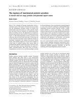
Cellulose-hemicellulose interactions - A nanoscale view
Bạn đang xem bản rút gọn của tài liệu. Xem và tải ngay bản đầy đủ của tài liệu tại đây (7.85 MB, 14 trang )
Carbohydrate Polymers 270 (2021) 118364
Contents lists available at ScienceDirect
Carbohydrate Polymers
journal homepage: www.elsevier.com/locate/carbpol
Cellulose-hemicellulose interactions - A nanoscale view
Ali Khodayari a, *, Wim Thielemans b, Ulrich Hirn c, Aart W. Van Vuure a, David Seveno a
a
Department of Materials Engineering, KU Leuven, Leuven, Belgium
Sustainable Materials Lab, Department of Chemical Engineering, KU Leuven, campus Kulak Kortrijk, Etienne Sabbelaan 53, 8500 Kortrijk, Belgium
c
Institute of Bioproducts and Paper Technology, TU Graz, Graz, Austria
b
A R T I C L E I N F O
A B S T R A C T
Keywords:
Cellulose
Hemicellulose
Free energy of adsorption
Shear
Hemicellulose folding on cellulose
Hydration effect
Molecular dynamics simulations
In this work, we study interactions of five different hemicellulose models, i.e. Galactoglucomannan, O-AcetylGalactoglucomannan, Fuco-Galacto-Xyloglucan, 4-O-Methylglucuronoxylan, and 4-O-Methylglucuronoar
abinoxylan, and their respective binding strength to cellulose nanocrystals by molecular dynamics simulations.
Glucuronoarabinoxylan showed the highest free energy of binding, whereas Xyloglucan had the lowest inter
action energies amongst the five models. We further performed simulated shear tests and concluded that failure
mostly happens at the inter-molecular interaction level within the hemicellulose fraction, rather than at the
interface with cellulose. The presence of water molecules seems to have a weakening effect on the interactions of
hemicellulose and cellulose, taking up the available hydroxyl groups on the surface of the cellulose for hydrogen
bonding. We believe that these studies can shed light on better understanding of plant cell walls, as well as
providing evidence on variability of the structures of different plant sources for extractions, purification, and
operation of biorefineries.
1. Introduction
Cellulose and its derivatives have been the target of numerous
experimental and numerical studies over the past decades because of
their excellent mechanical performance (Moon et al., 2011). Mechanical
properties of plant cell walls and hence elementary fibres are controlled
by several parameters including the structure of the secondary wall S2,
the microfibrillar angle (MFA), crystalline cellulose content, and the
ratio of other constituents such as hemicellulose, lignin, and pectin
(Eichhorn et al., 2010; Habibi et al., 2010). For instance, it is shown that
MFA inversely regulates the tensile behaviour of elementary fibres
(Bourmaud et al., 2013). In other words, fibres with lower MFA display
higher tensile moduli when stretched along the main axis. Moreover,
relative humidity (RH) has also proven to impose significant drifts on
the elastic modulus of plant fibres. While studies show that RH can lead
to swelling of fibres and according loss in mechanical strength, it is also
observed that certain percentages of RH can enhance the strength and
strain to failure of cellulosic fibres (Baley et al., 2005; Placet et al.,
2012).
Structural analysis of the plant cell wall proposes that the cell wall
assembly is mostly responsible for the mechanical properties of the fi
bres (Zhong et al., 2019). As an example, the secondary cell wall consists
of fibrils made up from cellulose nanofibrils, often referred to as cellu
lose microfibrils (CMF), which can be up to thousands of nanometers in
length, connected longitudinally through disordered regions (Khodayari
et al., 2021; Khodayari et al., 2020; Kontturi et al., 2016; Nishiyama
et al., 2003) and laterally through amorphous media including mostly
hemicellulose (Cosgrove, 2005; Gibson, 2012), as depicted in Fig. 1. The
content and ratio of the constituents can affect the moisture uptake, as
well as the mechanical interlocking of these constituents together. Due
to the alignment of the microfibrils along a director, the MFA, it is
proposed that when fibres are exposed to a tensile load, a shear force is
induced between the hemicellulose and the surface of the cellulose fi
brils (Placet et al., 2014). This shear force between the cellulose and
hemicellulose can be the reason for the non-linear shape of the stressstrain curves of elementary fibres.
One important matter of concern when studying interactions of cell
wall constituents, and resulting mechanical behaviour is water (Moore
et al., 2008; Tang et al., 1999). It has been shown that water-binding
capacity of cell walls can be modified by degradation of certain pectic
polysaccharide side-chains (Klaassen & Trindade, 2020). Water can alter
molecular conformations and mobility of pectic contents in the plant cell
wall. As minor changes of the rheological properties of the matrix, being
the stress transmitters between cellulose microfibrils, can lead to
* Corresponding author.
E-mail address: (A. Khodayari).
/>Received 7 April 2021; Received in revised form 14 June 2021; Accepted 17 June 2021
Available online 23 June 2021
0144-8617/© 2021 The Author(s). Published by Elsevier Ltd. This is an open access article under the CC BY license ( />
A. Khodayari et al.
Carbohydrate Polymers 270 (2021) 118364
modulations of the overall behaviour of plant cell walls (Ulvskov et al.,
2005), it is critical to account for the role of water on the structure of the
walls (Evered et al., 2007).
From a broader point of view, the type of hemicellulose could also
play a role in the strength of the cellulosic fibres. In particular, side
groups of the hemicellulose can make different types of bonds with the
free hydroxyl groups on the surface of the cellulose fibrils, provoking
specific shear behaviours. In terms of extraction of hemicellulose, this
leads to different yields and molecular weights depending on the plant
source (Gallina et al., 2018). The chemical composition of each hemi
cellulose and the ratio of its constituents depend on the plant species and
the stage of tissue development (Barbieri et al., 2017). The most abun
dant hemicellulose types are Xyloglucans (XyG), Glucuronoarabinox
ylans (GAX), Glucuronoxylans (GX), and Galactoglucomannan (GGM),
mostly present respectively in the primary cell walls of hardwood,
softwood, and grasses, in the secondary cell wall of grasses, in the sec
ondary cell wall of hardwood, and in the secondary cell wall of softwood
(Sorieul et al., 2016). The acetylated form of GGM (ACE-GGM) is
believed to be more abundant in softwoods (Hannuksela & Du Penhoat,
2004).
The diverse structures of XyG that have already been characterized
(Hsieh & Harris, 2009; Picard et al., 2000) are formed of β-D-glucopyr
anose (Glc) backbones, mostly branched with an α-D-xylopyranose (Xyl)
on the C6. The branched xylose units themselves also usually contain
galactosyl, fucosyls, or arabinosyl residues (Lerouxel et al., 2006; Pauly
et al., 2001). GAXs mainly consist of a β-D-xylopyranose backbone,
where α-L-arabinofuranosyl (Ara) residues are occasionally substituted
on the O3, and sometimes are branched with α-D-glucopyranosyl acid
(GlcA) on the O2, in a non-repeating manner. Ferulic acid is also
observed to reside on the arabinose groups (arabinofuranose) in a
random fashion, and the backbone is also acetylated to a minor extent
(Harris, 2006; Kozlova et al., 2012; Vogel, 2008). The Ara:Xyl ratio can
vary significantly as a function of the elongation phase or the plant
family. For instance the molar ratio of Xyl:Ara:GlcA was found to be
45:12:1 in wheat (Zeng et al., 2010), 100:28:8 in Guadua chacoensis
´ndez et al., 2019), or 100:67:8 in woody bamboo (Zelaya et al.,
(Ferna
2017). GX on the other hand has the same backbone, with more frequent
acetylations on either O2/O3 or both (Heinen et al., 2019). Minor 4-Omethyl-α-D-glucopyranosyl acids are substituted on the backbone as well
(de Carvalho et al., 2019). GGM consists of β-D-mannopyranose (Man)
and β-D-glucopyranose backbone, with occasional β-D-galactopyranose
(Gal) residues substituted on the O6 of the mannose units (Eichinger
et al., 2019). Acetylations in ACE-GGM occur on the mannose units, and
happen on either C2 or C3 (Berglund et al., 2019). The reported ratios
for Man:Glc:Gal also vary in different plant cells with Man being the
majority and Gal the minority in GGM structures (Barbieri et al., 2017;
Lundqvist et al., 2002; Sims et al., 1997; Yu et al., 2018). Note that,
despite concrete findings on the type and position of linkages and sub
stitutions on the backbone of each mentioned hemicellulose, the ratio of
the constituents in hemicelluloses differs from one plant to the other,
and also within one specific plant itself. For instance, it has been shown
that GAX substitution type and frequency of side chains differ within the
stem and leaf of grasses (Tryfona et al., 2019). In other words, compo
sition of the hemicelluloses has shown to be tissue specific (Pauly et al.,
2001). Degree of substitution and its pattern, playing an important role
on the functionality of hemicellulose models is also shown to differ in
plants, as well as their development stages (Scheller & Ulvskov, 2010) In
this study, we model the most abundant type of each hemicellulose from
the substitution point of view.
The Iβ cellulose crystal structure has been a source of debate for
years. Several structures, including different number of chains have
been already proposed. Proposed models for Iβ cellulose mainly include
18-chain (Kubicki et al., 2018; Nixon et al., 2016; Ros´
en et al., 2020;
Vandavasi et al., 2016; Zhong et al., 2019), 24-chain (Fernandes et al.,
2011; Willhammar et al., 2021), or 36-chain structures (Elazzouzihafraoui et al., 2008; Endler & Persson, 2011; Mutwil et al., 2008; C.
Zhang et al., 2021). Despite all, there are still discussions in the field,
whether one model should be favored over the other (Cosgrove, 2014;
Hill et al., 2014). Hill et al. (2014) did come up with a convincing
argument that from the possibilities of 12, 18, 24 and 36 chains, the 18
chain CMF was the most likely, while 36 would also be possible but less
probable, and that 12 and 24 would be the least likely to exist, also
supported by Cosgrove and Jarvis (2012). There is however also con
tradicting work. For instance, Wang and Hong (2016) provides NMR
Fig. 1. Structural hierarchy of plant fibres. Technical fibres are composed of elementary fibres. Elementary fibres consist of primary cell wall, secondary cell walls,
and a lumen at the core. Cellulose microfibrils connected through hemicellulose and lignin, mostly make up the secondary cell wall.
2
A. Khodayari et al.
Carbohydrate Polymers 270 (2021) 118364
evidence that the CMF consists of at least 24 chains in various wild type
and mutant Arabidopsis primary cell walls, which given the 18 enzyme
complex and the arguments of Hill et al. (2014) that 24 chains are not
possible might indicate 36 glucan chains crystals exist. To try to avoid
the complexity, we have chosen the hydrophilic surface of the 6 × 6
model to interact with hemicellulose, as the actual state of the surfaces
at the outer side of the crystals will be very similar to those of an 18chain model. Hence, the results of this study should remain intact,
whether a 36-chain or an 18-chain model is used.
Conformational analyses of crystalline cellulose and hemicellulose
have been already studied in detail by molecular dynamics simulations
in many studies (Berglund et al., 2020; Nishiyama et al., 2008; Nish
iyama et al., 2012). Flexibility of hemicelluloses has been a matter of
concern, as the molecular conformation, and consequently, function
ality of hemicelluloses would be directly affected. Berglund et al. (2016)
inspected the flexibility of different combinations of D-xylopyranosyl, Dmannopyranosyl, and D-glucopyranosyl units as di- and tetra
saccharides. The observation was that there are differences in the flex
ibility of different linkage types in their models. Particularly, the
backbone of the xylan seemed to show more flexibility that those seen in
cellulose, glucomannan, and xyloglucan. The higher flexibility is then
related to weaker interactions of xylan and cellulose, compared to glu
comannan and cellulose (Åkerholm & Salm´en, 2001).
From a modelling point of view, amongst the available force fields
parametrized to model carbohydrates, GLYCAM06 is shown to be a
proper choice to model cellulose and hemicellulose structures (Foley
et al., 2012). In a study performed by Matthews et al. (2012), authors
simulated Iβ crystalline cellulose structures with three different force
fields, all parametrized to model carbohydrates for near microseconds.
GLYCAM06 was shown to be capable of modelling cellulose, consistent
with experimentally observed structures.
It must be noted that, the complexes proposed in this study do not
aim to model the plant cell wall, as other important substances such as
pectin (McCann & Roberts, 1996) and lignin, which are inherently
determinant in defining the behaviour of the cell wall are not present
(Kang et al., 2019). These components, despite being minor in some
plant walls, can be crucial in determining the architecture, and even
´nchez et al.,
tually the mechanical behaviour of the cell walls (Moneo-Sa
2020). Moreover, we aim not to favor any particular plant cell wall
model, as it has been argued for quite a while that load-bearing segments
in the plant cell walls could be possibly formed of contacts made by
CMFs, bridged by hemicellulose matrices (Park & Cosgrove, 2012).
Therefore, our focus in this work is to model the shear behaviour of
cellulose and hemicellulose, and governing mechanisms controlling
their interactions.
We start by calculating and comparing the binding free energy be
tween five hemicellulose models and a cellulose nanocrystal (CNC) to
provide enough information on the way these hemicelluloses interact
with the surface of cellulosic fibrils. In particular, results of this study
tend to rationalize why different plant cell walls show different me
chanical properties, from the point of view of hemicellulose type con
tent. We further inspect the folding behaviour of hemicellulose models
onto cellulose hydrophilic surfaces and compare this with the folding
behaviour of the models in solvated states. Further, we investigate the
shear behaviour between CNCs and hemicellulose employing molecular
dynamics simulations. In particular, we model two types of shear: the
first model mimics the shear between two surfaces, one made of cellu
lose and the other made of hemicellulose, and the other model mimics
the situation where a CNC is pulled out of a hemicellulose bath,
mimicking the shear in elementary fibres. The relationship between the
observed stick-slip behaviour and the (de)formation of hydrogen bonds
is demonstrated and explained. These simulations are provided to shed
light on the internal mechanisms of natural fibres when mechanically
loaded. We believe this study could assist in better characterization and
modelling of the plant cell wall constituents and mechanisms of
deformation.
2. Materials and methods
2.1. Molecular dynamics simulations
Molecular dynamics simulations have been performed by GROnin
gen MAchine for Chemical Simulations (GROMACS) 2019.1 version
(van der Spoel et al., 2005). GLYCAM06 parameter sets (Kirschner et al.,
2008) from Amber (Case et al., 2018) are used, and conversions of the
Amber topology files to GROMACS format is done through Acpype py
thon code (Sousa Da Silva & Vranken, 2012). Leap-frog algorithm is
used to solve the Newton's equations of motion. Bonded hydrogens are
constrained throughout the ensemble and production runs with the
LINear Constraint Solver (LINCS) algorithm, which is performed to
speed up the calculations (Hess et al., 1997). All bonds were constrained
for the free energy calculations, allowing a time step of 2 fs. Verlet
scheme is used for neighbour searching with a cut-off of 1.4 nm for both
Coulombic and van der Waals interactions. Long range electrostatics are
treated by Particle Mesh Ewald (PME) method (Darden et al., 1993).
Nos´e-Hoover thermostat is used to keep the temperatures at 300 K with a
time constant of 1.0 (Hoover, 1985; Nos´
e, 1984), and Pressure coupling
is performed using Parrinello-Rahman barostat, applying isotropic
pressure at 1.0 bar with a time constant of 2.0, and compressibility of
4.5 × 10− 5 (Parrinello & Rahman, 1981). Periodic boundary conditions
are applied in all directions. Two box sizes were used for two sets of
simulations: a triclinic box of 17 × 17 × 30 nm3 for the first configu
rations, and a rhombic dodecahedron box of 25 nm, 25 nm, 17 nm being
v1(x), v2(y), and v3(z), respectively, and with v3(x) and v3(y) equal to
12.5 nm. TIP3P water molecules are used to solvate the simulation boxes
(Mark & Nilsson, 2001). Steepest descent minimization algorithm is
used to minimize the configurations. Molecular images in this work are
rendered by PyMOL (DeLano, 2009).
2.2. Cellulose model
6 × 6 Iβ crystalline cellulose models have been built according to the
crystallography data of (Nishiyama et al., 2002). Two finite degree of
polymerizations (DP) are used in these simulations, i.e. DP 10 for the
free energy calculations, and DP 30 for the second set of simulations
(Khodayari, Van Vuure, et al., 2020). Fig. 4a shows the CNC (DP 30)
structure.
2.3. Hemicellulose models
Five hemicellulose types are considered in this work, namely Gal
actoglucomannan (GGM), O-Acetyl-galactoglucomannan (ACE-GGM),
4-O-Methylglucuronoxylan (GX), 4-O-Methylglucuronoarabinoxylan
(GAX), and Fucogalactoxyloglucan (XyG). The hemicellulose models for
the free energy calculations all have 8 units of backbone (except for the
GGM model) as shown in Fig. 2. This is to eliminate length effects when
potential of mean forces are computed for each hemicellulose case.
Hence, comparison between the GGM free energy, and the rest of the
models might be biased in this work.
2.4. Free energy simulations
Free energy calculations are performed to measure the free energy of
(un)binding between each hemicellulose model and a CNC. A combi
nation of center-of-mass (COM) pulling and umbrella sampling is done
for this analysis (Lemkul & Bevan, 2010; Torrie & Valleau, 1977). The
CNC is considered to be short (10 DP), to decrease the computational
demand. Larger CNCs would have required bigger simulation box and
would not be cost-efficient to perform extended molecular dynamics.
Each hemicellulose model was positioned in the vicinity of the CNC,
on the (110) hydrophilic face with exposed hydroxyl groups (Besombes
& Mazeau, 2005), within the non-bonded cut-off range (<14 Å), and the
complex was put in the middle of a dodecahedron box. The box was
3
A. Khodayari et al.
Carbohydrate Polymers 270 (2021) 118364
Fig. 2. (a) GGM, (b) ACE-GGM, (c) GX, (d) GAX, and (e) XyG models. Glc: β-glucopyranose; Man: β-mannopyranose; Gal: β-galactopyranose; Ac: acetyl; Xyl:
β-xylopyranose; GlcA: α-glucopyranosyl acid; Met: methyl; Ara: α-L-arabinofuranose; Fuc: α-L-fucopyranose.
filled with water molecules and energy minimized. System was equili
brated in a NPT ensemble (constant pressure) for 100 ps, followed by a 2
ns production run. This ensures equilibration of energies, and attach
ment of the hemicellulose to the surface of the CNC. Hemicellulose was
then pulled out from the surface of the CNC in xyz (the reaction coor
dinate ξ) applying steered molecular dynamics (SMD). Each hemicel
lulose was pulled for 7 nm and 700 configurations were captured from
700 ps of SMD (every 1 ps of simulation), and COM-COM distances were
computed for each snapshot dropped out of the SMD trajectory. Equally
distanced snapshots with 0.2 nm COM-COM distances were chosen
leading to 36 configurations for each hemicellulose. More snapshots
were added further for the free energy calculations to ensure full overlap
of the probabilities, providing histograms covering all the reaction
coordinate.
Each chosen snapshot was sampled for different numbers of timesteps. Weighted histogram analysis method (WHAM) was used to
calculate the potential of mean forces (PMF) (Kumar et al., 1992). Free
energy ΔG was computed as the difference between the maximum and
the minimum of the PMF curve. In particular, snapshots for the umbrella
simulations of GGM complex were sampled for 135.5 ns, ACE-GGM for
130 ns, GX for 89.5 ns, GAX for 183.5 ns, and XyG for 170.5 ns, until the
convergence was insured. The first ns of the simulations was dropped
out for equilibration, whereas the rest of the sampling was considered
for the WHAM procedure. The sampling was stopped when free energy
was converged to a value, fluctuating with magnitudes lower than the
standard deviation calculated by the Bayesian bootstrapping of the
histograms (Hub et al., 2010). A full description of the convergence and
error analysis is brought in Section S.1 of the supplementary informa
tion. For this set of calculations a total of ca. 26.6 μs of umbrella sam
pling simulations was carried out. MD simulations (each snapshot) were
conducted on 9 CPU cores (Xeon 6104) and 1 GPU (Nvidia P100 SXM2)
leading to an average performance of 4.8 ns/day for the free energy
calculations.
4
A. Khodayari et al.
Carbohydrate Polymers 270 (2021) 118364
2.5. Cellulose-hemicellulose shear simulations
to the cellulose surface, i.e. for small reaction coordinates, and tend to
lessen in interaction strength when moving apart. The free energy of
(un)binding, ∆G, is calculated for each material as the difference be
tween the maximum and minimum of each curve and shown in Table 1.
According to the results, GAX shows the strongest interactions
amongst the five studied hemicelluloses, whereas XyG has the lowest
binding energy to cellulose. GGM displays a lower interaction strength
than GAX. Acetylations on the GGM model, i.e. ACE-GGM, seem to have
marginal effects on the binding free energy. Accordingly, three acety
lations on the backbone of the GGM model, lead to a decrease of only 3
kJ/mol in binding energy, weakening the interactions between cellulose
and ACE-GGM compared to that of the cellulose-GGM interaction
strength. However, it must be mentioned again that GGM with two
residues less might show lower free energy, if sampled. On the other
hand, adding arabinose substitutions on the GX backbone, alongside less
acetylations (model compound GAX), led to a significant increase in
interaction free energy of cellulose and GAX compared to GX. Results
here can be particularly interesting for extraction purposes of hemicel
lulose content from the plant cell wall. In other words, selective
extraction of hemicelluloses from plants can be performed considering
the insight on binding energies of different hemicellulose models. Our
results could confirm the observations by Tokoh et al. where they
showed that mannans are better in aggregating cellulose microfibrils
than xylans (Tokoh et al., 2002). Slightly stronger interaction forces
between (ACE-)GGM and cellulose compared to GX and cellulose ex
plains why (ACE-)GGM leads to better aggregation than GX. The abovementioned results could also explain the difference between the
enhanced mechanical properties of a group of samples, where CNFs
were mixed with GGM or XyG, producing all-polysaccharide composite
films (Lucenius et al., 2014). Particularly, addition of GGM has shown to
lead to a more profound increase in the tensile strength, Young's
modulus, and toughness of cellulosic composite films than XyG. In
another set of studies, authors investigated the effects of hemicellulose
composition on the interactions between the CMFs (Kumagai & Endo,
2020). Their experiments revealed that intensity of interactions between
CMFs was stronger in the presence of (ACE-)GGM, than in GX matrices.
On the other hand, it has been hypothesized more than a decade ago that
lower degrees of arabinose substitution on the backbone of xylose
should imply stronger cell wall properties (Ceusters et al., 2008). In
another similar argument, Harris proposed that a lower degree of sub
stitution can increase hydrogen bonds to cellulose microfibrils (Harris,
2006). However, our calculations provide evidence that these
The second set of simulations includes modelling the shear behaviour
of cellulosic fibrils and hemicellulose. A 30 DP CNC was positioned at
the center of a 15 × 15 × 15 nm3 box, and the box was randomly filled
by the 10 unit GGM model shown in Fig. 3. Simulations were repeated
three times to validate the shear behaviour. In particular, the CNC was
rotated along its axis, and the box was randomly filled to its maximum
capacity by 461, 463, and 473 GGM molecules. The configuration was
then positioned in the middle of a 17 × 17 × 30 nm3 box and the box was
solvated with water molecules. The system was energy minimized,
equilibrated in NVT ensemble (constant volume) for 1 ns, in NPT
ensemble for 2 ns, and a production run of 2 ns. Pulling the fibril out of
the hemicellulose bath was then performed under two conditions:
mimicking the shear of two surfaces of cellulose and hemicellulose by
restraining the hemicellulose molecules and pulling the cellulose with
respect to the center of the box; and pulling the cellulose out of the bath
without imposing any restraints, with respect to the hemicellulose. As
interactions between cellulose and hemicellulose is expected to be
stronger than inter-molecular interactions between hemicellulose mol
ecules, restraining the hemicellulose molecules can clearly illustrate
how hydrogen bonding can control the interactions between the CNC
and hemicellulose, without any creep occurring inside the hemicellulose
region. The latter case on the other hand, is to demonstrate the range of
forces acting in between hemicellulose molecules themselves in com
parison with the ones between the two molecules (see Fig. 4). Please
note that, the choice of GGM was made as it is one of the most abundant
types of hemicellulose in plant cell walls. The same configurations were
made and simulations were performed for GX as well, to show the
consistency of the observations.
To investigate the effects of water on the interactions between these
two substances, the same set of simulations was performed when water
molecules in the system were removed. The configuration was then
equilibrated for 2 ns, and the two pulling conditions were simulated.
3. Results and discussion
3.1. Free energy of (un)binding
The potential of mean forces (PMF) of unbinding hemicellulose
models, namely GGM, ACE-GGM, XyG, GX, and GAX are shown in Fig. 5.
Expectedly, hemicellulose models show strong interactions when closest
Fig. 3. Each hemicellulose is pulled starting from a binding configuration in xyz (along the reaction coordinate ξ).
5
Carbohydrate Polymers 270 (2021) 118364
A. Khodayari et al.
Fig. 4. (a) The lateral and cross-sectional view of the DP 30 CNC structure used in the simulations (b) The z and (c) xy view of the CNC embedded in the hemi
cellulose bath. Water molecules are not shown for clarity.
substitutions on GAX provoke stronger interactions between hemicel
lulose and cellulose than for GX.
Based on the above results, some remarks should be made. Free en
ergies reported here, exclusively represent the interaction energies be
tween cellulose and these specific hemicellulose models. For instance, it
has been shown how diverse XyG structures can be (Hsieh & Harris,
2009), and contribution of their chemical composition to free energy of
binding to cellulose is yet to be inspected. In a study performed by Zhang
et al. (2011), authors showed how variation of a single side chain can
deteriorate the structural properties of XyG, which we believe could
indirectly modify their affinity to cellulose. Zhao et al. (2014) inspected
interactions of XyG to different cellulose surfaces. Then, another factor
which can potentially modulate the free energy of binding is the cellu
lose surface on which hemicellulose is positioned. In particular,
depending on the composition of the hemicelluloses, while a specific
molecule might not bind well to a certain cellulose surface, its in
teractions with other surfaces can be more pronounced. An example of
such a behaviour was observed in lignin and cellulose interactions
(Besombes & Mazeau, 2005). According to those results, while a 20-unit
oligomer of lignin interacted the least with the hydrophobic (200) sur
face of cellulose compared to the other hydrophilic (110) and (1–10)
surfaces, a 10-unit lignin interacted the most with the (200) surface of
cellulose.
This would then open a whole new subject to see how other mech
anisms can alter the interactions within the plant cell wall. It can be a
valid question to see if the cellulose-hemicellulose interface is respon
sible in defining the overall mechanical properties of the fibrils, or the
interactions within the matrix itself. Furthermore, whether relative
humidity can induce significant shifts in the behaviour of the in
teractions within the hemicellulose fraction in the plant cell wall, or at
the interface of the cellulose and hemicellulose, can be relevant debates.
Results of the following sections try to answer these questions to some
extent for particular hemicellulose models.
Fig. 5. Potential of mean forces of unbinding for five hemicellulose models
from a cellulose nanocrystal surface. PMFs are at their maximum (here
expressed as negative value) when two species are bound together, and tend to
zero as hemicellulose is dissociated from the cellulose surface. ξ is the reaction
coordinate along which hemicellulose is separated from the surface of
the cellulose.
Table 1
Free energy of (un)binding hemicellulose models and cellulose nanocrystals, and
associated errors.
Hemicellulose model
ΔG [kJ/mol]
Err [kJ/mol]
GAX
GGM
ACE-GGM
GX
XyG
111.1
95.5
92.1
90.8
83.2
±1.9
±3.2
±1.5
±2.2
±3.4
3.2. Folding behaviour of hemicellulose onto cellulose
Folding behaviour of polysaccharides can be assessed through
6
A. Khodayari et al.
Carbohydrate Polymers 270 (2021) 118364
different methods, i.e. nuclear magnetic resonance (NMR) (Simmons
et al., 2016), preferential dihedral angles of the backbone (French et al.,
2021), Monte Carlo simulations (Levy et al., 1997), etc. It is particularly
shown that in β-(1-4)-linked glycans, a twofold helical screw (one 360◦
twist per two glycosidic bond) exists when summation of ϕ and ψ values
about the glycosidic linkage adds up to ~120◦ and glycosidic linkages
exhibit a threefold helical screw (one 360◦ twist per three glycosidic
bond) when the summation result is ~190◦ (left-handed screw) or ~50◦
(right-handed screw) (Busse-Wicher et al., 2014; French & Johnson,
2009; Mazeau et al., 2005). Here we evaluate the conformational
behaviour of hemicellulose models when solvated in water and when
bound to the hydrophilic surface of cellulose by the rotational angle
preferences around the glycosidic linkage. Fig. 6 shows the distribution
of ϕ + ψ values for all five models in this study, when solvated in water
in isolate form, and when bound to the surface of cellulose. The histo
grams are generated over the last 10 ns of the production runs. In
particular, not all the glycosidic linkages of the models show a perfect
threefold screw in water solution, as their behaviour is influenced by the
amount, size, and position of the side chain groups on the backbone.
Therefore, in each model only part of the chains are considered for
analysis. While majority of the models do not exhibit a permanent
twofold screw when bound to cellulose, the difference between con
formations they exhibit in isolation and when bound to cellulose is
distinguishable. In other words, all the models tend to show signs of
twofold helical screw conformations when adjacent to cellulose sur
faces. The reason is that different patterns of substitution on each
segment of the models interfere with the conformation of the adjacent
segments. This observation was well-discussed by Berglund et al.
(2020), where authors show how positioning of substitutions alters the
flexibility of different segments of hemicelluloses. Hence, these types of
analysis would be clearer, should the models only hold one type of
substitution at the same time, and be long enough to discard chain ends.
Additionally, a perfect two- or threefold helical screw in carbohydrate
chains is mainly seen for certain source materials with either unsub
stituted backbones, or when substitutions are not randomly positioned
on the backbone, having an evenly spaced distribution of substitutes on
the backbone of the hemicelluloses (Busse-Wicher et al., 2016). Uneven
distribution of acetylations has shown to disrupt twofold conformation
of xylan on the surface of cellulose, making its docking onto the hy
drophilic surface difficult (Grantham et al., 2017). Effect of substitution
can be seen for the threefold conformation of GGM model in isolated
form where only one galactose is substituted on the backbone. Berglund
et al. (2019), showed that presence of galactose stiffens the adjacent
glycosidic linkage within the glucomannan, which is reflected in slight
rigidity of our (ACE-)GGM model. (ACE-)GGM fluctuate between twoand threefold conformations due to presence of the galactose side
chains. Conformational behaviour of our GX model is comparable with
previous observations. Xylan is shown to exhibit a threefold helical
screw (being more flexible) in aqueous media, and a twofold helical
screw (being more restricted) when bonded to the surface of cellulose
(Simmons et al., 2016). Berglund et al. (2020) assessed the influence of
acetylation on the flexibility and solubility of mannans, and showed that
presence of acetylation on C2, C3, or both could noticeably alter the
flexibility of mannans. In particular, when substitutions take place on
C3, a more flexible conformation is expected, whereas, a substitution on
C2 leads to a more rigid, twofold conformation. The behaviour of sub
stitution on C3 was also similar to the case where a single residue was
simultaneously substituted at two different positions (C2 and C3). The
rigidity of chains increased when two adjacent residues were both
substituted (one on C2 and one on C3). This explains the threefold screw
on the GX/GAX end where acetyl groups are substituted on one single
residue, regardless of being isolated in solution or adhered on cellulose
(the left non-reducing end in Fig. 2c, linkages 4–7, see Section S.2,
Fig. S5). Slightly more rigidity is observed as a twofold screw on the GX/
GAX end where substitution is taken place on C2 and C3 from two
adjacent residues (right end of GX model in Fig. 2c, linkages 1–4).
Obviously, acetylation on C3 deteriorates formation of a stable two fold
screw imposed by the adjacent C2 substitution. Gupta et al. (2021) in a
recent work also showed that selective acetylation on xylans contributes
to their folding behaviour. Coherent with what seen for mannans,
acetylations on C3 leads to more flexible conformations, whereas C2
substitutions, irrespective of their nature, incorporate less flexibility,
and thus a twofold helical screw. This decrease in the flexibility is
interpreted to promote interaction with cellulose, and is reflected in the
flat conformation of the GX model on cellulose (see Fig. S3). Methylglucuronic acid was also shown to cause adoption of a threefold
conformation (Gupta et al., 2021), which explains the resistance to
twofold conformations in the middle of GX and GAX models. Therefore,
it can be concluded that for all hemicellulose models, acetylation, and in
particular different substitution types might also contribute to modu
lation of their adhesive properties. Moreover, the regioselectivity of
substitutions plays a major role in the behaviour of the hemicelluloses. It
must also be added that, our results suggest, aside from position of the
substitutions, helical conformation of hemicellulose models might be
affected by the size of the side chain residues, as seen for GAX and XyG,
where GAX cannot take a perfect two fold conformation on cellulose,
and XyG does not exhibit a perfect threefold conformation in aqueous
media, taking the most rigid conformation amongst all models (Fig. S4,
also see Levy et al., 1991). The low free energy of binding of XyG might
have arisen from its rigid backbone conformation (Picard et al., 2000).
Additionally, GAX and XyG models both struggle to perfectly lay on the
surface of cellulose, and high adsorption free energy of GAX compared
to XyG must majorly stem from the composition of the molecules rather
than mechanical interlocking. This proposes that, further investigation
must be carried out to see the effect of substitution regioselectivity, such
as acetyl groups, on the free energy of binding between cellulose and
hemicelluloses, as contribution of the chemical composition to binding
properties seems to be more pronounced than folding behaviour of the
hemicelluloses.
3.3. Stick-slip behaviour
To see how cellulose-hemicellulose surfaces interact when shear is
applied at the interface, movement of the GGM hemicellulose shown in
Fig. 4 was restrained as the cellulose crystal was pulled out of a hemi
cellulose bath. This guarantees the slippage to occur at the interface of
the crystal and the hemicellulose matrix. Simulations were done for a
system solvated in water in the first place. The required pulling force is
plotted versus the displacement of the cellulose in Fig. 7a. Force can be
seen to oscillate as the cellulose crystal is pulled out. The peak force
decreased with displacement of the crystal, because less interacting
surface between the CNC and the surrounding media is available as the
CNC is pulled out of the hemicellulose matrix. Normalizing the forces by
the interacting length of the crystal with the hemicellulose matrix
almost equalizes the force peaks, except for the first peak. This means
that the displaced crystal is not making the same amount of bonds as at
the beginning. To validate this behaviour, two other configurations with
random position of hemicellulose were also tested. The position of the
force peaks fully overlapped for all three cases, while their magnitude
was different, which corresponds to the amount of bonds between the
two phases. Moreover, the behaviour was found not to be hemicellulose
dependent. The behaviour was also observed when other hemicellulose
models were used (e.g. GX, GAX, or XyG). See Sections S.3 to S.6 of the
supplementary information for more details.
The stick-slip behaviour can be attributed to the hydrogen bonding
pattern at the interface, as also seen being the driving frictional force in
between cellulose nanocrystals (Zhang et al., 2021). To check the
hydrogen bonds, all possible donors and acceptors on both cellulose and
hemicellulose were considered, leading to 43 types of hydrogen bonds.
As shown in Fig. 7b, the hydrogen bonding pattern between the con
tacting parts of cellulose and the surrounding hemicellulose also oscil
lated with the same frequency, with a slight shift to lower displacements.
7
A. Khodayari et al.
Carbohydrate Polymers 270 (2021) 118364
Fig. 6. The frequency of the summation, ϕ + ψ for all the
hemicellulose models in water (left hand side plots,
named *-SOL) and when bound to the hydrophilic surface
of cellulose (right hand side plots, named *-CELL), where
* represents the name of the model. The numbers on top
right of each plot refer to the number of glycosidic link
ages considered for each model, where 1 is the first
glycosidic bond close to the reducing end and 7 would be
the last, adjacent to the non-reducing end of the hemi
cellulose. See Fig. S5 for more.
8
A. Khodayari et al.
Carbohydrate Polymers 270 (2021) 118364
Fig. 7. (a) Force-displacement curves and the normalized force per contact length. (b) Number of hydrogen bonds versus the displacement super-positioned on forcedisplacement trend. (c) A part of cellulose surface chains showing the distance of surface hydroxyl groups. All systems are solvated with water.
Fig. 8. (a) A simplified scheme showing that shear
can happen either at the interface of cellulose and
hemicellulose or within the hemicellulose matrix it
self. The phenomenon happens due to either a
decrease in the microfibrillar angle (θ), or presence of
any misalignment between fibrils, when they are
strained in the cell direction. (b) A simplified scheme
of the pulling out simulation to model the shear in the
hemicellulose matrix. (c) Force-displacement trend
required to apply shear to the system (pulling the
CNC out of the bath with no restraints).
9
A. Khodayari et al.
Carbohydrate Polymers 270 (2021) 118364
This is because hydrogen bonds tend to break before the force reaches its
maximum. As soon as a critical amount of hydrogen bonds is broken to
allow for displacement, the force decreases until new hydrogen bonds
reform at the interface between cellulose and hemicellulose. The trend
repeats itself with a smaller amount of hydrogen bonds and hence
smaller force as the CNC is pulled out further. Interestingly, the fre
quency of the peaks resembles the frequency of available donor/
acceptor pairs at the cellulose-hemicellulose interface. As hemicellulose
is restrained, it particularly makes similar bonding with available
oxygen-hydrogen pairs on the cellulose surface. The peak-peak distances
in Fig. 7a and b are on average 1.1 nm, equivalent to the length of a
cellobiose unit. In particular, Fig. 7c shows the distance between hy
droxyl groups on the surface of the cellulose, having a similar average of
1.1 nm.
In conclusion, moving a cellulosic surface along a hemicellulose
plane leads to a stick-slip movement due to formation, deformation, and
breaking of hydrogen bonds at the interface, governed by a cellobiose
length periodicity.
observed that indeed the interaction energies are higher both at the
interface and within the XyG matrix, when an aligned conformation is
considered. However, under any circumstances, the interactions at the
interface is noticeably larger than those in the matrix, which leads to the
fact that any possible failure in cell walls must first occur in the matrix.
See S.6 for figures and results.
3.5. Role of water
The models discussed in the previous sections were all solvated with
water. Water is expected to fill the gaps in the matrix, as well as at the
interface between cellulose and hemicellulose (Kulasinski et al., 2015).
To see how its presence can alter the behaviour or the magnitude of
forces, water was completely removed from the system, and the simu
lation box was equilibrated for 2 ns in constant temperature ensemble.
The same types of pulling were then applied and results are shown in
Fig. 9. According to Fig. 9a, the magnitude of forces are higher than
those for the case where water was present, i.e. ≈22,000 kJ/mol/nm in
solvated state compared to ≈27,000 kJ/mol/nm in the dry state. This is
also valid for hydrogen bond numbers being almost double the amount
than for the case where water is present (see Fig. 9b). The force required
to shear the matrix experienced also an increase, as depicted in Fig. 9c
(≈5000 kJ/mol/nm in the presence of water compared to ≈14,000 kJ/
mol/nm in a dry system). In other words, a higher force is required to
break the bonds at the interface of cellulose and hemicellulose, and also
within the matrix itself, in the absence of water. Considering GGM, there
is a slight increase in the force at the interface in the dry state, while
failure in the matrix takes place with a force more than twice that in the
solvated state. This is even slightly more pronounced when GAX is
considered (Figs. S8 and S9). In particular, the force required to break
the interfacial bonds in the presence of water is half of that in the dry
state. In the same manner, matrix failure in the presence of water hap
pens with a force almost one third of that when there is no water in the
system. These results evidence that water would not act as a bridge,
forming stronger bonds, but rather as a lubricant or plasticizer within
the matrix and at the interface (Hansen et al., 2011; Matveev et al.,
2000). These findings are in agreement with a large amount of in
vestigations on fibre mechanical properties at varying humidities
(Czibula et al., 2019; Ganser et al., 2014; Jajcinovic et al., 2018). This
observation should however be assessed by other simulations carried out
for different relative humidities, which can be investigated in further
works. It must also be noted that these comparisons are valid only when
two systems for the same hemicellulose are compared in the solvated
and the dry state. Quantitative comparison between the hemicellulose
models must not be made in this part of the study, as random generation
of the systems induces conformation and density variations between
models with different hemicellulose types, which could intrinsically
affect the results between different hemicelluloses.
3.4. Shear mechanism
As interactions between hemicellulose chains are expected to be
weaker than between hemicellulose and cellulose, due to fewer number
of available donor-acceptor pairs, shear failure should not occur at the
cellulose-hemicellulose interface. Cohesive failure can happen earlier
within the hemicellulose matrix, before adhesive failure at the interface.
Particularly, when elementary fibres are stretched, the micro-fibrillar
angle decreases (Baley, 2002). This leads to a shear movement be
tween the parallel cellulose microfibrils (see Fig. 8a). This shear is
mainly distributed in the matrix, with a weaker bonding system than at
the interface. To mimic this behaviour, the restraints on the configura
tion discussed in the previous section were removed from the hemicel
lulose, allowing it to reform as needed. The CNC was then pulled out of
the matrix while hemicellulose was pulled in the opposite direction (see
Fig. 8b). Fig. 8c shows the required force to remove the cellulose from
the hemicellulose bath. As can be seen, the force increases until it rea
ches a maximum at which point, the cellulose can be pulled out of the
hemicellulose matrix at a significantly lower force than the peak forces
seen in Fig. 7a. The force stays almost constant afterwards. This is due to
the fact that hemicellulose chains on the surface of cellulose remain
attached to the CNC, while bonds between the hemicellulose chains,
governed by smaller forces, tend to break sooner and more readily. Or in
other words, when elementary fibres are strained, the matrix fails first,
amplifying the importance of the hemicellulose inter-molecular binding
energy in the plant cell wall.
This behaviour is also shown to be hemicellulose-type independent
(see Sections S.3–S.6 from the supplementary information). On the other
hand, to compare the strength of the matrices in the plant cell wall, the
adsorption energies between hemicellulose molecules must be measured
and compared. Obviously, hemicellulose types with a higher intermolecular binding energy are expected to increase the mechanical
properties of the plant fibres. This would however be out of the scope of
this study, and will be investigated in further studies.
When studying plant cells assembly, disassembly, and plant expan
sion (Somerville et al., 2004), it is shown that arrangement of certain
hemicellulose models is expected to be in conformations where exten
sive hydrogen bonded networks control their interactions to cellulose
microfibrils. (Rose & Bennett, 1999). In particular, in dicots, XyG is
believed to cover the surface of the microfibrils, as well as providing
cross-links between them. This then provides another indication to
model structures where XyG is aligned around the surface of microfi
brils. This alignment can lead to two possible effects: an increase in
binding area, and a decrease in matrix entanglement. We hence
modelled two other structures where XyG is randomly inserted in the
simulation box, and where XyG models are all aligned alongside the
main axis of cellulose. After performing the same shear analysis, it is
4. Conclusions
We studied interactions of five hemicellulose models from the pri
mary and secondary cell walls of plants, namely Galactoglucomannan,
O-Acetyl-Galactoglucomannan, Fuco-Galacto-Xyloglucan, 4-O-Methyl
glucuronoxylan, and 4-O-Methylglucuronoarabinoxylan with cellulose
nanocrystals by molecular dynamics simulations. We measured free
energies of (un)binding by performing extensive molecular dynamics
simulations and showed that GAX has the strongest binding energy to
the hydrophilic surface of cellulose. GGM and ACE-GGM are slightly
weaker than GAX, whereas GX and XyG showed the lowest adsorption
free energy. Accordingly, acetylations seem to weaken the interactions
between hemicellulose and cellulose, by decreasing the number of
available hydroxyl groups for hydrogen bonding. On the other hand, in
contrast to the postulations of the role of arabinose substitutions in xy
lans, arabinose groups on the backbone of GAX tend to enhance the
interactions with cellulose. Our results agree with experimental
10
A. Khodayari et al.
Carbohydrate Polymers 270 (2021) 118364
Fig. 9. Results of removing water for (a) force-displacement curves and the normalized force per contact length, (b) number of hydrogen bonds versus the
displacement super-positioned on force-displacement trend, and (c) force-displacement trend required to apply shear to the system (pulling out the CNC out of the
bath with no restraints).
observations in the literature, explaining the differences in adhesion
force between cellulose and different hemicelluloses. Furthermore,
folding behaviour of the hemicellulose is inspected in absence and
presence of cellulose. Accordingly, majority of the models show enough
flexibility as a threefold helical screw when solvated in water, while
different segments are affected by the substitutions of neighbouring
residues. In particular, models tend to take threefold screw conforma
tion when substitutions take place on C3, and exhibit more rigidity when
backbone C2 is substituted. Analysis of folding conformations becomes
challenging when substitutions on C2 and C3 take place on single resi
dues, or positioned next to each other on two respective residues. GX/
GAX take threefold conformations in solvated states, and show signs of
rigidity when close to cellulose surfaces. XyG seems to show more ri
gidity both in isolated form and when adhered onto cellulose. This
stiffness of the backbone might contribute to XyG's lower affinity to
cellulose. Longer models and evenly spaced substitution patterns are
required to see more coherent folding behaviour, as random substitutes
seem to diverge the models to take perfect and permanent two- or
threefold conformations. More studies on the effect of substitutions
regioselectivity on the free energy of binding between cellulose and
particular models can be promising. Moreover, we illustrated that there
is a stick-slip behaviour when a cellulose and a hemicellulose surface
slide over each other, governed by the periodicity of the hydroxyl groups
on the surface of the cellulose crystals. Exhibiting large interaction
forces at the interface of cellulose and hemicellulose provokes shear
within the hemicellulose matrix surrounding the cellulose crystals. In
other words, this study shows that shear in the plant cell walls would
occur mostly within the hemicellulose matrix rather than at the interface
between cellulose and hemicellulose. This is due to weaker interaction
forces between hemicellulose molecules, i.e. weaker cohesion, than at
the interface between cellulose and hemicellulose, i.e. adhesion.
Furthermore, we demonstrated that water acts as a lubricant at the
interface as well as within the hemicellulose amorphous phase. In
submerged conditions, water can fill the gaps within the hemicellulose
matrix, as well as at the interface between the hemicellulose and cel
lulose molecules by forming hydrogen bonds, weakening the in
teractions of the constituents in the plant cell wall. These latter
simulations could explain the lower mechanical properties of xylans
with higher degrees of substitution in the plant cell walls. It must be
noted that, the conclusions made in this study are mainly based on the
assumption of a 6 × 6 cellulose crystal structure. While the interactions
of hemicellulose with the hydrophilic surface of cellulose will remain
intact, should the structure of the models change, possible alternation of
the binding free energy values can be expected. The conclusions on the
shear behaviour as well, most likely remain intact, as interactions at the
interface would noticeably be larger than those within the matrix, as
shown. These results can be used for extraction purposes of different
hemicelluloses from plant sources, purifications, and be applied in bio
refineries, as well as illustrate the inter-molecular mechanisms con
trolling the mechanical strength of the plant cell wall, in the presence or
absence of water.
CRediT authorship contribution statement
A.K. and D.S. conceived and designed the study; A.K. performed the
simulations and carried out the post-processes; all authors analyzed the
data; all authors contributed to write the paper.
Declaration of competing interest
Authors declare no conflict of interest.
Acknowledgement
The authors are grateful to Dr. Justin Lemkul for his invaluable
discussions. The computational resources and services used in this work
11
A. Khodayari et al.
Carbohydrate Polymers 270 (2021) 118364
were provided by the VSC (Flemish Supercomputer Center), funded by
the Research Foundation - Flanders (FWO) and the Flemish Government
– department EWI. This project has received funding from the European
Union's Horizon 2020 research and innovation programme under the
Marie Skłodowska-Curie grant agreement No 764713, project FibreNet.
Czibula, C., Ganser, C., Seidlhofer, T., Teichert, C., & Hirn, U. (2019). Transverse
viscoelastic properties of pulp fibers investigated with an atomic force microscopy
method. Journal of Materials Science, 54(17), 11448–11461. />10.1007/s10853-019-03707-1
Darden, T., York, D., & Pedersen, L. (1993). Particle mesh Ewald: An Nlog(N) method for
Ewald sums in large systems. The Journal of Chemical Physics, 98, 10089–10092.
DeLano, W. (2009). The PyMOL molecular graphics system. Palo Alto, CA: DeLano
Scientific LLC.
Eichhorn, S. J., Dufresne, A., Aranguren, M., Marcovich, N. E., Capadona, J. R.,
Rowan, S. J., … Peijs, T. (2010). Review: Current international research into cellulose
nanofibres and nanocomposites (Vol. 45). No. 1.
Eichinger, T., Rahkila, J., Willfӧr, S., & Xu, C. (2019). TEMPO-oxidized O-acetyl
galactoglucomannan oligomers: Isolation and comprehensive structural elucidation.
Wood Science and Technology, 53(1), 71–85. />Elazzouzi-hafraoui, S., Nishiyama, Y., Putaux, J.-l., Heux, L., Dubreuil, F., & Rochas, C.
(2008). The shape and size distribution of crystalline nanoparticles prepared by acid
hydrolysis of native cellulose. Biomacromolecules, 9, 57–65.
Endler, A., & Persson, S. (2011). Cellulose synthases and synthesis in arabidopsis.
Molecular Plant, 4(2), 199–211. />Evered, C., Majevadia, B., & Thompson, D. S. (2007). Cell wall water content has a direct
effect on extensibility in growing hypocotyls of sunflower (Helianthus annuus L.).
Journal of Experimental Botany, 58(12), 3361–3371. />erm183
Fernandes, A. N., Thomas, L. H., Altaner, C. M., Callow, P., Forsyth, V. T.,
Apperley, D. C., … Jarvis, M. C. (2011). Nanostructure of cellulose microfibrils in
spruce wood. Proceedings of the National Academy of Sciences of the United States of
America, 108(47). />Fern´
andez, P. V., Zelaya, V. M., Cobello, L., Vega, A. S., & Ciancia, M. (2019).
Glucuronoarabinoxylans and other cell wall polysaccharides from shoots of Guadua
chacoensis obtained by extraction in different conditions. Carbohydrate Polymers, 226
(May), Article 115313. />Foley, B. L., Tessier, M. B., & Woods, R. J. (2012). Carbohydrate force fields: A review.
Wiley Interdisciplinary Reviews: Computational Molecular Science, 2(4), 652–697.
/>French, A. D., & Johnson, G. P. (2009). Cellulose and the twofold screw axis: Modeling
and experimental arguments. Cellulose, 16(6), 959–973. />s10570-009-9347-4
French, A. D., Montgomery, D. W., Prevost, N. T., Edwards, J. V., & Woods, R. J. (2021).
Comparison of cellooligosaccharide conformations in complexes with proteins with
energy maps for cellobiose. Carbohydrate Polymers, 264, 118004. />10.1016/j.carbpol.2021.118004, 15 July 2021.
´ G´renman, H., Biasi, P., Gar´cıa-Serna, J., & Salmi, T. (2018).
Gallina, G., Cabeza, A.,
Hemicellulose extraction by hot pressurized water pretreatment at 160 ◦ C for 10
different woods: Yield and molecular weight. Journal of Supercritical Fluids, 133
(September 2017), 716–725. />Ganser, C., Hirn, U., Rohm, S., Schennach, R., & Teichert, C. (2014). AFM
nanoindentation of pulp fibers and thin cellulose films at varying relative humidity.
Holzforschung, 68(1), 53–60. />Gibson, L. J. (2012). The hierarchical structure and mechanics of plant materials. Journal
of the Royal Society Interface, 9(76), 2749–2766. />rsif.2012.0341
Grantham, N. J., Wurman-Rodrich, J., Terrett, O. M., Lyczakowski, J. J., Stott, K.,
Iuga, D., … Dupree, P. (2017). An even pattern of xylan substitution is critical for
interaction with cellulose in plant cell walls. Nature Plants, 3(11), 859–865. https://
doi.org/10.1038/s41477-017-0030-8
Gupta, M., Rawal, T. B., Dupree, P., Smith, J. C., & Petridis, L. (2021). Spontaneous
rearrangement of acetylated xylan on hydrophilic cellulose surfaces. Cellulose, 28(6),
3327–3345. />Habibi, Y., Lucia, L. A., & Rojas, O. J. (2010). Cellulose nanocrystals: Chemistry, selfassembly, and applications. Chemical Reviews, 110, 3479–3500.
Hannuksela, T., & Du Penhoat, C. H. (2004). NMR structural determination of dissolved
O-acetylated galactoglucomannan isolated from spruce thermomechanical pulp.
Carbohydrate Research, 339(2), 301–312. />carres.2003.10.025
Hansen, S. L., Ray, P. M., Karlsson, A. O., Jorgensen, B., Borkhardt, B., Petersen, B. L., &
Ulvskov, P. (2011). Mechanical properties of plant cell walls probed by relaxation
spectra. Plant Physiology, 155(1), 246–258. />Harris, P. J. (2006). Primary and secondary plant cell walls: A comparative overview.
New Zealand Journal of Forestry Science, 36(1), 36–53.
Heinen, P. R., Betini, J. H., & Polizeli, M. L. (2019). Xylanases. In Encyclopedia of
microbiology (pp. 604–615). />Hess, B., Bekker, H., Berendsen, H., & Fraaije, J. (1997). LINCS: A linear constraint solver
for molecular simulations. Journal of Computational Chemistry, 18, 1463–1472.
Hill, J. L., Hammudi, M. B., & Tien, M. (2014). The Arabidopsis cellulose synthase
complex: A proposed hexamer of CESA trimers in an equimolar stoichiometry. The
Plant Cell Online, 26(12), 4834–4842. />Hoover, W. (1985). Canonical dynamics: Equilibrium phase-space distributions. Physical
Review A, 31, 1695–1697.
Hsieh, Y. S., & Harris, P. J. (2009). Xyloglucans of monocotyledons have diverse
structures. Molecular Plant, 2(5), 943–965. />Hub, J. S., De Groot, B. L., & Van Der Spoel, D. (2010). G-whams-a free weighted
histogram analysis implementation including robust error and autocorrelation
estimates. Journal of Chemical Theory and Computation, 6(12), 3713–3720. https://
doi.org/10.1021/ct100494z
Data availability
All data that support this study are kept available at KU Leuven and
can be accessed upon request from the corresponding author.
Appendix A. Supplementary data
Supplementary data to this article can be found online at https://doi.
org/10.1016/j.carbpol.2021.118364.
References
Åkerholm, M., & Salm´en, L. (2001). Interactions between wood polymers studied by
dynamic FT-IR spectroscopy. Polymers, 42(3), 963–969. />S0032-3861(00)00434-1
Baley, C. (2002). Analysis of the flax fibres tensile behaviour and analysis of the tensile
stiffness increase. Composites - Part A: Applied Science and Manufacturing, 33(7),
939–948. />Baley, C., Morvan, C., & Grohens, Y. (2005). Influence of the absorbed water on the
tensile strength of flax fibers. Macro-molecular Symposia, 222, 195–201. https://doi.
org/10.1002/masy.200550425
Barbieri, S. F., Ruthes, A. C., Petkowicz, C. L.d. O., de Godoy, R. C. B., Sassaki, G. L.,
Santana Filho, A. P., & Silveira, J. L. M. (2017). Extraction, purification and
structural characterization of a galactoglucomannan from the gabiroba fruit
(Campomanesia xanthocarpa Berg), Myrtaceae family. Carbohydrate Polymers, 174,
887–895. />Berglund, J., Angles d’Ortoli, T., Vilaplana, F., Widmalm, G., Bergenstråhle-Wohlert, M.,
Lawoko, M., … Wohlert, J. (2016, Oct). A molecular dynamics study of the effect of
glycosidic linkage type in the hemicellulose backbone on the molecular chain
flexibility. The Plant Journal, 88(1), 56–70. />Berglund, J., Azhar, S., Lawoko, M., Lindstrӧm, M., Vilaplana, F., Wohlert, J., &
Henriksson, G. (2019). The structure of galactoglucomannan impacts the
degradation under alkaline conditions. Cellulose, 26, 2155–2175. />10.1007/s10570-018-1737-z
Berglund, J., Kishani, S., Morais De Carvalho, D., Lawoko, M., Wohlert, J.,
Henriksson, G., … Vilaplana, F. (2020). Acetylation and sugar composition influence
the (in)solubility of plant β-mannans and their interaction with cellulose surfaces.
ACS Sustainable Chemistry and Engineering, 8(27), 10027–10040. />10.1021/acssuschemeng.0c01716
Besombes, S., & Mazeau, K. (2005). The cellulose/lignin assembly assessed by molecular
modeling. Part 1: Adsorption of a threo guaiacyl β-O-4 dimer onto a Iβ cellulose
whisker. Plant Physiology and Biochemistry, 43(3), 299–308. />10.1016/j.plaphy.2005.02.005
Bourmaud, A., Morvan, C., Bouali, A., Placet, V., Per´re, P., & Baley, C. (2013).
Relationships between micro-fibrillar angle, mechanical properties and biochemical
composition of flax fibers. Industrial Crops and Products, 44, 343–351. https://doi.
org/10.1016/j.indcrop.2012.11.031
Busse-Wicher, M., Gomes, T. C., Tryfona, T., Nikolovski, N., Stott, K., Grantham, N. J., …
Dupree, P. (2014). The pattern of xylan acetylation suggests xylan may interact with
cellulose microfibrils as a twofold helical screw in the secondary plant cell wall of
Arabidopsis thaliana. Plant Journal, 79(3), 492–506. />tpj.12575
Busse-Wicher, M., Li, A., Silveira, R. L., Pereira, C. S., Tryfona, T., Gomes, T. C. F., …
Dupree, P. (2016). Evolution of xylan substitution patterns in gymnosperms and
angiosperms: Implications for xylan interaction with cellulose. Plant Physiology, 171
(August), 00539, 2016 />de Carvalho, D. M., Berglund, J., Marchand, C., Lindstrӧm, M. E., Vilaplana, F., &
Sevastyanova, O. (2019). Improving the thermal stability of different types of xylan
by acetylation. Carbohydrate Polymers, (May), 132–140. />carbpol.2019.05.063, 220.
Case, D. A., Ben-Shalom, I. Y., Brozell, S. R., Cerutti, D. S., E, C. T., Cruzerio, V. W. D., …
Kollman, P. A. (2018). AMBER 2018. San Francisco: University of California.
Ceusters, J., Londers, E., Brijs, K., Delcour, J. A., & De Proft, M. P. (2008).
Glucuronoarabinoxylan structure in the walls of Aechmea leaf chlorenchyma cells is
related to wall strength. Phytochemistry, 69(12), 2307–2311. />10.1016/j.phytochem.2008.06.002
Cosgrove, D. J. (2005). Growth of the plant cell wall. Nature Reviews Molecular Cell
Biology, 6(11), 850–861. />Cosgrove, D. J. (2014). Re-constructing our models of cellulose and primary cell wall
assembly. Current Opinion in Plant Biology, 22, 122–131. />pbi.2014.11.001
Cosgrove, D. J., & Jarvis, M. C. (2012). Comparative structure and biomechanics of plant
primary and secondary cell walls. Frontiers in Plant Science, 3(AUG), 1–6. https://doi.
org/10.3389/fpls.2012.00204
12
Carbohydrate Polymers 270 (2021) 118364
A. Khodayari et al.
Jajcinovic, M., Fischer, W. J., Mautner, A., Bauer, W., & Hirn, U. (2018). Influence of
relative humidity on the strength of hardwood and softwood pulp fibres and fibre to
fibre joints. Cellulose, 25(4), 2681–2690. />Kang, X., Kirui, A., Dickwella Widanage, M. C., Mentink-Vigier, F., Cosgrove, D. J., &
Wang, T. (2019). Lignin-polysaccharide interactions in plant secondary cell walls
revealed by solid-state NMR. Nature Communications, 10(1), 1–9. />10.1038/s41467-018-08252-0
Khodayari, A., Hirn, U., Spirk, S., Van Vuure, A. W., & Seveno, D. (2021).
Recrystallization and size distribution of dislocated segments in cellulose
microfibrils - a molecular dynamics perspective. Cellulose. />s10570-021-03906-7, 27 May 2021.
Khodayari, A., Hirn, U., Van Vuure, A., & Seveno, D. (2020). Inverse rule of mixtures at
the nanoscale: Prediction of elastic properties of cellulose nanofibrils. Composites
Part A: Applied Science and Manufacturing, 138(July), Article 106046. https://doi.
org/10.1016/j.compositesa.2020.106046
Khodayari, A., Van Vuure, A. W., Hirn, U., & Seveno, D. (2020). Tensile behaviour of
dislocated/crystalline cellulose fibrils at the nano scale. Carbohydrate Polymers, 235
(October 2019), Article 115946. />Kirschner, K. N., Yongye, A. B., Tschampel, S. M., Gonz´
alez-Outeirin˜
o, J., Daniels, C. R.,
Foley, B. L., & Woods, R. J. (2008). GLYCAM06: A generalizable biomolecular force
field. Carbohydrates. Journal of Computational Chemistry, 29, 622–655.
Klaassen, M. T., & Trindade, L. M. (2020). RG-I galactan side-chains are involved in the
regulation of the water-binding capacity of potato cell walls. Carbohydrate Polymers,
227(June 2019), Article 115353. />Kontturi, E., Meriluoto, A., Penttilă
a, P. A., Baccile, N., Malho, J. M., Potthast, A., …
Sixta, H. (2016). Degradation and crystallization of cellulose in hydrogen chloride
vapor for high-yield isolation of cellulose nanocrystals. Angewandte Chemie International Edition, 55(46), 14455–14458. />anie.201606626
Kozlova, L. V., Mikshina, P. V., & Gorshkova, T. A. (2012). Glucuronoarabinoxylan
extracted by treatment with endoxylanase from different zones of growing maize
root. Biochemistry (Moscow), 77(4), 395–403. />S0006297912040116
Kubicki, J. D., Yang, H., Sawada, D., O’Neill, H., Oehme, D., & Cosgrove, D. (2018). The
shape of native plant cellulose microfibrils. Scientific Reports, 8(1), 4–11. https://doi.
org/10.1038/s41598-018-32211-w
Kulasinski, K., Guyer, R., Derome, D., & Carmeliet, J. (2015). Water adsorption in wood
microfibril-hemicellulose system: Role of the crystalline-amorphous interface.
Biomacromolecules, 16(9), 2972–2978. />biomac.5b00878
Kumagai, A., & Endo, T. (2020). Effects of hemicellulose composition and content on the
interaction between cellulose nanofibers. Cellulose, 28(1), 259–271. />10.1007/s10570-020-03530-x
Kumar, S., Rosenberg, J. M., Bouzida, D., Swendsen, R. H., & Kollman, P. A. (1992). The
weighted histogram analysis method for free-energy calculations on biomolecules. I.
The method. Journal of Computational Chemistry, 13(8), 1011–1021. />10.1002/jcc.540130812
Lemkul, J. A., & Bevan, D. R. (2010). Assessing the stability of Alzheimer’s amyloid
protofibrils using molecular dynamics. Journal of Physical Chemistry B, 114(4),
1652–1660. />Lerouxel, O., Cavalier, D. M., Liepman, A. H., & Keegstra, K. (2006). Biosynthesis of plant
cell wall polysaccharides - A complex process. Current Opinion in Plant Biology, 9(6),
621–630. />Levy, S., Maclachlan, G., & Staehelin, L. A. (1997). Xyloglucan sidechains modulate
binding to cellulose during in vitro binding assays as predicted by conformational
dynamics simulations. Plant Journal, 11(3), 373–386. />j.1365-313X.1997.11030373.x
Levy, S., York, W. S., Stuike-Prill, R., Meyer, B., & Staehelin, A. (1991). Simulations of the
static and dynamic molecular conformations of xyloglucan. The sole of the
fucosylated sidechain in surface-specific sidechain folding. The Plant Journal, 1(2),
195215.
ă
Lucenius, J., Parikka, K., & Osterberg,
M. (2014). Nanocomposite films based on
cellulose nanofibrils and water-soluble polysaccharides. Reactive and Functional
Polymers, 85, 167–174. />Lundqvist, J., Teleman, A., Junel, L., Zacchi, G., Dahlman, O., Tjerneld, F., &
St◦ albrand, H. (2002). Isolation and characterization of galactoglucomannan from
spruce (Picea abies). Carbohydrate Polymers, 48(1), 29–39. />S0144-8617(01)00210-7
Mark, P., & Nilsson, L. (2001). Structure and dynamics of the tip3p, spc, and spc/e water
models at 298 k. The Journal of Physical Chemistry A, 105(43), 9954–9960.
Matthews, J. F., Beckham, G. T., Bergenstr◦ ahle-Wohlert, M., Brady, J. W.,
Himmel, M. E., & Crowley, M. F. (2012). Comparison of cellulose Iβ simulations with
three carbohydrate force fields. Journal of Chemical Theory and Computation, 8(2),
735–748. />Matveev, Y. I., Grinberg, V. Y., & Tolstoguzov, V. B. (2000). The plasticizing effect of
water on proteins, polysaccharides and their mixtures. Glassy state of biopolymers,
food and seeds. Food Hydrocolloids, 14(5), 425–437. />Mazeau, K., Moine, C., Krausz, P., & Gloaguen, V. (2005). Conformational analysis of
xylan chains. Carbohydrate Research, 340(18), 2752–2760. />j.carres.2005.09.023
McCann, M., & Roberts, K. (1996). Plant cell wall architecture: The role of pectins.
Progress in Biotechnology, 14, 91–107.
Moneo-S´
anchez, M., Vaquero-Rod´rıguez, A., Hern´
andez-Nistal, J., Albornos, L., Knox, P.,
́ I. (2020). Pectic galactan affects cell wall architecture during
Dopico, B., … Martın,
secondary cell wall deposition. Planta, 251(5), 1–15. Retrieved from doi:10.1007/
s00425-020-03394-2 />Moon, R. J., Martini, A., Nairn, J., Simonsen, J., & Youngblood, J. (2011). Cellulose
nanomaterials review: Structure, properties and nanocomposites. Chemical Society
Reviews, 40(7), 3941–3994.
Moore, J. P., Vic´re-Gibouin, M., Farrant, J. M., & Driouich, A. (2008). Adaptations of
higher plant cell walls to water loss: Drought vs desiccation. Physiologia Plantarum,
134(2), 237–245. />Mutwil, M., Debolt, S., & Persson, S. (2008). Cellulose synthesis: a complex complex.
Current Opinion in Plant Biology, 11(3), 252–257. />pbi.2008.03.007
Nishiyama, Y., Johnson, G. P., & French, A. D. (2012). Diffraction from nonperiodic
models of cellulose crystals. Cellulose, 19(2), 319–336. />s10570-012-9652-1
Nishiyama, Y., Johnson, G. P., French, A. D., Forsyth, V. T., & Langan, P. (2008). Neutron
crystallography, molecular dynamics, and quantum cellulose I. Biomacromolecules, 9
(11), 3133–3140. />Nishiyama, Y., Kim, U. J., Kim, D. Y., Katsumata, K. S., May, R. P., & Langan, P. (2003).
Periodic disorder along ramie cellulose microfibrils. Biomacromolecules, 4(4),
1013–1017. />Nishiyama, Y., Langan, P., & Chanzy, H. (2002). Crystal structure and hydrogen-bonding
system in cellulose Iβ from synchrotron X-ray and neutron fiber diffraction. Journal
of the American Chemical Society, 124(31), 9074–9082.
Nixon, B. T., Mansouri, K., Singh, A., Du, J., Davis, J. K., Lee, J. G., … Haigler, C. H.
(2016). Comparative structural and computational analysis supports eighteen
cellulose synthases in the plant cellulose synthesis complex. Scientific Reports, 6
(March), 1–14. />Nos´
e, S. (1984). A molecular dynamics method for simulations in the canonical
ensemble. Journal of Molecular Physics, 52, 255–268.
Park, Y. B., & Cosgrove, D. J. (2012). A revised architecture of primary cell walls based
on biomechanical changes induced by substrate-specific endoglucanases. Plant
Physiology, 158(4), 1933–1943. />Parrinello, M., & Rahman, A. (1981). Polymorphic transitions in single crystals: A new
molecular dynamics method. Journal of Applied Physics, 52, 7182–7190.
Pauly, M., Qin, Q., Greene, H., Albersheim, P., Darvill, A., & York, W. S. (2001). Changes
in the structure of xyloglucan during cell elongation. Planta, 212(5–6), 842–850.
/>Picard, C., Gruza, J., Derouet, C., Renard, C. M., Mazeau, K., Koca, J., & Herv´e Du
Penhoat, C. (2000). A conformational study of the xyloglucan oligomer, XXXG, by
NMR spectroscopy and molecular modeling. Biopolymers, 54(1), 11–26. https://doi.
org/10.1002/(SICI)1097-0282(200007)54:1¡11::AID-BIP20¿3.0.CO;2-D
Placet, V., Cisse, O., & Boubakar, M. L. (2012). Influence of environmental relative
humidity on the tensile and rotational behaviour of hemp fibres. Journal of Materials
Science, 47(7), 3435–3446. />Placet, V., Ciss´e, O., & Lamine Boubakar, M. (2014). Nonlinear tensile behaviour of
elementary hemp fibres. Part I: Investigation of the possible origins using repeated
progressive loading with in situ microscopic observations. Composites Part A: Applied
Science and Manufacturing, 56, 319–327. />compositesa.2012.11.019
Rose, J. K., & Bennett, A. B. (1999). Cooperative disassembly of the cellulose-xyloglucan
network of plant cell walls: Parallels between cell expansion and fruit ripening.
Trends in Plant Science, 4(5), 176–183. />01405-3
Ros´
en, T., He, H. R., Wang, R., Zhan, C., Chodankar, S., Fall, A., … Hsiao, B. S. (2020).
Cross-sections of nanocellulose from wood analyzed by quantized polydispersity of
elementary microfibrils. ACS Nano, 14(12), 16743–16754. />acsnano.0c04570
Scheller, H. V., & Ulvskov, P. (2010). Hemicelluloses. Annual Review of Plant Biology, 61,
263–289. />Simmons, T. J., Mortimer, J. C., Bernardinelli, O. D., Pă
oppler, A. C., Brown, S. P.,
DeAzevedo, E. R., Dupree, P. (2016). Folding of xylan onto cellulose fibrils in
plant cell walls revealed by solid-state NMR. Nature Communications, 7, 13902.
/>Sims, I. M., Craik, D. J., & Bacic, A. (1997). Structural characterisation of
galactoglucomannan secreted by suspension-cultured cells of Nicotiana
plumbaginifolia. Carbohydrate Research, 303(1), 79–92. />S0008-6215(97)00144-4
Somerville, C., Bauer, S., Brininstool, G., Facette, M., Hamann, T., Milne, J., …
Youngs, H. (2004). Toward a systems approach to understanding plant cell walls.
Science, 306(5705), 2206–2211. />Sorieul, M., Dickson, A., Hill, S. J., & Pearson, H. (2016). Plant fibre: Molecular structure
and biomechanical properties, of a complex living material, influencing its deconstruction
towards a biobased composite (Vol. 9). No. 8.
Sousa Da Silva, A. W., & Vranken, W. F. (2012). ACPYPE - AnteChamber PYthon Parser
interfacE. BMC Research Notes, 5(367).
van der Spoel, Y., Lindahl, E., Hess, B., Groenho, G., Mark, A. E., & Berendsen, H. J. C.
(2005). GROMACS: Fast, flexible and free. Journal of Computer Chemistry, 19(6),
1701–1718.
Tang, H., Belton, P. S., Ng, A., & Ryden, P. (1999). 13C MAS NMR studies of the effects of
hydration on the cell walls of potatoes and Chinese water chestnuts. Journal of
Agricultural and Food Chemistry, 47(2), 510–517. />Tokoh, C., Takabe, K., Sugiyama, J., & Fujita, M. (2002). Cellulose synthesized by
Acetobacter xylinum in the presence of plant cell wall polysaccharides. Cellulose, 9
(1), 65–74. />
13
A. Khodayari et al.
Carbohydrate Polymers 270 (2021) 118364
seed mucilage. Plant Physiology, 178(3), 1011–1026. />pp.18.00709
Zelaya, V. M., Fern´
andez, P. V., Vega, A. S., Mantese, A. I., Federico, A. A., & Ciancia, M.
(2017). Glucuronoarabinoxylans as major cell walls polymers from young shoots of
the woody bamboo Phyllostachys aurea. Carbohydrate Polymers, 167, 240–249.
/>Zeng, W., Jiang, N., Nadella, R., Killen, T. L., Nadella, V., & Faik, A. (2010). A Glucurono
(arabino)xylan synthase complex from wheat contains members of the GT43, GT47,
and GT75 families and functions cooperatively. Plant Physiology, 154(1), 78–97.
/>Zhang, C., Keten, S., Derome, D., & Carmeliet, J. (2021). Hydrogen bonds dominated
frictional stick-slip of cellulose nanocrystals. Carbohydrate Polymers, 258, Article
117682. Retrieved from doi:10.1016/j.carbpol.2021.117682 />16/j.carbpol.2021.117682.
Zhang, Q., Brumer, H., Ågren, H., & Tu, Y. (2011). The adsorption of xyloglucan on
cellulose: Effects of explicit water and side chain variation. Carbohydrate Research,
346(16), 2595–2602. />Zhao, Z., Crespi, V. H., Kubicki, J. D., Cosgrove, D. J., & Zhong, L. (2014). Molecular
dynamics simulation study of xyloglucan adsorption on cellulose surfaces: Effects of
surface hydrophobicity and side-chain variation. Cellulose, 21(2), 1025–1039.
/>Zhong, R., Cui, D., & Ye, Z. H. (2019). Secondary cell wall biosynthesis. New Phytologist,
221(4), 1703–1723. />
Torrie, G. M., & Valleau, J. (1977). Nonphysical sampling distributions in Monte Carlo
free-energy estimation: Umbrella sampling. Journal of Computational Physics, 23,
187–199.
Tryfona, T., Sorieul, M., Feijao, C., Stott, K., Rubtsov, D. V., Anders, N., & Dupree, P.
(2019). Development of an oligosac-charide library to characterise the structural
variation in glucuronoarabinoxylan in the cell walls of vegetative tissues in grasses.
Biotechnology for Biofuels, 12(1), 1–19. />Ulvskov, P., Wium, H., Bruce, D., Jørgensen, B., Qvist, K. B., Skjøt, M., & Sørensen, S. O.
(2005). Biophysical consequences of remodeling the neutral side chains of
rhamnogalacturonan I in tubers of transgenic potatoes. Planta, 220(4), 609–620.
/>Vandavasi, V. G., Putnam, D. K., Zhang, Q., Petridis, L., Heller, W. T., Nixon, B. T., …
O’Neill, H. (2016). A structural study of CESA1 catalytic domain of Arabidopsis
cellulose synthesis complex: Evidence for CESA trimers. Plant Physiology, 170(1),
123–135. />Vogel, J. (2008). Unique aspects of the grass cell wall. Current Opinion in Plant Biology, 11
(3), 301–307. />Wang, T., & Hong, M. (2016). Solid-state NMR investigations of cellulose structure and
interactions with matrix polysaccharides in plant primary cell walls. Journal of
Experimental Botany, 67(2), 503–514. />Willhammar, T., Daicho, K., Johnstone, D. N., Kobayashi, K., Liu, Y., Midgley, P. A., …
Saito, T. (2021). Local crystallinity in twisted cellulose nanofibers. ACS Nano, 15(2),
2730–2737. />Yu, L., Lyczakowski, J. J., Pereira, C. S., Kotake, T., Yu, X., Li, A., … Dupree, P. (2018).
The patterned structure of galactoglucomannan suggests it may bind to cellulose in
14