S-protected thiolated hyaluronic acid: In-situ crosslinking hydrogels for 3D cell culture scaffold
Bạn đang xem bản rút gọn của tài liệu. Xem và tải ngay bản đầy đủ của tài liệu tại đây (2 MB, 9 trang )
Carbohydrate Polymers 237 (2020) 116092
Contents lists available at ScienceDirect
Carbohydrate Polymers
journal homepage: www.elsevier.com/locate/carbpol
S-protected thiolated hyaluronic acid: In-situ crosslinking hydrogels for 3D
cell culture scaffold
T
Mulazim Hussain Asima,b, Stefanie Silberhumera, Iram Shahzadia, Aamir Jalila,
Barbara Matuszczakc, Andreas Bernkop-Schnürcha,*
a
Center for Chemistry and Biomedicine, Department of Pharmaceutical Technology, Institute of Pharmacy, University of Innsbruck, Innrain 80/82, 6020 Innsbruck,
Austria
b
Department of Pharmaceutics, Faculty of Pharmacy, University of Sargodha, 40100 Sargodha, Pakistan
c
Center for Chemistry and Biomedicine, Department of Pharmaceutical Chemistry, Institute of Pharmacy, University of Innsbruck, Innrain 80/82, 6020 Innsbruck, Austria
A R T I C LE I N FO
A B S T R A C T
Keywords:
Thiolated
Disulfide-crosslinked hydrogels
S-protected thiolated hyaluronic acid
Hyaluronic acid
Cell proliferation
Tissue engineering
3D scaffold
Thiol-disulfide exchange
Rheological properties
The purpose of this study was to synthesize S-protected thiolated hyaluronic acid (HA) and to evaluate its
potential for 3D cell culture scaffold. S-protected thiolated HA was synthesized by the covalent attachment of Nacetyl-S-((3-((2,5-dioxopyrrolidin-1-yl)oxy)-3-oxopropyl)thio)cysteine hydrazide ligand to the HA. Hydrogels
were characterized for texture, swelling behavior and rheological properties. Furthermore, the potential of Sprotected thiolated HA hydrogels as a scaffold for tissue engineering was evaluated by cell proliferation studies
with Caco-2 and NIH 3T3 cells. It showed enhanced cohesion upon addition of N-acetyl cysteine (NAC). Dynamic
viscosity of S-protected thiolated HA hydrogel was increased up to 19.5-fold by addition of NAC and 10.1-fold
after mixing with mucus. Furthermore, Caco-2 and NIH 3T3 cells encapsulated into hydrogels proliferated invitro. As this novel S-protected thiolated HA is stable towards oxidation and forms highly cohesive gels when
getting into contact with endogenous thiols due to disulfide-crosslinking, it is a promising tool for 3D cell culture
scaffold.
1. Introduction
Hyaluronic acid (HA), a non-sulfated, glycosaminoglycan (GAG) is
found in the extracellular matrix (ECM) of many soft connective tissues
(Prestwich, 2011; Shu, Liu, Luo, Roberts, & Prestwich, 2002). HA is
ubiquitous in the body with many beneficial properties such as hydrophilicity and viscoelasticity (He, Zhao, Yin, Tang, & Yin, 2009). Due
to these characteristics, HA has become an important building block for
new biomaterials with utility in tissue engineering and regenerative
medicine (Bian et al., 2016; Burdick & Prestwich, 2011). As only low
viscous HA hydrogels can be injected into the target tissue providing
insufficient stiffness to control scaffold architecture and being rapidly
diluted in-situ, however, its application in regenerative medicine is still
limited (Xu, Jha, Harrington, Farach-Carson, & Jia, 2012). Chemically
modified HAs addressing this material design challenge are therefore in
focus of many research groups and are used in hydrogels with improved
mechanical properties. In particular, hydrogels that are able to increase
their viscosity after injection are of interest for 3D cell culture scaffold.
A lot of methods such as Michael-type addition reaction (Jin et al.,
2010), Schiff-base reaction (Li et al., 2014), photo polymerization
⁎
(Gramlich, Kim, & Burdick, 2013; Lee & Park, 2009), thiol-ene reaction
(Yu et al., 2014) and oxidizing reaction of tyramine (Burdick &
Prestwich, 2011) are employed to achieve that goal. Although these
methods show potential, there are still a lot of shortcomings such as
poor gelation efficiency, uncontrollable gelation processes (Shoham
et al., 2013) and safety issues (Lin & Stern, 2001). More recently, disulfide-cross-linked HA hydrogels have been introduced in regenerative
medicine as they are easy to synthesize with convenient gelation
properties and minor safety concerns due to biodegradability (Lee et al.,
2010).
Thiolated HA hydrogels increase their viscosity by a crosslinking of
free –SH groups mediated by oxygen or catalyzed by strong oxidants
(Khutoryanskiy, 2011; Shu et al., 2002). These auto-oxidation reactions, however, are hard to control. Furthermore, these thiol oxidation
reactions are often taking place too slowly in the target tissue, as oxidizing conditions are there insufficient to cause a rapid crosslinking.
These shortcomings limit the use of such hydrogels, especially when
used for in-situ gelation and encapsulation of cells.
To overcome above-mentioned limitations, a new type of thiolated
HA, S-protected thiolated HA was synthesized using SPDP-NAC
Corresponding author.
E-mail address: (A. Bernkop-Schnürch).
/>Received 22 January 2020; Received in revised form 27 February 2020; Accepted 28 February 2020
Available online 05 March 2020
0144-8617/ © 2020 Elsevier Ltd. All rights reserved.
Carbohydrate Polymers 237 (2020) 116092
M.H. Asim, et al.
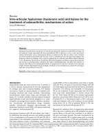
Fig. 1. -A. Synthesis of S-protected thiolated
hyaluronic acid: First N-acetyl cysteine (NAC)
was linked to succinimidyl 3-(2-pyridyldithio)
propionate (SPDP) via disulfide bond formation
to generate SPDP-NAC ligand that was conjugated with hydrazine to hyaluronic acid (HA)
in order to obtain S-protected thiolated HA
under inert conditions.
B. Schematic representation of mediated thiol/
disulfide exchange reactions taking place in Sprotected thiolated HA hydrogel upon addition
of N-acetyl cysteine (NAC) or endogenous thiol
(Endog-SH).
Austria.
hydrazides attached to the HA backbone as outlined in Fig. 1-A. In
contrast to already established thiolated HAs, the crosslinking of this
novel polymer is not triggered by an oxidation process but by endogenous thiols as illustrated in Fig. 1-B. Subsequently, this newly
developed S-protected thiolated HA was formulated to a hydrogel and
tested for texture, swelling ratio, cytotoxicity and viscoelastic behavior.
Furthermore, the potential of S-protected thiolated HA hydrogel for 3D
cell culture scaffold was evaluated using different cell lines.
2.2. Methods
2.2.1. Synthesis of SPDP-NAC hydrazide ligand
First, 50 mg of SPDP was dissolved in 5 mL of anhydrous THF and
30 mg of NAC dissolved in 5 mL of anhydrous THF was added drop-wise
under constant stirring. The reaction was carried out under nitrogen to
avoid oxidation of NAC. Within 10 min reaction, the mixture turned
yellow as the pyridine-2-thione (PDT) group of SPDP was replaced by
NAC. The concentration of the dye was determined by measuring absorbance at 343 nm. The reaction mixture was further stirred for 18 h
under inert conditions at room temperature in dark (Sharma et al.,
2018). The reaction product was analyzed by thin layer chromatography (TLC) to confirm the entire derivatization of SPDP. The solvent
was removed by evaporation and the crude target compound was dissolved in anhydrous DCM. PDT and other impurities were removed by
column chromatography using silica gel with a mobile phase of DCM,
ethyl acetate and methanol in a ratio of 7:2:1. The purified SPDP-NAC
ligand was further reacted with hydrazine (NH2–NH2) to introduce a
primary amino group on the ligand for amide bond formation with
hyaluronic acid. Briefly, 40 mg of SPDP-NAC ligand was dissolved in
2 mL of THF and 0.2 mL of 1 M hydrazine solution in THF was added
drop-wise. After 30 min of stirring at 0 °C, the solvent was evaporated
and SPDP-NAC hydrazide was obtained as a white powder that was
stored at −20 °C until further use.
2. Materials and methods
2.1. Materials
Hyaluronic acid sodium salt (low molecular mass 10−50 kDa),
succinimidyl 3-(2-pyridyldithio)propionate (SPDP), N-acetyl- L-cysteine
(NAC), tetrahydrofuran anhydrous (THF), ethyl acetate, dichloromethane (DCM), methanol, hydrazine (NH2–NH2), N-(3-dimethylaminopropyl)-N'-ethylcarbodiimide hydrochloride (EDAC), Nhydroxysuccinimide (NHS), N,N-dimethylformamide anhydrous (DMF),
sodium chloride (NaCl), L-cysteine hydrochloride hydrate, 5,5′-dithiobis
(2-nitrobenzoic acid) (DTNB, Ellman’s reagent), potassium dihydrogen
phosphate (KH2PO4), disodium hydrogen phosphate (Na2HPO4), tris
(hydroxymethyl)aminomethane hydrochloride (Tris HCl), sodium borohydride (NaBH4), potassium phosphate dibasic (K2HPO4), hydrochloric acid (HCl), silica gel (high purity grade), dialysis tubing MWCO
(molecular weight cut-off) 3.5 kDa, resazurin (7-hydroxy-3H-phenoxazin-3-one sodium salt), minimum essential medium eagle (MEM) and
Triton™ X-100 were all purchased from Merck, Austria. Human colorectal carcinoma cells (Caco-2) were acquired by the European
Collection of Cell Cultures (Salisbury, United Kingdom) and embryonic
fibroblast cells (NIH 3T3) were donated by the Universitätsklinik für
Dermatologie, Venerologie und Allergologie, Innsbruck, Austria.
The cell culture medium was made of MEM powder 9.66 g/L
(modified with Earle’s salts, nineteen amino acids and the non-essential
amino acids L-asp, L-asn, L-glu, L-ser, L-ala, L-gly, and L-pro), 2.2 g/L
sodium bicarbonate, phenol red, 2 mM L-glutamine, 10 % fetal bovine
serum (FBS) and 1 % penicillin-streptomycin solution. 100 mM phosphate-buffered saline (PBS) and FBS were purchased from Invitrogen,
2.2.2. Synthesis of S-protected thiolated hyaluronic acid
S-protected thiolated HA was synthesized by the covalent attachment of the ligand described above to the carboxyl groups of hyaluronic
acid according to a previously described method with minor modifications (de Sousa, Suchaoin, Zupančič, Leichner, & BernkopSchnürch, 2016). In detail, 50 mg of HA was dissolved in 10 mL of
demineralized water under permanent shaking. The pH of the gel was
adjusted at 4.5 with 1 M HCl. To activate the carboxyl groups of hyaluronic acid, 233 mg of EDAC and 46 mg of NHS were added and pH
was re-adjusted to 4.5. The reaction mixture was stirred for 1 h and then
neutralized with 1 M NaOH. 50 mg of SPDP-NAC hydrazide ligand
2
Carbohydrate Polymers 237 (2020) 116092
M.H. Asim, et al.
and removed immediately with the same velocity. The study was conducted at the acquisition rate of 500 points/s with a relaxation period of
30 s between compression cycles. From force-distance plots, results
were calculated employing Texture Exponent software (Version 6,
Stable Micro Systems Ltd) (Tai, Bianchini, & Jachowicz, 2014).
Stickiness and work of shear of S-protected thiolated HA hydrogel
was measured using the same apparatus equipped with a TTC
Spreadability rig (HDP/SR, Stable Micro Systems Ltd) at room temperature. The female sample holder was filled with S-protected thiolated HA hydrogel samples and the male probe was brought downwards
at a speed of 3 mm/s with an angle of 45° up to a distance of 2 mm
above the bottom and then raised upwards with speed of 10 mm/s.
Unmodified HA served as a control. The stickiness was determined as
the maximum negative force and work of shear as the positive area
under the force-distance curve (Tai et al., 2014).
dissolved in 10 mL of anhydrous DCM was drop-wise added to the HAsolution under stirring. After 24 h of stirring at room temperature in the
dark under nitrogen, the mixture was evaporated by using Hei-VAP
Value Digital Rotary Evaporator (Heidolph Instruments GmbH & CO.
Schwabach, Germany. In last, the mixture was transferred into dialysis
tubes (MWCO: 3.5 kDa) and dialyzed for 24 h against 1 % NaCl (m/v)
and then for further 48 h against demineralized water. Finally, the
dialyzed solution was freeze-dried to obtain pure S-protected thiolated
HA.
2.3. Characterization of S-protected thiolated HA by FT-IR and 1H NMR
IR spectra of S-protected thiolated HA were recorded using Bruker
ALPHA FT-IR apparatus (Billerica, MA, USA). FT-IR spectra of test
compounds were collected by placing these compounds on the tip of
platinum ART (attenuated total reflection) module. Results were obtained by 32 scans at a scanning speed of 4 cm−1 between 4000 cm−1
and 500 cm−1. FT-IR spectra were presented as the average of these 32
scans.
1
H NMR spectra were recorded using a Bruker-400 spectrometer (1H
:400 MHz) at 30 °C in 5 mm tubes containing the compounds dissolved
in DMSO-d6. As internal standard served the center of the DMSO-d6
multiplet that was correlated to TMS (tetramethylsilane) with δ
2.49 ppm (1H).
2.7. Swelling ratio of S-protected thiolated hyaluronic acid hydrogels
To study the swelling capabilities of the S-protected thiolated HA
hydrogels, equilibrium swell experiments were performed (Ding, He,
Zhou, Tang, & Yin, 2012). The S-protected thiolated HA hydrogel (1 %
m/v) was washed with demineralized water, freeze-dried and weighed
(Wd). Then 25 mg of freeze-dried hydrogel immersed in 500 μL of
10 mM PBS was mixed with 0.1 and 1 % (m/v) NAC, incubated at 37 °C
and shaken at 300 rpm. After defined time intervals the medium was
carefully removed and swollen hydrogel was weighed again (Ws). Then,
500 μL of buffer was added and the hydrogel was incubated again. This
process was repeated after 5, 10, 20, 30, 60, and 120 min. The swelling
ratio was calculated using the following equation:
2.4. Quantification of the degree of modification
The amount of free thiol groups immobilized on the backbone of Sprotected thiolated HA was determined photometrically with Ellman’s
reagent as already described by our research group (Asim et al., 2019).
The amount of disulfide substructures on the polymer was quantified
via Ellman’s reagent as described above but after reduction with NaBH4
as described before (Asim et al., 2018). Furthermore, to confirm the
lack of unbound SPDP-NAC hydrazide ligand, free primary amines were
determined using TNBS assay as previously described (Suchaoin et al.,
2016). All experiments were performed in triplicate.
Swelling ratio=
(Ws − Wd )
Wd
2.8. Safety screening
To ensure that the S-protected thiolated HA is non-cytotoxic, a resazurin assay was performed (Shahzadi et al., 2019). Cell viability was
evaluated on human colorectal carcinoma cells (Caco-2). Cells were
cultivated in a 24-well plate at a density of 2.5 × 10⁴ cells/well for 10
days in 500 μL of MEM with Earle's salts supplemented with 10 % (v/v)
fetal bovine serum (FBS), 2.0 mM L-glutamine and 1 % penicillinstreptomycin solution (100 units/0.1 mg/L) at 37 °C in an environment
of 5% CO2 and 95 % humidity. Every second day, the growing medium
was changed.
When the cells were approximately 80 % confluent, the red MEM
was removed and the cells were washed twice with pre-warmed PBS at
pH 7.4. Thereafter, 500 μL of unmodified and S-protected thiolated HA
(0.5 % m/v) in white MEM without phenol red, 500 μL of Triton™ X-100
(4 % v/v) as negative control and 500 μL of white MEM without phenol
red as positive control were added to the cell culture in triplicate.
The treated cells were incubated at 37 °C for further 3 and 24 h at an
atmosphere of 5 % CO2 and 95 % relative humidity (Heracell™ 150i
CO2 Incubator, Thermo Fisher Scientific Inc. USA). Afterwards, the test
solutions were removed and the cells were washed again with prewarmed PBS pH 7.4 and finally 500 μL of a 2.2 μM resazurin solution
was added to each well. After 3 h of incubation at 37 °C, 100 μL of supernatant was transferred to a black 96-well plate and the fluorescence
intensity was measured using Spark® multifunctional microplate reader
(Tecan Austria, GmbH) at a wavelength of 540 nm with background
subtraction at 590 nm. Cell viability was calculated following the
under-mentioned equation:
2.5. Molecular weight determination of S-protected hyaluronic acid by gel
permeation chromatography
The molecular weight (MW) of S-protected thiolated HA were calculated by GPC using Merck Hitachi LaChrom Elite® HPLC-System
equipped with Merck Hitachi L-2130 Pump, Merck Hitachi L-2200
Autosampler, Merck Hitachi L-2450 Diode Array Detector (DAD) and
VWR Hitachi Refractive Index (RI) detector by following previous established methods with minor modification (Yang et al., 2012; Yang,
Guo, Zang, & Liu, 2015). A chromatographic analytical column PSS
SUPREMA 3000 (8 × 300, 10 μ) (PSS-Polymer Standards Service-USA
Inc.) with an injection volume of 20 μL was used. The mobile phase
consisted of phosphate buffer saline (PBS) pH 7.4 filtered through
0.2 μm Millipore filter was used with flow rate of 0.5 mL/min. The
detection wavelength was 220 nm and the column was maintained at
35 °C. For calibration curve, HAs of different molecular weights (10–50
KDa, 1,500–1,800 KDa and 2,000–2,400 KDa) were used.
2.6. Surface properties and spreadability of S-protected thiolated hyaluronic
acid hydrogels
The texture properties of hydrogels were analyzed with a TA.XTplus
texture analyser with a 5 kg load cell (Stable Micro Systems Ltd, Surrey,
UK). To evaluate the effect of NAC on hydrogel cohesion and stickiness
increasing concentrations of NAC (0–1 % m/v) in a vial were added to Sprotected thiolated and unmodified HA (1 % m/v) for half an hour at
25 °C (Rupenthal, Green, & Alany, 2011). A 10 mm cylindrical probe
(SMSP/10, Stable Micro Systems Ltd) was pushed down into hydrogel
samples at room temperature to a depth of 5 mm at a rate of 2 mm/s
Cell Viability [%] =
3
Experimental value− Negative control
× 100
Positive control− Negative control
Carbohydrate Polymers 237 (2020) 116092
M.H. Asim, et al.
2.9. Rheological studies of S-protected thiolated hyaluronic acid hydrogels
3. Results and discussion
S-protected thiolated HA (1.5 % m/v) was dissolved in sterile demineralized water. Then, 0.1, 0.3, 0.5, 0.8 and 1 % (m/v) solutions of
NAC dissolved in 0.01 M PBS pH 7.4 were added to the above solutions
in 1:2 ratio. The viscosity of all samples was measured after incubation
at 37 °C for 3 h. The same procedure was followed for unmodified HA
serving as a control. Moreover, the gelation time of S-protected thiolated HA with increasing concentrations of NAC was noted.
Rheological studies such as dynamic viscosity (η), apparent elastic
modulus (G′) and viscous modulus (G″) were measured on a thermostatically controlled plate-plate rheometer (Thermo Scientific™
HAAKE™ MARS™ rheometer, Thermo Fisher Scientific, Karlsruhe,
Germany). Besides, the phase angle (δ), calculated by δ = tan−1 G″/G′,
was used for differentiation between gel and solution, while tan δ < 1
is characterizing as gel and tan δ > 1 as a solution. The linear viscoelastic region of all samples was determined through initial oscillatory strain sweep measurements at a frequency of 1 Hz at 37 °C with a
shear stress of 0.5 Pa.
Furthermore, the dynamic viscosity of S-protected thiolated HA
hydrogel in the presence of freshly collected porcine mucus was evaluated. The mucus was collected and purified as previously described
(Asim, Nazir, Jalil, Matuszczak, & Bernkop-Schnürch, 2020). Samples
of unmodified and S-protected thiolated HA (1 % m/v) in 200 mM
phosphate buffer pH 7.4 were mixed with porcine intestinal mucus in
1:2 ratio. Mucus mixed with phosphate buffer in the same ratio served
as control. Samples were incubated for 1, 2 and 3 h at 37 °C and dynamic viscosity (η), apparent elastic modulus (G′) and viscous modulus
(G″) of hydrogels were determined as a function of frequency using
HAAKE RheoWin 3 software.
3.1. Synthesis and characterization of S-protected thiolated hyaluronic acid
HA served as a polymeric backbone for the immobilization of thiol
moieties via amide bond formation. For the synthesis of S-protected
thiolated HA, the SPDP-NAC ligand was synthesized by a thiol-disulfide
exchange reaction. Then, it was reacted with hydrazine to introduce an
eNH2 group for amide bond formation with hyaluronic acid to generate
SPDP-NAC-azide with 77.8 % yield. A carbodiimide was used for activation ofeCOOH groups of HA under acidic conditions for protonation
of the carbodiimide nitrogens (Shu et al., 2002). The activated eCOOH
groups of HA reacted with SPDP-NAC hydrazide ligand via amide bond
formation to gain S-protected thiolated HA with 81.4 % yield as displayed in Fig. 1-A.
The successful immobilization of the ligand was confirmed via FT-IR
spectroscopy. The FT-IR spectrum of S-protected thiolated HA are given
in Supplementary Fig. S1. The IR spectrum of the S-protected thiolated
HA significantly differed from that of the unmodified HA. In detail, the
characteristic peak of eNH group at 1556 cm−1 confirmed amide bond
formation (Du, Fu, Shi, & Yin, 2015), whereas peak located at
3260 cm−1 is associated with stretching vibration of eOH groups.
Moreover, the SeS stretching peaks in the frequency range of
560−610 cm−1 confirmed disulfide bonds of the ligand attached to the
hyaluronic acid backbone. Furthermore, the chemical structure of Sprotected thiolated HA was confirmed via 1H NMR spectral analysis.
The 1H NMR spectrum of S-protected thiolated HA showed signals at
1.9 ppm that are assigned to eCH3 protons of NAC and amide protons at
9.3 ppm as shown in Supplementary Fig. S2. Moreover, the eCH3
protons of the N-acetyl group of HA merged with eCH3 protons of NAC
resulting in just one peak for both sub-structures (Li, Yu, Jin, & Yin,
2012; Shu et al., 2002). For molecular weight determination of modified HA, the retention time of unmodified HA was 35 min with MW of
10–50 KDa that was shorten with S-protected thiolated HA to 31 min
indicating increase in MW of modified HA. The MW of modified HA was
calculated to 15–69 KDa.
S-protected thiolated HA showed a negligible amount of free eSH
groups and eNH2 groups as confirmed by Ellman’s test and TNBS test,
respectively. These results confirm the purification of the product from
unbound ligands and the successful protection of eSH groups. In total,
320.55 μmol/g disulfide bonds were detected in the product that corresponds to 7.4 % of the total moles of carboxylic acid groups.
2.10. In-situ cell encapsulation
S-protected thiolated HA was sterilized under UV radiation for
30 min under N2 protection and hydrated in a final concentration of 1.5
% (m/v) in 1 mL of sterile red MEM (3 % m/v) at pH 7.4. To this Sprotected thiolated HA hydrogel, 100 μL of Caco-2 and NIH 3T3 cell
suspensions were added to obtain a final concentration of 5 × 10⁶ cells/
mL, respectively. Due to the addition of 100 μL of NAC solution (1 % m/
v) viscosity of the S-protected thiolated HA hydrogels was increased via
thiol-disulfide exchange reactions encapsulating the dispersed cells.
The hydrogels were then immersed into 500 μL red MEM with Earle's
salts supplemented with 10 % (v/v) fetal bovine serum (FBS), 2.0 mM Lglutamine and 1 % penicillin-streptomycin solution (100 units/0.1 mg/
L) in an environment of 5 % CO2 and 95 % humidity (Heracell™ 150i
CO2 Incubator, Thermo Fisher Scientific Inc. USA) at 37 °C. S-protected
thiolated HA hydrogels containing Caco-2 and NIH 3T3 cells were
cultured at 37 °C and 5 % CO2 and observed after 3, 7, 12 and 15 days
using Motic AE31 inverted microscope (Motic Deutschland GmbH,
Wetzlar, Germany) equipped with the ProgRes® series camera (JENOPTIK, Goeschwitzer, Germany). Pictures were taken by using ProgRes®
CapturePro 2.7 software.
3.2. Texture analysis and spreadability of S-protected thiolated HA
hydrogels
For the effective application of hydrogels suitable texture properties
are essential (Kong, Kim, & Park, 2016) as cells can adapt to substrate
mechanics by adjusting their proliferation and migration (Ghosh et al.,
2007). Moreover, stiffness of scaffolds plays a key role as it does not
only modulate biological processes but has even an impact on the fate
of cells (Shi et al., 2012). Surface properties of S-protected thiolated HA
hydrogels were investigated by exposure to increasing concentrations of
NAC serving as a representative model for endogenous thiols at the
target tissue. The resulting work of cohesion is shown in Fig. 2.
S-protected thiolated HA hydrogel remained cohesive over a broad
range of added NAC concentrations. These textural properties revealed
also a high resistance to shear for S-protected thiolated HA hydrogels,
which was in good agreement with their high stickiness. The stiffness of
hydrogels can control scaffold architecture and can facilitate cell
growth and survival within the three dimensional network. Changes in
rheological behavior by altering the storage modulus lead to a change
in the morphology of cells during proliferation. Therefore, the mechanical properties of hydrogels could potentially be adjusted to meet
the required stiffness to aid repairment of injured tissues (da Cunha
et al., 2014). High cohesiveness of hydrogels corresponds enhanced
2.11. Statistical analyses
Statistical analyses of all data were performed by using the student's
t-test. A confidence interval of p < 0.05 was used for the analysis of
two groups. One-way ANOVA was applied to compare different groups
difference with 95 % CI (confident interval). (GraphPad Prism®,
GraphPad Software, Inc.). All results were expressed as the mean of at
least three experiments ± SD.
4
Carbohydrate Polymers 237 (2020) 116092
M.H. Asim, et al.
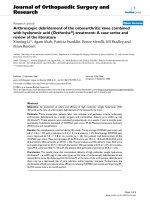
Fig. 3. The swelling ratio of S-protected thiolated HA with NAC (0.1 % m/v)
(black bar) in comparison to S-protected thiolated HA with NAC (1 % m/v)
(white bar) in 0.01 M PBS pH 7.4 at 37 °C. Indicated values represent an
average of at least three experiments ± SD (***P < 0.001).
shown poly(propylene fumarate) crosslinked hydrogels and Arg-GlyAsp (RGD)-modified PEG hydrogels (Burdick & Anseth, 2002; He,
Yaszemski, Yasko, Engel, & Mikos, 2000). Furthermore, these previously synthesized hydrogels have free –SH groups that form disulfide
bonds upon exposure to air. This crosslinking process, however, is
difficult to control and proceeding too slowly so that a required fast
increase in viscosity cannot be achieved (Shu et al., 2002). In contrast,
the crosslinking process of S-protected thiolated HA hydrogels can be
controlled to a higher extent.
3.4. Cytotoxic investigation
Fig. 2. Analysis of gel texture and spreadabiliy. [A] Work of cohesion determined as the negative area under the force/distance curve of mixtures of Sprotected thiolated HA and increasing concentrations of N-acetyl cysteine
(NAC) at room temperature. All values are means of at least three
experiments ± SD. [B] Work of shear (light grey bars) determined as positive
area under the force/distance curve and stickiness (dark grey bars) determined
as maximum negative force of S-protected thiolated HA hydrogels. All values
are means of at least three experiments ± SD.
Cytotoxicity of S-protected thiolated HA was quantified by resazurin
assay. Resazurin is a redox indicator that provides information about
the viability of cells. Cytotoxic compounds reduce the redox potential of
cells, whereby no reduction takes place with non-toxic compounds
(Borra, Lotufo, Gagioti, Barros, & Andrade, 2009). Caco-2 cells were
treated with unmodified HA and S-protected thiolated HA in a concentration of 0.5 % (m/v). After 3 and 24 h of incubation, cells showed
viability over 80 % on Caco-2 cells as illustrated in Fig. 4.
interactions in the polymer network and an increased area for possible
interactions with cells for improved proliferation.
3.3. Swelling behavior and stability of S-protected thiolated hyaluronic acid
hydrogels
The ability of hydrogels to held water in their hydrophilic matrix is
influenced by the chemical structure, crosslinking density and molecular weight of polymers (Collins & Birkinshaw, 2013). The swelling
ratio plays an important role in maintaining the structure of hydrogels.
The swelling ratio of S-protected thiolated HA was compared after
addition of 0.1 and 1 % (m/v) NAC. Consequently, both polymers were
crosslinked during the swelling process explaining why they do not
simply dissolve. The initial increase of weight could be due to the
polymeric network stretching that allowed an increasing influx of fluid.
Maximum fluid uptake took place within 30 min and then no further
increase took place as shown in Fig. 3. These results correlate with
previously shown results by Bian et al. for self-crosslinking smart hyaluronic acid hydrogels exhibiting swelling equilibrium within 30 min
(Bian et al., 2016).
For use in tissue engineering, HA-based hydrogels have a great
advantage due to their high water content as shown by swelling
properties. Moreover, these hydrogels have high permeability for
oxygen and other water-soluble compounds, as compare to previously
Fig. 4. Cell viability in the presence of S-protected thiolated HA (0.5 % m/v)
compared to unmodified HA (0.5 % m/v) after 3 and 24 h. Cytotoxicity studies
were performed using resazurin on Caco-2 cells. MEM without phenol red was
used as negative control, and Triton™ X-100 (4% m/v) were used as positive
control. Indicated values represent an average of at least three
experiments ± SD. (For interpretation of the references to colour in this figure
legend, the reader is referred to the web version of this article).
5
Carbohydrate Polymers 237 (2020) 116092
M.H. Asim, et al.
The minor cytotoxic effect of unmodified HA as shown in Fig. 4 is in
agreement with previous studies demonstrating a similar effect of HA
on various cultured cells (Bodo et al., 2002; Boeckel, Shinkai, Grossi, &
Teixeira, 2014; Wiig, Abrahamsson, & Lundborg, 1996). Moreover, HAbased thiomers are considered safe for in-vivo application as various
thiolated HAs are already subject to clinical trials showing no safety
concerns (Safety of Hyaluronan Thiomer i.o, 2020) and even some are
already on the market such as HyStem™ hyaluronic acid based-hydrogels from Merck, Glycosil® from ESI BIO (Zarembinski et al., 2014).
Furthermore, even the S-protecting leaving group NAC does not seem to
reach toxic levels of concern as marketed products containing high
amounts of NAC are generally regarded as safe.
3.5. Viscoelastic behavior of S-protected thiolated hyaluronic acid hydrogels
As the stiffness of hydrogels has a substantial impact on cell migration and proliferation, the characterization of rheological properties
of hydrogels used as a scaffold material for tissue engineering is of high
relevance (da Cunha et al., 2014). As thiol-disulfide exchange reactions
triggered by endogenous free thiols will cause a crosslinking of HA via
disulfide bond formation and consequently gelation, increasing concentrations of NAC were added to S-protected thiolated HA to simulate
this process. Results of this study are listed in Table 1.
The gelation process in S-protected thiolated HA occurred via thioldisulfide exchange reaction. It was confirmed by mixing S-protected
thiolated HA with freshly collected mucus serving as a model for extracellular proteins such as mucins building up the glycocalyx of cells or
cysteine-rich membrane bound keratins. Mucus consists of glycoproteins with cysteine-rich side chains. These thiol groups allow mucin
monomers to form disulfide bonds with the S-protected thiomer as
shown in Fig. 5.
Moreover, as NAC concentration in hydrogel increases, dynamic
viscosity (η) increases from 1.2 Pa (0.1 % m/v NAC) to a maximum of
23.5 Pa (1 % m/v NAC) confirming gel formation as shown in Fig. 5.
Elastic modulus (G′) was also increased with increasing concentration of NAC (Fig. 6A) characterizing energy recovery stored in the
system (Valenta, Kast, Harich, & Bernkop-Schnürch, 2001). A high
elastic modulus (G′) of S-protected thiolated HA hydrogel indicates high
mechanical strength and rigidity and further denotes more solid-like
properties. Moreover, a high elastic modulus (G′) shows the polymer’s
ability to store deformation energy in an elastic manner. The higher the
degree of cross-linking was, the greater was the storage modulus. Furthermore, the viscous modulus (G″) of the S-protected thiolated HA was
also higher than the one of unmodified HA as depicted in Fig. 6B. In the
presence of NAC, S-protected thiolated HA hydrogels showed a tan δ
value of 0.2 indicating gel formation, whereas unmodified HA hydrogel
did not show any gel formation.
Similarly, in rheological studies with mucus elastic modulus (G′)
and viscous modulus (G″) of the S-protected thiolated HA increased
over time as shown in Fig. 6-C and D.
Mucus consists of glycoproteins with cysteine-rich side chains.
These thiol groups allow mucin monomers to form disulfide bonds with
the S-protected thiomer (Kaldybekov, Filippov, Radulescu, &
Fig. 5. Dynamic viscosity (Pas) [A] of unmodified and S-protected thiolated
HA (1.5 % m/v) in the presence of indicated concentrations of N-acetyl cysteine
(NAC) and dynamic viscosity [B] of unmodified HA (1 % m/v) and S-protected
thiolated HA (1 % m/v) in the presence of 500 μL of mucus in 200 mM phosphate buffer at pH 7 at a frequency of 1 Hz on a plate-plate viscometer at 37 °C
for 3 h. Indicated values represent an average of at least three
experiments ± SD.
Khutoryanskiy, 2019; Kolawole, Lau, & Khutoryanskiy, 2018). Unmodified hyaluronic acid/mucus mixture showed almost the same
viscosity as mucus, whereas the viscosity of S-protected thiolated HA
was 10.1-fold higher.
3.6. In-vitro cell proliferation and morphology
After 3, 7, 12 and 15 days proliferation of Caco-2 and NIH 3T3 cells
were monitored as shown in Figs. 7 and 8.
Results showed that both cell lines having been encapsulated in the
hydrogel proliferated within the culture medium. After 7 days of culture, cells bilayer was observed whereas after 12 days cell clusters
appeared. Due to the hydrophilic surface of HA, no cell attachment was
observed. Spherical cell clusters of Caco-2 cells and elongated, spindlelike morphology of NIH 3T3 cells reflect proper gel stiffness as high
hydrogel stiffness leads to change in morphology of cells during proliferation (da Cunha et al., 2014). Moreover, the number and size of cell
clusters increased with the increase of incubation time.
For cell proliferation, Cys/CySS redox potential (Eh) has an important role in redox exchange between cells and organs. Ramirez et al.
explored that in lung fibroblasts the more oxidizing Eh values of Cys/
CySS produced intracellular signals that stimulate cell proliferation.
These oxidizing Eh values increased extracellular fibronectin production
in NIH 3T3 fibroblasts via the protein kinase C pathway and induction
of transforming growth factor-β1 (Ramirez et al., 2007). S-protected
thiolated HAs having NAC are also predicted to follow the same
Table 1
Gelation time of S-protected thiolated HA (1.5 % m/v) in
the presence of indicated concentrations of NAC at 37 °C.
Indicated values are mean ± SD (n = 3).
NAC concentration
(% m/v)
Gelation time
(min)
0.1
0.3
0.5
0.8
1.0
20 ± 6
18 ± 4
11 ± 3
9±2
5±1
6
Carbohydrate Polymers 237 (2020) 116092
M.H. Asim, et al.
Fig. 6. Rheological measurements of S-protected thiolated HA in terms of [A] elastic
modulus (G´) and [B] viscous modulus (G″)
after incubation with indicated concentrations
of NAC and [C] elastic modulus (G´) and [D]
viscous modulus (G″) after incubation with
500 μL of mucus in 200 mM phosphate buffer
at 37 °C for 3 h at a frequency of 1 Hz on a
plate-plate viscometer. All the results are expressed as the mean of at least three
experiments ± SD, (***P < 0.001).
endogenous thiols on the surface of surrounding cells. The intracellular
concentration of thiols is in the millimolar range, whereas free thiols in
the extracellular environment are in the micromolar range depending
on the target compartment (Hu, 1994; Yi & Khosla, 2016). The thiol/
disulfide balance within the extracellular environment is kept constant
via various mechanisms. As due to the addition of S-protected HA this
balance is strongly shifted towards disulfides the concentration of free
mechanism for proliferation of cells. S-protected thiolated HAs are
advantageous over just thiolated HAs that are unstable in aqueous solutions of pH ≥ 5 due to oxidation of thiol groups (de Sousa et al.,
2016). On contrary, S-protected thiolated HA hydrogels react in a pHindependent manner being stable towards oxidation. Moreover, S-protected thiolated HA hydrogels will not just crosslink but will also be
anchored in the target tissue by disulfide bond formation with
Fig. 7. The S-protected thiolated HA hydrogels
(1.5 % m/v)) cultured with Caco-2 cells
(5 × 10⁶ cells/mL) incubated at 37 °C in an
environment of 95 % humidity and 5% CO2.
Inverted phase contrast micrographs of Caco-2
cells encapsulated in S-protected thiolated HA
hydrogel after co-culture for 3 days (A1), 7
days (B1), 12 days (C1) and 15 days (D1). All
the micrographs are of 10 μM magnification.
7
Carbohydrate Polymers 237 (2020) 116092
M.H. Asim, et al.
Fig. 8. The S-protected thiolated HA hydrogels
(1.5 % m/v)) cultured with NIH 3T3 (5 × 10⁶
cells/mL) incubated at 37 °C in an environment
of 95 % humidity and 5% CO2. Inverted phase
contrast micrographs of NIH 3T3 cells encapsulated in S-protected thiolated HA hydrogel after co-culture for 3 days (A2), 7 days
(B2), 12 days (C2) and 15 days (D2). All the
micrographs are of 10 μM magnification.
thiols will likely rapidly raise in order to compensate this imbalance.
Furthermore, as the crosslinking of S-protected thiolated HA is
triggered by free thiols, whereas the crosslinking of thiolated HA is
triggered by oxygen, a direct comparison in their in-situ gelling properties is not feasible. The concentration of available free thiols and
oxygen is highly variable depending on the type of target tissue and
disorders such as inflammation or necrosis. In case of S-protected
thiolated HA, however, an unintended gelation during production,
storage and application can be excluded, as this type of thiolated HA is
stable towards oxidation. These results confirm that S-protected thiolated HA hydrogels enhance the proliferation of encapsulated cells and
can be helpful in 3D cell culture scaffold.
analysis. Andreas Bernkop-Schnürch: Conceptualization, Resources,
Funding acquisition, Supervision, Project administration.
4. Conclusion
This research work was supported by the Higher Education
Commission (HEC), Pakistan and the Austrian Agency for International
Cooperation in Education and Research (OeAD), Austria.
Declaration of Competing Interest
The authors report no conflicts of interest. The authors have no
relevant affiliations or financial involvement with any organization or
entity with a financial interest in or financial conflict with the subject
matter or materials discussed in the manuscript apart from those disclosed.
Acknowledgments
In this study, S-protected thiolated HA was synthesized by conjugating a SPDP-NAC hydrazide ligand to the backbone of HA. S-protected thiolated HA showed enhanced cohesion and stickiness upon
addition of N-acetyl cysteine (NAC). The crosslinking process of previously synthesized thiolated HA hydrogels was slow and un-controlled.
In contrast, this novel S-protected thiolated HA is stable towards oxidation and forms highly cohesive gels when getting into contact with
endogenous thiols due to disulfide-crosslinking. In addition, S-protected
thiolated HA hydrogels were non-toxic and showed enhanced dynamic
viscosity in the presence of extracellular model proteins via thiol-disulfide exchange reactions. Furthermore, S-protected thiolated HA hydrogel showed proliferation of Caco-2 and NIH 3T3 cells encapsulated
in hydrogels. Because of all these properties, it could be concluded that
these S-protected thiolated HA hydrogels have potential in the area of
3D cell culture scaffold and tissue engineering as matrices for repairing
and regenerating tissues and organs.
Appendix A. Supplementary data
Supplementary material related to this article can be found, in the
online version, at doi: />References
Asim, M. H., Moghadam, A., Ijaz, M., Mahmood, A., Götz, R. X., Matuszczak, B., ...
Bernkop-Schnürch, A. (2018). S-protected thiolated cyclodextrins as mucoadhesive
oligomers for drug delivery. Journal of Colloid and Interface Science, 531, 261–268.
Asim, M. H., Jalil, A., Shahzadi, I., Khan, M., Matuszczak, B., & Bernkop-Schnürch, A.
(2019). Mucoadhesive S-protected thiolated cyclodextrin-iodine complexes: A promising strategy to prolong mucosal residence time of iodine. Future Microbiology,
14(5), 411–424.
Asim, M. H., Nazir, I., Jalil, A., Matuszczak, B., & Bernkop-Schnürch, A. (2020).
Tetradeca-thiolated cyclodextrins: Highly mucoadhesive and in-situ gelling oligomers
with prolonged mucosal adhesion. International Journal of Pharmaceutics, 119040.
Bian, S., He, M., Sui, J., Cai, H., Sun, Y., Liang, J., & Zhang, X. (2016). The self-crosslinking smart hyaluronic acid hydrogels as injectable three-dimensional scaffolds for
cells culture. Colloids and Surfaces B, Biointerfaces, 140, 392–402.
Bodo, M., Pezzetti, F., Baroni, T., Carinci, F., Arena, N., Nicoletti, I., ... Becchetti, E.
(2002). Hyaluronic acid modulates growth, morphology and cytoskeleton in embryonic chick skin fibroblasts. The International Journal of Developmental Biology,
37(2), 349–352.
Boeckel, D. G., Shinkai, R. S. A., Grossi, M. L., & Teixeira, E. R. (2014). In vitro evaluation
of cytotoxicity of hyaluronic acid as an extracellular matrix on OFCOL II cells by the
CRediT authorship contribution statement
Mulazim Hussain Asim: Investigation, Methodology, Validation,
Visualization, Writing - original draft. Stefanie Silberhumer:
Investigation, Writing - review & editing. Iram Shahzadi:
Investigation. Aamir Jalil: Investigation. Barbara Matuszczak: Formal
8
Carbohydrate Polymers 237 (2020) 116092
M.H. Asim, et al.
Li, L., Wang, N., Jin, X., Deng, R., Nie, S., Sun, L., & Gong, C. (2014). Biodegradable and
injectable in situ cross-linking chitosan-hyaluronic acid based hydrogels for postoperative adhesion prevention. Biomaterials, 35(12), 3903–3917.
Lin, G., & Stern, R. (2001). Plasma hyaluronidase (Hyal-1) promotes tumor cell cycling.
Cancer Letters, 163(1), 95–101.
Prestwich, G. D. (2011). Hyaluronic acid-based clinical biomaterials derived for cell and
molecule delivery in regenerative medicine. Journal of Controlled Release, 155(2),
193–199.
Ramirez, A., Ramadan, B., Ritzenthaler, J. D., Rivera, H. N., Jones, D. P., & Roman, J.
(2007). Extracellular cysteine/cystine redox potential controls lung fibroblast proliferation and matrix expression through upregulation of transforming growth factorβ. American Journal of Physiology-Lung Cellular and Molecular Physiology, 293(4),
L972–L981.
Rupenthal, I. D., Green, C. R., & Alany, R. G. (2011). Comparison of ion-activated in situ
gelling systems for ocular drug delivery. Part 1: Physicochemical characterisation and
in vitro release. International Journal of Pharmaceutics, 411(1–2), 69–77.
Safety of Hyaluronan Thiomer i.o (2020). Implant during combined phacoemulsification non penetrating deep sclerectomy. Croma-Pharma GmbH.
Shahzadi, I., Asim, M., Dizdarević, A., Wolf, J., Kurpiers, M., Matuszczak, B., ... BernkopSchnürch, A. (2019). Arginine-based cationic surfactants: Biodegradable auxiliary
agents for the formation of hydrophobic ion pairs with hydrophilic macromolecular
drugs. Journal of Colloid and Interface Science, 552, 287–294.
Sharma, A., Liaw, K., Sharma, R., Zhang, Z., Kannan, S., & Kannan, R. M. (2018).
Targeting mitochondrial dysfunction and oxidative stress in activated microglia using
dendrimer-based therapeutics. Theranostics, 8(20), 5529.
Shi, J., Ouyang, J., Li, Q., Wang, L., Wu, J., Zhong, W., ... Xing, M. M. (2012). Cellcompatible hydrogels based on a multifunctional crosslinker with tunable stiffness for
tissue engineering. Journal of Materials Chemistry, 22(45), 23952–23962.
Shoham, N., Sasson, A. L., Lin, F.-H., Benayahu, D., Haj-Ali, R., & Gefen, A. (2013). The
mechanics of hyaluronic acid/adipic acid dihydrazide hydrogel: Towards developing
a vessel for delivery of preadipocytes to native tissues. Journal of the Mechanical
Behavior of Biomedical Materials, 28, 320–331.
Shu, X. Z., Liu, Y., Luo, Y., Roberts, M. C., & Prestwich, G. D. (2002). Disulfide crosslinked hyaluronan hydrogels. Biomacromolecules, 3(6), 1304–1311.
Suchaoin, W., Bonengel, S., Grießinger, J. A., de Sousa, I. P., Hussain, S., Huck, C. W., ...
Bernkop-Schnürch, A. (2016). Novel bioadhesive polymers as intra-articular agents:
Chondroitin sulfate-cysteine conjugates. European Journal of Pharmaceutics and
Biopharmaceutics, 101, 25–32.
Tai, A., Bianchini, R., & Jachowicz, J. (2014). Texture analysis of cosmetic/pharmaceutical raw materials and formulations. International Journal of Cosmetic Science, 36(4),
291–304.
Valenta, C., Kast, C. E., Harich, I., & Bernkop-Schnürch, A. (2001). Development and in
vitro evaluation of a mucoadhesive vaginal delivery system for progesterone. Journal
of Controlled Release, 77(3), 323–332.
Wiig, M., Abrahamsson, S.-O., & Lundborg, G. (1996). Effects of hyaluronan on cell
proliferation and collagen synthesis: A study of rabbit flexor tendons in vitro. The
Journal of Hand Surgery, 21(4), 599–604.
Xu, X., Jha, A. K., Harrington, D. A., Farach-Carson, M. C., & Jia, X. (2012). Hyaluronic
acid-based hydrogels: From a natural polysaccharide to complex networks. Soft
Matter, 8(12), 3280–3294.
Yang, J.-A., Kim, E.-S., Kwon, J. H., Kim, H., Shin, J. H., Yun, S. H., ... Hahn, S. K. (2012).
Transdermal delivery of hyaluronic acid–human growth hormone conjugate.
Biomaterials, 33(25), 5947–5954.
Yang, B., Guo, X., Zang, H., & Liu, J. (2015). Determination of modification degree in
BDDE-modified hyaluronic acid hydrogel by SEC/MS. Carbohydrate Polymers, 131,
233–239.
Yi, M. C., & Khosla, C. (2016). Thiol–Disulfide exchange reactions in the mammalian
extracellular environment. Annual Review of Chemical and Biomolecular Engineering, 7,
197–222.
Yu, F., Cao, X., Li, Y., Zeng, L., Zhu, J., Wang, G., ... Chen, X. (2014). Diels–Alder
crosslinked HA/PEG hydrogels with high elasticity and fatigue resistance for cell
encapsulation and articular cartilage tissue repair. Polymer Chemistry, 5(17),
5116–5123.
Zarembinski, T. I., Doty, N. J., Erickson, I. E., Srinivas, R., Wirostko, B. M., & Tew, W. P.
(2014). Thiolated hyaluronan-based hydrogels crosslinked using oxidized glutathione: An injectable matrix designed for ophthalmic applications. Acta
Biomaterialia, 10(1), 94–103.
MTT assay. Oral Surgery, Oral Medicine, Oral Pathology and Oral Radiology, 117(6),
e423–e428.
Borra, R. C., Lotufo, M. A., Gagioti, S. M., Barros, F. M., & Andrade, P. M. (2009). A simple
method to measure cell viability in proliferation and cytotoxicity assays. Brazilian
Oral Research, 23(3), 255–262.
Burdick, J. A., & Anseth, K. S. (2002). Photoencapsulation of osteoblasts in injectable
RGD-modified PEG hydrogels for bone tissue engineering. Biomaterials, 23(22),
4315–4323.
Burdick, J. A., & Prestwich, G. D. (2011). Hyaluronic acid hydrogels for biomedical applications. Advanced Materials, 23(12), H41–H56.
Collins, M. N., & Birkinshaw, C. (2013). Hyaluronic acid based scaffolds for tissue
engineering—A review. Carbohydrate Polymers, 92(2), 1262–1279.
da Cunha, C. B., Klumpers, D. D., Li, W. A., Koshy, S. T., Weaver, J. C., Chaudhuri, O., ...
Mooney, D. J. (2014). Influence of the stiffness of three-dimensional alginate/collagen-I interpenetrating networks on fibroblast biology. Biomaterials, 35(32),
8927–8936.
de Sousa, I. P., Suchaoin, W., Zupančič, O., Leichner, C., & Bernkop-Schnürch, A. (2016).
Totally S-protected hyaluronic acid: Evaluation of stability and mucoadhesive
properties as liquid dosage form. Carbohydrate Polymers, 152, 632–638.
Ding, J., He, R., Zhou, G., Tang, C., & Yin, C. (2012). Multilayered mucoadhesive hydrogel films based on thiolated hyaluronic acid and polyvinylalcohol for insulin delivery. Acta Biomaterialia, 8(10), 3643–3651.
Du, J., Fu, F., Shi, X., & Yin, Z. (2015). Controlled release of a model protein drug
ovalbumin from thiolated hyaluronic acid matrix. Journal of Drug Delivery Science and
Technology, 30, 74–81.
Ghosh, K., Pan, Z., Guan, E., Ge, S., Liu, Y., Nakamura, T., & Clark, R. A. (2007). Cell
adaptation to a physiologically relevant ECM mimic with different viscoelastic
properties. Biomaterials, 28(4), 671–679.
Gramlich, W. M., Kim, I. L., & Burdick, J. A. (2013). Synthesis and orthogonal photopatterning of hyaluronic acid hydrogels with thiol-norbornene chemistry.
Biomaterials, 34(38), 9803–9811.
He, S., Yaszemski, M. J., Yasko, A. W., Engel, P. S., & Mikos, A. G. (2000). Injectable
biodegradable polymer composites based on poly (propylene fumarate) crosslinked
with poly (ethylene glycol)-dimethacrylate. Biomaterials, 21(23), 2389–2394.
He, M., Zhao, Z., Yin, L., Tang, C., & Yin, C. (2009). Hyaluronic acid coated poly (butyl
cyanoacrylate) nanoparticles as anticancer drug carriers. International Journal of
Pharmaceutics, 373(1-2), 165–173.
Hu, M.-L. (1994). 41] measurement of protein thiol groups and glutathione in plasma. Methods
in enzymology. Elsevier380–385.
Jin, R., Teixeira, L. M., Krouwels, A., Dijkstra, P. J., Van Blitterswijk, C., Karperien, M., ...
Feijen, J. (2010). Synthesis and characterization of hyaluronic acid–poly (ethylene
glycol) hydrogels via Michael addition: An injectable biomaterial for cartilage repair.
Acta Biomaterialia, 6(6), 1968–1977.
Kaldybekov, D. B., Filippov, S. K., Radulescu, A., & Khutoryanskiy, V. V. (2019).
Maleimide-functionalised PLGA-PEG nanoparticles as mucoadhesive carriers for intravesical drug delivery. European Journal of Pharmaceutics and Biopharmaceutics, 143,
24–34.
Khutoryanskiy, V. V. (2011). Advances in mucoadhesion and mucoadhesive polymers.
Macromolecular Bioscience, 11(6), 748–764.
Kolawole, O. M., Lau, W. M., & Khutoryanskiy, V. V. (2018). Methacrylated chitosan as a
polymer with enhanced mucoadhesive properties for transmucosal drug delivery.
International Journal of Pharmaceutics, 550(1-2), 123–129.
Kong, B. J., Kim, A., & Park, S. N. (2016). Properties and in vitro drug release of hyaluronic acid-hydroxyethyl cellulose hydrogels for transdermal delivery of isoliquiritigenin. Carbohydrate Polymers, 147, 473–481.
Lee, H., & Park, T. G. (2009). Photo‐crosslinkable, biomimetic, and thermo‐sensitive
pluronic grafted hyaluronic acid copolymers for injectable delivery of chondrocytes.
Journal of Biomedical Materials Research Part A: An Official Journal of The Society for
Biomaterials, The Japanese Society for Biomaterials, and The Australian Society for
Biomaterials and the Korean Society for Biomaterials, 88(3), 797–806.
Lee, Y., Chung, H. J., Yeo, S., Ahn, C.-H., Lee, H., Messersmith, P. B., ... Park, T. G. (2010).
Thermo-sensitive, injectable, and tissue adhesive sol–gel transition hyaluronic acid/
pluronic composite hydrogels prepared from bio-inspired catechol-thiol reaction. Soft
Matter, 6(5), 977–983.
Li, X., Yu, G., Jin, K., & Yin, Z. (2012). Hyaluronic acid L-cysteine conjugate exhibits
controlled-release potential for mucoadhesive drug delivery. Die Pharmazie-An
International Journal of Pharmaceutical Sciences, 67(3), 224–228.
9