An in-depth study of molecular and supramolecular structures of bamboo cellulose upon heat treatment
Bạn đang xem bản rút gọn của tài liệu. Xem và tải ngay bản đầy đủ của tài liệu tại đây (4.75 MB, 9 trang )
Carbohydrate Polymers 241 (2020) 116412
Contents lists available at ScienceDirect
Carbohydrate Polymers
journal homepage: www.elsevier.com/locate/carbpol
An in-depth study of molecular and supramolecular structures of bamboo
cellulose upon heat treatment
T
Qiuqin Lin, Yuxiang Huang*, Wenji Yu
Research Institute of Wood Industry, Chinese Academy of Forestry, Haidian 100091, Beijing, China
A R T I C LE I N FO
A B S T R A C T
Keywords:
Bamboo
Cellulose
Heat treatment
Molecular structure
Supramolecular structure
Hydrogen bonding system
In this study, two methods including a common method using high concentration of alkali solution and a mild
extraction method at ambient conditions were used to extract cellulose from bamboo. The results showed that
two methods affected the initial state of the cellulose. Celluloses obtained by the former was a hybrid of cellulose
I and II while the latter was pure cellulose I. However, their heat treatment results indicated that the heat
treatment (≤200 °C) would not change the aggregation structure of bamboo cellulose, but it will cause the
change of intramolecular and intermolecular hydrogen bonds, and the break of glycosidic bonds in the amorphous region and part of the crystalline region of cellulose. Accordingly, the crystallinity of bamboo cellulose
will decrease slightly after heat treatment. Finally, the macroscopic morphology change of bamboo cellulose
caused by heat treatment was the thermal expansion in the width direction instead of distort.
1. Introduction
Bamboo, as a biological eco-friendly material, is growing to a top
interest due to the special natural functional gradient structure and
superior mechanical properties. In order to meet the various demands
of bamboo products, heat treatment is extensively used to give bamboo
a new darken color, improve its dimensional stability and durability
(Cheng, Jiang, & Zhang, 2013), but at the same time reduce the strength
properties (Zhang, Yu, & Yu, 2013). Many efforts have been put into the
exploration of heat treatment process (Cheng et al., 2013) and its effect
on the macroscopic performance, such as physical-mechanical properties (Boonstra, Van Acker, Tjeerdsma, & Kegel, 2007; Zhang et al.,
2013), color traits (Meng, Yu, Zhang, Yu, & Gao, 2016) and chemical
contents (Ma et al., 2014; Meng et al., 2016; Sharma et al., 2018). The
crystallinity of thermal modified bamboo increased gradually in the
temperature range of 170–210 °C but decreased above 210 °C
(Maheswari, Reddy, Muzenda, Guduri, & Rajulu, 2012). The phenonmenon that the cleavage of cellulose chain started as the temperature exceeded 150 °C was observed in the hydrothermal treated bamboo
(Ma et al., 2013). The process of heat treatment was accompanied by
the alterations of chemical composition and supramolecular structure
in lignified cell walls (Huang, Meng, Liu, Yu, & Yu, 2019; Mehrotra,
Singh, & Kandpal, 2010). Most studies attributed the decrement of
mechanical properties to the degradation of hemicellulose, but little
focus has been put on the changes in molecular and supramolecular
⁎
structure of bamboo cellulose during the heat treatment.
Cellulose is the structure and skeleton material of bamboo cell wall,
which directly affects the physical and mechanical properties of
bamboo. Whereas, since the experimental samples were usually raw
bamboo, it was difficult to ignore the other chemical contents, such as
hemicellulose and lignin, when studying the effects of the heat treatment on cellulose. Therefore, it is necessary to study the response of
native cellulose to heat treatment. Researches have noted that lignocellulose underwent an enormous changes during chemical and
physical treatment, especially heating (Cai et al., 2015; Weimer,
Hackney, & French, 1995). Irreversible transformation tended to occur
in native aspen wood cellulose while exposing to elevated temperatures
(R. S. Atalla, Crowley, Himmel, & Atalla, 2014). Moreover, anisotropic
thermal expansion was observed in tunicate cellulose Iβ while heating
(Wada, 2002). The changing average crystallite size upon heating has
been reported in previous literature (R. S. Atalla et al., 2014;
Kuribayashi et al., 2016). In many studies, triclinic structure (Iα) has a
tendency to convert into a more stable monoclinic structure (Iβ) during
heat modification, as a result of the formation of new types of hydrogen
bonds (Ma et al., 2013; M. Wada, Kondo, & Okano, 2003; Yildiz &
Gumuskaya, 2007). However, there is a gap in knowledge about the
understanding in molecular-level of bamboo cellulose when it exposing
to elevated temperatures.
While exploring the effects of heat treatment on the supramolecular
structure of bamboo cellulose in our previous study, it was found that
Corresponding author.
E-mail addresses: , (Y. Huang).
/>Received 24 February 2020; Received in revised form 17 April 2020; Accepted 30 April 2020
Available online 11 May 2020
0144-8617/ © 2020 Elsevier Ltd. All rights reserved.
Carbohydrate Polymers 241 (2020) 116412
Q. Lin, et al.
The reaction system was put on a magnetic stirrer at 300 rpm. The same
dosage of NaClO2 was added to the solution once a week and the pH
was also adjusted to 4.0 every time. During the delignified process, the
solution was saturated with chlorine dioxide, causing the dispersion
presented in the color of canary yellow. The delignified procedure
lasted for 2 months at ambient conditions. The residue was filtered
thoroughly with DW to neutrality. Then the delignified bamboo was
further treated with 4 wt% NaOH solution for 72 h at 300 rpm at
ambient temperature. After that, the cellulose was filtered and washed
with DW to a neutrality and the final native cellulose was obtained after
freeze drying.
the cellulose extracted by 17.5 % caustic soda from heat-treated (180 °C
and 200 °C) bamboo was more prone to distort and shrink with the
changes of aggregation structure (Huang et al., 2019). It was likely that
efficient transformation of bamboo cellulose was due to the improving
Na+ accessibility to crystalline lattice as the degradation of lignin and
hemicellulose upon heating, promoting swelling actions within the internal molecular chain (Das & Chakraborty, 2006; Ma et al., 2013).
Whereas, whether the heat treatment itself transforms the structure of
cellulose or not is still uncertain. Furthermore, the drastic structural
changes of alkali-extracted cellulose from heat-treated bamboo have
not excluded the effects of temperature history on supramolecular
structure so far.
For further explaining the above two doubts, two types of cellulose
were extracted from bamboo by two methods including high concentration of alkali solution and a mild extraction method at ambient
conditions, and then they were heat treated at 180 °C and 200 °C by
exposing in superheated steam environment. The purposes of this study
are as follows: 1) to explore structure transformation of alkali-extracted
cellulose from heat-treated bamboo was ascribed to heat history or
alkali treatment; 2) to deeply study the effects of heat treatment on the
molecular and supramolecular structure of cellulose, especially the
important hydrogen-bonding system.
2.3. Heat treatment of cellulose
Two types of extracted cellulose were placed in the glass container
in an oven saturated with overheated steam at specified temperature
(180 °C and 200 °C) for 8 h. The samples of alkali-extracted cellulose
and the subsequent heat-treated cellulose were denoted as A-Cell-Co, ACell-180 °C and A-Cell-200 °C, respectively. And the cellulose isolated at
ambient conditions and the subsequent heat-treated samples were denoted as N-Cell-Co, N-Cell-180 °C and N-Cell-200 °C, respectively.
2.4. Characterization
2. Experimental
The morphology of all the cellulose samples were imaged by SEM
(Hitachi SU8020, Japan). The crystal structure and crystallinity of
cellulose was determined by XRD (Bruker, D8 ADVANCE). The crystalline index (CrI) of alkali-extracted cellulose and the heat-treated
samples were calculated using the formula CrI (%) = (I200 - Iam)/I200,
where I200 and Iam are the intensity of the crystalline portion at about 2θ
= 22.4° and the amorphous portion at about 2θ = 18°, respectively. In
the case of the cellulose samples isolated at ambient conditions, the
degree of crystallinity was determined by amorphous subtraction
method using a software named “maud”. The surface chemical groups
of samples were recorded by FT-IR (Nicolet IS10, USA). The spectra
were detected in the range of 4000 cm−1 to 500 cm−1.
2.1. Materials
Moso bamboo plant (Phyllostachys edulis) obtained from a forest
farm in Anji, Zhejiang, China was selected as the raw material for the
study. These bamboo culms were obtained with the diameters of 7–10
cm and the thickness of 7–10 mm. After air-drying, the culms were
machined into smaller strips upon peeling the outer and inner layer.
The moisture content of bamboo strips was dried in an oven at 85 °C for
24 h to reach about 10 % and then the strips were conditioned in a
room at 20 °C and 50 % relative humidity. Finally, the bamboo strips
were processed into bamboo powder (40 mesh) using a grinder.
Benzene (C6H6, 99.5 %, AR), ethanol (C2H6O, 95 %, AR) and acetic
acid (CH3COOH, 99.5 %, AR) were purchased from Beijing chemical.
Sodium chlorite (NaClO2, 80 %, AR), sodium hydroxide (NaOH, 96 %,
AR) were purchased from Aladdin.
3. Results and discussion
3.1. Heat treatment of alkali extracted cellulose
3.1.1. Cellulose morphology by SEM
Fig. 1 shows the changes of morphology of alkali-extracted bamboo
cellulose before and after heat treatment. Bamboo cellulose remains in
the shape of fiber cells and parenchyma cells after removing the lignin
and hemicellulose. As seen from Fig. 1, the morphology of cellulose
after heat treatment basically remained unchanged, which presented
particularly well ordered, parallel and rigid structure. Meanwhile, the
typical aggregation of microfibers on the surface of wrinkles after
treatment with high concentration of alkali solution was observed
(Chen et al., 2016; Das & Chakraborty, 2006). In our previous study,
bamboo was heat treated at 180 °C and 200 °C first and then the cellulose was extracted from the heat-treated bamboo by 17.5 wt% NaOH
solution. The results showed that the cellulose from heat-treated
bamboo at high temperature was prone to distort and shrink (Huang
et al., 2019). As is known to all, mercerization phenomenon would
appear during the process of extraction by alkali with above 15 %
concentration, in which the morphology of fibers could change (Das &
Chakraborty, 2006; Eronen, Osterberg, & Jaaskelainen, 2009). Because
all the samples were treated with the same alkali extraction conditions,
the mercerization was not the reason for the morphology change of
bamboo cellulose. At that time, two reasons were speculated. The first
was that the heat treatment of bamboo directly resulted in the morphological changes of cellulose, and the other was that the history of
heat treatment would promote the degree of mercerization in the subsequent alkali extraction process. In this study, the raw bamboo and
2.2. Extraction of cellulose
2.2.1. Alkali-extracted cellulose
The cellulose was extracted from the natural moso bamboo according to GB/T 2677.10−1995 and GB/T 744−1989. And the details
of the operation process and reagent dosage have been described in our
previous paper (Huang et al., 2019). Benzene-ethanol extraction, lignin
removal and hemicellulose removal have been carried out successively
to obtain the finally alkali-extracted cellulose. 2.0 g bamboo powders
was extracted by benzene-ethanol (2:1, v: v) first. Then, the residue was
surrounded by 65 mL distilled water (DW) in a container in 75 °C water
bath. 0.5 mL CH3COOH and 0.6 g 80 % NaClO2 were added to the
solution per hour until the powders become white. After washing to
neutrality, the mixture was further treated by 17.5 wt% NaOH in 25 °C
water bath. After washing to a neutral pH and drying in an oven at
105 ± 2 °C, the cellulose was isolated from the bamboo. All the above
operations were carried out in a fume hood.
2.2.2. Cellulose extracted at ambient conditions
According to the reported study (R. S. Atalla et al., 2014), the native
cellulose was isolated at ambient conditions. 10 g bamboo powder was
used to be delignified at ambient temperature with 18 g 80 % NaClO2
and 400 mL DW in a 400 mL beaker. Notably, the pH of the solution
should be adjusted to 4.0 with CH3COOH. The beaker was capped with
glass-surface vessel for the release of the redundant chlorine dioxide.
2
Carbohydrate Polymers 241 (2020) 116412
Q. Lin, et al.
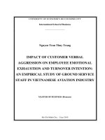
Fig. 1. The morphology of alkali-extracted cellulose before (a1 overall; a2 cellulose in fiber; a3 cellulose in parenchyma cell) and after the heat treatment at 180 °C (b1
overall; b2 cellulose in fiber; b3 cellulose in parenchyma cell) and 200 °C (c1 overall; c2 cellulose in fiber; c3 cellulose in parenchyma cell).
methods for extraction and heat treatment were the same as our previous study and the only difference was the order of processing. The
first step was to extract cellulose from bamboo, and then heat treated
the cellulose. The results showed that heat treatment alone did not
change the morphology of cellulose. The reason why heat-treated
bamboo was more prone to deformation after cellulose extraction can
only be attributed to the promoting effect of heat treatment history.
Although there were no obvious changes in the morphology of
cellulose, elevated temperature could cause the thermal expansion of
cellulose (Fig. 1b2 and c2) and also exacerbate the surface aggregation
of microfibers. However, it seemed that heat treatment had little effect
on the morphology of parenchyma cells (Fig. 1a3, b3 and c3), which
could be ascribed to the different cell wall structure and microfibril
arrangement of fiber cells and parenchyma cells. The average width of
cellulose before heat treatment was 12.02 ± 2.2 μm while the diameter
of fibers became slightly larger after heat treatment at 180 °C
(14.4 ± 1.5 μm). Previous study has shown that the thermal expansion
behaviors of cellulose was ascribed to the intermolecular hydrogen
bonding systems (Hori & Wada, 2005; Wada, 2002), which will be
further analyzed in section 3.1.3.
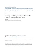
Fig. 2. XRD diffractograms of the natural bamboo, alkali-extracted cellulose
and its heat-treated samples at 180 °C and 200 °C.
cannot change the structural type of cellulose but the process of heat
treatment was an efficient manner for transforming the cellulose I to II
during conventional alkali treatment, which was of great significance to
the mercerization of cellulose (Huang et al., 2019).
The empirical measurements of CrI were used to allow rapid comparison of the changing of crystallinity of cellulose samples upon heat
treatment. According to Table 1, the CrI of A-Cell-Co (69.70 %)
3.1.2. Crystal structure and crystallinity of cellulose by XRD
In addition to morphology, we are most concerned about whether
heat treatment will affect the supramolecular structure of bamboo
cellulose or not. Fig. 2 shows the X-ray diffractograms of natural
bamboo, alkali-extracted cellulose and the cellulose with heat treatment at 180 °C and 200 °C. The typical peaks of both cellulose I and II
are observed, indicating that the alkali extraction process changed the
cellulose crystal type. For natural bamboo, three major diffraction
planes of cellulose I named 200, 110 and 004 were presented at 2θ =
22.23°, 15.74° and 34.51° (Maheswari et al., 2012). For other cellulose
samples, their X-ray diffractograms presented two additional diffraction
planes of 1–10 (2θ = 12.23°) and 110 (2θ = 19.54°), which belonged
to typical cellulose II structure. This indicated that the alkali extraction
process indeed changed the supramolecular structure of cellulose.
There was no new peak in the diffractogram of heat-treated cellulose, nor the dramatic changes from cellulose I to II seen in our previous
study. The results confirmed that heat treatment (≤200 °C) itself
Table 1
The CrI (%) of natural bamboo, alkali-extracted cellulose and its heat-treated
samples at 180 °C and 200 °C.
3
Sample
Iam
I200
CrI (%)
Bamboo
A-Cell-Co
A-Cell-180 °C
A-Cell-200 °C
7547
6602
5940
4222
15,756
21,783
19,689
16,780
52.10
69.70
69.83
74.84
Carbohydrate Polymers 241 (2020) 116412
Q. Lin, et al.
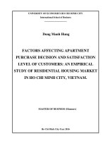
Fig. 3. Hydrogen-bonding schemes between center chains in cellulose I and II (a), origin chains in cellulose II (b) and center chain and origin chain in cellulose II (c).
schemes of 200 crystal plane, in which the scheme A was the typical
form for cellulose I. In the case of scheme A, the intramolecular hydrogen bonding (intra eOH) formed in intramolecular chain, involving
O3 and O2 as donors and O5 and O6 as acceptors (O3H—O5 and O2H—
O6), respectively. Meanwhile, the intramolecular hydrogen bonding
(inter eOH) of O6H—O3 was found between adjacent center chains,
which was almost the main form of inter eOH in cellulose I. However,
some studies considered that there was a possible inter eOH in cellulose I, which involved O6 as donor and O2 as accepter (Oh, Yoo, Shin, &
Seo, 2005). Even so, O6H—O2 was energetically less competitive, that
is, little contribution can be made to the stabilization of the supramolecular organization due to the very weak interaction between two
groups (Mazeau, 2005). As for cellulose II in the scheme B, only one
intra eOH and one inter eOH were formed: (1) between the O3H group
and the neighboring O5 atom (intra eOH); (2) between the O6H group
and the O2 atom of the glucose ring of the adjacent chain (inter eOH).
This was because the alteration of cellulose conformation happened
during the mercerization process, causing more O6H-O2 generated or
retained (Sturcova, His, Wess, Cameron, & Jarvis, 2003). In addition,
another specific inter eOH (O2H—O2) was formed between the origin
increased by 17.6 % with the removal of lignin and hemicelluloses,
compared with that of natural bamboo (52.10 %). Heat treatment,
within a certain range, can improve the crystallinity of cellulose
(Weimer et al., 1995). The heat treatment at 200 °C improved the CrI of
cellulose from 69.70 % to 74.84 %. This was a consequence of the
partial recrystallization of amorphous regions or the partial co-crystallization of crystalline zones in adjacent fibrils (Wan, Wang, & Xiao,
2010). From the FT-IR spectrum in Fig. 4a, the bands at approximately
1736 cm−1 and 1512 cm−1 were assigned to the hemicellulose (C]O
stretching in unconjugated ketones) and lignin (C]C stretching of the
aromatic ring). After extraction, the peak of lignin disappeared while
there was a residual peak at hemicellulose peak. The residual hemicellulose, as the amorphous substance, degraded during the subsequent
heat treatment, resulting in an increase in the relative proportion of
crystalline zone.
3.1.3. Hydrogen-bonding system of cellulose by FT-IR
Figs. 3 and 4 shows the three kinds of hydrogen-bonding patterns in
both cellulose I and II, which are defined as scheme A, B and C, respectively. The scheme A and B were the two hydrogen-bonding
Fig. 4. The FT-IR absorbance spectra of bamboo and alkali-extracted cellulose and its heat-treated samples at 180 and 200 °C (4000 - 500 cm−1) (a), the peak
separation of hydrogen bonding OH stretching of all the samples (3800 - 3000 cm−1) (b) and the content and wavenumber assignments of free eOH (peak 1), intra
OeH (peak 2) and inter eOH (peak 3 and 4) (c).
4
Carbohydrate Polymers 241 (2020) 116412
Q. Lin, et al.
into a more swollen state with a rough surface when exposing at elevated temperatures (Hori & Wada, 2005, 2006; Wada, 2002). The hydrogen bond system in cellulose has a great correlation with the lateral
size, which will be discussed in following Section 3.4. More importantly, the cellulose extracted by the two methods used in this paper
did not distort and shrink after heat treatment, proving that heat
treatment did not have a great impact on the morphology and structure
of cellulose again.
chain and center chain in 110 crystal plane (Scheme C).
Fig. 4a displays the infrared spectrum of natural bamboo, alkaliextracted cellulose and heat-treated samples. As discussed above, the
lignin was almost completely removed, but some hemicellulose remained in the extracted cellulose. Fig. 4b shows the peak separation of
hydrogen bonding OH stretching of samples ranging from 3800 to 3000
cm−1, and the corresponding parameters of different hydrogen bond
types are shown in Fig. 4c. For the pure cellulose I, it was generally
considered that inter eOH was stronger than intra eOH. In other
words, the former was more difficult to be broken during the heat
treatment (El Oudiani, Msahli, & Sakli, 2017; Popescu, Popescu, &
Vasile, 2011). Nevertheless, the mercerizing cellulose extracted by high
concentration of alkali solution was the mixture of cellulose I and II.
Therefore, the contribution of hydrogen-bonding system of mercerized
cellulose should be considered. Moreover, it has generally been deemed
that the length between donor and acceptor atom was inversely proportional to the bond energy. Inter eOH had a higher average distance
than that of intra eOH in mercerized cellulose, thus, part of inter eOH
tended to be destroyed prior to intra eOH during the heating process.
Therefore, the above two conditions competed during the process.
Concerning these experimental results, the alkali extraction process
made the situation from intra eOH dominating to inter eOH taking
majority. The sum number of inter-chains slightly decreased after 180
°C and 200 °C treatment (Fig. 4c), which indicated that the heat
treatment mainly influenced the inter eOH of alkali-extracted cellulose.
In the case of 200 °C treated cellulose, the contents of the intermolecular and intramolecular hydrogen bonds were approximately the
same.
3.2.2. Crystal structure and crystallinity of cellulose by XRD
Fig. 6 shows the x-ray diffraction patterns of untreated cellulose by
mild extraction method and heat-treated cellulose. It can be seen from
Fig. 6a that the cellulose obtained by the mild extraction method had a
typical cellulose I structure, and there was no diffraction peak of cellulose II. This indicated that this extraction method would not destroy
the original structure of cellulose. Compared with the N-Cell-Co, there
were no new peak and no large shift of peak in the diffraction curves
after heat treatment, which suggested that heat-treated cellulose samples remained typical cellulose I structure. The results indicated that the
process of high temperature did not lead to the transformation of cellulose from parallel-chain structure to anti-parallel-chain of structure.
The degree of crystallinity was further obtained by amorphous
subtraction method, that is, crystallinity was determined by subtracting
the amorphous contribution from diffraction spectra via an amorphous
standard. The crystallinity of N-cell-Co was 79.68 % and then an approximately 12 % decrease of degree of crystallinity was occurred after
the heat treatment at 180 °C (70.08 %) and 200 °C (69.06 %). It was
worth noting that the crystallinity of cellulose extracted by the first
method with high concentration of alkali solution increased after the
heat treatment. This difference may come from two aspects. On the one
hand, the cellulose obtained from the first extraction method was a
hybrid structure of cellulose I and cellulose II, and the crystallinity
calculated by using the empirical formula was not very accurate. On the
other hand, as mentioned above, the cellulose obtained by the first
method still contained residual hemicellulose, thus affecting the results.
Therefore, the crystallinity obtained by the second mild extraction
method was more reliable. That is, the crystallinity of pure cellulose
would decrease after the heat treatment at 180 and 200 °C.
3.2. Heat treatment of cellulose extracted at ambient conditions
The above experiments have proved that a single heat treatment
process will not result in the transformation of the crystal structure of
cellulose. However, due to the influence of alkali concentration (17.5
%) and temperature (75 °C) in the extraction method, the obtained
cellulose was a hybrid structure of cellulose I and cellulose II and had a
heating history, which brought complexity to the subsequent structural
analysis. Therefore, another mild extraction method was adopted,
which was carried out at room temperature and would not change the
structure of cellulose.
3.2.3. Hydrogen-bonding system of cellulose by FT-IR
Fig. 7 shows the FTIR spectra of bamboo, untreated cellulose by the
mild extraction method, and heat-treated cellulose. Compared with the
spectrum of bamboo, in the case of N-Cell-Co, the functional groups at
1738 cm−1 and 1505 cm−1 assigned to hemicellulose and lignin
(Popescu et al., 2011), respectively, were totally disappeared. This illustrated that pure cellulose can be obtained by this mild extraction
method. However, the band at 1738 cm−1 appeared again after the
heat treatment at 180 °C and a more prominent band at 200 °C. The
reappearance of the C]O stretching peak indicated treatment at high
temperature lead to the cleavage of cellulose and a rise of soluble
species. The active hydroxyl groups on the glucose ring in cellulose’s
macromolecular chain were oxidized to aldehyde, ketone and carboxyl
groups at high temperature. The cleavage of cellulose chains had occurred when the temperature reached 150 °C (Ma et al., 2013). This
may be the reason for the decrease of crystallinity of cellulose after the
heat treatment.
The peaks at 1425 cm−1 and 897 cm−1 are generally called “crystallinity band” and “amorphous band” in cellulose, respectively, and
the absorbance ratio A1425/A897 is considered as CrI (El Oudiani
et al., 2017; Oh et al., 2005). Compared with the N-Cell-Co, the CrI of
N-Cell-180 °C have decreased by 39 %. The results again proved that
high temperatures (≥180 °C) destroy not only the amorphous regions,
but also the crystalline regions of cellulose.
The large band from 3600 to 3200 cm−1 was ascribed to the different types of hydrogen bonds. Fig. 8a shows the 2nd derivative of this
region that can improve the resolution considerably and Table 2
3.2.1. Cellulose morphology by SEM
Fig. 5 shows the micrographs of untreated and heat-treated bamboo
cellulose derived from bamboo by mild extraction method. Since the
effects of stirring force caused by the magnetic stirrer, it can be seen
that the morphology of fibers and parenchyma cells was damaged to
some extent by mechanical effect, resulting cellulose clustered together
(Fig. 5a1). However, the presence of cellulose in the form of a single
fiber (Fig. 5a2) and a single parenchyma cell (Fig. 5a3) was still observed at high magnification. After the heat treatment at 180 and 200
°C, the morphology of cellulose remained essentially unchanged
(Fig. 5b1−3 and c1−3). Notably, a lot of pores with diameter about 40
nm–110 nm are observed in the N-Cell-200 °C sample (Fig. 5c2). Cellulose, as a natural biopolymer, is composed with glucose units connected by β-1,4-glycosidic bonds (Osullivan, 1997). The active hydroxyl groups on the glucose ring tended to be oxidized at high
temperatures, causing the degradation of cellulose (Ma et al., 2013;
Yousefifar, Baroutian, Farid, Gapes, & Young, 2017). Therefore, these
pores may be derived from the degradation of cellulose in the amorphous region under high temperature treatment. This unique porous
structure provided new possibilities for studying more potential applications of bamboo fibers, such as energy storage, drug or cell delivery,
catalysis, separation, etc.
By repeated measurements of the diameter of the single fiber, the
average diameters of N-Cell-Co, N-Cell-180 °C and N-Cell-200 °C was
6.01 μm, 9.20 μm and 9.87 μm, respectively. The cellulose was changed
5
Carbohydrate Polymers 241 (2020) 116412
Q. Lin, et al.
Fig. 5. The morphology of cellulose extracted by mild extraction method before (a1 overall; a2 cellulose in fiber; a3 cellulose in parenchyma cell) and after the heat
treatment at 180 °C (b1 overall; b2 cellulose in fiber; b3 cellulose in parenchyma cell) and 200 °C (c1 overall; c2 cellulose in fiber; c3 cellulose in parenchyma cell).
Fig. 6. XRD diffractograms of cellulose extracted by mild extraction method and its heat-treated samples at 180 and 200 °C (a) and the amorphous subtraction
analysis of N-Cell-Co (b), N-Cell-180 °C (c) and N-Cell-200 °C (d).
6
Carbohydrate Polymers 241 (2020) 116412
Q. Lin, et al.
Fig. 7. The FT-IR absorbance spectra of bamboo, the cellulose extracted by mild extraction method and its heat-treated samples at 180 and 200 °C (4000–500 cm−1)
(a), the peak separation of hydrogen bonding OH stretching of all the samples (3800–3000 cm−1) (b) and the crystallinity of all the samples, content and wavenumber assignments of free eOH (peak 1), intra OeH (peak 2) and inter eOH (peak 3) (c).
inter eOH (O6H‑‑‑O3).
After the extraction, the contents of intra eOH and inter eOH in NCell-Co were similar. However, inter eOH gradually dominated in
cellulose after the thermal treatment. When the temperature reached
200 °C, the contents of inter eOH, intra eOH and free eOH were 67.11
%, 28.32 % and 4.57 %, respectively. In cellulosic structure, the hydrogen bonds with high energy tended to form between cellulose molecules and chains (intermolecular) rather than in the same molecule
(intramolecular) (El Oudiani et al., 2017; Popescu et al., 2011).
Therefore, intra eOH was more easily to be damaged during heat
treatment, bringing about the decreasing of its relative content. From
the changes of the content ratio of OH bands and CrI after the heat
treatment, it can be inferred that there was no correlation between the
crystallization degree of cellulose and the content ratio of the inter-H
bonds and the intra eOH.
From the Fig. 8a, the blue shifts were observed in all the absorption
frequencies of the free eOH, intra eOH and inter eOH, which indicated
an increase in bond energy and a decrease in bond length. It could be
considered that the alkali treatment had positive influence on the hydrogen bond energy of cellulose. Nevertheless, the effect of temperature
summarized the IR assignments of main functional groups for OH bond
regions. It was worth noting that the peak of cellulose Iβ (at 3273 cm−1)
had a blue shift after 200 °C heat treatment, indicating an increase in
bond energy and the formation of more stable groups. Meanwhile, the
absorbance at 3234 cm−1 assigned to cellulose Iα almost disappeared.
The crystalline dimorphism of native cellulose (R. H. Atalla &
Vanderhart, 1984; Atalla et al., 2014), cellulose Iα and Iβ were considered to have different hydrogen bonds rather than the conformation
(Janardhnan & Sain, 2011; Michell, 1993; Sugiyama, Persson, &
Chanzy, 1991). Cellulose Iα (one-chain triclinic structure) can be irreversibly converted into cellulose Iβ (two-chain monoclinic structure)
upon heating (Sun, Sun, Fowler, & Baird, 2004). According to Wada
(Wada et al., 2003), the high temperature would induce the rearrangement of hydrogen bonds in cellulose, causing the transition
from Iα to Iβ.
Hydroxyl degradation in cellulose mainly begins from the amorphous region and follows to the semi-crystalline and crystalline region
(Mitsui, Inagaki, & Tsuchikawa, 2008). In order to explore the changes
of hydrogen-bonding system in cellulose, Fig. 8c displays the curvefitted OH bands of free eOH, intra eOH (O2H‑‑‑O6 and O3H‑‑‑O5) and
Fig. 8. The 2nd derivative of FT-IR spectra (3800–3200 cm−1) for bamboo, cellulose extracted by mild extraction method and its heat-treated samples at 180 and 200
°C (a) and the hydrogen-bonding scheme in cellulose I (b).
7
Carbohydrate Polymers 241 (2020) 116412
Q. Lin, et al.
Table 2
IR assignments of eOH bond region in the 2nd derivative of FT-IR spectrum (3800–3200 cm−1).
Wave number (cm−1)
Band assignment
Reference
3582, 3539
3558
3458, 3411
3337
3273(3234)
Free-OH
Absorbed water weakly bound
O2H‑‑‑O6 intramolecular hydrogen bonding in cellulose
O3H‑‑‑O5 intramolecular hydrogen bonding in cellulose
O6H‑‑‑O3 intermolecular hydrogen bonding in cellulose Iβ(Iα)
(El Oudiani
(Popescu et
(El Oudiani
(El Oudiani
(El Oudiani
on the bond energy of three types of hydrogen bonds was different. The
peak at 3576 cm−1 in cellulose shifted to 3565 cm−1 after 200 °C
treatment, representing the decrement of bond energy of free eOH.
Meanwhile, the blue shifts from 3455 cm−1 and 3264 cm−1 to 3462
cm−1 and 3316 cm−1, respectively, indicated the both shortening of
the intra eOH and inter eOH length and the increasing bond energy.
Studies has shown that the lateral behavior of thermal expansion
was ascribed to the inter-molecular hydrogen bonding system in cellulose. Plenty of inter eOH existed along the parallel direction to the
glucose ring (b-axis) but little existed along the perpendicular direction
(a-axis) (Hori & Wada, 2005; Mazeau, 2005; Wada, 2002; Wada et al.,
2003). Thus, due to the decrement of the hydrogen bonds after the heat
treatment, there was also a lack of inter eOH with higher bond energy
in a-axis. These overall changes caused a strain along b-axis and an
expanse along a-axis in the cellulose unit cell.
et al., 2017)
al., 2011)
et al., 2017; Yue et al., 2015)
et al., 2017; He, Tang, & Wang, 2007; Popescu et al., 2011)
et al., 2017; He et al., 2007; Popescu et al., 2011)
of thermally modified softwoods and its relation to polymeric structural wood constituents. Annals of Forest Science, 64(7), 679–690.
Cai, M., Takagi, H., Nakagaito, A. N., Katoh, M., Ueki, T., Waterhouse, G. I. N., et al.
(2015). Influence of alkali treatment on internal microstructure and tensile properties
of abaca fibers. Industrial Crops and Products, 65, 27–35.
Chen, H., Yu, Y., Zhong, T., Wu, Y., Li, Y., Wu, Z., et al. (2016). Effect of alkali treatment
on microstructure and mechanical properties of individual bamboo fibers. Cellulose,
24(1), 333–347. />Cheng, D. L., Jiang, S. X., & Zhang, Q. S. (2013). Mould resistance of Moso bamboo
treated by two step heat treatment with different aqueous solutions. European Journal
of Wood and Wood Products, 71(1), 143–145.
Das, M., & Chakraborty, D. (2006). Influence of alkali treatment on the fine structure and
morphology of bamboo fibers. Journal of Applied Polymer Science, 102(5), 5050–5056.
El Oudiani, A., Msahli, S., & Sakli, F. (2017). In-depth study of agave fiber structure using
Fourier transform infrared spectroscopy. Carbohydrate Polymers, 164, 242–248.
Eronen, P., Osterberg, M., & Jaaskelainen, A. S. (2009). Effect of alkaline treatment on
cellulose supramolecular structure studied with combined confocal Raman spectroscopy and atomic force microscopy. Cellulose, 16(2), 167–178.
He, J. X., Tang, Y. Y., & Wang, S. Y. (2007). Differences in morphological characteristics
of bamboo fibres and other natural cellulose fibres: Studies on X-ray diffraction, solid
state C-13-CP/MAS NMR, and second derivative FTIR spectroscopy data. Iranian
Polymer Journal, 16(12), 807–818.
Hori, R., & Wada, M. (2005). The thermal expansion of wood cellulose crystals. Cellulose,
12(5), 479–484.
Hori, R., & Wada, M. (2006). The thermal expansion of cellulose II and IIIII crystals.
Cellulose, 13(3), 281–290.
Huang, Y., Meng, F., Liu, R., Yu, Y., & Yu, W. (2019). Morphology and supramolecular
structure characterization of cellulose isolated from heat-treated moso bamboo.
Cellulose, 26(12), 7067–7078.
Janardhnan, S., & Sain, M. (2011). Isolation of cellulose nanofibers: Effect of biotreatment
on hydrogen bonding network in wood fibers. International Journal of Polymer
Science, 6.
Kuribayashi, T., Ogawa, Y., Rochas, C., Matsumoto, Y., Heux, L., & Nishiyama, Y. (2016).
Hydrothermal transformation of wood cellulose crystals into pseudo-orthorhombic
structure by cocrystallization. ACS Macro Letters, 5(6), 730–734.
Ma, X. J., Cao, S. L., Lin, L., Luo, X. L., Hu, H. C., Chen, L. H., et al. (2013). Hydrothermal
pretreatment of bamboo and cellulose degradation. Bioresource Technology, 148,
408–413.
Ma, X. J., Yang, X. F., Zheng, X., Lin, L., Chen, L. H., Huang, L. L., et al. (2014).
Degradation and dissolution of hemicelluloses during bamboo hydrothermal pretreatment. Bioresource Technology, 161, 215–220.
Maheswari, C. U., Reddy, K. O., Muzenda, E., Guduri, B. R., & Rajulu, A. V. (2012).
Extraction and characterization of cellulose microfibrils from agricultural residue Cocos nucifera L. Biomass & Bioenergy, 46, 555–563.
Mazeau, K. (2005). Structural micro-heterogeneities of crystalline I beta-cellulose.
Cellulose, 12(4), 339–349.
Mehrotra, R., Singh, P., & Kandpal, H. (2010). Near infrared spectroscopic investigation
of the thermal degradation of wood. Thermochimica Acta 507-08, 60-65.
Meng, F. D., Yu, Y. L., Zhang, Y. M., Yu, W. J., & Gao, J. M. (2016). Surface chemical
composition analysis of heat-treated bamboo. Applied Surface Science, 371, 383–390.
Michell, A. J. (1993). 2nd-derivative ftir spectra of native celluloses from valonia and
tunicin. Carbohydrate Research, 241, 47–54.
Mitsui, K., Inagaki, T., & Tsuchikawa, S. (2008). Monitoring of hydroxyl groups in wood
during heat treatment using NIR spectroscopy. Biomacromolecules, 9(1), 286–288.
Oh, S. Y., Yoo, D. I., Shin, Y., Kim, H. C., Kim, H. Y., Chung, Y. S., et al. (2005). Crystalline
structure analysis of cellulose treated with sodium hydroxide and carbon dioxide by
means of X-ray diffraction and FTIR spectroscopy. Carbohydrate Research, 340(15),
2376–2391.
Oh, S. Y., Yoo, D. I., Shin, Y., & Seo, G. (2005). FTIR analysis of cellulose treated with
sodium hydroxide and carbon dioxide. Carbohydrate Research, 340(3), 417–428.
/>Osullivan, A. C. (1997). Cellulose: The structure slowly unravels. Cellulose, 4(3), 173–207.
Popescu, C. M., Popescu, M. C., & Vasile, C. (2011). Structural analysis of photodegraded
lime wood by means of FT-IR and 2D IR correlation spectroscopy. International
Journal of Biological Macromolecules, 48(4), 667–675.
Sharma, B., Shah, D. U., Beaugrand, J., Janecek, E. R., Scherman, O. A., & Ramage, M. H.
(2018). Chemical composition of processed bamboo for structural applications.
Cellulose, 25(6), 3255–3266.
Sturcova, A., His, I., Wess, T. J., Cameron, G., & Jarvis, M. C. (2003). Polarized vibrational
Spectroscopy of fiber polymers: Hydrogen bonding in cellulose II. Biomacromolecules,
4(6), 1589–1595. />Sugiyama, J., Persson, J., & Chanzy, H. (1991). Combined infrared and electron-diffraction study of the polymorphism of native celluloses. Macromolecules, 24(9),
4. Conclusion
In this study, two methods were used to extract cellulose from
bamboo, and then heat treatment (180 °C and 200 °C) was conducted
on the two types of celluloses. Their results were basically the same,
that is, the heat treatment (≤ 200 °C) would not change the aggregation structure of bamboo cellulose, but it will cause the change of intermolecular and intermolecular hydrogen bonds, and the break of
glycosidic bonds in the amorphous region and part of the crystalline
region of cellulose. Take the cellulose isolated at ambient conditions for
example, after the heat treatment at 180 and 200 °C, the cellulose
samples remained typical cellulose I structure. The breaking of glycoside bond lead to more C]O bond formation. Accordingly, their degree
of crystalline decreased by 12 %. Intermolecular hydrogen bonds gradually dominated with the content from 42 % to 67 %. These changes in
molecular and supramolecular structures of cellulose samples were ultimately reflected in the changes in their morphology as thermal expansion in the width direction.
5. Credit author statement
Qiuqin Lin: Investigation, Formal analysis, Writing- Original draft
preparation.
Yuxiang Huang: Conceptualization, Methodology, WritingReviewing and Editing.
Wenji Yu: Resources, Data curation, Supervision.
Acknowledgements
This work was financially supported by the National Natural Science
Foundation of China (No. 31890771). The authors thank Professor
Alfred D. French for providing the Maud software and its instructions.
References
Atalla, R. H., & Vanderhart, D. L. (1984). Native cellulose: a composite of two distinct
crystalline forms. Science (New York, N.Y.), 223(4633), 283–285.
Atalla, R. S., Crowley, M. F., Himmel, M. E., & Atalla, R. H. (2014). Irreversible transformations of native celluloses, upon exposure to elevated temperatures.
Carbohydrate Polymers, 100, 2–8.
Boonstra, M. J., Van Acker, J., Tjeerdsma, B. F., & Kegel, E. V. (2007). Strength properties
8
Carbohydrate Polymers 241 (2020) 116412
Q. Lin, et al.
48, 169–178.
Yildiz, S., & Gumuskaya, E. (2007). The effects of thermal modification on crystalline
structure of cellulose in soft and hardwood. Building and Environment, 42(1), 62–67.
Yousefifar, A., Baroutian, S., Farid, M. M., Gapes, D. J., & Young, B. R. (2017).
Hydrothermal processing of cellulose: A comparison between oxidative and nonoxidative processes. Bioresource Technology, 226, 229–237.
Yue, Y. Y., Han, J. Q., Han, G. P., Zhang, Q. G., French, A. D., & Wu, Q. L. (2015).
Characterization of cellulose I/II hybrid fibers isolated from energycane bagasse
during the delignification process.: Morphology, crystallinity and percentage estimation. Carbohydrate Polymers, 133, 438–447.
Zhang, Y. M., Yu, Y. L., & Yu, W. J. (2013). Effect of thermal treatment on the physical
and mechanical properties of phyllostachys pubescen bamboo. European Journal of
Wood and Wood Products, 71(1), 61–67.
2461–2466.
Sun, X. F., Sun, R. C., Fowler, P., & Baird, M. S. (2004). Isolation and characterisation of
cellulose obtained by a two-stage treatment with organosolv and cyanamide activated hydrogen peroxide from wheat straw. Carbohydrate Polymers, 55(4), 379–391.
Wada, M. (2002). Lateral thermal expansion of cellulose Iβ And IIII polymorphs. Journal
of Polymer Science Part B: Polymer Physics, 40(11), 1095–1102.
Wada, M., Kondo, T., & Okano, T. (2003). Thermally induced crystal transformation from
cellulose I-alpha to I-beta. Polymer Journal, 35(2), 155–159.
Wan, J. Q., Wang, Y., & Xiao, Q. (2010). Effects of hemicellulose removal on cellulose
fiber structure and recycling characteristics of eucalyptus pulp. Bioresource
Technology, 101(12), 4577–4583.
Weimer, P. J., Hackney, J. M., & French, A. D. (1995). Effects of chemical treatments and
heating on the crystallinity of celluloses and their implications for evaluating the
effect of crystallinity on cellulose biodegradation. Biotechnology and Bioengineering,
9