First report of electrospun cellulose acetate nanofibers mats with chitin and chitosan nanowhiskers: Fabrication, characterization, and antibacterial activity
Bạn đang xem bản rút gọn của tài liệu. Xem và tải ngay bản đầy đủ của tài liệu tại đây (6.2 MB, 9 trang )
Carbohydrate Polymers 250 (2020) 116954
Contents lists available at ScienceDirect
Carbohydrate Polymers
journal homepage: www.elsevier.com/locate/carbpol
First report of electrospun cellulose acetate nanofibers mats with chitin and
chitosan nanowhiskers: Fabrication, characterization, and
antibacterial activity
Antonio G.B. Pereira a, e, f, *, Andr´e R. Fajardo b, Adriana P. Gerola c, Jean H.S. Rodrigues d,
Celso V. Nakamura d, Edvani C. Muniz e, You-Lo Hsieh f
a
Universidade Tecnol´
ogica Federal do Paran´
a (UTFPR), Campus Dois Vizinhos (DV), Engenharia de Bioprocessos e Biotecnologia, Dois Vizinhos, PR, Brazil
Universidade Federal de Pelotas, Campus Cap˜
ao do Le˜
ao, Laborat´
orio de Tecnologia e Desenvolvimento de Comp´
ositos e Materiais Polim´ericos (LaCoPol), Pelotas, RS,
Brazil
c
Universidade Federal de Santa Catarina, Departamento de Química, Florian´
opolis, SC, Brazil
d
Universidade Estadual de Maring´
a, Departamento de An´
alises Clínicas, Maring´
a, PR, Brazil
e
Universidade Estadual de Maring´
a, Grupo de Materiais Polim´ericos e Comp´
ositos (GMPC), Departamento de Química, Av. Colombo 5790, 87020-900, Maring´
a, PR,
Brazil
f
University of California, Davis, Biological and Agricultural Engineering, One Shields Avenue, Davis, CA, 95616, USA
b
A R T I C L E I N F O
A B S T R A C T
Keywords:
Chitosan nanowhiskers
Chitin nanowhiskers
Electrospun nanofibers mats
Cellulose acetate
Physical adsorption
Physical adsorption has shown to be facile and highly effective to deposit chitosan nanowhiskers (CsNWs, 60 %
deacetylated, length: 247 nm, thickness: 4–12 nm, width:15 nm) on electrospun cellulose acetate nanofibers
(CANFs, 560 nm) to effect complete surface charge reversal from negatively charged CANFs (− 40 mV) to
positively charged CsNWs-adsorbed CANFs (+8 mV). The CsNWs coverage did not alter the smooth and ho
mogeneous morphology of fibers, as observed from SEM images. Biological assays showed the CsNWs covered
nanofibers were effective against the Gram-negative bacterium E. coli, reducing 99 % of colony forming units
(CFU) in 24 h and atoxic to healthy Vero cells. The use of CsNWs to modify cellulose fiber surfaces has been
proved to be efficient and may be applied to a broad scope of fields, especially as biomaterials and biomedical
applications.
Chemical compounds studied in this article:
Cellulose acetate (PubChem CID: 139600838)
Chitin (PubChem CID: 6857375)
Chitosan (PubChem CID: 71853)
3-(4,5-Dimethylthiazol-2-yl)-2,5diphenyltetrazolium bromide (PubChem CID:
64965)
Sodium hydroxide (PubChem CID: 14798)
Hydrochloric acid (PubChem CID: 313)
1. Introduction
Electrospinning has been recognized as a versatile technique in the
preparation of ultra-fine fibrous mats (Doshi & Reneker, 1995; Xue, Wu,
Dai, & Xia, 2019). Due to the ease to generate nanometer to submicron
wide fibers from a great variety of polymers as well as the intrinsically
high specific surface and widely possible porosity, electrospun fibers
have been investigated for many applications including tissue engi
neering (Orlova, Magome, Liu, Chen, & Agladze, 2011; Zhang, Ven
ugopal et al., 2008), filtration (Beier, Guerra, Garde, & Jonsson, 2006),
metal ion removal (Haider & Park, 2009), drug release (Ma et al., 2011)
and catalysis (Yousef et al., 2012). Among naturally derived polymers,
one of particular interest is the cellulose acetate (CA), a soluble
esterified-derivative of the biopolymer cellulose that can be easily
electrospun into nanofibers mats (Liu & Hsieh, 2002). Not only the
versatile solvent systems allow CA to be mixed with a large number of
polymers and compounds (Du & Hsieh, 2009; Zhang, Hsieh, Zhang, &
Hsieh, 2008), but easy hydrolysis of CA to cellulose also enables further
chemical reactions to functional materials (Chen & Hsieh, 2005; Wang &
Hsieh, 2004).
* Corresponding author at: Universidade Tecnol´
ogica Federal do Paran´
a (UTFPR), Campus Dois Vizinhos (DV), Engenharia de Bioprocessos e Biotecnologia, Dois
Vizinhos, PR, Brazil.
E-mail address: (A.G.B. Pereira).
/>Received 12 April 2020; Received in revised form 6 August 2020; Accepted 13 August 2020
Available online 19 August 2020
0144-8617/© 2020 Elsevier Ltd. This article is made available under the Elsevier license ( />
A.G.B. Pereira et al.
Carbohydrate Polymers 250 (2020) 116954
Different approaches such as addition of fillers, preparation of
bicomponent fibers, surface modification, among others, have been used
to provide or further improve some properties of electrospun nanofibers.
Multiwalled carbon nanotubes were incorporated into cellulose nano
fibers rendering fibers with improved water wettability, higher specific
surface, and mechanical properties (Lu & Hsieh, 2010). The addition of
ZnO nanoparticles into electrospun CA nanofibers showed improvement
in both hydrophobicity and antibacterial activity (Anitha, Brabu, Thir
uvadigal, Gopalakrishnan, & Natarajan, 2012). Phase-separated core-
shell bicomponent nanofibers were produced by the electrospinning of
CA and polyethylene oxide (Zhang & Hsieh, 2008). Cellulose fibrous
membrane, obtained from hydrolysis of electrospun CA, was success
fully functionalized with Cibracon Blue F3GA for lipase enzyme immo
bilization to enhance high catalytic rate and persistent activity
compared to that from free form of lipase (Lu & Hsieh, 2009).
Surface modification takes the advantage of the high specific surface
of electrospun fibers and is attractive. The negatively charged nature of
CA has been utilized to deposit positive species by physical adsorption
ˇ cík, & Lyutakov, 2019), such
(Elashnikov, Rimpelov´
a, Dˇekanovský, Svorˇ
as alternating assembling of positively charged polyethyleneimine and
negatively charged graphene oxide as an ammonium sensor (Jia, Yu,
Zhang, Dong, & Li, 2016) and hydroxyapatite and chitosan as corrosion
resistant and bioactive coating agents in metallic implants (Zhong, Qin,
& Ma, 2015). Chitin nanocrystals were used as surface modifying agents
to reverse the hydrophobic nature of CA mats to render super hydro
philic electrospun mats to be used as water filtration system (Goetz,
Jalvo, Rosal, & Mathew, 2016). Moreover, the biofouling and biofilm
formation were significantly reduced in the coated membranes while the
material presented a web-like structure with reduced pore size.
The use of bioactive chitosan is attractive due to its many attractive
physicochemical and biological properties well-documented in the
literature (Berger, Reist, Mayer, Felt, & Gurny, 2004). Chitosan is a
linear polysaccharide composed of randomly distributed β-(1→4)-linked
D-glucosamine and N-acetyl- d-glucosamine units derived from the
biopolymer chitin (Berger et al., 2004), to exhibit polycationic behavior
in pH conditions lower than the pKa of its amino groups (~6.5) (Dash,
Chiellini, Ottenbrite, & Chiellini, 2011). This feature endows chitosan
with a self-assembling ability triggered by the formation of poly
˜ ones, Peniche, & Peniche,
electrolyte complexes with polyanions (Quin
2018). Due to this ability, chitosan has been extensively studied in the
modification of negatively charged surfaces due to electrostatic inter
action (Antunes et al., 2011; Tu et al., 2019). Multiple alternating bi
layers based on chitosan and sodium alginate (SA) can be easily
assembled on CA fibers (Ding, Du, & Hsieh, 2011). Increasing such bi
layers has shown to reduce the permeability of pure water and NaCl
solution (Ritcharoen, Supaphol, & Pavasant, 2008).
Although the preparation of both chitin and chitosan nanocrystals,
highly crystalline spindle-like material with nanometric dimensions, is
well established (Bai et al., 2020; Pereira, Muniz, & Hsieh, 2014; Per
eira, Muniz, & Hsieh, 2015), the use of chitosan nanowhiskers (CsNWs)
to modify the surface of CA nanofibers mats have not been reported yet.
Therefore, this study develops processes to fabricate electrospun CA
nanofibrous mats with CtNWs incorporated in the spin dope or CsNWs
adsorbed on the fiber surfaces. The focus includes how this embodiment
of CsNWs in electrospun CA fibers and the effect on their surface charge
properties. We hypothesize that the CsNWs coating may enhance the
biological activity of the CA nanofibers, which potentiate their further
use in biomedical applications.
dimethylacetamide (DMAc, 99.8 %), acetone (P.A.), sodium hydroxide
(NaOH, 97 %), potassium hydroxide (KOH, 85 %), sodium chlorite
(NaClO2, 80 %), hydrochloric acid (HCl, 36.5 %) were purchased from
EMD Chemicals (USA). Phosphate buffer solution pH 4.0 was purchased
from Dinˆ
amica (Brazil). Dulbecco’s Modified Eagle Medium (DMEM)
and fetal bovine serum (FBS) were purchased from Gibco (USA). 3-(4,5dimethylthiazol-2-yl)-2,5-diphenyltetrazolium bromide (MTT) was
purchased from Gen-View Scientific Inc. (USA). All chemicals were used
as received, without further purification.
2.2. Isolation of chitin nanowhiskers
Chitin nanowhiskers (CtNWs) were prepared using the same protocol
described by Pereira et al. (Pereira et al., 2014, 2015) without modifi
cations. Commercial chitin was firstly purified by removing residual
proteins followed by bleaching. Proteins were removed by heating 5 g of
chitin in 150 mL of KOH solution (5 w/v-%) at boil under vigorous
stirring for 6 h. The suspension was kept under stirring at 25 ◦ C for
another 12 h, filtered and washed with water. Next, the collected solid
was bleached in 150 mL of 1.7 % NaClO2 in pH 4 buffer acetate at 80 ◦ C
for 2 h, then filtered and washed with water. The bleaching reaction was
performed twice. Finally, the bleached solid was re-suspended in 150 mL
of KOH solution (5 w/v-%) for 48 h, then centrifuged, washed, and oven
dried (50 ◦ C) to yield 71 % (~3.6 g).
CtNWs were obtained by hydrolyzing the purified chitin in 3 mol/L
HCl at boil for 90 min under stirring. The ratio chitin/volume of HCl
solution (g/mL) was fixed at 1:30. The reaction was stopped by adding
50 mL of cold water and centrifuged (3400 rpm for 15 min). The pre
cipitate was re-suspended in 200 mL of distilled water followed by
centrifugation. This procedure was repeated three times. Next, the
precipitate was re-suspended in distilled water (200 mL) and dialyzed
(molecular weight cut-off 12,000 g/mol) against water at room tem
perature (~25 ◦ C) up to neutral pH. The suspension was sonicated (40 %
maximum amplitude) for a total of 20 min with 5 min of interval be
tween every 5 min of sonication cycle, followed by centrifugation (3000
rpm, 10 min) for removing any remaining precipitate. Finally, the
CtNWs suspension was stored at 8 ◦ C. The yield was 65 % (~2.3 g)
2.3. Synthesis of chitosan nanowhiskers
Chitosan nanowhiskers (CsNWs) were synthesized via deacetylation
of the as-prepared CtNWs. For this, 50 mL of an aqueous suspension
containing ~ 500 mg of CtNWs were diluted with a NaOH solution (100
mL, 50 w/v-%) under stirring at 50 ◦ C for 48 h. Next, 100 mL of distilled
water was added to the system, which was centrifuged (5000 rpm for 10
min) to collect the precipitate, and the water-adding and centrifugation
process was repeated two more times. The CsNWs suspension was dia
lyzed against distilled water (molecular weight cut-off 12,000 g/mol) for
72 h at room temperature (~25 ◦ C) until neutral pH. The pH of this
aqueous CsNWs supernatant was adjusted to 3 using 1 mol/L HCl, then
homogenized by sonication. Finally, the suspension was centrifuged
(3000 rpm for 5 min) to remove last remaining precipitate. The CsNWs
yield was 74 % as compared with the CtNWs initial mass. CsNWs sus
pension (1.0 w/v-% or 10 mg/mL) was stored in a fridge (8 ◦ C) prior to
its use.
2.4. Fabrication of the nanofiber mats
2.4.1. CA nanofibers
CA homogeneous solution was prepared by dissolving it in 2:1 v/v
acetone:DMAc solution (total volume 10 mL) under stirring for 24 h at
room temperature (~25 ◦ C). The solution concentration was fixed at 15
w/v-% of CA (i.e., 1.5 g of CA in 10 mL of acetone:DMAc). Next, this
solution was electrospun using the same protocol described by Liu et al.
(Liu & Hsieh, 2002) with slight modification. The CA solution (10 mL)
was put into a 20 mL syringe (Henk Sass Wolf, Germany) equipped with
2. Materials and methods
2.1. Materials
Cellulose acetate (CA, 39.8 % acetyl content, and Mn ≈30,000 Da);
chitin from crab shells (practical grade), tryptic soy broth (TSB) and
tryptic soy agar (TSA) were purchased from Sigma-Aldrich (USA). N,N2
A.G.B. Pereira et al.
Carbohydrate Polymers 250 (2020) 116954
a metal 21 or 24-gauge needle. Then, the solution was spun under a 14
kV using a DC power supply (0–30 kV, Gamma High Voltage Research
Inc., USA) and a flow rate of 1 mL/h controlled by a syringe pump (KD
Scientific, model KDS 200, USA). The electrospun nanofibers mats were
collected in a vertically positioned grounded aluminum plate (30 × 30
cm) located at 25 cm (horizontal direction) from the tip of the needle.
The electrospun mats (labeled as CANFs) were vacuum dried at ambient
temperature (~25 ◦ C) with 85 % of yield (~1.3 g).
2.6. Antibacterial activity
The antibacterial activity of nanofiber mats was assessed using
Escherichia coli (E. coli) ATCC 26922 as model microorganism. The
number of living cells was determined by the viable counting method
(Rauf et al., 2019; Xu et al., 2011). Firstly, 950 μL of nanofibers mats or
300 μL CsNWs suspensions were transferred to Eppendorfs containing
50 μL of E. coli (5 × 10⁷ CFU/mL). The volume was adjusted to 1 mL
using a physiological saline solution. Then, the samples were incubated
at 37 ◦ C for 1 and 24 h. Aliquots were collected from the supernatant and
diluted with Tryptic Soy Broth (TSB) to a final concentration of 104
CFU/mL. Then, 30 μL were added to Tryptic Soy Agar (TSA) plates and
incubated at 37 ◦ C for 24 h, prior CFU counting. A sterile physiological
saline solution was used as control. The minimum inhibitory concen
tration (MIC) for CsNWs was 117 μg/mL and for CANFs the MIC was
negligible. All experiments were performed in triplicates.
2.4.2. CA nanofibers filled with CtNWs
The nanofibers filled with CtNWs were fabricated using a similar
protocol; however, specific amounts of CtNWs (0.5 or 2.5 w% in relation
to CA mass) were added in CA solutions before the electrospun process.
These mats were labeled as CANFs-CtNWs0.5 and CANFs-CTNWs2.5,
respectively.
2.4.3. CA nanofibers coated by CsNWs
The as-fabricated CANFs were coated with CsNWs via a physical
adsorption. Briefly, CANFs samples (5 mg) were immersed in a CsNWs
aqueous suspension (20 mL, 1 mg/mL, pH 3) for 3 h at room tempera
ture (~25 ◦ C). Next, the coated nanofibers mats (labeled as CANFsCsNWs) were recovered and rinsed in distilled water for 5 min. This
rinsing process was repeated three times. Finally, the CANFs-CsNWs
were vacuum dried at ambient temperature prior to characterization.
2.7. Evaluation of cytotoxicity
Vero cells (kidney epithelial cells extracted from an African green
monkey) were cultivated in DMEM supplemented with 2 mmol/L of Lglutamine and 10 % of fetal bovine serum (FBS). The cells were quan
tified and seed to 24 wells plates at 2.5 × 10⁵ cells/mL and incubated at
37 ◦ C and 5 % CO2. After 12 h, the culture medium was substituted by
DMEM free of serum, then polymer fragments (1 cm2) were added,
followed by 72 h of incubation. Cell viability was determined by MTT
assay (Mosmann, 1983). Cells cultivated in the absence of membranes
were used as control.
2.5. Characterization techniques
The chemical nature of the electrospun nanofibers was examined by
Fourier Transform Infrared (FTIR) spectroscopy. The spectra of samples
pressed with KBr were obtained in a Nicolet 6700 (Thermo Electron
Corporation, USA) spectrophotometer operating in the region from 400
to 4000 cm− 1, at a resolution of 4 cm− 1 and 64 scan acquisitions. FieldEmission Scanning Electron Microscopy (FE-SEM) was used to investi
gate the morphology of the nanofibers. Herein, dried samples were
sputtered coated with gold, then, imaged by FE-SEM (FI/Philips model
XL 30-SFEG, USA) operating at a 5 mm working distance and 5-kV
accelerating voltage. Fiber diameter distribution was measured using
ImageJ® software from 100 randomly fibers in different FE-SEM images
of the same sample. The X-ray diffraction (XRD) patterns of the nano
fiber samples were obtained in a Sintag powder diffractometer (model
XDS 2000, USA) equipped with a Ni-filtered Cu-Kα radiation source
operating at an anode voltage of 45 kV and a current of 40 mA. XRD
patterns were obtained in a scanning range of 5–50◦ with a scanning rate
of 1◦ /min. The crystallinity was calculated per Eq. (1):
C=
AC
x 100
AT
2.8. Statistical analysis
The data were analyzed by one-way analysis of variance (ANOVA)
followed by Newman-Keuls test. All analyses were performed using the
OriginPro® software (version 8.5, USA). Data are expressed as mean ±
standard error of the mean. Also, p < 0.05 was considered statistically
significant.
3. Results and discussion
3.1. Characterization of the nanofibers mats
CA dissolved easily in 2:1 v/v acetone/DMAc mixture, forming a
clear solution at 15 w%. Aqueous CtNWs suspension (up to 7.5 w%) at
pH 3 appeared homogeneous and slightly translucent, indicating
excellent dispersibility without precipitates (Pereira et al., 2014). Mix
ing aqueous CtNWs suspension with CA solution caused slight opales
cence, due to the presence of 5 % water that is a non-solvent for CA. The
CtNWs containing solutions remained homogeneous and with no pre
cipitation, indicating the solubility of CA was not significantly affected
by the addition of such a small percentage of water. Pure CA solutions
could be electrospun smoothly and continuously under the conditions
used. Electrospinning of CA/CtNWs mixtures, however, showed
considerable gelation at the needle tip in ca. 20 min that was resolved by
reducing the needle size from 21 to 24 gauge, or ca. 0.5 mm to 0.3 mm
inner diameter.
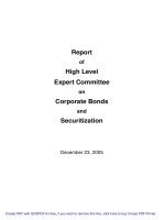
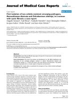
Electrospun CANFs appears as a white mat with uniform texture and
could be easily detached from the aluminum foil collector. SEM images
show the CANFs to be straight and uniform along the lengths of fibers
and well separated individual fibers with average diameter of 563 ± 222
nm and lengths at least several millimeters (Fig. 1a and b). The addition
of CtNWs did not change the gross appearance of the mats as evident of
well spatially distributed fibers (Fig. 1c and e); however, the fiber di
ameters were significantly reduced to 223 ± 76 nm for the CANFsCtNWs0.5 (Fig. 1c and d) and 240 ± 102 nm for CANFs-CtNWs2.5
(Fig. 1e and f). It is evident that the addition of CtNWs reduced fiber
diameters as the extent of diameter reduction exceeds the reduced
(1)
where AC is the total area under the crystalline diffraction peaks and AT
is the total area under the curve 2θ = 5◦ to 30◦ . The deconvolution
method was used to resolve the individual peaks. The data was
smoothed using 10 points in a second-order regression based on the
Savitzky-Golay filter, then deconvoluted based on Gaussian or Lor
entzian functions in the OriginPro® software (version 8.5, USA). Ther
mogravimetric analyses (TGA) were performed in a Shimadzu TGA50
Analyzer (Japan) equipment operating in a temperature range of
30–550 ◦ C at a heating rate of 10 ◦ C/min under N2(g) atmosphere (flow
of 50 mL/min). Differential Scanning Calorimetry (DSC) thermograms
were recorded in a Shimadzu DSC-60 calorimeter (Japan) operating in a
temperature range of 30–550 ◦ C at a heating rate of 10 ◦ C/min under N2
atmosphere (flow of 30 mL/min). Zeta potential measurements were
done in a Malvern ZetaSizer (model NanoZS90, USA) equipped with an
auto-titrator device (MPT-2). The nanofibers samples (~10 mg) were
immersed in an HCl solution (20 mL, pH 2) and sonicated for 6 min. By
adding 0.5 (mol/L) NaOH, different pH values (from 2 to 12) were
achieved in which the zeta potential was measured.
3
A.G.B. Pereira et al.
Carbohydrate Polymers 250 (2020) 116954
Fig. 1. SEM images of (a,b) CANFs, (c,d) CANFs-CtNWs0.5, and (e,f) CANFs-CtNWs2.5.
spinneret size. This effect resulted from the charged nature of CtNWs,
which increased the electrical conductivity of the CtNWs-containing CA
solution. Therefore, the polymer jet in the electrospinning process was
accelerated and stretched more than the jet in pure CA solution, leading
to decreased diameter of final fibers. This effect is consistent with what
has been reported (Haider, Haider, & Kang, 2018). Besides, the as-spun
mats at the higher 2.5 w% CtNWs showed more varying fiber size as well
as some beads, indicative of impaired electrospinning due to the higher
amounts of CtNWs.
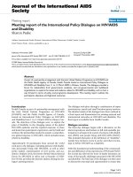
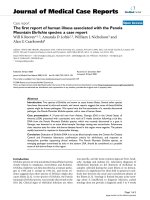
FTIR spectra of CANFs, CANFs-CtNWs0.5, CANF-CtNWS2.5, and
CtNWs to confirm the presence of the CtNWs filler within the nanofibers
mats (Fig. 2a). The spectrum of CANFs exhibited a broad band centered
at 3475 cm− 1 (O–H stretching of hydroxyl groups), bands in
2950–2890 cm− 1 region (C-H stretching of CHx groups), bands at 1744
cm− 1 (C=O stretching of carbonyl group), and bands at 1372 cm− 1 (CCH3stretching), 1244 cm− 1–(C-O-C anti-symmetric stretching ester
group) and 906 cm− 1 (a combination of –C-O stretching and CH2
rocking vibrations) (Rieger, Porter, & Schiffman, 2016). Also, the band
at 1646 cm− 1 can be associated with the presence of water molecules
(Sudiarti, Wahyuningrum, Bundjali, & Made Arcana, 2017). CtNWs
spectrum showed the chitin characteristic absorption bands at 3450
cm− 1–(O-H stretching), 3264 cm− 1 and 3103 cm− 1 (N-H stretching),
2900–2800 cm− 1 region (–C-H stretching), 1655 cm− 1 (amide I), 1560
cm− 1 (amide II), 1166 cm− 1– (C-N stretching), and 1070 cm− 1 (C-O
stretching) (Pereira et al., 2014). With low CtNWs added, the FTIR of
CANFs-CtNWs0.5 showed no noticeable change from CANFs, suggesting
the filler to be below the limit of detection and without observable
interaction with CA, and at this small concentration, it is not perceptible.
On the other hand, the CANFs-CtNWs2.5 spectrum exhibited the pres
ence of CtNWs with chitin characteristic bands at 3264 cm− 1 and 3105
cm− 1 (N-H stretching, 1560 cm− 1 (amide II), and 1070 cm− 1 (C-O
stretching). Furthermore, no changes in the position of the bands asso
ciated with CA were observed, suggesting weak interaction between the
CA matrix and the CtNWs filler.
4
A.G.B. Pereira et al.
Carbohydrate Polymers 250 (2020) 116954
Fig. 2. (a) CANFs, CANFs-CtNWs0.5, CANF-CtNWS2.5, and CtNWs. (b) XRD patterns of CtNWs, CANFs, and CANFs-CtNWS2.5.
The XRD pattern of CtNWs exhibited diffraction peaks at 2θ≈9.3◦ ,
19.1◦ , 20.7◦ , 23.2◦ , and 26.2◦ (Fig. 2b) corresponding to the (020),
(110), (120), (130), and (013) crystallographic planes of chitin (Beibei
Ding et al., 2012; Minke & Blackwell, 1978; Pereira et al., 2014). The
crystallinity of CtNWs was calculated to be 86 %, which corroborates
with our previous study (Pereira et al., 2014). The CANFs pattern did not
exhibit any diffraction peak due to the amorphous nature of the nano
fibers (Hamano et al., 2016). For the CANF-CtS2.5 mats, CA in the
CtNWs-containing nanofibers were also amorphous, similar to CANFs,
and the diffraction peaks of CtNWs were not observed, due likely to the
extent below the detection level.
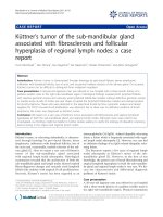
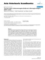
Thermal analysis (DSC and TGA/DTG) were used to examine the
effect of CtNWs addition on the thermal stability of CANFs. At compo
sitions up to 2.5 w% CtNWs, no significant effect was observed (Fig. 3).
DSC curve of CtNWs showed one exothermic broad peak in the tem
perature range of 250–450 ◦ C (Fig. 3a) associated with its thermal
degradation. The DSC curve of CANFs showed four thermal transitions.
The first transition occurred in the temperature range of 50–100 ◦ C due
Fig. 3. (a) DSC, (b) TGA, and (c) DTG curves obtained for CANFs, CtNWs, CANFs-CtNWS0.5, and CANFs-CtNWS2.5.
5
A.G.B. Pereira et al.
Carbohydrate Polymers 250 (2020) 116954
to the evaporation of adsorbed water. Besides, a minimal baseline
change (endothermic shoulder) around 225 ◦ C is attributed to Tg of CA
(Kendouli et al., 2014), followed by two endothermic peaks centered at
332 ◦ C and 400 ◦ C attributed to degradation stages of the poly
saccharide. The DSC curve of CANFs filled with CtNWs (0.5 or 2.5 w%)
showed reduced moisture endothermic peaks and barely distinguishable
endothermic transitions. For the mat containing the lowest amount of
CtNWs, the endothermic peak around 225 ◦ C was slightly reduced as
compared to CANFs. In comparison, the exothermic peak associated
with the degradation of CtNWs was sharpened and shifted to a
high-temperature range (maximum at 388 ◦ C). Moreover, the first
endothermic peak ascribed to the decomposition of CA (at 332 ◦ C) was
reduced. For the CANFs-CtNWS2.5 sample, this endothermic peak was
not observed, which may indicate an interaction between CA and CtNWs
by hydrogen bonding or hydrophobic interactions, considering the
chemical nature of these two compounds. Again, an intense endothermic
peak is still observed at 388 ◦ C due to the thermal degradation of CtNWs
filled in CANFs-CtNWS2.5.
TGA/DTG curves of pure CtNWs, CANFs, CANFs-CtNWS0.5, and
CANFs-CtNWS2.5 shown in Fig. 3b and c. For CANFs, a one stage
weight-loss of ~85 % was noticed occurring from 200 to 450 ◦ C with a
maximum temperature at 373 ◦ C. The TGA curve of CtNWs also
exhibited one weight loss state with a maximum temperature at 381 ◦ C
(weight loss ~85 %). Although it is expected some interaction between
CtNWs and CANFs, as noticed from the TGA/DTG curves, the addition of
different amounts of CtNWs did not change the thermal stability of
CANFs mats, independent of loading level. Similar to CANFs mat, the
TGA curves for CANFs-CtNWS0.5 and CANFs-CtNWS2.5 exhibited major
weight loss at maximum temperature around 373 ◦ C.
From these preliminary analyses, it was concluded that the addition
of CtNWs on the bulk phase of CANFs exerted only a slight effect on the
properties examined . Focusing on the modification of surface properties
of the CANFs, an alternative approach to took advantage of the fact that
CANFs are negatively charged at surface was investigated by depositing
CsNWs, a deacetylated CtNW-derivative, that can be positively charged
by protonating surface amino groups under acidic conditions (Pereira
et al., 2014).
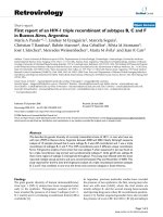
The surface charge properties of CANFs as is and with coated
nanowhiskers were measured to derive their zeta potential (ζ) values
under a full range of pH. CANFs exhibited ζ around − 40 mV from pH 4 to
pH 10 as expected (Fig. 4). With coated CsNWs, the CANFs-CsNWs
showed complete reversal to ζ around +8 mV from pH 2 to pH 10,
confirming the successful adsorption of cationic CsNWs on anionic
CANFs surfaces by electrostatic interactions. The ζ for aqueous CsNWs
suspension was around +40 mV (at pH < 6), which is an indicative of
their high stability under neutral and acidic conditions (Pereira et al.,
2014). While the surface adsorbed CsNWs more than neutralized the
negative charged CANFs, the lower positive zeta potential of
CANFs-CsNWs than CsNWs suggests CANF surfaces to be partially
covered with CsNWs under the condition studied. -However, complete
reversal of ζ was not observed for electrospun CA coated with chitin
nanocrystals (Goetz et al., 2016). The ζ of CtNWs and CANFs-CtNWs2.5
were also measured for comparison. While lower than CsNWs, the pre
dominant positively charged CtNWs under acidic conditions (pH < 6)
suggest partial hydrolysis the chitin moieties on their surfaces. However,
when CtNWs were internally doped, the resulting CANFs-CtNWs2.5 had
similarly negative charges as CANFs, indicating CtNWs to be imbedded
in the bulk of the fiber thus ineffective in altering surface charge char
acteristics. Intriguingly, upon heating at 180 ◦ C for 4 h,
CANFs-CtNWs2.5(180◦ ) also exhibited positive zeta potential similar to
CANFs-CsNWs. This confirms that the initially embedded inside the
nanofibers surfaced upon heating to be responsible for the positive
charge over a large pH range (CANFs-CtNWs2.5(180◦ )).
These zeta potential data are also useful to determine the isoelectric
point (IP) on pH values where there is no net surface charge. The IPs
derived were pHs at 2.7, 7.3 and 8.3, 9.9, 10.3, and 10.5 for CANFs,
CtNWs, CsNWs, CANFs-CsNWs, CANFS-CtNWs2.5, and CANFSCtNWs2.5(180◦ ), respectively. These surface charge characteristics
showed the surface adsorption of CsNWs approach to be indeed most
effective to modify the surface properties of the fibers. It is essential to
highlight that surface properties could be changed by merely immersing
CANFs in diluted CsNW suspensions. These drastic alterations of surface
charge nature of cellulose fibrous mats by surface adsorption or simple
dip coating with either chitin or chitosan nanowhiskers are successfully
demonstrated and reported for the first time.
The SEM of CANFs-CsNWs mat showed similar smooth morphology
and texture as CANFs and without any change in overall fiber distri
bution nor porosity, Fig. 5. The average diameter of the nanofibers was
430 ± 194 nm, statistically the same as that of CANFs.
Fig. 4. Zeta potential (ζ) data over pH for whiskers and nanofibrous mats.
6
A.G.B. Pereira et al.
Carbohydrate Polymers 250 (2020) 116954
3.2. Antibacterial activity and cytotoxicity studies
interaction with the bacterial cell membrane. Goetz et al. also demon
strated the antibacterial activity induced by chitin nanocrystals on
coated CA electrospun mats (Goetz et al., 2016).
The antibacterial activity of CANFs-CsNWs against E. coli is similar to
that of other electrospun CA or cellulose that contained conventional
metal nanoparticles (Ag, Ni, Co, Cu) and metal oxides (ZnO, CuO) as
antimicrobial agents. Most importantly, CANFs-CsNWs is advantageous
over the use of toxic and expensive reducing agents, advanced tech
niques (laser ablation) or complicated steps in their preparation
(Ahmed, Menazea, & Abdelghany, 2020; Demirdogen et al., 2020; Jatoi,
Kim, & Ni, 2019; Wu, Qiu, Wang, Zhang, & Qin, 2019). This fact high
lights the potential of the CANFs-CsNWs for more sustainable and
biocompatible applications.
Another crucial aspect of materials in the biomedical field is the
cytotoxic effects on healthy cells. Herein, the cytotoxicity of CANFs and
CANFs-CsNWs towards Vero cells were assessed by cell viability after 72
h of incubation (Fig. 7b). For all samples studied, the cell viability was
higher than the control (absence of fibrous mats), indicating the samples
not to be toxic to the healthy cells. Also, the higher cell viability when
incubated with CANFs and CANFs-CsNWs than that with the control
indicates that both mats increase cell density. In summary, the absence
of cytotoxicity in combination with the remarkable antibacterial prop
erties ranks CANFs-CsNWs as a promising material for medical
applications.
The antibacterial assays of CsNWs, CANFs, and CANFs-CsNWs
against E. coli, common and naturally occurring bacillar bacteria in
the human intestine that cause serious infections when present in food,
water, and bloodstream (Katouli, 2010), are displayed in Fig. 6. As
shown in agar plates (Fig. 6), both CsNWs and CtNWs reduced E. coli cell
viability slightly while CANFs-CsNWs showed greater reduction in 1 h
than CANFs that were not biologically active. These antibacterial ac
tivities were more pronounced observed for CsNWs and CtNWs and, for
CANFs-CsNWs mats after 24 h. Although CtNWs and CsNWs presented
antibacterial activity, it was lower than that observed for CANFs-CsNWs.
That anchoring at the CANFs surfaces provided CsNWs stability to
continuous interact with E. coli cells for longer times and optimize the
bacteriostatic effect is a significant finding. The saline solution used in
the culture medium as well as the liberation of cell components may
shield the charged groups of CsNWs to promote aggregation, decreasing
antibacterial effect and/or reducing suspension of free CsNWs as
compared to those bound to CANF membrane.
The colony-forming units (CFU) of samples as a function of time are
presented in Fig. 7a. CsNWs inhibited around 34 % of E. coli for 1 h of
contact. The inhibitory effect increased to 85 % for the longest contact
time (24 h). The minimum inhibitory concentration (MIC) was calcu
lated to be 117 μg/mL of CsNWs. The antibacterial activity of CANFs was
minimum even after 24 h of contact (~35 % of reduction); however, it
was calculated a decrease of 99 % of CFU when it was coated with
CsNWs.
The antibacterial activity efficiency of chitin/chitosan depends on
several factors: i) microorganism type; ii) charge density, molar mass,
and concentration; iii) physical state such as a solid or in solution; iv)
environmental condition such as pH, ionic strength, temperature and
contact time (Kong, Chen, Xing, & Park, 2010; Martins et al., 2014).
Gram-negative bacteria, like E. coli, have an external lipopolysaccha
rides (LPS) cell layer, which consists of lipidic compounds, and an inner
LPS layer, that bear anionic carboxylate and phosphate groups to sta
bilize the membrane by interacting with divalent ions. The antimicrobial
effect of chitin and chitosan is thus attributed to the electrostatic
attraction between their positively charged–NH+
3 with those negatively
charged (R-COO-, R-OPO(O2)2-) bacterial external cellular membrane to
destabilize and damage leading to cell death (Helander,
Nurmiaho-Lassila, Ahvenainen, Rhoades, & Roller, 2001). At above IP or
pH > pKa of amino groups, in which there are no positive charges on
chitin/chitosan, the action mode of such molecules on bacteria is
different. The amino groups act as chelating agents binding to the
divalent cations of the cellular membrane promoting the antibacterial
´sson, 2017). The fact that both CtNWs and
activity (Sahariah & Ma
CsNWs presented significant inhibition effect against E. coli may also
relate to their nano-scale dimensions and high specific surface and a
large number of surface amino groups available, increasing the
4. Conclusion
This study has validated the hypothesis that chitin (CtNWs) and
chitosan nanowhiskers (CsNWs) change the surface properties of elec
trospun cellulose acetate nanofibers (CANFs) to induce biological ac
tivity. Physical adsorption of CsNWs on the as-prepared CANFs was
proven to be most effective and facile, switching the negatively charged
CANFs (ζ = − 40 mV) to positively charged CANFs-CsNWs (ζ = +8 mV).
While CANFs-CtNWs prepared by doping CtNWs in CA did not produce
any effect, heat treatment has shown to mobilize CtNWs to fiber surfaces
to exert similar surface charge effect. Although zeta potential of CANFsCsNWs was not as highly positive as pure CsNWs, suggesting the CANFs
surfaces not to be fully covered by CsNWs, the coverage was sufficient to
promote significant changes in biological features, i.e., effective in
reducing 99 % of CFU of Gram-negative bacterium, E. coli, in 24 h and
atoxic to Vero cells. SEM images showed the CsNWs coverage did not
change the smooth and homogeneous morphology of fibers.
The concept of modifying materials surfaces by electrostatic attrac
tion of cationically charged CsNWs to anionically charged cellulose fi
bers has been proven and validated. This facile approach has been
demonstrated to be effective in inducing biological properties to present
enormous potential to be applied to a wide scope of fields including
tissue engineering, wound dressing, filtration systems, diapers, and
hygienic products, among others. Moreover, the developed material is
Fig. 5. SEM images of CANFs-CsNWs at different magnifications. (a) Mag ×1000 and (b) Mag ×5000.
7
A.G.B. Pereira et al.
Carbohydrate Polymers 250 (2020) 116954
Fig. 6. Antibacterial activities of CsNWs, CANFs, and CANFs-CsNWs against E. coli tested in Agar plates for (A) 1 h and (B) 24 h of contact time.
Fig. 7. (a) Effect of CsNWs, CANFs, CANFs-CsNWs against E. coli for different time intervals. (b) Cytotoxic of CANFs and CANFs-CsNWs against Vero cells line after 72
h of incubation. Data represent the mean ± S.E.M. (one-way ANOVA). *p < 0.05 compared with the control group, #p < 0.05 compared CANFs group, &p < 0.05
compared with CsNWs group.
based on the two most abundant polysaccharides, i.e., cellulose, and
chitin, to be biocompatible, biodegradable, and renewable. Further
studies on the effect of different degrees of CsNWs deposition on charge
nature and antibacterial activity as well as on mechanical properties of
electrospun mats will enable more potential applications of CANFsCsNWs.
References
Ahmed, M. K., Menazea, A. A., & Abdelghany, A. M. (2020). Blend biopolymeric
nanofibrous scaffolds of cellulose acetate/ε-polycaprolactone containing metallic
nanoparticles prepared by laser ablation for wound disinfection applications.
International Journal of Biological Macromolecules. />IJBIOMAC.2020.03.257.
Anitha, S., Brabu, B., Thiruvadigal, D. J., Gopalakrishnan, C., & Natarajan, T. S. (2012).
Optical, bactericidal and water repellent properties of electrospun nano-composite
membranes of cellulose acetate and ZnO. Carbohydrate Polymers, 87(2), 1065–1072.
/>Antunes, J. C., Pereira, C. L., Molinos, M., Ferreira-da-Silva, F., Dessı ̀, M., Gloria, A., et al.
(2011). Layer-by-layer self-assembly of chitosan and poly(γ-glutamic acid) into
polyelectrolyte complexes. Biomacromolecules, 12(12), 41834195. />10.1021/bm2008235.
Bai, L., Kă
amă
ară
ainen, T., Xiang, W., Majoinen, J., Seitsonen, J., Grande, R., et al. (2020).
Chirality from cryo-electron tomograms of nanocrystals obtained by lateral
disassembly and surface etching of never-dried chitin. ACS Nano, 14(6), 6921–6930.
/>Beier, S. P., Guerra, M., Garde, A., & Jonsson, G. (2006). Dynamic microfiltration with a
vibrating hollow fiber membrane module: Filtration of yeast suspensions. Journal of
Membrane Science, 281(1–2), 281–287. />MEMSCI.2006.03.051.
Berger, J., Reist, M., Mayer, J., Felt, O., & Gurny, R. (2004). Structure and interactions in
chitosan hydrogels formed by complexation or aggregation for biomedical
applications. European Journal of Pharmaceutics and Biopharmaceutics, 57(1), 35–52.
/>Chen, H., & Hsieh, Y.-L. (2005). Enzyme immobilization on ultrafine cellulose fibers via
poly(acrylic acid) electrolyte grafts. Biotechnology and Bioengineering, 90(4),
405–413. />Dash, M., Chiellini, F., Ottenbrite, R. M., & Chiellini, E. (2011). Chitosan—A versatile
semi-synthetic polymer in biomedical applications. Progress in Polymer Science, 36
(8), 981–1014. />˙ Yesilkaynak, T., et al.
Demirdogen, R. E., Kilic, D., Emen, F. M., Asákar, Sá., Karaỗolak, A.I.,
(2020). Novel antibacterial cellulose acetate fibers modified with 2-fluoropyridine
CRediT authorship contribution statement
Antonio G.B. Pereira: Formal analysis, Methodology, Writing ´ R. Fajardo: Methodology, Writing - original draft.
original draft. Andre
Adriana P. Gerola: Formal analysis. Jean H.S. Rodrigues: Formal
analysis. Celso V. Nakamura: Supervision. Edvani C. Muniz: Super
vision. You-Lo Hsieh: Conceptualization, Supervision.
Declaration of Competing Interest
The authors report no declarations of interest.
Acknowledgments
A.G.B. Pereira is grateful to Dr. F. Jiang for the assistance on SEM and
XRD and to CAPES for the fellowship (Process BEX 2394/11-1) as a
visiting scholar at University of California, Davis, USA. A.R.F is thankful
to CNPq for his PQ fellowship (Process 304711/2018-7).
8
A.G.B. Pereira et al.
Carbohydrate Polymers 250 (2020) 116954
Minke, R., & Blackwell, J. (1978). The structure of α-chitin. Journal of Molecular Biology,
120(2), 167–181. />Mosmann, T. (1983). Rapid colorimetric assay for cellular growth and survival:
Application to proliferation and cytotoxicity assays. Journal of Immunological
Methods, 65(1–2), 55–63. />Orlova, Y., Magome, N., Liu, L., Chen, Y., & Agladze, K. (2011). Electrospun nanofibers as
a tool for architecture control in engineered cardiac tissue. Biomaterials, 32(24),
5615–5624. />Pereira, A. G. B., Muniz, E. C., & Hsieh, Y.-L. (2014). Chitosan-sheath and chitin-core
nanowhiskers. Carbohydrate Polymers, 107, 158–166. />CARBPOL.2014.02.046.
Pereira, A. G. B., Muniz, E. C., & Hsieh, Y.-L. (2015). 1H NMR and 1H–13C HSQC surface
characterization of chitosan–chitin sheath-core nanowhiskers. Carbohydrate
Polymers, 123, 46–52. />Qui˜
nones, J. P., Peniche, H., & Peniche, C. (2018). Chitosan based self-assembled
nanoparticles in drug delivery. Polymers, 10(3), 235. />polym10030235.
Rauf, A., Ye, J., Zhang, S., Qi, Y., Wang, G., Che, Y., et al. (2019). Copper(ii)-based
coordination polymer nanofibers as a highly effective antibacterial material with a
synergistic mechanism. Dalton Transactions, 48(48), 17810–17817. />10.1039/C9DT03649K.
Rieger, K., Porter, M., & Schiffman, J. (2016). Polyelectrolyte-functionalized nanofiber
mats control the collection and inactivation of Escherichia coli. Materials, 9(4), 297.
/>Ritcharoen, W., Supaphol, P., & Pavasant, P. (2008). Development of polyelectrolyte
multilayer-coated electrospun cellulose acetate fiber mat as composite membranes.
European Polymer Journal, 44(12), 3963–3968. />EURPOLYMJ.2008.09.023.
Sahariah, P., & M´
asson, M. (2017). Antimicrobial chitosan and chitosan derivatives: A
review of the structure-activity relationship. Biomacromolecules, 18(11), 3846–3868.
/>Sudiarti, T., Wahyuningrum, D., Bundjali, B., & Made Arcana, I. (2017). Mechanical
strength and ionic conductivity of polymer electrolyte membranes prepared from
cellulose acetate-lithium perchlorate. IOP Conference Series: Materials Science and
Engineering, 223, Article 012052. />012052.
Tu, H., Wu, G., Yi, Y., Huang, M., Liu, R., Shi, X., et al. (2019). Layer-by-layer
immobilization of amphoteric carboxymethyl chitosan onto biocompatible silk
fibroin nanofibrous mats. Carbohydrate Polymers, 210, 9–16. />10.1016/J.CARBPOL.2019.01.047.
Wang, Y., & Hsieh, Y.-L. (2004). Enzyme immobilization to ultra-fine cellulose fibers via
amphiphilic polyethylene glycol spacers. Journal of Polymer Science Part A: Polymer
Chemistry, 42(17), 4289–4299. />Wu, J., Qiu, Q., Wang, Y., Zhang, H., & Qin, X. (2019). Asymmetric water affinity on
antibacterial electrospun sub-micro cellulose acetate Janus membrane. Materials
Letters, 256, Article 126607. />Xu, T., Xin, M., Li, M., Huang, H., Zhou, S., & Liu, J. (2011). Synthesis, characterization,
and antibacterial activity of N,O-quaternary ammonium chitosan. Carbohydrate
Research, 346(15), 2445–2450. />Xue, J., Wu, T., Dai, Y., & Xia, Y. (2019). Electrospinning and electrospun nanofibers:
Methods, materials, and applications. Chemical Reviews, 119(8), 5298–5415. https://
doi.org/10.1021/acs.chemrev.8b00593.
Yousef, A., Barakat, N. A. M., Khalil, K. A., Unnithan, A. R., Panthi, G., Pant, B., et al.
(2012). Photocatalytic release of hydrogen from ammonia borane-complex using Ni
(0)-doped TiO2/C electrospun nanofibers. Colloids and Surfaces A: Physicochemical
and Engineering Aspects, 410, 59–65. />COLSURFA.2012.06.017.
Zhang, L., & Hsieh, Y.-L. (2008). Ultra-fine cellulose acetate/poly(ethylene oxide)
bicomponent fibers. Carbohydrate Polymers, 71(2), 196–207. />10.1016/j.carbpol.2007.05.031.
Zhang, L., Hsieh, Y.-L., Zhang, L., & Hsieh, Y.-L. (2008). Ultrafine cellulose acetate fibers
with nanoscale structural features. Journal of Nanoscience and Nanotechnology, 8(9),
4461–4469. />8/00000009/art00023.
Zhang, Y., Venugopal, J. R., El-Turki, A., Ramakrishna, S., Su, B., & Lim, C. T. (2008).
Electrospun biomimetic nanocomposite nanofibers of hydroxyapatite/chitosan for
bone tissue engineering. Biomaterials, 29(32), 4314–4322. />j.biomaterials.2008.07.038.
Zhong, Z., Qin, J., & Ma, J. (2015). Cellulose acetate/hydroxyapatite/chitosan coatings
for improved corrosion resistance and bioactivity. Materials Science and Engineering:
C, 49, 251–255. />
complexes. Journal of Molecular Structure, 1204, Article 127537. />10.1016/J.MOLSTRUC.2019.127537.
Ding, B., Cai, J., Huang, J., Zhang, L., Chen, Y., Shi, X., et al. (2012). Facile preparation of
robust and biocompatible chitin aerogels. Journal of Materials Chemistry, 22(12),
5801–5809. />Ding, B., Du, J., & Hsieh, Y.-L. (2011). Tubular multi-bilayer polysaccharide biofilms on
ultra-thin cellulose fibers. Journal of Applied Polymer Science, 121(5), 2526–2534.
/>Doshi, J., & Reneker, D. H. (1995). Electrospinning process and applications of
electrospun fibers. Journal of Electrostatics, 35(2), 151–160. />10.1016/0304-3886(95)00041-8.
Du, J., & Hsieh, Y.-L. (2009). Cellulose/chitosan hybrid nanofibers from electrospinning
of their ester derivatives. Cellulose, 16(2), 247–260. />s10570-008-9266-9.
ˇ cík, V., & Lyutakov, O. (2019).
Elashnikov, R., Rimpelov´
a, S., Dˇekanovský, L., Svorˇ
Polypyrrole-coated cellulose nanofibers: Influence of orientation, coverage and
electrical stimulation on SH-SY5Y behavior. Journal of Materials Chemistry B, 7(42),
6500–6507. />Goetz, L. A., Jalvo, B., Rosal, R., & Mathew, A. P. (2016). Superhydrophilic anti-fouling
electrospun cellulose acetate membranes coated with chitin nanocrystals for water
filtration. Journal of Membrane Science, 510, 238–248. />memsci.2016.02.069.
Haider, S., & Park, S.-Y. (2009). Preparation of the electrospun chitosan nanofibers and
their applications to the adsorption of Cu(II) and Pb(II) ions from an aqueous
solution. Journal of Membrane Science, 328(1–2), 90–96. />MEMSCI.2008.11.046.
Haider, A., Haider, S., & Kang, I.-K. (2018). A comprehensive review summarizing the
effect of electrospinning parameters and potential applications of nanofibers in
biomedical and biotechnology. Arabian Journal of Chemistry, 11(8), 1165–1188.
/>Hamano, F., Seki, H., Ke, M., Gopiraman, M., Lim, C. T., & Kim, I. S. (2016). Cellulose
acetate nanofiber mat with honeycomb-like surface structure. Materials Letters, 169,
33–36. />Helander, I., Nurmiaho-Lassila, E.-L., Ahvenainen, R., Rhoades, J., & Roller, S. (2001).
Chitosan disrupts the barrier properties of the outer membrane of Gram-negative
bacteria. International Journal of Food Microbiology, 71(2–3), 235–244. https://doi.
org/10.1016/S0168-1605(01)00609-2.
Jatoi, A. W., Kim, I. S., & Ni, Q. Q. (2019). A comparative study on synthesis of AgNPs on
cellulose nanofibers by thermal treatment and DMF for antibacterial activities.
Materials Science and Engineering: C, 98, 1179–1195. />MSEC.2019.01.017.
Jia, Y., Yu, H., Zhang, Y., Dong, F., & Li, Z. (2016). Cellulose acetate nanofibers coated
layer-by-layer with polyethylenimine and graphene oxide on a quartz crystal
microbalance for use as a highly sensitive ammonia sensor. Colloids and Surfaces B:
Biointerfaces, 148, 263–269. />Katouli, M. (2010). Population structure of gut Escherichia coli and its role in
development of extra-intestinal infections. Iranian Journal of Microbiology, 2(2),
59–72. />Kendouli, S., khalfallah, O., Sobti, N., Bensouissi, A., Avci, A., Eskizeybek, V., et al.
(2014). Modification of cellulose acetate nanofibers with PVP/Ag addition. Materials
Science in Semiconductor Processing, 28, 13–19. />MSSP.2014.03.010.
Kong, M., Chen, X. G., Xing, K., & Park, H. J. (2010). Antimicrobial properties of chitosan
and mode of action: A state of the art review. International Journal of Food
Microbiology, 144(1), 51–63. />IJFOODMICRO.2010.09.012.
Liu, H., & Hsieh, Y.-L. (2002). Ultrafine fibrous cellulose membranes from
electrospinning of cellulose acetate. Journal of Polymer Science Part B: Polymer
Physics, 40(18), 2119–2129. />Lu, P., & Hsieh, Y.-L. (2009). Lipase bound cellulose nanofibrous membrane via Cibacron
Blue F3GA affinity ligand. Journal of Membrane Science, 330(1), 288–296. https://
doi.org/10.1016/j.memsci.2008.12.064.
Lu, P., & Hsieh, Y.-L. (2010). Multiwalled carbon nanotube (MWCNT) reinforced
cellulose fibers by electrospinning. ACS Applied Materials & Interfaces, 2(8),
2413–2420. />Ma, G., Liu, Y., Peng, C., Fang, D., He, B., & Nie, J. (2011). Paclitaxel loaded electrospun
porous nanofibers as mat potential application for chemotherapy against prostate
cancer. Carbohydrate Polymers, 86(2), 505–512. />CARBPOL.2011.04.082.
Martins, A. F., Facchi, S. P., Follmann, H. D. M., Pereira, A. G. B., Rubira, A. F., &
Muniz, E. C. (2014). Antimicrobial activity of chitosan derivatives containing Nquaternized moieties in its backbone: A review. International Journal of Molecular
Sciences, 15(11). />
9