Enhanced blood coagulation and antibacterial activities of carboxymethyl-kappa-carrageenan-containing nanofibers
Bạn đang xem bản rút gọn của tài liệu. Xem và tải ngay bản đầy đủ của tài liệu tại đây (5.19 MB, 12 trang )
Carbohydrate Polymers 273 (2021) 118541
Contents lists available at ScienceDirect
Carbohydrate Polymers
journal homepage: www.elsevier.com/locate/carbpol
Enhanced blood coagulation and antibacterial activities of
carboxymethyl-kappa-carrageenan-containing nanofibers
Liszt Y.C. Madruga a, b, Ketul C. Popat c, d, e, Rosangela C. Balaban b, Matt J. Kipper a, c, e, *
a
Department of Chemical and Biological Engineering, Colorado State University, Fort Collins, CO, United States
Institute of Chemistry, Federal University of Rio Grande do Norte (UFRN), Natal, RN, Brazil
c
School of Advanced Materials Discovery, Colorado State University, Fort Collins, CO, United States
d
Department of Mechanical Engineering, Colorado State University, Fort Collins, CO, United States
e
School of Biomedical Engineering, Colorado State University, Fort Collins, CO, United States
b
A R T I C L E I N F O
A B S T R A C T
Keywords:
Carboxymethyl-kappa-carrageenan
Polysaccharides
Platelet adhesion
Protein interaction
Antibacterial activity
Wound dressings
Ideal wound dressings should be biocompatible, exhibit high antibacterial activity, and promote blood coagu
lation. To impart these imperative functions, carboxymethyl-kappa-carrageenan was incorporated into poly
(vinyl alcohol) nanofibers (PVA-CMKC). The antibacterial activity of the nanofibers was evaluated. Adsorption of
two important blood proteins, fibrinogen and albumin, was also assessed. The adhesion and activation of
platelets, and the clotting of whole blood were evaluated to characterize the ability of the nanofibers to promote
hemostasis. Adhesion and morphology of both Staphylococcus aureus and Pseudomonas aeruginosa were evaluated
using fluorescence microscopy and scanning electron microscopy. CMKC-containing nanofibers demonstrated
significant increases in platelet adhesion and activation, percentage of coagulation in whole blood clotting test
and fibrinogen adsorption, compared to PVA nanofibers, showing blood coagulation activity. Incorporating
CMKC also reduces adhesion and viability of S. aureus and P. aeruginosa bacteria after 24 h of incubation. PVACMKC nanofibers show potential application as dressings for wound healing applications.
1. Introduction
Skin is an important barrier, providing protection from bacterial
infection and environmental damage (Mogos¸anu & Grumezescu, 2014).
Skin damage caused by burns, chemicals, and accidents can lead to
wounds with delayed healing and elevated risk of infection. (Dumont
et al., 2018). However, wound healing is a complex sequence involving
multiple cell types, which is coordinated by dynamic cytokine signal
ling. Wound dressings that promote wound healing and prevent infec
tion are an essential resource for wound treatment.
Wound dressings represent a significant component of the healthcare
market (Homaeigohar & Boccaccini, 2020). Ideal wound dressings
should be biocompatible and should support the healing process, while
preventing bacterial infection. Wound dressings should also provide
stable coverage, promote coagulation of the blood to accelerate closure
of the wound, absorb wound exudate while maintaining moisture, and
exhibit low adherence to the wound surface, enabling removal without
causing additional trauma (Chattopadhyay & Raines, 2014).
Many currently available wound dressings are films, foams, and
´var et al., 2013; Bajpai & Daheriya, 2014; da Cruz
hydrogels (Almodo
et al., 2020; Das et al., 2019; Fujiwara et al., 2012; Yegappan, Selvap
rithiviraj, Amirthalingam, & Jayakumar, 2018; Zia et al., 2017).
Nanofibrous materials have emerged as new wound dressings, due to
their notably large exposed surface area and nanoporosity, normally on
the scale of nanometers. These characteristics can mimic the extracel
lular matrix (ECM) structure, facilitating interactions with cells in the
wound bed (Bhattacharjee, Clark, Gentry-Weeks, & Li, 2020; Guo et al.,
2016; Sadeghi, Zandi, Pezeshki-Modaress, & Rajabi, 2019; Truong,
Glattauer, Briggs, Zappe, & Ramshaw, 2012; Unnithan et al., 2015; Xu,
Weng, Gilkerson, Materon, & Lozano, 2015). Electrospinning is a wellestablished technique for the production of nanoscale fibers. Electro
spun nanofibers comprise highly porous 3D structures, that enhance
cell-material and cell-cell interactions, while maintaining or enhancing
the biological properties of the material used for nanofiber preparation.
Moreover, the simplicity and low operating cost make electrospinning a
compelling method for production of nanostructured materials
(Madruga, Balaban, Popat, & Kipper, 2021; Mogos¸anu & Grumezescu,
2014). Electrospun nanofibers can be modified to incorporate biological
* Corresponding author at: Department of Chemical and Biological Engineering, Colorado State University, Fort Collins, CO, United States.
E-mail address: (M.J. Kipper).
/>Received 21 May 2021; Received in revised form 3 August 2021; Accepted 5 August 2021
Available online 11 August 2021
0144-8617/© 2021 Elsevier Ltd. This article is made available under the Elsevier license ( />
L.Y.C. Madruga et al.
Carbohydrate Polymers 273 (2021) 118541
signals that promote healing. However, incorporation of all functions
necessary to promote wound healing into synthetic polymers increases
the complexity and cost of the process, reducing manufacturability. On
the other hand, natural polymers with inherent biocompatibility and
biological activities, combined with the favorable wound healing
properties introduced via electrospinning can overcome many of these
challenges (de Oliveira et al., 2021; do Nascimento Marques et al., 2020;
Miguel et al., 2018; Zahedi, Rezaeian, Ranaei-Siadat, Jafari, & Supa
phol, 2010; Zhao et al., 2014).
Nanofibers can be prepared from natural polymers that possess
similar chemical compositions to components of the extracellular ma
trix, facilitating the manufacture of fibers similar to the ECM (Young
et al., 2017). Nanofiber-based dressings for wound healing should
possess favorable biological properties, including cytocompatibility,
moisture retention, blood coagulant activity, antibacterial activity, nontoxicity, and low cost (Fahimirad & Ajalloueian, 2019; Felgueiras &
Amorim, 2017; Haider, Haider, & Kang, 2018; Nas, Abrigo, McArthur, &
Kingshott, 2014; Trinca, Westin, da Silva, & Moraes, 2017; Zia et al.,
2017).
Carrageenan and derivatives of carrageenan are attractive bio
materials. Carrageenans are sulfated polysaccharides, affording the op
portunity to introduce biochemical functionality of sulfated polymers,
without requiring harsh and hazardous sulfation/sulfonation chemis
tries. Previous work from our labs has shown that carboxymethyl kappacarrageenan (CMKC) exhibits high cell viability, no cytotoxicity toward
human adipose-derived stem cells (ADSCs), and no hemolytic activity
toward human red blood cells. Furthermore, these materials exhibit
increased antioxidant activity and they inhibit Staphylococcus aureus,
B. cereus, E. coli, and P. aeruginosa (Madruga et al., 2020).
Electrospinning of CMKC is difficult because it is a strong poly
electrolyte. Therefore, we blended CMKC with poly(vinyl alcohol)
(PVA) to form PVA-CMKC aqueous solutions, to improve the spinn
ability of CMKC, and successfully produced nanofibers. Both PVA and
CMKC are hydrophilic, making the electrospun fibers water soluble as
well, and therefore unsuitable for wound dressing applications, since
they need to be able to absorb the exudate of the wounds. Thermal
crosslinking for 8 h at 180 ◦ C induces ester bond formation between
carboxyl groups in CMKC and hydroxyl groups in PVA making them
insoluble in water (Madruga, Balaban, Popat, & Kipper, 2021). The
CMKC-containing nanofibers exhibit high cytocompatibility, cell growth
and cell adhesion of ADSCs, biodegradability in a lysozyme solution, and
enhanced ADSC response with respect to osteogenic differentiation
(Madruga, Balaban, Popat, & Kipper, 2021). These properties suggest
that CMKC-containing nanofibers are excellent candidate biomaterials
for tissue engineering. However, the hemostatic property and antibac
terial activity of these nanofibers, which are important properties for
wound healing, have not been reported.
Based on our previous work, we hypothesize that antimicrobial ac
tivity and procoagulant activity can be introduced into nanofibers by
blending CMKC with PVA. In this work, we evaluated the antibacterial
activity and blood protein interactions with PVA-CMKC electrospun
nanofibers (0, 25, 50 and 75 wt.% CMKC). In this work, the nanofibers
were exposed to protein solutions (fibrinogen and albumin), plateletrich plasma (PRP), human whole blood, and bacteria inocula. Protein
adsorption was evaluated by X-ray photoelectron spectroscopy (XPS).
The amount of adhered platelets and blood clotting index were analysed
by scanning electron microscopy (SEM), fluorescence microscope im
ages, and absorbance measures. The adhesion and cellular integrity of
S. aureus and P. aeruginosa on the nanofibers were evaluated by SEM
images and fluorescence microscope images using live/dead staining.
PVA-CMKC nanofibers may have improved features and functions
compared to other wound dressing formulations (e.g., hydrogels), such
as increased surface area, nanoscale topographic features, the ability to
absorb the exudate of the wounds, hemostatic activity, and antibacterial
activity. PVA-CMKC nanofibers may therefore be used as dressings for
wound healing applications.
2. Experimental section
2.1. Materials
Poly(vinyl alcohol) 87–89% hydrolyzed (PVA) of Mw 1.46–1.86 ×
105 g mol− 1, kappa-carrageenan (KC) of Mw 3.9 × 10⁵ g mol− 1 [deter
ˆmara, Marques, &
mined previously by our group (Madruga, da Ca
Balaban, 2018)] and monochloroacetic acid (MCA) were purchased
from Sigma-Aldrich (USA). LB broth (Miller) was purchased from Fisher
(USA). Millipore water was used in the preparation of all aqueous
solutions.
2.2. Carboxymethylation of kappa-carrageenan
Williamson's ether synthesis procedure was followed to carbox
ymethylate KC, according to previous protocols (Madruga et al., 2020;
Madruga, Balaban, Popat, & Kipper, 2021). Briefly, KC (10 g) was sus
pended in an aqueous solution (200 mL) containing 80% (w/v) of 2propanol in a three-necked glass flask coupled with a reflux
condenser. A 20% (w/v) NaOH aqueous solution (20 mL) was added
dropwise over 15 min. The reaction mixture was kept at 40 ◦ C for 1 h
with vigorous stirring. A solution of monochloroacetic acid (8.75 g in 20
mL of 20% NaOH aqueous solution) was added dropwise with a syringe
over 20 min to the KC solution, and the temperature was maintained at
55 ◦ C for 4 h with stirring. The product was recovered through vacuum
filtration and washed three times with 80% 2-propanol aqueous solution
and pure 2-propanol. The precipitate was dissolved in deionized water
(300 mL) overnight. The solution was dialyzed against water through a
membrane (7000 Da maximum molecular weight cutoff) until the con
ductivity was below 20 mS⋅cm− 1, measured with a conductivity meter
from Thermo Orion, model Orion 145A+, with conductivity cell Orion
011510 (USA). Finally, the material was freeze-dried in a ModulyoD
lyophilizer from ThermoSavant. The reaction was conducted with the
molar ratio of MCA:KC monomer of 3.5:1, yielding CMKC with a degree
of substitution (DS) of 1.1 (Mw 4.3 × 10⁵ g mol− 1). This DS was chosen
based on our previous evaluation of different CMKC DS and biological
assays (Madruga et al., 2020; Madruga, Balaban, Popat, & Kipper,
2021). The modified KC is referred to as carboxymethyl-kappacarrageenan (CMKC).
2.3. Electrospinning of PVA-CMKC nanofibers
Nanofibers were fabricated by electrospinning following procedures
from our previous report (Madruga, Balaban, Popat, & Kipper, 2021).
Briefly, the solutions were prepared by blending PVA and CMKC at
different weight ratios in water (5.0 mL) and stirring overnight. The
CMKC content (wt%) is reported relative to the total polymer concen
tration (which is 5% w/v for all samples) in the final solution. Four
compositions were used in this study, with 0, 25, 50 and 75 wt% CMKC.
The blend solutions were pumped (at 1.0 mL h− 1 for 5 h), using a syringe
pump (Genie Plus, Kent Scientific, Torrington, CT), through a 19-gauge
needle (0.686 mm inner diameter). Electrospinning was carried out at
ambient conditions (19 ± 1 ◦ C and 18% relative humidity), at a field
strength of 1 kV cm− 1 provided by a DC power supply (Gama High
Voltage Research, Ormond Beach, FL). Nanofibers were collected on
aluminum foil on a copper plate. The nozzle-to-collector distance was set
as 15 cm. The nanofibers were cut into 8-mm diameter circles for all
subsequent assays. For crosslinking, heat treatment of the nanofibers in
a vacuum oven at 180 ◦ C for 10 h was performed (Madruga, Balaban,
Popat, & Kipper, 2021).
2.4. Characterization of PVA-CMKC nanofibers
Nanofiber chemical composition was characterized by X-ray photo
electron spectroscopy (XPS) (5800 spectrometer, Physical Electronics,
Chanhassen, MN). Survey spectra were collected from 0 to 1100 eV, with
2
L.Y.C. Madruga et al.
Carbohydrate Polymers 273 (2021) 118541
2.5.3. Whole blood clotting
Human blood from healthy donors was drawn into 3 mL vacuum
tubes with no anticoagulants by a trained phlebotomist. To evaluate
whole blood clotting kinetics, sterilized nanofiber samples were placed
in a 24-well plate and 5.0 μL of whole blood was dropped on each sample
and allowed to clot for 15 and 30 min. In a different 24-well plate, with
500 μL DI water, the nanofibers were gently agitated for 5 min on a
shaker to lyse the red blood cells and release free hemoglobin. The
absorbance of free hemoglobin was measured using a plate reader
(Molecular Devices Spectra Max M3) at 540 nm. The control for 100%
free hemoglobin was obtained from a sample solubilized in water and
ˆmara et al., 2020;
measured immediately after collection (0 min) (da Ca
Sabino & Popat, 2020).
Table 1
Elemental composition of the nanofibers.
PVA
PVA-CMKC 25%
PVA-CMKC 50%
PVA-CMKC 75%
% C1s
% N1s
% O1s
% S2p
70.53
66.78
60.99
65.15
0.00
0.00
0.00
0.00
29.47
32.79
38.50
34.18
0.00
0.43
0.51
0.67
a pass energy of 187 eV. The C1s peak (284.8 eV) was used as reference.
High-resolution spectra of the C1s envelopes were also acquired with
0.1 eV steps and an X-ray spot of 800 μm. Origin and Multipak Software
were used for performing the curve fitting of all presented spectra.
2.5. Hemostatic activity
2.6. Antibacterial activity
2.5.1. Protein adsorption on the nanofibers
The adsorption of fibrinogen (FIB) and albumin (ALB) to nanofibers
was investigated following the procedure reported previously (da
Cˆ
amara et al., 2020; Sabino et al., 2020; Sabino, Kauk, Movafaghi, Kota,
& Popat, 2019). The nanofibers were sterilized by immersion in 70%
ethanol for 15 min and washed 3 times with sterile phosphate-buffered
saline (PBS). Sterilized nanofibers were incubated in a 48-well plate
with 100 μg mL− 1 solution of human fibrinogen or albumin at 37 ◦ C for
2 h with 100 rpm shaking. All samples were rinsed with PBS and water
before analysis. The surface composition of adsorbed samples before and
after protein adsorption was characterized by the C1s envelope using
high-resolution XPS spectra, by evaluating the C–N peaks.
A standardized inoculum of each strain (Pseudomonas aeruginosa P01
and Staphylococcus aureus ATCC 6538) was prepared by suspending
colonies directly in a nutrient broth media solution (LB-Miller — 25 mg
mL− 1) diluted to obtain a concentration of 106 CFU/mL. To evaluate the
antibacterial activity, 500 μL of bacteria solution was added to the
sterilized nanofibers for 6 and 24 h.
2.6.1. Bacteria adhesion and morphology on the nanofibers
The adhesion of live and dead bacteria to the nanofibers was eval
uated using a live/dead stain (3 μL/mL of propidium iodide and Syto 9
stain 1:1 in PBS), following the protocol of the manufacturer, and
quantified from fluorescence microscope images. The nanofibers were
rinsed with PBS three times after the incubation period, and the stain
solution was added and allowed to react with the samples for 20 min.
Then the nanofibers were rinsed with PBS and imaged on a Zeiss Axio
vision fluorescence microscope. The percentage of live and dead bac
teria on the nanofibers was determined by analyzing the fluorescence
microscopy images in ImageJ. Five images from randomly selected lo
cations were taken from each of three samples per condition.
Scanning electron microscopy was used to investigate the
morphology of the adhered bacteria and biofilm formation on the
nanofibers. After incubation for 6 and 24 h in bacteria broth, the
nanofibers were rinsed with PBS to remove non-adhered bacteria. The
samples were fixed and dehydrated as described above for the platelet
SEM images (Section 2.5.2).
2.5.2. Platelet adhesion and activation
For this study two healthy individuals consented to donate blood via
venous phlebotomy, using procedures approved by the Colorado State
University Institutional Review Board, in accordance with the National
Institutes of Health's “Guiding Principles for Ethical Research.” Blood
was drawn by a phlebotomist (into 10 mL EDTA-coated vacuum tubes).
Whole blood was centrifuged (100 ×g for 15 min). The plasma con
taining the platelets and leukocytes was removed and allowed to rest for
10 min before use, to obtain platelet-rich plasma (PRP). Fluorescence
microscopy was used to evaluate the platelet adhesion on the nanofibers
ˆmara et al., 2020; Sabino et al., 2020). Six separate samples of
(da Ca
each nanofiber were used for fluorescence microscopy. Each sample was
placed in the well of a 48-well plate and incubated with 500 μL of PRP
(37 ◦ C for 2 h with 100 rpm shaking). Following incubation with PRP,
samples were rinsed with PBS and water before analysis, to remove nonadhered platelets. The samples were then stained with calcein-AM live
stain (Invitrogen) in PBS (2 μM) for 30 min with 100 rpm shaking at
room temperature, protected from light. The samples were imaged using
a Zeiss Axiovision fluorescence microscope using a 493/514 nm filter,
and five images from randomly selected locations were taken from each
of three samples per condition. ImageJ software was used to calculate
the percentage of the area with adhered platelets.
Platelet activation was also characterized by scanning electron mi
croscopy (SEM) on three separate samples of each nanofiber type. The
nanofibers were incubated for 2 h in PRP, then rinsed twice with PBS
and were fixed with primary fixative (3.0% glutaraldehyde, 0.1 M so
dium cacodylate, and 0.1 M sucrose) for 45 min. Primary fixation was
followed by a 10-min secondary fixation (using primary fixative without
glutaraldehyde). After fixation, the nanofibers were dehydrated with
consecutive solutions of ethanol (35, 50, 70, and 100%, respectively) for
10 min each. All samples were sputter-coated with gold (15 nm) and
imaged via SEM (JSM-6500F JEOL, Tokyo, Japan) using an accelerating
voltage of 15 kV. Five images of randomly selected locations were taken
from each of three samples per condition. The SEM images were used to
visualize platelet adhesion and morphology, indicative of platelet
activation.
2.7. Statistical analysis
At least three different samples of each nanofiber type were used in
all experiments; results are presented as mean ± standard deviation.
Differences were determined using one-way ANOVA (p = 0.05) with a
post-hoc Tukey's honest significant difference test.
3. Results and discussion
3.1. Characterization of PVA/CMKC nanofibers
The SEM images agree with the fiber morphology of our previous
study, showing that the thermal crosslinking maintains the morphology
of all nanofibers and makes them insoluble in water (Fig. S1 in the
supplementary information).
XPS data confirm the chemical composition of the crosslinked
nanofibers. Survey spectra of the nanofibers have oxygen (O1s) and
carbon (C1s) peaks, and CMKC-PVA nanofiber spectra also have sulfur
(S2s and S2p) peaks, from the sulfate groups in CMKC (Fig. S2 — sup
plementary information). From survey XPS scans, elemental composi
tion of the nanofibers was obtained, and the data are shown in Table 1.
The CMKC-containing nanofibers have increasing sulfur content with
increasing concentration of CMKC in the samples. High-resolution XPS
C1s spectra were also collected (Fig. 1a). The CMKC-containing
3
L.Y.C. Madruga et al.
Carbohydrate Polymers 273 (2021) 118541
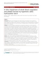
Fig. 1. XPS high-resolution C1s spectra for crosslinked nanofibers (a); high-resolution C1s spectra for FIB and ALB adsorbed on nanofibers showing C–H, C–C,
–O and C–O–C signals (b).
C–OH, N–C–
nanofibers have a significant increase in –COOH groups, compared to
the PVA nanofibers, due to the presence of the carboxymethyl group on
CMKC. The crosslinked nanofibers contain ether and ester bonds
resulting in peaks in the region of 286 eV and overlap with the C–OH
bonds. However, previously reported infrared spectra confirmed the
presence of the crosslinked sites with peaks between 1700 and 1750
cm− 1 (Madruga, Balaban, Popat, & Kipper, 2021). The incorporation of
CMKC is therefore confirmed by the XPS spectra and agrees with the
FTIR data from our previous study.
3.2. Hemostatic activity
3.2.1. Protein adsorption on the nanofibers
Blood clot formation results from the activation and aggregation of
platelets, and a multistep coagulation cascade, culminating with the
polymerization of fibrinogen and formation of a network of crosslinked
fibrin fibers (Hedayati, Neufeld, Reynolds, & Kipper, 2019). The
monolayer of proteins that adsorbs on the surface of a biomaterial is a
mediator to the formation of a clot, and its composition can dictate
subsequent biological protein processes (Prawel et al., 2014). Albumin
(ALB) is one of the most abundant proteins in the blood. Albumin
adsorption can block or promote coagulation, depending on whether it is
4
L.Y.C. Madruga et al.
Carbohydrate Polymers 273 (2021) 118541
peak in the C1s spectra for both the PVA-CMKC 25% and 75% (Fig. 1b).
This suggests that adding CMKC to nanofibers may promote higher
coagulation and blood clot formation, due to higher protein adsorption.
Even after crosslinking, all nanofibers still present some hydrophilicity,
however, the nanofibers containing CMKC present more crosslinking
sites, due to presence of the carboxymethyl groups, which make them a
little more hydrophobic when compared to pure PVA. The pure PVA
nanofibers had the highest amount of proteins adsorbed, which can be
attributed to the high surface area of this fiber and to the hydroxyl and
ester groups that can promote protein adsorption and changes in protein
conformation (Sivaraman & Latour, 2010; Yang, Han, Liu, Xu, & Jia,
2017). The high adsorption of albumin in PVA nanofibers might block
platelet adhesion decreasing clot formation, and the hydrophilicity can
lead to a decrease of the platelet binding sites of the fibrinogen adsorbed
(Zhang et al., 2017). On the other hand, increasing the concentration of
CMKC in the nanofibers up to 50% decreases the albumin adsorption and
increases the fibrinogen adsorption, which promotes more sites for
platelets to bind and form clots. The chemical similarity of CMKC to
biological molecules, such as glycosaminoglycans found in the human
body, as well as the large number of hydrogen-bonding groups present
on the molecule may promote protein-material interactions (Rodrigues,
Gonỗalves, Martins, Barbosa, & Ratner, 2006). The PVA-CMKC 75%
nanofibers had less fibrinogen and more albumin adsorbed when
compared to the PVA-CMKC 50% nanofibers, which could lead to
reduced platelet adhesion and activation. The smaller amount of pro
teins adsorbed can be correlated to the higher dispersity in the fiber
diameter, due to the higher instability when electrospinning high
charge-density solutions (Haider, Haider, & Kang, 2018; Merkle et al.,
2015a).
Table 2
Nitrogen content of the nanofibers before and after protein adsorption experi
ments, obtained from XPS survey scans.
PVA
PVA-CMKC 25%
PVA-CMKC 50%
PVA-CMKC 75%
% N (before)
% N (fibrinogen)
% N (albumin)
0.00
0.00
0.00
0.00
5.28
3.38
4.69
3.13
3.03
1.83
0.17
0.61
in its native conformation or denatured (Paar et al., 2017). Fibrinogen
(FIB) is spindle or rod-shaped protein that is converted to the poly
merizable form, fibrin, in the blood coagulation cascade. As the pre
cursor of the polymerizable fibrin, FIB is essential for the formation of
blood clots and provides binding sites for platelets (da Cˆ
amara et al.,
2020; Sabino, Kauk, Movafaghi, Kota, & Popat, 2019).
High-resolution XPS spectra of the C1s envelope and survey spectra
were obtained for the nanofibers after incubation in human albumin and
fibrinogen solutions. The amount of proteins adsorbed to the nanofibers
was estimated by the elemental composition. Since the nanofibers have
no nitrogen in their structure (Table 1), the increase in nitrogen
elemental composition obtained from the XPS survey scans on the fibers
is evidence of protein adsorption (Table 2). The adsorption of FIB and
ALB on the fibers was evaluated from the high-resolution spectra for the
C1s envelope by analyzing the increment of the amide carbonyl
– O) peaks (Fig. 1b).
(N–C–
FIB promotes platelet adhesion and activation, by exposing binding
sites to platelets. Thus, an increase in the adsorption of fibrinogen on the
nanofibers can be correlated with increasing pro-coagulant capacity.
ALB, on the other hand, can block or promote the formation of clots,
depending on the conformation adopted or denaturation. The highresolution XPS spectra of the C1s envelope (Fig. 1b) shows similar
– O) increases following adsorption of both proteins
amide peak (N–C–
to PVA nanofibers. PVA-CMKC nanofibers all exhibit larger nitrogen
content increases following fibrinogen adsorption compared to albumin
adsorption. PVA nanofibers have the highest nitrogen content following
FIB adsorption. The same trend is observed when comparing the amide
3.2.2. Platelet adhesion and activation
Platelet adhesion on the surfaces of biomaterials is an indicator of
thrombogenicity and pro-coagulant activity, leading to platelet activa
tion, which can initiate the coagulation cascade (Hedayati, Neufeld,
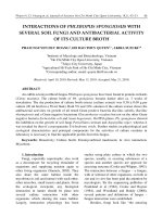
Reynolds, & Kipper, 2019). Fig. 2 illustrates the adhesion of platelets
(green) on the surface of the nanofibers and tissue culture polystyrene
(control) following 2 h incubation in human PRP. Nanofibers exhibit a
Fig. 2. Percentage area of adhered platelets on nanofibers and fluorescence microscopy images of adhered platelets stained with calcein-AM on the nanofibers after
2 h of incubation in platelet-rich plasma. CMKC-containing nanofibers have significantly higher platelet adhesion compared to control. ****p ≤ 0.0001, **p ≤ 0.01,
*p ≤ 0.05 and “ns” p ≥ 0.05.
5
L.Y.C. Madruga et al.
Carbohydrate Polymers 273 (2021) 118541
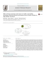
Fig. 3. SEM micrographs of adhered platelets on the nanofibers after 2 h of incubation in platelet-rich plasma.
Platelet adhesion to surfaces can lead to rapid platelet activation.
Activated platelets undergo a series of morphological changes, including
spreading, dendrite formation and then aggregation (da Cˆ
amara et al.,
2020; Sabino, Kauk, Movafaghi, Kota, & Popat, 2019). While nonactivated platelets are spherical, platelets undergoing activation
exhibit long, finger-like extensions. Fully activated platelets are char
acterized as having a “fried egg” appearance (Simon-Walker et al., 2017;
Vlcek, Hedayati, Melvin, Reynolds, & Kipper, 2021). The morphology of
the platelets adhered on the nanofibers was evaluated by SEM images
(Fig. 3). The high number of adhered platelets on the CMKC-containing
nanofibers seen in the SEM images confirms the observations in the
fluorescence micrographs, demonstrating that CMKC promotes platelet
adhesion. All platelets show dendrite formation and a very small number
are in a round (unactivated) morphology. Heparin, another sulphated
polysaccharide can have anticoagulant activity, through interactions
with antithrombin III and other components of the coagulation cascade.
Nonetheless, when adsorbed to a surface heparin can also promote
platelet activation on nanostructured surfaces, as its negatively charged
sulfate groups form complexes with positively charged platelet factor 4,
which can result in immune complexes that activate platelets (Krauel,
Hackbarth, Fürll, & Greinacher, 2012; Vlcek, Hedayati, Melvin, Rey
nolds, & Kipper, 2021).
Platelets have negatively charged membranes. Since the CMKC is
also negatively charged, electrostatic forces alone would cause CMKC to
repel platelets from the nanofibers. However, this is not what is observed
from the results on Fig. 2. In fact, studies have shown that carboxyl
groups, which are also present in CMKC, have relatively little impact on
platelet adhesion and aggregation (Dorahy, Thorne, Fecondo, & Burns,
1997; Wilner, Nossel, & LeRoy, 1968). However, studies have shown
that negatively charged surfaces can activate factor XII and platelet
factor 3, leading to intrinsic blood coagulation (Tranquilan-Aranilla,
Barba, Vista, & Abad, 2016). We suggest that the processes that lead to
platelet adhesion and activation on CMKC-containing nanofibers are
related to attachment of plasma proteins and interactions of the platelets
with these proteins attached to the nanofibers (Rodrigues, Gonỗalves,
Martins, Barbosa, & Ratner, 2006). Since this work used PRP, all the
proteins present on the plasma (such as fibrinogen and complement
Fig. 4. Whole blood clotting measured by the normalized amount of free he
moglobin in human whole blood incubated with nanofibers for 15 and 30 min.
Reduced blood clotting index indicates increased clotting. * p ≤ 0.05 and “ns” p
≥ 0.05 compared to the PVA control.
significant increase in platelet adhesion compared to the control, which
increases with increasing CMKC content. The difference in the number
of adhered platelets between the fibers and the control can be attributed
partially to the relatively high specific surface area and nanoscale
topography of the nanofibers compared to the two-dimensional control
surface. Because they have a three-dimensional structure and a rough
surface with pores, nanofibers tend to have a higher deposition of
platelets and proteins on their surfaces (Zeng et al., 2016). Moreover,
when compared with PVA nanofibers, CMKC-containing nanofibers also
have higher platelet adhesion (Fig. 2). This suggests that CMKC en
hances platelet adhesion. The formation of ester groups by crosslinking
with PVA may also contribute to increased platelet adhesion (Ma et al.,
2015; Madruga et al., 2020).
6
L.Y.C. Madruga et al.
Carbohydrate Polymers 273 (2021) 118541
Fig. 5. Fluorescence microscopy images of S. aureus on the nanofibers. Live bacteria are represented in green (SYTO 9 stain) and dead bacteria in red (propidium
iodide stain) (a). Percentage of coverage for live and dead S. aureus adhered to the nanofibers (b). Inset shows the percentage of coverage for live bacteria on an
expanded y-axis for comparison. ****p ≤ 0.0001, ***p ≤ 0.001, **p ≤ 0.01, and “ns” p ≥ 0.05 compared to the PVA control.
proteins) can attach to the nanofibers and provide sites for the platelets
to interact and attach. The presence of fibrinogen on the nanofibers
shown on Fig. 1 and Table 2 corroborates these results. Data from the
literature shows that fibrinogen adsorption is related to high platelet
adhesion and activation and the conformation of the protein is relevant
to this mechanism (Chiumiento, Lamponi, & Barbucci, 2007; Rodrigues,
Gonỗalves, Martins, Barbosa, & Ratner, 2006). Zhang et al. (2017)
observed that on hydrophilic surfaces the γ400–411 platelet-binding
dodecapeptide on the D region of fibrinogen is exposed, leading to for
mation of uniform monolayers of activated platelets on the surface
(Zhang et al., 2017). Similar phenomena could be responsible for the
observed platelet activation on the CMKC nanofibers reported here. In
addition, the similarity of CMKC to biological molecules can promote
biochemical signals and sites for the deposition and activation of
platelets (Merkle et al., 2015b). Increasing the amount of CMKC to 75%
made the fibers more unstable, due to the high presence of charges in
solution when electrospinning, resulting in the highest fiber roughness
and fiber porosity, and perhaps lower surface area for protein adsorption
and subsequent platelet adhesion. This explains why the nanofibers with
75% CMKC presented lower number of platelets adhered, when
compared to the ones with 50% CMKC. This trend also correlates to the
higher amount of albumin and lower amount of fibrinogen on the 75%
CMKC samples, compared to the 50% CMKC samples. Nonetheless, the
difference in area of adhered platelets between the 50% and 75% CMKC
nanofibers is not statistically significant.
characterize the biochemical reactions involved in the hemostatic
response. Although the investigation of single components of the coag
ulation cascade can provide information on specific interactions be
tween blood components and the biomaterial, whole blood clotting
offers the most accurate and clinically relevant thrombogenicity index,
presenting the combined effects of all components (Sabino & Popat,
2020).
Human blood droplets were applied to the nanofibers and the clot
formation after 15 and 30 min were analysed by absorbance measure
ments of the samples for the free hemoglobin released from the unclotted blood (Fig. 4). The blood clotting index (BCI) was calculated
for all samples and the values of a blood sample in water at time 0 (as
soon as the blood is collected) (Barba et al., 2018; Zhao et al., 2018).
Absorbance measurements were scaled from 0% to 100% free hemo
globin. According to the absorbance values, the percentage of free he
moglobin for each sample was calculated and reported as blood clotting
index, as shown in Fig. 4. A reduction in the free hemoglobin indicates
an increase in the procoagulant activity. These results agree with the
results from serum protein adsorption and from platelet adhesion and
activation. All nanofiber samples exhibit some non-zero pro-coagulation
activity; nanofibers with higher CMKC content (50 and 75%) resulted in
significantly lower BCI than PVA nanofibers, reaching values close to
20%, with no statistically significant difference between the two.
Therefore both the nanoscale features of the fibers and their chemistry
ăgtle et al., 2019; Xu, Weng, Gilkerson, Materon,
promote coagulation (Vo
& Lozano, 2015). The hemostatic effects of CMKC hydrogels are similar
to the ones observed in CMKC nanofibers in terms of BCI and platelet
adhesion, confirming the contribution of CMKC to the hemostatic
3.2.3. Whole blood clotting
Blood clotting tests using human blood (plasma and erythrocytes)
7
L.Y.C. Madruga et al.
Carbohydrate Polymers 273 (2021) 118541
Fig. 6. Fluorescence microscopy images of P. aeruginosa on the nanofibers. Live bacteria are represented in green (SYTO 9 stain) and dead bacteria in red (propidium
iodide stain) (a). Percentage of coverage for live and dead S. aureus adhered to the nanofibers (b). Inset shows the percentage of coverage for live bacteria on an
expanded y-axis for comparison. ****p ≤ 0.0001, **p ≤ 0.01, *p ≤ 0.05 and “ns” p ≥ 0.05, compared to the PVA control.
Conversely, P. aeruginosa is a Gram-negative, bacillus (rod-shaped), with
a complex and thin cell wall. In general, higher adhesion of P. aeruginosa
bacteria is observed in all nanofibers, compared to S. aureus, which can
be explained by the greater mobility of the bacteria, due to their flagella
(Fredua-Agyeman, Gaisford, & Beezer, 2018). Despite the higher adhe
sion on the nanofibers, after 6 h of growth, almost all the P. aeruginosa
adhered to the CMKC-containing nanofibers were stained red, which
characterizes dead bacteria. After 24 h, the PVA nanofibers have a sig
nificant increase in the amount of live bacteria for both bacteria types.
The CMKC-containing nanofibers with higher CMKC content have
reduced live bacteria compared to the PVA nanofibers after 24 h for both
types of bacteria. Furthermore, the 50% and 75% CMKC nanofibers have
an increased number of dead bacteria compared to the PVA nanofibers
after 24 h for both bacteria. Therefore, the CMKC-containing nanofibers
do not provide a favorable environment for bacteria, even in a nutrientrich broth condition.
behavior (Tranquilan-Aranilla, Barba, Vista, & Abad, 2016). CMKCcontaining nanofibers with greater than 50% CMKC are strong candi
dates for application in wound dressings based on the observed procoagulant activity.
3.3. Antibacterial activity
3.3.1. Bacteria adhesion on the nanofibers
Exposed wounds are viable environments for the colonization of
bacteria, especially those present on the skin. Wound dressings that can
repel or kill bacteria can help obviate the overuse of antibiotics (Vallet´lez, & Izquierdo-Barba, 2019). Fluorescence images were
Regí, Gonza
used to assess the bacteria that were deposited on the nanofibers. The
green dye (SYTO9) permeates the bacterial membranes, indicating live
bacteria, while the red dye (propidium iodide), does not permeate live
bacteria, only staining the bacteria that have some defect or failure in
their membrane, staining only dead bacteria (Stiefel, Schmidt-Emrich,
Maniura-Weber, & Ren, 2015). Quantifying bacterial adhesion is pref
erable over zone-of-inhibition tests on the nanofibers, due to the simi
larity with the conditions in a wound bed. The antibacterial effect
observed here is not due to the release and diffusion of an antibacterial
agent (measured by the zone-of-inhibition test). Rather, the antimicro
bial activity is present on the fiber surface, making the evaluation of
live/dead bacteria on the surface and bacterial morphology ideal for this
material. Figs. 5 and 6 show fluorescence microscopy images and per
centage coverage of live and dead S. aureus and P. aeruginosa, respec
tively, on the nanofibers after 6 h and 24 h. S. aureus is a coccal (round)
Gram-positive bacterium, with a thick peptidoglycan-rich cell wall.
3.3.2. Bacteria morphology and biofilm formation
SEM images of the nanofibers after 6 and 24 h of incubation in
bacteria broth were used to evaluate the morphology of adhered bac
teria and biofilm formation. The images agree with the results from
fluorescence microscopy. After 6 h, adhered S. aureus on the nanofibers
(Fig. S4 — supplementary information) have a spherical morphology
similar to “grape bunches,” characteristic of Staphylococcus, and CMKCcontaining nanofibers show a lower number of bacteria attached
compared to PVA. Moreover, some bacteria on CMKC 75% nanofibers
begin to exhibit morphological changes. After 24 h, PVA nanofibers
show a high number of adhered S. aureus (Fig. 7), as well as colony
8
L.Y.C. Madruga et al.
Carbohydrate Polymers 273 (2021) 118541
Fig. 7. False colored SEM images of S. aureus on the nanofibers after 24 h of incubation.
respiratory enzymes, as well as the integrity of the membrane itself,
causing the death of the bacteria (Pajerski et al., 2019).
formation and aggregation. PVA-CMKC nanofibers show a low number
of adhered bacteria and few colony formations, except on 50% CMKC,
which may be due to the higher hydrophilicity. Confirming the fluo
rescence microscopy data, some bacteria on CMKC-containing fibers
have an elliptical shape, and some defective membranes. These bacteria
are probably dead. No biofilm formation was observed on any of the
fibers.
It is important to note that P. aeruginosa is a biofilm-forming bacteria,
a defense mechanism that makes it a pathogen that is difficult to fight
(Madruga et al., 2020; Reynolds & Neufeld, 2016). After 6 h, adhered
P. aeruginosa on the nanofibers (Fig. S5 — supplementary information)
have a bacillus morphology, and all nanofibers have a high number of
bacteria attached. However, some disruptions of the morphology can be
observed, indicating dead bacteria. After 24 h, PVA nanofibers show a
higher number of adhered P. aeruginosa (Fig. 8), as well as colony for
mation and some biofilm formation. PVA-CMKC nanofibers also have
bacteria attached, but with defective morphology and no biofilm for
mation, corroborating the fluorescence microscopy and indicating sig
nificant antimicrobial activity.
CMKC-containing nanofibers have multiple features that may impart
antibacterial activity. Because they have rigid cell walls, gram-positive
and gram-negative bacteria cannot adapt easily to the nanoscale fea
tures, which can lead to cell death on nanostructured surfaces (ValletRegí, Gonz´
alez, & Izquierdo-Barba, 2019). The increased hydrophilicity
introduced by crosslinking the PVA with CMKC can promote the for
mation of a water layer on the surface, generating a physical and en
ergetic barrier for the deposition of bacteria (Wang, Hu, & Shao, 2017).
The charged carboxylate and sulfate groups in CMKC can also interact
with the bacterial cell wall and membrane, affecting ion channels and
4. Conclusions
In this study, electrospun PVA-CMKC nanofibers show enhanced
blood coagulation and antibacterial activity, compared to PVA nano
fibers. PVA-CMKC nanofibers preferentially adsorb fibrinogen compared
to albumin, promote platelet adhesion and activation, and promote
coagulation in contact with human whole blood. CMKC-containing
nanofibers also exhibit superior antibacterial activity against both
Staphylococcus aureus and Pseudomonas aeruginosa compared to PVA
nanofibers. These favorable biological properties can be modulated by
tuning the CMKC content. These properties are achieved due to a com
bination of the nanometer-scale features of the fibers and the biologi
cally active biopolymer containing carboxyl, ether, and sulfate groups.
PVA-CMKC nanofibers are non-cytotoxic, biodegradable, low-cost, and
prepared following green manufacturing methods. PVA-CMKC nano
fibers show potential for application as dressings for wound healing
applications.
CRediT authorship contribution statement
Liszt Y.C. Madruga: Conceptualization, Methodology, Validation,
Formal analysis, Investigation, Data curation, Writing – original draft,
Writing – review & editing, Visualization, Funding acquisition. Ketul C.
Popat: Conceptualization, Resources, Writing – review & editing.
Rosangela C. Balaban: Conceptualization, Methodology, Resources,
Writing – review & editing, Supervision, Project administration,
9
L.Y.C. Madruga et al.
Carbohydrate Polymers 273 (2021) 118541
Fig. 8. False colored SEM images of P. aeruginosa on the nanofibers after 24 h of incubation.
Funding acquisition. Matt J. Kipper: Conceptualization, Methodology,
Resources, Writing – review & editing, Supervision, Project adminis
tration, Funding acquisition.
Bhattacharjee, A., Clark, R., Gentry-Weeks, C., & Li, Y. V. (2020). A novel receptor-free
polydiacetylene nanofiber biosensor for detecting E. coli via colorimetric changes.
Materials Advances, 1(9), 3387–3397. />da Cˆ
amara, P. C. F., Madruga, L. Y. C., Sabino, R. M., Vlcek, J., Balaban, R. C.,
Popat, K. C., … Kipper, M. J. (2020). Polyelectrolyte multilayers containing a tannin
derivative polyphenol improve blood compatibility through interactions with
platelets and serum proteins. Materials Science and Engineering: C, 112(March),
Article 110919. />Chattopadhyay, S., & Raines, R. T. (2014). Collagen-based biomaterials for wound
healing. Biopolymers, 101(8), 821–833. />Chiumiento, A., Lamponi, S., & Barbucci, R. (2007). Role of fibrinogen conformation in
platelet activation. Biomacromolecules, 8(2), 523–531. />bm060664m
da Cruz, J. A., da Silva, A. B., Ramin, B. B. S., Souza, P. R., Popat, K. C., Zola, R. S., …
Martins, A. F. (2020). Poly(vinyl alcohol)/cationic tannin blend films with
antioxidant and antimicrobial activities. Materials Science and Engineering C, 107
(August 2019), Article 110357. />Das, A., Abas, M., Biswas, N., Banerjee, P., Ghosh, N., Rawat, A., … Sen, C. K. (2019).
A modified collagen dressing induces transition of inflammatory to reparative
phenotype of wound macrophages. Scientific Reports, 9(1), 1–10. />10.1038/s41598-019-49435-z
do Nascimento Marques, N., dos Santos Alves, K., Vidal, R. R. L., da Silva Maia, A. M.,
Madruga, L. Y. C., Curti, P. S., … Taft, C. (2020). Chemical modification of
polysaccharides and applications in strategic areas. In Emerging research in science
and engineering based on advanced experimental and computational strategies.
Engineering materials (pp. 433–472). Cham: Springer. />Dorahy, D. J., Thorne, R. F., Fecondo, J. V., & Burns, G. F. (1997). Stimulation of platelet
activation and aggregation by a carboxyl-terminal peptide from thrombospondin
binding to the integrin-associated protein receptor. Journal of Biological Chemistry,
272(2), 1323–1330. />Dumont, M., Villet, R., Guirand, M., Montembault, A., Delair, T., Lack, S., Barikosky, M.,
Crepet, A., Alcouffe, P., Laurent, F., & David, L. (2018). Processing and antibacterial
properties of chitosan-coated alginate fibers. Carbohydrate Polymers, 190(December
2016), 31–42. />Fahimirad, S., & Ajalloueian, F. (2019). Naturally-derived electrospun wound dressings
for target delivery of bio-active agents. International Journal of Pharmaceutics, 566
(May), 307328. />
Acknowledgements
o de Aperfeiỗoaư
This study was financed in part by the Coordenaỗa
mento de Pessoal de Nớvel Superior Brasil (CAPES) – Finance Code 001.
Also, the authors gratefully acknowledge the financial support from the
National Science Foundation (award number 1933552).
Appendix A. Supplementary data
Supplementary data to this article can be found online at https://doi.
org/10.1016/j.carbpol.2021.118541.
References
Almod´
ovar, J., Mower, J., Banerjee, A., Sarkar, A. K., Ehrhart, N. P., & Kipper, M. J.
(2013). Chitosan-heparin polyelectrolyte multilayers on cortical bone: Periosteummimetic, cytophilic, antibacterial coatings. Biotechnology and Bioengineering, 110(2),
609–618. />Bajpai, S. K., & Daheriya, P. (2014). Kappa-carrageenan/PVA films with antibacterial
properties: Part 1. Optimization of preparation conditions and preliminary drug
release studies. Journal of Macromolecular Science, Part A, 51(4), 286–295. https://
doi.org/10.1080/10601325.2014.882687
Barba, B. J. D., Aranilla, C. T., Relleve, L. S., Cruz, V. R. C., Vista, J. R., & Abad, L. V.
(2018). Hemostatic granules and dressing prepared from formulations of
carboxymethyl cellulose, kappa-carrageenan and polyethylene oxide crosslinked by
gamma radiation. Radiation Physics and Chemistry, 144(August 2017), 180–188.
/>
10
L.Y.C. Madruga et al.
Carbohydrate Polymers 273 (2021) 118541
Felgueiras, H. P., & Amorim, M. T. P. (2017). Functionalization of electrospun polymeric
wound dressings with antimicrobial peptides. Colloids and Surfaces B: Biointerfaces,
156, 133–148. />Fredua-Agyeman, M., Gaisford, S., & Beezer, A. E. (2018). Observation with
microcalorimetry: Behaviour of P. aeruginosa in mixed cultures with S. aureus and
E. coli. Thermochimica Acta, 663(March), 93–98. />tca.2018.03.009
Fujiwara, T., Nishimoto, S., Ishise, H., Kawai, K., Fukuda, K., & Kakibuchi, M. (2012).
Comparative study of the antibacterial penetrating effects of wound dressings.
Journal of Plastic Surgery and Hand Surgery, 46(1), 2–7. />2000656X.2011.644939
Guo, J., Zhou, H., Akram, M. Y., Mu, X., Nie, J., & Ma, G. (2016). Characterization and
application of chondroitin sulfate/polyvinyl alcohol nanofibres prepared by
electrospinning. Carbohydrate Polymers, 143, 239–245. />carbpol.2016.02.013
Haider, A., Haider, S., & Kang, I.-K. (2018). A comprehensive review summarizing the
effect of electrospinning parameters and potential applications of nanofibers in
biomedical and biotechnology. Arabian Journal of Chemistry, 11(8), 1165–1188.
/>Hedayati, M., Neufeld, M. J., Reynolds, M. M., & Kipper, M. J. (2019). The quest for
blood-compatible materials: Recent advances and future technologies. Materials
Science and Engineering: R: Reports, 138(July), 118–152. />mser.2019.06.002
Homaeigohar, S., & Boccaccini, A. R. (2020). Antibacterial biohybrid nanofibers for
wound dressings. Acta Biomaterialia, 107(2020), 25–49. />actbio.2020.02.022
Krauel, K., Hackbarth, C., Fürll, B., & Greinacher, A. (2012). Heparin-induced
thrombocytopenia: In vitro studies on the interaction of dabigatran, rivaroxaban,
and low-sulfated heparin, with platelet factor 4 and anti-PF4/heparin antibodies.
Blood, 119(5), 1248–1255. />Ma, N., Liu, X.-W., Yang, Y.-J., Li, J.-Y., Mohamed, I., Liu, G.-R., & Zhang, J.-Y. (2015).
Preventive effect of aspirin eugenol ester on thrombosis in κ-carrageenan-induced rat
tail thrombosis model. PLoS One, 10(7), Article e0133125. />journal.pone.0133125
Madruga, L. Y. C., Balaban, R. C., Popat, K. C., & Kipper, M. J. (2021). Biocompatible
crosslinked nanofibers of poly(vinyl alcohol)/carboxymethyl-kappa-carrageenan
produced by a green process. Macromolecular Bioscience, 21(1), Article 2000292.
/>Madruga, L. Y. C., da Cˆ
amara, P. C. F., Marques, N.d. N., & Balaban, R.d. C. (2018). Effect
of ionic strength on solution and drilling fluid properties of ionic polysaccharides: A
comparative study between Na-carboxymethylcellulose and Na-kappa-carrageenan
responses. Journal of Molecular Liquids, 266, 870–879. />molliq.2018.07.016
Madruga, L. Y. C., Sabino, R. M., Santos, E. C. G., Popat, K. C., Balaban, R.d. C., &
Kipper, M. J. (2020). Carboxymethyl-kappa-carrageenan: A study of
biocompatibility, antioxidant and antibacterial activities. International Journal of
Biological Macromolecules, 152, 483–491. />ijbiomac.2020.02.274
Merkle, V. M., Martin, D., Hutchinson, M., Tran, P. L., Behrens, A., Hossainy, S., …
Slepian, M. J. (2015a). Hemocompatibility of poly(vinyl alcohol)-gelatin core-shell
electrospun nanofibers: A scaffold for modulating platelet deposition and activation.
ACS Applied Materials and Interfaces, 7(15), 8302–8312. />acsami.5b01671
Merkle, V. M., Martin, D., Hutchinson, M., Tran, P. L., Behrens, A., Hossainy, S., …
Slepian, M. J. (2015b). Hemocompatibility of poly(vinyl alcohol)–gelatin core–shell
electrospun nanofibers: A scaffold for modulating platelet deposition and activation.
ACS Applied Materials & Interfaces, 7(15), 8302–8312. />acsami.5b01671
Miguel, S. P., Figueira, D. R., Sim˜
oes, D., Ribeiro, M. P., Coutinho, P., Ferreira, P., &
Correia, I. J. (2018). Electrospun polymeric nanofibres as wound dressings: A
review. Colloids and Surfaces B: Biointerfaces, 169, 60–71. />colsurfb.2018.05.011
Mogos¸anu, G. D., & Grumezescu, A. M. (2014). Natural and synthetic polymers for
wounds and burns dressing. International Journal of Pharmaceutics, 463(2), 127–136.
/>Nas, F. S., Abrigo, M., McArthur, S. L., & Kingshott, P. (2014). Electrospun nanofibers as
dressings for chronic wound care: Advances, challenges, and future prospects.
Macromolecular Bioscience, 14(6), 772–792. />mabi.201300561
de Oliveira, M., Madruga, L., de Lima, B., Villetti, M., de Souza Filho, M., Kipper, M., …
Balaban, R. (2021). Agro-industrial waste valorization: Transformation of starch
from mango kernel into biocompatible, thermoresponsive and high swelling
nanogels. Journal of the Brazilian Chemical Society, 00(00), 1–10. />10.21577/0103-5053.20210059
Paar, M., Rossmann, C., Nusshold, C., Wagner, T., Schlagenhauf, A., Leschnik, B.,
Hallstră
om, S. (2017). Anticoagulant action of low, physiologic, and high albumin
levels in whole blood. PLoS One, 12(8), 1–12. />pone.0182997
Pajerski, W., Ochonska, D., Brzychczy-Wloch, M., Indyka, P., Jarosz, M., Golda-Cepa, M.,
Sojka, Z., & Kotarba, A. (2019). Attachment efficiency of gold nanoparticles by
Gram-positive and Gram-negative bacterial strains governed by surface charges.
Journal of Nanoparticle Research, 21(8). />Prawel, D. A., Dean, H., Forleo, M., Lewis, N., Gangwish, J., Popat, K. C., … James, S. P.
(2014). Hemocompatibility and hemodynamics of novel hyaluronan–polyethylene
materials for flexible heart valve leaflets. Cardiovascular Engineering and Technology,
5(1), 70–81. />
Reynolds, B. H. N. M., & Neufeld, B. H. (2016). Critical nitric oxide concentration for
Pseudomonas aeruginosa biofilm reduction on polyurethane substrates, Article 031012.
/>Rodrigues, S. N., Gonỗalves, I. C., Martins, M. C. L., Barbosa, M. A., & Ratner, B. D.
(2006). Fibrinogen adsorption, platelet adhesion and activation on mixed hydroxyl-/
methyl-terminated self-assembled monolayers. Biomaterials, 27(31), 5357–5367.
/>Sabino, R. M., Kauk, K., Madruga, L. Y. C., Kipper, M. J., Martins, A. F., & Popat, K. C.
(2020). Enhanced hemocompatibility and antibacterial activity on titania nanotubes
with tanfloc/heparin polyelectrolyte multilayers. Journal of Biomedical Materials
Research — Part A, 108(4), 992–1005. />Sabino, R. M., Kauk, K., Movafaghi, S., Kota, A., & Popat, K. C. (2019). Interaction of
blood plasma proteins with superhemophobic titania nanotube surfaces.
Nanomedicine: Nanotechnology, Biology and Medicine, 21, Article 102046. https://doi.
org/10.1016/j.nano.2019.102046
Sabino, R. M., & Popat, K. C. (2020). Evaluating whole blood clotting in vitro on
biomaterial surfaces. Bio-Protocol, 10(3), Article e3505. />BioProtoc.3505
Sadeghi, A., Zandi, M., Pezeshki-Modaress, M., & Rajabi, S. (2019). Tough, hybrid
chondroitin sulfate nanofibers as a promising scaffold for skin tissue engineering.
International Journal of Biological Macromolecules, 132, 63–75. />10.1016/j.ijbiomac.2019.03.208
Simon-Walker, R., Romero, R., Staver, J. M., Zang, Y., Reynolds, M. M., Popat, K. C., &
Kipper, M. J. (2017). Glycocalyx-inspired nitric oxide-releasing surfaces reduce
platelet adhesion and activation on titanium. ACS Biomaterials Science & Engineering,
3(1), 68–77. />Sivaraman, B., & Latour, R. A. (2010). The relationship between platelet adhesion on
surfaces and the structure versus the amount of adsorbed fibrinogen. Biomaterials, 31
(5), 832–839. />Stiefel, P., Schmidt-Emrich, S., Maniura-Weber, K., & Ren, Q. (2015). Critical aspects of
using bacterial cell viability assays with the fluorophores SYTO9 and propidium
iodide. BMC Microbiology, 15(1), 36. />Tranquilan-Aranilla, C., Barba, B. J. D., Vista, J. R. M., & Abad, L. V. (2016). Hemostatic
efficacy evaluation of radiation crosslinked carboxymethyl kappa-carrageenan and
chitosan with varying degrees of substitution. Radiation Physics and Chemistry, 124,
124–129. />ˆ M. (2017). Electrospun
Trinca, R. B., Westin, C. B., da Silva, J. A. F., & Moraes, A.
multilayer chitosan scaffolds as potential wound dressings for skin lesions. European
Polymer Journal, 88, 161–170. />Truong, Y. B., Glattauer, V., Briggs, K. L., Zappe, S., & Ramshaw, J. A. M. (2012).
Collagen-based layer-by-layer coating on electrospun polymer scaffolds.
Biomaterials, 33(36), 9198–9204. />12.
Unnithan, A. R., Sasikala, A. R. K., Murugesan, P., Gurusamy, M., Wu, D., Park, C. H., &
Kim, C. S. (2015). Electrospun polyurethane-dextran nanofiber mats loaded with
Estradiol for post-menopausal wound dressing. International Journal of Biological
Macromolecules, 77, 1–8. />Vallet-Regí, M., Gonz´
alez, B., & Izquierdo-Barba, I. (2019). Nanomaterials as promising
alternative in the infection treatment. International Journal of Molecular Sciences, 20
(15). />Vlcek, J. R., Hedayati, M., Melvin, A. C., Reynolds, M. M., & Kipper, M. J. (2021). Bloodcompatible materials: Vascular endothelium-mimetic surfaces that mitigate multiple
cell-material interactions. Advanced Healthcare Materials, 10(7), Article 2001748.
/>Vă
ogtle, T., Sharma, S., Mori, J., Nagy, Z., Semeniak, D., Scandola, C., … Senis, Y. A.
(2019). Heparan sulfates are critical regulators of the inhibitory megakaryocyteplatelet receptor G6b-B. ELife, 8, 1–43. />Wang, L., Hu, C., & Shao, L. (2017). The-antimicrobial-activity-ofnanoparticles—Present-situati. International Journal of Nanomedicine, 12,
1227–1249. />Wilner, G. D., Nossel, H. L., & LeRoy, E. C. (1968). Aggregation of platelets by collagen.
Journal of Clinical Investigation, 47(12), 2616–2621. />JCI105944
Xu, F., Weng, B., Gilkerson, R., Materon, L. A., & Lozano, K. (2015). Development of
tannic acid/chitosan/pullulan composite nanofibers from aqueous solution for
potential applications as wound dressing. Carbohydrate Polymers, 115, 16–24.
/>Yang, L., Han, L., Liu, Q., Xu, Y., & Jia, L. (2017). Galloyl groups-regulated fibrinogen
conformation: Understanding antiplatelet adhesion on tannic acid coating. Acta
Biomaterialia, 64, 187–199. />Yegappan, R., Selvaprithiviraj, V., Amirthalingam, S., & Jayakumar, R. (2018).
Carrageenan based hydrogels for drug delivery, tissue engineering and wound
healing. Carbohydrate Polymers, 198(June), 385–400. />carbpol.2018.06.086
Young, B. M., Shankar, K., Allen, B. P., Pouliot, R. A., Schneck, M. B., Mikhaiel, N. S., &
Heise, R. L. (2017). Electrospun decellularized lung matrix scaffold for airway
smooth muscle culture. ACS Biomaterials Science & Engineering, 3(12), 3480–3492.
/>Zahedi, P., Rezaeian, I., Ranaei-Siadat, S.-O., Jafari, S.-H., & Supaphol, P. (2010).
A review on wound dressings with an emphasis on electrospun nanofibrous
polymeric bandages. Polymers for Advanced Technologies, 21(2), 77–95. https://doi.
org/10.1002/pat.1625
Zeng, Q., Qin, J., Yin, X., Liu, H., Zhu, L., Dong, W., & Zhang, S. (2016). Preparation and
hemocompatibility of electrospun O-carboxymethyl chitosan/PVA nanofibers.
Journal of Applied Polymer Science, 133(26), 2–9. />app.43565
11
L.Y.C. Madruga et al.
Carbohydrate Polymers 273 (2021) 118541
Zhang, L., Casey, B., Galanakis, D. K., Marmorat, C., Skoog, S., Vorvolakos, K., …
Rafailovich, M. H. (2017). The influence of surface chemistry on adsorbed fibrinogen
conformation, orientation, fiber formation and platelet adhesion. Acta Biomaterialia,
54, 164–174. />Zhao, R., Li, X., Sun, B., Zhang, Y., Zhang, D., Tang, Z., Chen, X., & Wang, C. (2014).
Electrospun chitosan/sericin composite nanofibers with antibacterial property as
potential wound dressings. International Journal of Biological Macromolecules, 68,
92–97. />
Zhao, X., Gao, J., Hu, X., Guo, H., Wang, F., Qiao, Y., & Wang, L. (2018). Collagen/
polyethylene oxide nanofibrous membranes with improved hemostasis and
cytocompatibility for wound dressing. Applied Sciences, 8(8), 1226. />10.3390/app8081226
Zia, K. M., Tabasum, S., Nasif, M., Sultan, N., Aslam, N., Noreen, A., & Zuber, M. (2017).
A review on synthesis, properties and applications of natural polymer based
carrageenan blends and composites. International Journal of Biological
Macromolecules, 96, 282–301. />
12


![Synthesis and evaluation of antimicrobial, antitubercular and anticancer activities of 2-(1-benzoyl-1H-benzo[d] imidazol-2-ylthio)-N-substituted acetamides](https://media.store123doc.com/images/document/2020_05/29/medium_rqw1590732393.jpg)