Chitosan – Rosmarinic acid conjugates with antioxidant, anti-inflammatory and photoprotective properties
Bạn đang xem bản rút gọn của tài liệu. Xem và tải ngay bản đầy đủ của tài liệu tại đây (2.28 MB, 12 trang )
Carbohydrate Polymers 273 (2021) 118619
Contents lists available at ScienceDirect
Carbohydrate Polymers
journal homepage: www.elsevier.com/locate/carbpol
Chitosan – Rosmarinic acid conjugates with antioxidant, anti-inflammatory
and photoprotective properties
˜ al a, Javier Caro-Leo
´n b, Eva Espinosa-Cano a, c, María Rosa Aguilar a, c, *,
Miguel Huerta-Madron
a, c
Blanca V´
azquez-Lasa
a
b
c
Group of Biomaterials, Institute of Polymer Science and Technology ICTP-CSIC, Madrid, Spain
Grupo de Investigaci´
on en Biopolímeros, Centro de Investigaci´
on en Alimentaci´
on y Desarrollo A.C., Sonora, Mexico
Networking Biomedical Research Centre in Bioengineering, Biomaterials and Nanomedicine, CIBER-BBN, Madrid, Spain
A R T I C L E I N F O
A B S T R A C T
Keywords:
Chitosan
Rosmarinic acid
Conjugates
Antioxidant activity
Photoprotective properties
Wound healing
Rosmarinic acid is an attractive candidate for skin applications because of its antioxidant, anti-inflammatory, and
photoprotective functions, however, its poor bioavailability hampers its therapeutic outcome. In this context,
synthesis of polymer conjugates is an alternative to enlarge its applications. This work describes the synthesis of
novel water-soluble chitosan – rosmarinic acid conjugates (CSRA) that have great potential for skin applications.
Chitosan was functionalized with different contents of rosmarinic acid as confirmed by ATR-FTIR, 1H NMR and
UV spectroscopies. CSRA conjugates presented three-fold radical scavenger capacity compared to the free
phenolic compound. Films were prepared by solvent-casting procedure and the biological activity of the lixiv
iates was studied in vitro. Results revealed that lixiviates reduced activation of inflamed macrophages, improved
antibacterial capacity against E. coli with respect to native chitosan and free rosmarinic acid, and also attenuated
UVB-induced cellular damage and reactive oxygen species production in fibroblasts and keratinocytes.
1. Introduction
Phytochemical is a broad term meaning plant (phyto) chemical that
refers to a wide variety of plant-derived compounds with beneficial
therapeutic activities on human health such as anticarcinogenic, anti
mutagenic, anti-inflammatory, and antioxidant properties (El-Sherbiny
et al., 2016; Huang et al., 2016; Shahidi & Ambigaipalan, 2015; Tsao,
2010; Vuolo et al., 2019). The most common phytochemical found in
human diet are polyphenols (Mrduljas et al., 2017). These compounds
are one of the most widespread groups of bioactive molecules distrib
uted almost ubiquitously in nature; they can be found in fruits, cereals,
vegetables, tea, coffee and cocoa among others (Abbas et al., 2017;
Shahidi & Ambigaipalan, 2015; Souto et al., 2019).
It is generally accepted that the primary cause of aging and agerelated diseases as well as cancer is the cellular damage exerted by
aberrant production of reactive oxygen and nitrogen species, resulting
from an imbalance in cellular metabolism (Fachel et al., 2019; Vittorio
et al., 2017). In this sense, the attributed anti-inflammatory, car
dioprotective, neuroprotective, and antiaging properties of polyphenols
are related to their potent antioxidant capacity which directly arises
from their chemical structure (Mrduljas et al., 2017; Shahidi & Ambi
gaipalan, 2015; Singla et al., 2019; Souto et al., 2019), can play an
important role in the treatment of these pathological processes.
Chitosan (CS) is a cationic polysaccharide that presents many
promising properties for biomedical applications such as excellent
biocompatibility and biodegradability, abundance and low cost, besides
other well-known biological activities: antibacterial, antifungal among
ˆme, 2013; Islam et al., 2017; Rinaudo, 2006). It is
others (Croisier & J´ero
obtained from the alkaline deacetylation of chitin and consists of Dglucosamine and N-acetyl-D-glucosamine units linked by β-1, 4 glyco
sidic linkage (Muxika et al., 2017). The difference between chitin and
chitosan relies on the content of acetylated groups, expressed as degree
of acetylation, as well as the distribution of the acetyl groups along its
structure, known as degree of acetylation. These characteristics strongly
affect chitosan properties and open the door to chemical modifications
to broaden its application area. In fact, chemical modifications of its
functional groups have led to numerous useful biopolymers with
different fields of application, such as cosmetics, wound healing,
* Corresponding author at: ICTP-CSIC, 28006 Madrid, Spain.
E-mail addresses: (M. Huerta-Madro˜
nal), (J. Caro-Le´
on), (E. EspinosaCano), (M.R. Aguilar), (B. V´
azquez-Lasa).
/>Received 24 May 2021; Received in revised form 24 August 2021; Accepted 26 August 2021
Available online 1 September 2021
0144-8617/© 2021 The Author(s). Published by Elsevier Ltd. This is an open access article under the CC BY license ( />
M. Huerta-Madro˜
nal et al.
Carbohydrate Polymers 273 (2021) 118619
pharma, biosensors, packaging or agriculture (Boeriu & van den Broek,
2019; Islam et al., 2017).
Several examples of polyphenols-chitosan derivatives with physico
chemical and biological improvements, such as superior water solubility
or radical scavenger activity, can be found in the literature following
different strategies (Aytekin et al., 2010; Curcio et al., 2009; Fan et al.,
2017; Hu & Luo, 2016; Ilyasoglu & Guo, 2019; S. Kim, 2018; Vittorio
et al., 2017; Xu et al., 2015). Polyphenols have been grafted into chi
tosan backbone through several techniques including enzyme-mediated
modification, free radical induced grafting reaction and activated estermediated modification (Hu & Luo, 2016). Polyphenol oxidases, as
tyrosinase and laccase, are able to convert phenols in highly reactive
species that covalently bind to chitosan amine groups. The free radical
induced grafting method involves the use of a redox pair to generate
hydroxyl chitosan radicals in which polyphenols are inserted. In acti
vated ester-mediated modification, different coupling agents have been
used to covalently conjugate a phenolic acid to chitosan. Among them,
the most widely used coupling agent to link phenolic carboxylic groups
to amine moieties of chitosan is the water-soluble 1-ethyl-3-(3-dimethy
laminopropyl)carbodiimide (EDC).
Rosmarinic acid (RA) corresponds to the hydroxycinnamic acid
family and it is an ester of 3,4-dihydroxyphenyllactic acid and caffeic
acid (Silveira Fachel et al., 2019; Fadel et al., 2011). It is a ubiquitous
phenolic compound found in more than 30 families of plants with many
remarkable biological and pharmacological activities. The well-known
antioxidant potential of RA (Amoah et al., 2016; Fadel et al., 2011;
Kim et al., 2015; Qiao et al., 2005; Tache et al., 2012), consequence of
the two catechol groups present in its structure, give rise to other
extensively studied biological properties such as anti-inflammatory
(Amoah et al., 2016; Luo et al., 2020; Qiao et al., 2005), antiviral
(Amoah et al., 2016; Kim et al., 2015), antitumoral (Amoah et al., 2016;
Fachel et al., 2019), neuroprotective (Amoah et al., 2016; Fachel et al.,
2019; Silveira Fachel et al., 2019), photoprotective (Cutrim & Cortez,
´nchez et al., 2016), and wound
2018; Osakabe et al., 2004; P´
erez-Sa
healing (Amoah et al., 2016; Chhabra et al., 2020; Küba et al., 2020;
Wani et al., 2019). These characteristics have led to its pharmaceutical
and analytical development as a natural molecule of interest in
biomedical applications. For example, in skin applications, topical or
local delivery of rosmarinic acid has shown potential to reduce the risk
of skin cancer preventing tissue damage by oxidative stress, and to
accelerate wound healing in murine models (Chhabra et al., 2020;
Hossan et al., 2014; Küba et al., 2020; Osakabe et al., 2004; Wani et al.,
2019). However, its poor bioavailability due to high instability, ineffi
cient permeability through biological barriers and poor water solubility
hamper its therapeutic outcome (Amoah et al., 2016; Fachel et al., 2019;
Kim et al., 2015). In this context, nanotechnology-based drug delivery
systems have been proposed to overcome these limitations (Chhabra
et al., 2020; da Silva et al., 2016; Kuo & Rajesh, 2017; Vittorio et al.,
2017; Wani et al., 2019). RA encapsulation in nanostructures has been
proved to allow a spatio-temporal controlled release increasing its
bioavailability while reducing the cytotoxicity effects (Baptista da Silva
et al., 2014; Bastos et al., 2016; da Silva et al., 2016; Fachel et al., 2019).
Another strategy to solve low bioavailability issues, consists on the
synthesis of polymer conjugates composed of a drug covalently linked to
a macromolecular system to develop a high effective therapy using the
favourable biological properties of polyphenols (Aytekin et al., 2010; Hu
& Luo, 2016; Kim, 2018; Pokhrel & Yadav, 2019; Ryu et al., 2011; Xu
et al., 2015). This approach can be also exploited to prepare RA conju
gates with different polymers (Calzoni et al., 2019; Ge et al., 2018; Parisi
et al., 2017) that may overcome bioavailability issues and adverse ef
fects of free administered rosmarinic acid. In this context new materials
with multiple bioactivities to stimulate wound healing or protect skin
from exogenous damage are being sought. Our hypothesis establishes
that the chitosan-RA conjugate (CSRA) could give rise to a new material
with properties already described for rosmarinic acid (i.e. antioxidant,
anti-inflammatory, and photoprotective) and for chitosan (i.e.
antimicrobial activity and biodegradability) that would benefit in skin
applications.
2. Materials and methods
2.1. Synthesis and characterization of rosmarinic acid-chitosan
conjugates
Rosmarinic acid (RA, 96% pure, Merck KGaA, Darmstadt, Germany)
was conjugated to chitosan (CS, 90/200, 90% degree of deacetylation,
viscosity 151–350 mPas (1% in 1% acetic acid, 20◦ )) (Chitoscience,
Halle, Germany) backbone by carbodiimide coupling using (1-ethyl-3(3-dimethylaminopropyl)carbodiimide hydrochloride (EDC), commer
cial grade, Merck KGaA, Darmstadt, Germany) as a coupling agent.
Reaction was performed at pH 5.0 and room temperature, as reported by
Aytekin and colleagues (Aytekin et al., 2010), avoiding light exposure to
minimize phenol oxidation. Briefly, chitosan (290 mg) was dissolved in
a mixture of 2.5 mL acetic acid (0.1%) (Merck KGaA, Darmstadt, Ger
many) solution and 22.75 mL Milli-Q water. The resulting solution was
adjusted to pH 5.0 with addition of NaOH (0.2 M) (Merck KGaA,
Darmstadt, Germany) dropwise. Different CS:EDC:RA molar ratios were
used in order to obtain conjugates with varying RA content. Reacting
mixture was left under stirring overnight at room temperature. Sepa
rately, RA and EDC were dissolved in 13.5 mL ethanol (Merck KGaA,
Darmstadt, Germany) and 17 mL Milli-Q water respectively and then
added drop-by-drop to the chitosan solution. Afterwards, pH was read
justed to 5.0 with NaOH (0.2 M) and mixture was stirred for 3 h at room
temperature and in darkness. At the end of the reaction, the resultant
solution was dialyzed (dialysis membrane MWCO 3.5 kDa, Merck KGaA,
Darmstadt, Germany) against acid Milli-Q water (pH adjusted to 5.0) for
3 days and Milli-Q water for another 24 h to eliminate rests of RA, NaOH
and isourea. Upon dialysis, the solution was frozen and lyophilized to
obtain CSRA conjugates as yellow powders which were stored at 4 ◦ C
and avoiding light exposure until used. In the present paper CSRA
conjugates will be designated as CS-XRA, X being the effective per
centage of rosmarinic acid in chitosan polysaccharide rings obtained by
UV spectroscopy (see code samples in Table 1 in Subsection 3.1.3).
2.2. Physicochemical characterization
2.2.1. ATR-FTIR spectroscopy
ATR-FTIR spectra of lyophilised CSRA conjugates were recorded in
the mid-infrared absorbance region (4000–1000 cm− 1) using a PerkinElmer (Spectrum One) spectrometer equipped with an ATR accessory
using 32 scans and a resolution of 4 cm− 1.
2.2.2. 1H NMR spectroscopy
1
H NMR spectra were recorded in a Varian Mercury equipment
operating at 500 MHz at 45 ◦ C in presaturated conditions. Conjugates
and native chitosan were dissolved in a 49:1 (v/v) solvent mixture
deuterium oxide (D2O, Merck KGaA, Darmstadt, Germany):deuterium
chloride (DCl, Merck KGaA, Darmstadt, Germany) at 25 ◦ C. RA was
dissolved in deuterated DMSO (DMSO‑d6, Merck KGaA, Darmstadt,
Germany). Spectral analysis and proton identification were performed
using MestreNova 9.0.
2.2.3. UV spectrophotometry
UV spectra of conjugates and RA dissolved in acetic acid (0.1%) were
recorded at 25 ◦ C using a NanoDrop One spectrophotometer (Thermo
Fisher Scientific) to determine the degree of conjugation of RA in the
corresponding conjugate.
2.2.4. TGA
TGA diagrams were obtained in a thermogravimetric TGA Q500 (TA
instruments) apparatus. Samples were analysed in a range of 30–600 ◦ C
under nitrogen at a heating rate of 10 ◦ C/min. Maximum thermal
2
M. Huerta-Madro˜
nal et al.
Carbohydrate Polymers 273 (2021) 118619
Table 1
Sample codes, CS:EDC:RA feed molar ratio x 103, theoretical and effective percentage of rosmarinic acid conjugated to each CSRA (*obtained by UV spectrophotometry
(λ = 325 nm), and effective RA:CSRA mass ratio (μg:mg) of conjugates.
Sample
CS-10RA
CS-5RA
CS-0.8RA
CS-0.4RA
CS:EDC:RA feed molar ratio × 103
1.580:0.449:0.766
1.580:0.449:0.383
1.580:0.449:0.192
1.580:0.449:0.096
RA conjugation (%)
Effective RA:CSRA mass ratio (μm:mg)
Theoretical
Effective*
48
24
12
6
10.0
5.0
0.8
0.4
decomposition temperature (Tmax) as well as weight loss and residue,
both at 600 ◦ C, were calculated from TGA and derivative curves (DTG),
respectively. Each experiment was repeated three times for each sample.
173.5
99.0
17.5
8.0
polyphenol was dissolved in DMEM without phenol red (250 μg/mL).
Serial dilutions were performed to obtain the different RA concentra
tions and pH was measured (pH 7). Likewise, RSA of films lixiviates with
different RA concentrations (Table S1) were tested. RSA was obtained
following the protocol described above for RA and CSRA conjugates. All
results are given as mean ± SD (n = 8).
2.3. Film preparation and release kinetics
Thin films (around 200 μm thickness) of the CSRA samples were
obtained by a solvent casting methodology; 2.5 mL of a Milli-Q water
solution of the corresponding conjugate polymer (2.5 mg/mL) was
poured to a P12 glass plate (22.4 mm diameter) at room temperature.
Films were left to dry at room temperature avoiding exposure to light.
Release kinetics were obtained by immersion of the corresponding
conjugate film in high-glucose Dulbecco's Modified Eagle's Medium
(DMEM) without phenol red (Gibco, Waltham, MA USA) at 37 ◦ C. Lix
iviates were collected at several time points for 2 days. Appropriate
calibration curves of RA in DMEM without phenol red (2.5–30 μg/mL)
with R2 = 0.9997 (Abs (325 nm) = 0.0397*[RA] (μg/mL) + 0.0188)
were prepared in order to determine the amount of catechol species
released from each sample. Each experiment was repeated three times
and results are given as mean ± standard deviation (SD).
Lixiviates collected in DMEM were used to analyse the effect of the
conjugates in the free radical and reactive oxygen scavenger capacity
(see Subsections 2.4 and 2.5.3, respectively), and in the cytotoxicity,
nitric oxide reduction, and photoprotective assays (Subsection 2.5). For
the antibacterial capacity assay, the corresponding conjugate film was
immersed in bacteria culture broth (Merck KGaA, Darmstadt, Germany)
and lixiviates collected at 1 h were used in these experiments (Subsec
tion 2.5.4).
2.5. Cell culture experiments
Murine macrophages (RAW 264.7) and human dermal fibroblasts
(FBH) cell lines were purchased from Merck (Merck KGaA, Darmstadt,
Germany). Human epidermal keratinocytes (HEK) cell line was obtained
from Innoprot (Derio, Bizkaia, Spain). RAW 264.7 and FBH cells were
maintained over permissive conditions in high-glucose DMEM supple
mented with 10% Fetal Bovine Serum (FBS) (Gibco Waltham, MA USA),
2% L-Glutamine (Merck KGaA, Darmstadt, Germany) and Penicillin-G
(Merck KGaA, Darmstadt, Germany) at 37 ◦ C in a humidified incu
bator with 5% CO2. The HEK cell line was cultured in the Keratinocytes
Medium Kit from Innoprot (Derio, Bizkaia, Spain) and maintained over
permissive conditions in a humidified incubator with 5% CO2. In vitro
cell culture experiments were performed with lixiviates of CSRA film
samples collected in DMEM at 1 h at 37 ◦ C (Table S1) and after sterili
zation by 0.22 μm polyether sulfone (PES) filtration (Merck KGaA,
Darmstadt, Germany) before use.
2.5.1. Cytotoxicity assay
In order to evaluate the toxicity of the CSRA film lixiviates Alamar
Blue Reagent (AB, Invitrogen) was used to determine cell viability. RAW
264.7, FBH and HEK were seeded in 96 well-plates under permissive
conditions at 200,000, 90,000 and 100,000 live cells/mL (100 μL per
well). After 24 h, cells were treated with either fresh DMEM (as positive
control) or the corresponding lixiviate sample. Then, upon 24 h of
exposure to lixiviates, cellular viability was determined using AB.
Absorbance at 570 nm was measured by a Multi-Detection Microplate
Reader Synergy HT (BioTek Instruments). Percentage of cell viability
was expressed with respect to the positive control (fresh DMEM). All
results are given as mean ± SD (n = 16).
2.4. Radical scavenger activity
Radical scavenger activity (RSA) of CSRA conjugates was determined
by measuring the decolorization of 1,1-diphenyl-2-picrylhydrazyl
radical (DPPH) (Merck KGaA, Darmstadt, Germany) from the trapping
of its unpaired electron, according to the method reported by Qiao et al.
(2009) with slight modification. In addition, RSA of free RA was eval
uated for comparison purposes. Stock solutions of RA (25 μg/mL) and
CSRA conjugates (1000 μg/mL) were prepared in acetic acid (0.1%) (pH
4) and successively diluted. Serial dilutions containing different con
centrations of free RA and polymer conjugate were tested. Briefly, 100
μL DPPH ethanol solution (0.25 mM) were added to 100 μL of the cor
responding CSRA conjugate sample, lixiviate or free RA. Mixture was
allowed to react under stirring for 30 min in dark and room temperature
conditions. Then, the absorbance was measured at 515 nm against a
blank (100 μL of acetic acid solution (0.1%) and 100 μL of DPPH solu
tion) with a Multi-Detection Microplate Reader Synergy HT (BioTek
Instruments; Vermont, USA). The RSA was calculated as: radical scav
enging capacity (%) = (((A0 − (A1 − A2)) / A0) * 100, where A0 is the
absorbance of the blank, A1 is the absorbance of the sample and A2 is the
absorbance of the sample under identical conditions as A1 with ethanol
instead of DPPH solution. Therefore, the smaller the absorbance of the
mixture, the higher the radical scavenger activity of the tested sample.
RSA was expressed in percentage and results are given as mean ± SD (n
= 16).
To study the influence of pH in the RSA of rosmarinic acid, the
2.5.2. Nitric oxide release assay
The anti-inflammatory activity of CSRA conjugate lixiviates was
investigated using nitric oxide (NO) assay. RAW 264.7 cells were seeded
in a 96 well-plate under permissive conditions at a concentration of
200,000 live cells/mL (100 μL per well). After seeding and 24 h incu
bation, cells were treated with only DMEM (negative control), DMEM
with 5 μg/mL lipopolysaccharide (positive control) (LPS, from E. coli
O111:B4; CAS Number: 297-473-0, Merck KGaA, Darmstadt, Germany)
or non-toxic CSRA lixiviates with 5 μg/mL LPS. Upon 24 h of treatment,
LPS-induced NO release was measured for each condition using Griess
Reagent kit (Merck KGaA, Darmstadt, Germany) following manufac
turer specifications. Absorbance was measured at 540 nm by a MultiDetection Microplate Reader Synergy HT, and data were expressed as
percentage of NO production with respect to the positive control (100%
NO production). Results are given as mean ± SD (n = 16).
3
M. Huerta-Madro˜
nal et al.
Carbohydrate Polymers 273 (2021) 118619
2.5.3. UVB irradiation and reactive oxygen species assay
For the UV irradiation, an Ultraviolet Crosslinker (Model CL-1000 L,
UVP) with a bank of 5 × 0.4 mW/cm2 tubes was used. The emission
spectrum was in the UVB range (280–320 nm) with an emission peak at
313 nm. RAW 264.7, HFB and HEK were seeded in 96-well plates at
150,000, 90,000 and 100,000 live cells/mL respectively, and main
tained in medium for 24 h. For the treatment, cells were PBS-washed and
covered with a thin layer (50 μL) of DMEM without phenol red (positive
control) or CSRA lixiviates. Well plates were placed at 25 cm from the
lamps and the irradiation dose consisted of one single pulse of 100, 140
and 140 mJ/cm2 for RAW 264.7, HFB and HEK, respectively. Nonirradiated cells (negative controls) were treated similarly and covered
with a black panel barrier to eliminate unnecessary stimulation. After
wards, the medium was replaced with fresh media and cells were either
incubated for 24 h for the viability assay (see Subsection 2.5.1) or
treated with the H2DCF-DA probe, after 1 h incubation in case of HFB
and HEK, or 24 h incubation in case of RAW 264.7 for intracellular
reactive oxygen species (ROS) imaging. Percentage of cell viability after
UV irradiation was expressed with respect to non-irradiated cells and
results are given as mean ± SD (n = 24). The total ROS free radical
activity was fluorometrically measured using 2′ ,7′ -dichlorofluorescin
diacetate (H2DCF-DA, Merck KGaA, Darmstadt, Germany). After UVB
irradiation and incubation, the cell medium was removed and 100 μL/
well of a 20 μM H2DCF-DA solution in PBS was added to the cells. Then,
cells were incubated at 37 ◦ C in dark conditions for 20 min and washed
twice with PBS before imaging.
Rosmarinic acid is a phenolic compound with remarkable biological
activities and well-known antioxidant potential that have been used
lately in its free form, encapsulated in different drug delivery systems or
conjugated to several polymers (i.e. gelatin, poly(lactic-co-glycolic acid)
or dextran) to fully exploit its pharmacological potential (Calzoni et al.,
2019; Ge et al., 2018; Parisi et al., 2017). However, to the best of our
knowledge, derivatives of chitosan and rosmarinic acid have not been
published yet, so, our goal was to prepare and characterize for the first
time, chitosan-RA conjugates that combine the properties of each indi
vidual component in a novel functionalized polymer.
3.1. Chitosan – Rosmarinic acid conjugate synthesis and characterization
CSRA conjugates with varying composition were successfully syn
thetized via carbodiimide coupling in a one-step reaction. EDC activates
RA carboxylic groups to form an O-acylisourea intermediate which will
couple to primary amines of chitosan via amide bond formation. No
precipitates or drastic pH changes were observed during the whole
process. After dialysis and lyophilisation, the product had a light-yellow
colour consequence of the presence of RA elucidating a positive chitosan
derivatization. The more RA reacted with chitosan, the higher colour
intensity showed the resulting polymer. ATR-FTIR and 1H NMR spectra
further confirmed RA inclusion into chitosan backbone.
3.1.1. ATR-FTIR analysis
Spectra of native and functionalized chitosan are compared in
Fig. S1. The characteristic chitosan pattern was observed in all spectra:
N–H and O–H stretching vibrations at 3370 cm− 1, C–H stretching
vibration of methylene at 2870 cm− 1, N–H bending vibration at 1600
cm− 1 in the CS spectrum, shifted to 1605 cm− 1 in the CSRA sample
spectra, and C–O stretching involved in chitosan skeleton vibration at
1070 cm− 1 (Tan et al., 2018) (Sajomsang et al., 2009). In addition,
chitosan derivatives spectra showed the typical vibrational bands of
rosmarinic acid between 1700 and 1000 cm− 1 (Stehfest et al., 2004): a
band at 1690 cm− 1 attributed to CO stretching vibration in associated
ester groups which increased with content of conjugated RA, two bands
at 1605 and 1520 cm− 1 attributed to aromatic ring stretching and two
other signals at 1380 cm− 1 and 1160 cm− 1 due to O–H and C–O
stretching, respectively. All these bands overlapped with those of chi
tosan, except for the 1520 cm− 1 peak which confirmed RA conjugation
in CSRA samples. In addition, another band at 1260 cm− 1 attributed to
C–N stretching vibrations (amide III) was observed in the conjugate
sample spectra that may result from the new amide bond formation
(Singh, 1999). Therefore, it can be said that successful chitosan func
tionalization is validated and supported by the characteristic chitosan
peaks together with bands observed at 1520 and 1260 cm− 1, attributed
to RA and amide III of the newly formed amide bond, respectively.
2.5.4. Antibacterial capacity
E. coli (CECT DH5α) and S. epidermidis (CECT 232T) were obtained
from the Spanish Type Culture Collection (CECT). LB Broth and bacte
riological agar were purchased from Merck (Merck KGaA, Darmstadt,
Germany). Bacterial density was standardized to OD (optical density)
value by using NanoDrop One spectrophotometer (ThermoFisher Sci
entific) at 600 nm wavelength. Dynamic growth of bacteria in the
presence of CSRA lixiviates was evaluated by obtaining the OD at 600
nm after 24 h incubation, following a previously described method
(Matejczyk et al., 2018). Briefly, Gram-negative E. coli and Grampositive S. epidermidis bacteria were seeded initially at 0.1 OD and
their respective bacterial cell density measured after 24 h of incubation
in the presence of either free RA, or native CS, CS-0.8RA and CS-0.4RA
lixiviate samples collected in broth culture, or bacterial growth media.
All samples were compared to bacteria incubated under permissive
conditions in bacterial growth media (negative control). The determi
nation of bacterial growth inhibition (GI) was obtained as GI (%) =
ODcontrol (%) − ODsample (%), where ODcontrol was the bacterial density of
the control sample (negative control), which was equal to 100%, and
ODsample corresponded to the decrease in optical density of bacteria in
the presence of studied samples with respect to the ODcontrol value.
Gentamicin (Acofarma, Madrid, Spain) and Ampicillin (Merck KGaA,
Darmstadt, Germany) were used as growth inhibition controls (positive
controls) for E. coli and S. epidermidis respectively, and results are given
as mean ± SD (n = 16).
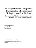
3.1.2. 1H NMR analysis
To further confirm rosmarinic acid conjugation into chitosan back
bone, 1H NMR analysis of derivatives and initial RA was conducted and
main signals of chitosan and the polyphenolic compound were identified
and assigned. Spectrum of RA showed the typical resonance signals
described in literature (Charisiadis et al., 2012) (Fig. S2). All CSRA
sample spectra exhibited the characteristic chitosan pattern (Sajomsang
et al., 2009) (Lavertu et al., 2003) (Fig. 1): a singlet at 5.2 ppm due to
anomeric proton, a multiplet between 4.2 and 3.2 ppm corresponding to
protons H3-H6 of the polysaccharide ring, and two singlet signals at 3.2
and 2.1 due to H2 proton of the amine group (H-amine) and the acetyl
group protons (H-Ac), respectively. Furthermore, 1H NMR spectra of
CSRA derivatives showed a broad multiplet signal between 7.5 and 6.7
ppm assigned to aromatic protons (e–j) of conjugated RA that was
shifted to lower field respect to that in the RA spectrum (between 6.4
and 5.5 ppm); a signal due to proton m of RA (7.7 ppm), a signal
attributed to proton n of RA (6.5 ppm), and a triplet signal (proton k)
slightly displaced and overlapped with the chitosan H2 signal at 3.2 ppm
2.5.5. Statistical analysis
Statistical analysis (ANOVA) with a significance level of *p < 0.05
between controls and samples or #p < 0.05 among samples was per
formed using Origin 8 Pro software (Origin Lab, USA) and Tukey
grouping method.
3. Results and discussion
Due to an increasing need of functionalized polymers, in the past
years several conjugates of chitosan and different polyphenols have been
reported with improved properties such as superior water solubility,
increased biocompatibility or higher radical scavenger activity (Hu &
Luo, 2016; Ilyasoglu & Guo, 2019; Kim, 2018; Xu et al., 2015).
4
M. Huerta-Madro˜
nal et al.
Carbohydrate Polymers 273 (2021) 118619
Fig. 1. 1H NMR spectra of native chitosan (CS) and CSRA samples in a 1:49 (v/v) D2O:DCl solvent mixture at 45 ◦ C. CSRA samples were named as CS-XRA, where X
denotes the effective percentage of RA conjugation into chitosan determined by UV spectroscopy analysis.
consequence of amide bond formation.
crystallinity and looseness of packing structure. This translates into the
lower thermal stability as a result of the grafting process produced in the
CSRA conjugates.
3.1.3. UV spectroscopy analysis
Rosmarinic acid UV spectrum shows maximum absorbance at
325–330 nm wavelength as already described (Saltas et al., 2013).
Therefore, absorbance at that specific wavelength was used to determine
RA concentrations in chitosan derivatives. It is worth mentioning that
chitosan is a polysaccharide obtained from the partial deacetylation of
chitin. Due to chitosan structure, RA can only be conjugated to the
primary amine group of the N-glucosamine rings. The percentage of
chitosan polysaccharide rings to which RA was conjugated (i.e. % RA
conjugation) was calculated for all chitosan derivatives which were
named as CS-XRA, where X denotes the effective percentage of RA
conjugated to chitosan. Codes of samples and results of CSRA compo
sition are shown in Table 1.
3.2. Release kinetics of CSRA conjugate films
Release of catechol-bearing species from film samples was analysed
in culture media (DMEM) without phenol red at 37 ◦ C. Given that chi
tosan derivatization was achieved via amide bond formation, as
confirmed previously by ATR-FTIR and 1H NMR spectroscopies, and
amide bonds are highly stable linkages resistant to hydrolysis at physi
ological pH and body temperatures, it is very unlikely that RA molecules
will be released from films to the culture media. In fact, reflux, high
temperatures and strong acid or basic solutions are used for its cleavage
(Mahesh et al., 2018; Ouellette & Rawn, 2018; Pill et al., 2019). Because
of this, lixiviates from films will be composed of catechol species most
likely consisting of chitosan molecules with RA attached via their pri
mary amine groups. Fig. 2 shows the release profiles of catechol-bearing
species of each sample at different time points. All curves followed a
similar pattern: a first initial burst release and a controlled release
pattern approaching to a plateau of RA-bearing species release. They
showed that CS-0.4RA and CS-0.8RA reached a plateau within the first
hour reaching final concentrations of 11 and 28 μg/mL (49.4 ± 2.8%
and 65.9 ± 2.4% of the initial RA content) respectively, while in the case
of CS-5RA and CS-10RA conjugates, both showed maximum release at 4
h achieving 115 μg/mL and 316 μg/mL (41.7 ± 1.6% and 57.7 ± 1.5% of
the initial RA amount) correspondingly.
3.1.4. Thermal stability study
Fig. S3 shows the thermogravimetric (A) and derivative thermog
ravimetry (DTG) (B) curves of CSRA conjugates compared to those of
initial chitosan and RA. Weight loss of chitosan underwent in two steps.
The first step observed below 150 ◦ C might be consequence of water
molecules entrapped into the carbohydrate chains (Tan et al., 2018) and
corresponded to 6.1% of weight loss. The second step and main degra
dation stage, in which chitosan decomposition and scission of the
polymer chain occurred (Jana et al., 2015), went from 260 ◦ C (onset) to
400 ◦ C with a 69.5% weight loss at 600 ◦ C (Table S2). DTG analysis
showed a maximum thermal decomposition temperature (Tmax) at
309 ◦ C for native chitosan. Similarly, CSRA derivatives presented a first
step of weight loss (in the range 5.0–6.8%) consequence of water
evaporation, however, CSRA derivatives started to degrade at lower
temperature than that of chitosan. The main thermal degradation event
started at 200 ◦ C and prolonged up to 400 ◦ C with a weight loss at 600 ◦ C
in the range of 62.3–65.5%. DTG thermographs showed that the highest
decomposition rate (Tmax) arose in the range of 245–247 ◦ C for all CSRA
derivatives. Grafting of RA, as occur when other polyphenols are con
jugated to chitosan (Hu & Luo, 2016), may cause a disruption of chi
tosan intermolecular hydrogen bonds, resulting in a remarkably reduced
3.3. Antioxidant capacity
3.3.1. CSRA conjugates
The radical scavenging activity of antioxidants against species such
as 1,1-diphenyl-2-picrylhydrazyl radical (DPPH), 2,2′ -azino-bis(3-eth
ylbenzothiazoline-6-sulfonic acid) (ABTS) or the superoxide anion
radical (O2⋅-) is currently measured to study the capacity of molecules to
act as free radical terminators or hydrogen donors (de Vega et al., 2020;
Ji et al., 2019). Of these methods, DPPH radical scavenger activity is
5
M. Huerta-Madro˜
nal et al.
Carbohydrate Polymers 273 (2021) 118619
Table 2
EC50 values of CSRA conjugates and calculated RA concentrations corresponding
to them.
Sample
CSRA EC50
(μg/mL)
[RA] for CSRA EC50 values
(μg/mL)
CS-10RA
CS-5RA
CS-0.8RA
CS-0.4RA
16
38
210
370
2.8
3.7
3.6
2.9
pKa4 = 10.62 (Danaf et al., 2016), which probably will burden its
antioxidant capacity at physiological pH. In order to investigate this
effect, initially the RA radical scavenger activity was evaluated at pH 4
and 7 and results are represented in Fig. 4. It can be observed that RSA
notably reduced at pH 7 compared to pH 4 in the concentration range
between 5 and 250 μg/mL. Separately, DPPH scavenger activity of film
lixiviates obtained at pH 7 was evaluated and results were compared
respect to DMEM without phenol red alone (control) in Fig. 4B. Inter
estingly, any CSRA conjugate lixiviate exerted similar antioxidant
properties giving RSA values around 40% independently of the degree of
functionalization. Therefore, it can be said that at pH 7 antioxidant
properties of RA may be hampered caused by deprotonation of one of its
catechol motives due to the proximity to pKa2 = 8.36 (Danaf et al.,
2016).
Fig. 2. RA-bearing species release profiles of CSRA films in culture medium
at 37 ◦ C.
widely used as a rapid, simple and inexpensive method. In this work,
antioxidant capacity of CSRA samples was initially studied versus con
centration and compared to that of free RA, whose potent radical
scavenger has been reported by different authors (Adomako-Bonsu
et al., 2017; Ji et al., 2019; Zhu et al., 2014) (Fig. 3). In this work, for
rosmarinic acid a half-maximum effective concentration (EC50) of 9.6
μg/mL (26.6 μM) was determined (Fig. 3A). As it can be noticed in
Fig. 3B, native chitosan exerted no antioxidant capacity at any con
centration while for the different conjugates, the higher the RA content,
the higher the antioxidant capacity. EC50 values of conjugates were
obtained from the curves represented in Fig. 5B and they are summa
rized in Table 2 along with the free RA concentrations corresponding to
these CSRA EC50 values. Interestingly, it can be observed that RA
immobilization into the chitosan backbone improves its antioxidant
activity, as free rosmarinic acid presents an EC50 value of 10 μg/mL
while CSRA EC50 concentrations correspond to 2.8–3.7 μg/mL of free
RA. Therefore, CSRA presents an antioxidant activity significantly
higher than free RA.
3.4. Cytotoxicity of CSRA conjugates
Cytotoxicity of CSRA lixiviates was monitored in HFB, HEK and RAW
264.7 cultures under ISO 10993-5:2009 and results are shown in
Fig. 5A–C respectively. As can be observed in the graphs, lixiviates from
CS-5RA and CS-10RA films resulted toxic for all three cell lines. How
ever, lixiviates from the CS-0.4RA and CS-0.8RA samples showed
absence of cytotoxicity. Biocompatibility of free RA was also assessed in
the three cell lines in the concentration range of those of lixiviates
(Fig. 5D–F). The selected concentrations for free RA were the results of a
wider experiment in which a larger range of concentrations were tested.
Those presented in the manuscript are the ones that allow determining
the EC50 easier. Free RA displayed cytotoxic effects at the highest
concentrations, showing viability values lower than 70% at 50 μg/mL
for HFB and at 75 μg/mL for both HEK and RAW 264.7 lines. Interest
ingly, similar cell viability (around 100%) were observed in the three
cell lines when compared lixiviates of the CS-0.8RA and CS-0.4RA
samples with their equivalent free RA concentrations (i.e., 28 and 11
μg/mL free RA respectively).
3.3.2. Influence of pH on antioxidant capacity
RA excellent antioxidant properties are mainly attributed to their
two catechol groups. However, they present several dissociated forms
depending on pH. According to Danaf et al., RA catechol groups pro
gressively lose their hydrogen atoms as pH increases from pKa1 = 2.92 to
Fig. 3. Radical scavenger activity of (A) free RA and (B) CSRA derivatives and native chitosan versus concentration.
6
M. Huerta-Madro˜
nal et al.
Carbohydrate Polymers 273 (2021) 118619
Fig. 4. (A) DPPH scavenger capacity of rosmarinic acid samples at different concentrations and at pH 4 or 7 and (B) CSRA lixiviates at pH = 7. Results are the mean
± SD (n = 8). Panel B includes the ANOVA results (*p < 0.05) comparing samples against DMEM without phenol red (control).
Fig. 5. Cell viability human epidermal fibroblasts (HFB), human epidermal keratinocytes (HEK) and murine macrophages (RAW 264.7) exposed to CSRA lixiviates
(A–C) or and free RA (D–F). The diagrams include the mean, SD (n = 16), and the ANOVA results (*p < 0.05 statistically significant difference between the cells in
DMEM (control) and treated cells, and #p < 0.05 between the cells treated with different CSRA conjugates or RA concentrations (brackets)).
3.5. Anti-inflammatory capacity
were tested in a previous experiment, however, they were not included
in the anti-inflammatory test since they showed to reduce RAW viability
below 50%. Almost null NO release was observed in non-stimulated cells
(negative control). Only 5 μg/mL of RA was enough to reduce nitric
oxide production in half compared to the positive control (Control +
LPS) (Fig. 6A). Likewise, lixiviates of both conjugates reduced NO levels
below 40%, showing similar effects than their corresponding free RA
concentrations, which confirms our initial hypothesis. CSRA derivatives
with higher grafting of RA suppressed in a greater manner NO release,
however no significant differences were observed among the two of
them (Fig. 6B).
The anti-inflammatory capacity of biocompatible lixiviates (i.e. CS0.8RA and CS-0.4RA samples) was assessed by means of the NO
release assay in macrophages RAW 264.7 cell line. Also, RA ability to
reduce LPS-induced nitric oxide levels was assessed and used for
comparative purposes. Different authors already proved the capacity of
rosmarinic acid and catechol bearing formulations to attenuate nitric
oxide production after activation with LPS (Silveira Fachel et al., 2019;
Puertas-Bartolom´
e et al., 2018; Qiao et al., 2005). Fig. 6 shows the total
amount of NO released by LPS-stimulated cells expressed in percentage
after treatment with RA samples at different concentrations (Fig. 6A) or
lixiviate samples (Fig. 6B). RA concentrations higher than 50 μg/mL
7
M. Huerta-Madro˜
nal et al.
Carbohydrate Polymers 273 (2021) 118619
Fig. 6. Nitric oxide release by RAW 264.7 macrophages after 24 h with: (A) treatment with LPS (positive control) and different rosmarinic acid (RA) concentrations,
and (B) treatment with LPS (positive control), no treatment (negative control), and treatment with LPS and 1 h lixiviates of CS-0.4RA and CS-0.8RA. The diagrams
include the mean, SD (n = 16), and the ANOVA results (*p < 0.05 statistically significant difference between positive control and tested samples, and #p < 0.05
between untreated cells and cells treated with different CSRA conjugates or RA concentrations (brackets)).
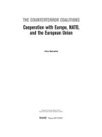
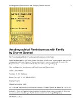
3.6. Photoprotective capacity of CSRA conjugates
contributions at this wavelength, and therefore if considered, they
would provide incorrect RA growth inhibition values. Fig. 8 shows the
growth inhibition capacity of the different samples compared to nega
tive control (incubated bacteria under permissive conditions in bacterial
growth media). The obtained data revealed that RA had stronger
bactericidal effect against S. epidermidis than to E. coli. CSRA conjugates
presented similar bacterial inhibition against S. epidermidis than their
corresponding free RA concentrations (1 μg/mL and 0.3 μg/mL, for CS0.8RA and CS-0.4RA, respectively). Nevertheless, in the case of E. coli,
both CSRA showed a two-fold GI value when compared to free RA.
Therefore, we conclude that CSRA derivatives elicited interesting
bactericidal effect similar to that of native chitosan and corresponding
concentration of free RA in the case of S. epidermidis. Noteworthy, a
slight synergistic effect was shown in the case of CS-0.8RA against E. coli,
since higher GI can be seen when it is compared to unmodified CS, and
free RA's growth inhibition capacity is doubled. In the same way against
E. coli, superior bactericidal effect of CS-0.4RA was evidenced, however,
no significant differences were observed when it was compared to native
CS.
Rosmarinic acid has shown photoprotective capacity, increasing the
cellular viability and reducing the oxidative stress of UV-irradiated cells
(Fernando et al., 2016; Lembo et al., 2014; Osakabe et al., 2004; P´
erezS´
anchez et al., 2014; P´erez-S´
anchez et al., 2016). In order to study the
photoprotective ability of CSRA polymers, lixiviates from biocompatible
conjugates were tested to evaluate their capacity to increase cell
viability and attenuate UVB-induced ROS effects after irradiation. After
UVB exposure, cells treated with CS-0.8RA or CS-0.4RA showed a sig
nificant increase in the percentage of cell viability with respect to pos
itive control (untreated and irradiated cells) (Fig. 7A–C). Interestingly, a
greater photoprotection was achieved with the polymer conjugate
incorporating the highest amount of RA. Moreover, Fig. 7D–G shows
results on the ROS production of HFB, HEK and RAW 264.7 upon UVB
irradiation in the presence or absence of CSRA. Irradiated cells without
CSRA conjugates were taken as 100% of ROS production. As it can be
observed, due to cellular basal metabolism, a certain fluorescence signal
is emitted in non-irradiated cells (negative control). When cells were
exposed to UVB in the presence of CSRA lixiviates, the production of
intracellular ROS was similar to the negative control. Notably again, the
chitosan derivative with the highest RA content was capable of reducing
in a greater manner UVB-induced radical oxygen species production.
This fact also supports our initial hypothesis, since it proves that chi
tosan conjugates maintained the photoprotective capacity of RA.
4. Conclusions
Chitosan functionalization with RA was successfully carried out as
confirmed by an extensive physicochemical characterization including
ATR-FTIR, 1H NMR, UV spectroscopy and TGA analysis. The resultant
water-soluble conjugates have demonstrated a three-fold increase in
radical scavenger activity and improved antimicrobial properties over
free RA. Moreover, they attenuated inflammatory activation of macro
phages and reduced UVB-induced damage and ROS production in fi
broblasts, keratinocytes and macrophages. Altogether these data
confirm our working hypothesis proving that the novel CSRA conjugates
present the bioactivity attributed to the original compounds (i.e. chito
san and rosmarinic acid), as demonstrated in vitro using skin-derived cell
cultures. CSRA conjugates possess desirable properties for skin appli
cations such as the treatment of age-related diseases and healing of
chronic wounds.
3.7. Antibacterial activity
The antibacterial properties of chitosan are extensively reported in
the literature since its broad-spectrum of antibacterial activity was first
explained by Allan and Hardwiger (Jana & Jana, 2019). Since then,
several mechanisms of antibacterial action for chitosan have been pro
posed, however, the topic is still a matter of discussion. On the other
hand, rosmarinic acid antibacterial activity has already been reported by
several authors (Abedini et al., 2013; Adamczak et al., 2019; Matejczyk
et al., 2018; Nieto et al., 2018) as well as the synergistic bactericidal
effect of chitosan when it is functionalized with different phytochemi
cals bearing catechol groups (Amato et al., 2018; Kim et al., 2017; Qin &
Li, 2020). Therefore, in this work the growth inhibition capacity of CSRA
conjugates was evaluated and compared to unmodified chitosan and
free RA. Notably, since bacterial concentrations were determined by
optical density at 600 nm, free RA concentrations were limited by the
experiment itself. Concentrations higher than 3.9 μg/mL led to
Credit authorship contribution statement
˜ al: Conceptualization, Methodology, Formal
M. Huerta-Madron
analysis, Investigation, Writing – original draft, Writing – review &
´ n: Conceptualization, Methodology,
editing, Visualization. J. Caro-Leo
Formal analysis, Investigation, Writing – review & editing,
8
M. Huerta-Madro˜
nal et al.
Carbohydrate Polymers 273 (2021) 118619
A
C
B
D
E
F
120
HFB
HEK
RAW
G
ROS production (%)
100
80
60
40
20
*
*
*
*
*
*
*
*
*
0
Negative
control
Positive
control
CS-0.8RA
CS-0.4RA
Fig. 7. Photo-protective capacity of CSRA 1 h lixiviates. (A–C) cell viability after 24 h, and (D–G) intracellular reactive oxygen species production of HFB, HEK and
RAW 264.7 upon UVB irradiation in presence of 1 h lixiviates from rosmarinic acid-chitosan (CSRA) films. The diagrams include the mean, SD (n = 24), and the
ANOVA results (*p < 0.05 statistically significant difference between untreated and either control or treated cells, and #p < 0.05 between control and CSRA treated
cells (brackets)).
9
M. Huerta-Madro˜
nal et al.
Carbohydrate Polymers 273 (2021) 118619
Fig. 8. Growth inhibition capacity of (A) different rosmarinic acid (RA) concentrations, and (B) 1 h lixiviates from native chitosan (CS) and rosmarinic-acid chitosan
conjugates (CSRA) films on S. epidermidis and (C) E. coli. Ampicillin and Gentamicin were used as specific antibiotics. The diagrams include the mean, SD (n = 16),
and the ANOVA results (*p < 0.05 statistically significant difference between negative control and samples, and #p < 0.05 between the different samples (brackets)).
Visualization. E. Espinosa-Cano: Conceptualization, Methodology,
Formal analysis, Investigation. M.R. Aguilar: Conceptualization,
Methodology, Writing – review & editing, Supervision, Project admin
´zquez-Lasa: Conceptualization,
istration, Funding acquisition. B. Va
Methodology, Writing – review & editing, Supervision, Project admin
istration, Funding acquisition.
References
Abbas, M., Saeed, F., Anjum, F., Afzaal, M., Tufail, T., Bashir, M., … Suleria, H. A. R.
(2017). Natural polyphenols: An overview. International Journal of Food Properties,
20. />Abedini, A., Roumy, V., Mahieux, S., Biabiany, M., Standaert, A., Rivi`
ere, C., …
Hennebelle, T. (2013). Rosmarinic acid and its methyl ester as antimicrobial
components of the hydromethanolic extract of Hyptis atrorubens Poit. (Lamiaceae).
Evidence-Based Complementary and Alternative Medicine: ECAM, 2013, Article 604536.
/>Adamczak, A., O˙zarowski, M., & Karpi´
nski, T. M. (2019). Antibacterial activity of some
flavonoids and organic acids widely distributed in plants. Journal of Clinical Medicine,
9(1), 109. />Adomako-Bonsu, A. G., Chan, S. L., Pratten, M., & Fry, J. R. (2017). Antioxidant activity
of rosmarinic acid and its principal metabolites in chemical and cellular systems:
Importance of physico-chemical characteristics. Toxicology In Vitro, 40, 248–255.
/>Amato, A., Migneco, L. M., Martinelli, A., Pietrelli, L., Piozzi, A., & Francolini, I. (2018).
Antimicrobial activity of catechol functionalized-chitosan versus Staphylococcus
epidermidis. Carbohydrate Polymers, 179, 273–281. />carbpol.2017.09.073
Amoah, S. K., Sandjo, L. P., Kratz, J. M., & Biavatti, M. W. (2016). Rosmarinic
acid–Pharmaceutical and clinical aspects. Planta Medica, 82(5), 388–406. https://
doi.org/10.1055/s-0035-1568274
Aytekin, A., Morimura, S., & Kida, K. (2010). Synthesis of chitosan-caffeic acid
derivatives and evaluation of their antioxidant activities. Journal of Bioscience and
Bioengineering, 111, 212–216. />Baptista da Silva, S., Amorim, M., Fonte, P., Madureira, A., Ferreira, D., Pintado, M., &
Sarmento, B. (2014). Natural extracts into chitosan nanocarriers for rosmarinic acid
Acknowledgements
This work was supported by MICINN (Spain) (MAT2017-84277-R,
and PRE2018-083873 M. Huerta's scholarship). F. J. Caro acknowledge
financial support from CONACyT (Mexico) through the scholarship
‘Apoyo para estancias postdoctorales en el extranjero vinculadas a la
´n de grupos de investigacio
´n y fortalecimiento del Posgrado
consolidacio
´zquez-Lasa are members of the Sus
nacional’. M.R. Aguilar and B. Va
Plast platform from CSIC.
Appendix A. Supplementary data
Supplementary data to this article can be found online at https://doi.
org/10.1016/j.carbpol.2021.118619.
10
M. Huerta-Madro˜
nal et al.
Carbohydrate Polymers 273 (2021) 118619
drug delivery. Pharmaceutical Biology, 1–11. />13880209.2014.935949
Bastos, F., Estevinho, B., & Santos, L. (2016). Preliminary studies of rosmarinic acid
microencapsulation with chitosan and modified chitosan for topical delivery. Powder
Technology, 297. />Boeriu, C. G., & van den Broek, L. A. M. (2019). Chemical and enzymatic modification of
chitosan to produce new functional materials with improved properties. In Chitin and
chitosan (pp. 245–258). />Calzoni, E., Cesaretti, A., Polchi, A., Di Michele, A., Tancini, B., & Emiliani, C. (2019).
Biocompatible polymer nanoparticles for drug delivery applications in cancer and
neurodegenerative disorder therapies. Journal of Functional Biomaterials, 10(1).
/>Charisiadis, P., Tsiafoulis, C. G., Exarchou, V., Tzakos, A. G., & Gerothanassis, I. P.
(2012). Rapid and direct low micromolar NMR method for the simultaneous
detection of hydrogen peroxide and phenolics in plant extracts. Journal of
Agricultural and Food Chemistry, 60(18), 4508–4513. />jf205003e
Chhabra, P., Chauhan, G., & Kumar, A. (2020). Augmented healing of full thickness
chronic excision wound by rosmarinic acid loaded chitosan encapsulated graphene
nanopockets. Drug Development and Industrial Pharmacy, 1–33. />10.1080/03639045.2020.1762200
Croisier, F., & J´erˆ
ome, C. (2013). Chitosan-based biomaterials for tissue engineering.
European Polymer Journal, 49(4), 780–792. />eurpolymj.2012.12.009
Curcio, M., Puoci, F., Iemma, F., Parisi, O. I., Cirillo, G., Spizzirri, U. G., & Picci, N.
(2009). Covalent insertion of antioxidant molecules on chitosan by a free radical
grafting procedure. Journal of Agricultural and Food Chemistry, 57(13), 5933–5938.
/>Cutrim, C., & Cortez, M. (2018). A review on polyphenols: Classification, beneficial
effects and their application in dairy products. International Journal of Dairy
Technology, 71. />da Silva, S. B., Ferreira, D., Pintado, M., & Sarmento, B. (2016). Chitosan-based
nanoparticles for rosmarinic acid ocular delivery—In vitro tests. International Journal
of Biological Macromolecules, 84, 112–120. />ijbiomac.2015.11.070
Danaf, N., Abi Melhem, R., Assaf, K., Nau, W., & Patra, D. (2016). Photophysical
properties of neutral and dissociated forms of rosmarinic acid. Journal of
Luminescence, 175. />de Vega, M. J., Moreno-Fern´
andez, S., Pontes-Quero, G. M., Gonz´
alez-Amor, M.,
V´
azquez-Lasa, B., Sabater-Mu˜
noz, B., … Gonz´
alez-Mu˜
niz, R. (2020).
Characterization of novel synthetic polyphenols: Validation of antioxidant and
vasculoprotective activities. Antioxidants, 9(9). />antiox9090787
El-Sherbiny, I., Elbaz, N., & Hefnawy, A. (2016). Potential of nanotechnology in
nutraceuticals delivery for the prevention and treatment of cancer. In Nutraceuticals
(pp. 117–152). />Fachel, F. N. S., Schuh, R. S., Veras, K. S., Bassani, V. L., Koester, L. S., Henriques, A. T.,
… Teixeira, H. F. (2019). An overview of the neuroprotective potential of rosmarinic
acid and its association with nanotechnology-based delivery systems: A novel
approach to treating neurodegenerative disorders. Neurochemistry International, 122,
47–58. />Fadel, O., El Kirat, K., & Morandat, S. (2011). The natural antioxidant rosmarinic acid
spontaneously penetrates membranes to inhibit lipid peroxidation in situ. Biochimica
et Biophysica Acta (BBA) - Biomembranes, 1808(12), 2973–2980. />10.1016/j.bbamem.2011.08.011
Fan, Y., Yi, J., Zhang, Y., & Yokoyama, W. (2017). Improved chemical stability and
antiproliferative activities of curcumin-loaded nanoparticles with a chitosan
chlorogenic acid conjugate. Journal of Agricultural and Food Chemistry, 65. https://
doi.org/10.1021/acs.jafc.7b04451
Fernando, P. M. D. J., Piao, M. J., Kang, K. A., Ryu, Y. S., Hewage, S. R. K. M.,
Chae, S. W., & Hyun, J. W. (2016). Rosmarinic acid attenuates cell damage against
UVB radiation-induced oxidative stress via enhancing antioxidant effects in human
HaCaT cells. Biomolecules & Therapeutics, 24(1), 75–84. />biomolther.2015.069
Ge, L., Zhu, M., li, X., Xu, Y., Ma, X., Shi, R., Li, D., & Mu, C. (2018). Development of
active rosmarinic acid-gelatin biodegradable films with antioxidant and long-term
antibacterial activities. Food Hydrocolloids, 83. />foodhyd.2018.04.052
Hossan, M. S., Rahman, S., Bashar, A. B. M., Jahan, R., Nahian, A., & Rahmatullah, M.
(2014). Rosmarinic acid: A review of its anticancer action. World Journal of
Pharmacy and Pharmaceutical Science, 3, 57–70.
Hu, Q., & Luo, Y. (2016). Polyphenol-chitosan conjugates: Synthesis, characterization,
and applications. Carbohydrate Polymers, 151, 624–639. />carbpol.2016.05.109
Huang, Y., Xiao, D., Burton-Freeman, B., & Edirisinghe, I. (2016). Chemical changes of
bioactive phytochemicals during thermal processing. In Reference module in food
science. />Ilyasoglu, H., & Guo, Z. (2019). Water soluble chitosan-caffeic acid conjugates as a dual
functional polymeric surfactant. Food Bioscience, 29. />fbio.2019.04.007
Islam, S., Bhuiyan, M. A. R., & Islam, M. N. (2017). Chitin and chitosan: Structure,
properties and applications in biomedical engineering. Journal of Polymers and the
Environment, 25(3), 854–866. />Jana, S., Trivedi, M., Tallapragada, R. M., Branton, A., Trivedi, D., Nayak, G., &
Mishra, R. (2015). Characterization of physicochemical and thermal properties of
chitosan and sodium alginate after biofield treatment. Pharmaceutica Analytica Acta,
6. />Jana, S., & Jana, S. (2019). Functional chitosan: Drug delivery and biomedical applications.
Functional Chitosan.
Ji, H.-S., Li, H., Mo, E.-J., Kim, U.-H., Kim, Y.-H., Park, H.-Y., & Jeong, T.-S. (2019). Lowdensity lipoprotein-antioxidant flavonoids and a phenolic ester from Plectranthus
hadiensis var. tomentosus. Applied Biological Chemistry, 62(1), 58. />10.1186/s13765-019-0464-y
Kim, G. D., Park, Y. S., Jin, Y. H., & Park, C. S. (2015). Production and applications of
rosmarinic acid and structurally related compounds. Applied Microbiology and
Biotechnology, 99(5), 2083–2092. />Kim, J.-H., Yu, D., Eom, S.-H., Kim, S.-H., Oh, J., Jung, W.-K., & Kim, Y.-M. (2017).
Synergistic antibacterial effects of chitosan-caffeic acid conjugate against antibioticresistant acne-related Bacteria. Marine Drugs, 15(6), 167. />md15060167
Kim, S. (2018). Competitive biological activities of chitosan and its derivatives:
Antimicrobial, antioxidant, anticancer, and anti-inflammatory activities.
International Journal of Polymer Science, 2018, 113. />1708172
ă Tatl, F. (2020).
Kỹba, M. C., Türko˘
glu, A., O˘
guz, A., Tuncer, M. C., Kaya, S¸., Bas¸ol, O.,
Comparison of local rosmarinic acid and topical dexpanthenol applications on
wound healing in a rat experimental wound model. Folia Morphologica. https://doi.
org/10.5603/FM.a2020.0097
Kuo, Y.-C., & Rajesh, R. (2017). Targeted delivery of rosmarinic acid across the bloodbrain barrier for neuronal rescue using polyacrylamide-chitosan-poly(lactide-coglycolide) nanoparticles with surface cross-reacting material 197 and apolipoprotein
E. International Journal of Pharmaceutics, 528. />ijpharm.2017.05.039
Lavertu, M., Xia, Z., Serreqi, A. N., Berrada, M., Rodrigues, A., Wang, D., … Gupta, A.
(2003). A validated 1H NMR method for the determination of the degree of
deacetylation of chitosan. Journal of Pharmaceutical and Biomedical Analysis, 32(6),
1149–1158. />Lembo, S., Balato, A., Di Caprio, R., Cirillo, T., Giannini, V., Gasparri, F., &
Monfrecola, G. (2014). The modulatory effect of ellagic acid and rosmarinic acid on
ultraviolet-B-induced cytokine/chemokine gene expression in skin keratinocyte
(HaCaT) cells. BioMed Research International, 2014, Article 346793. />10.1155/2014/346793
Luo, C., Zou, L., Sun, H., Peng, J., Gao, C., Bao, L., … Sun, S. (2020). A review of the antiinflammatory effects of Rosmarinic acid on inflammatory diseases. In , 11. Frontiers
in pharmacology (p. 153). />0.00153.
Mahesh, S., Tang, K.-C., & Raj, M. (2018). Amide bond activation of biological molecules.
Molecules (Basel, Switzerland), 23(10), 2615. />molecules23102615
´
Matejczyk, M., Swisłocka,
R., Golonko, A., Lewandowski, W., & Hawrylik, E. (2018).
Cytotoxic, genotoxic and antimicrobial activity of caffeic and rosmarinic acids and
their lithium, sodium and potassium salts as potential anticancer compounds.
Advances in Medical Sciences, 63(1), 14–21. />advms.2017.07.003
Mrduljas, N., Kresic, G., & Biluˇsi´c, T. (2017). Polyphenols: Food sources and health
benefits. In Functional food. />Muxika, A., Etxabide, A., Uranga, J., Guerrero, P., & de la Caba, K. (2017). Chitosan as a
bioactive polymer: Processing, properties and applications. International Journal of
Biological Macromolecules, 105(Pt 2), 1358–1368. />ijbiomac.2017.07.087
Nieto, G., Ros, G., & Castillo, J. (2018). Antioxidant and antimicrobial properties of
rosemary (Rosmarinus officinalis, L.): A review. Medicines (Basel, Switzerland), 5(3),
98. />Osakabe, N., Yasuda, A., Natsume, M., & Yoshikawa, T. (2004). Rosmarinic acid inhibits
epidermal inflammatory responses: Anticarcinogenic effect of Perilla frutescens
extract in the murine two-stage skin model. Carcinogenesis, 25(4), 549–557. https://
doi.org/10.1093/carcin/bgh034
Ouellette, R. J., & Rawn, J. D. (2018). Carboxylic acid derivatives. In R. J. Ouellette, & J.
D. B. T.-O. C (Eds.), Organic chemistry (Second E. Rawn, pp. 665–710). Academic
Press. />Parisi, O. I., Malivindi, R., Amone, F., Ruffo, M., Malanchin, R., Carlomagno, F., …
Puoci, F. (2017). Safety and efficacy of dextran-rosmarinic acid conjugates as
innovative polymeric antioxidants in skin whitening: What is the evidence?
Cosmetics, 4(3). />P´
erez-S´
anchez, A, Barraj´
on-Catal´
an, E., Caturla, N., Castillo, J., Benavente-García, O.,
Alcaraz, M., & Micol, V. (2014). Protective effects of citrus and rosemary extracts on
UV-induced damage in skin cell model and human volunteers. Journal of
Photochemistry and Photobiology. B, Biology, 136C, 12–18. doi: />1016/j.jphotobiol.2014.04.007.
P´
erez-S´
anchez, A., Barraj´
on-Catal´
an, E., Herranz Lopez, M., Castillo, J., & Micol, V.
(2016). Lemon balm extract (Melissa officinalis, L.) promotes melanogenesis and
prevents UVB-induced oxidative stress and DNA damage in a skin cell model. Journal
of Dermatological Science, 84. />Pill, M., East, A., Marx, D., Beyer, M., & Clausen-Schaumann, H. (2019). Mechanical
activation drastically accelerates amide bond hydrolysis, matching enzyme activity.
Angewandte Chemie International Edition, 58. />anie.201902752
Pokhrel, S., & Yadav, P. N. (2019). Functionalization of chitosan polymer and their
applications. Journal of Macromolecular Science, Part A, 56(5), 450–475. https://doi.
org/10.1080/10601325.2019.1581576
11
M. Huerta-Madro˜
nal et al.
Carbohydrate Polymers 273 (2021) 118619
Singla, R. K., Dubey, A., Garg, A., Sharma, R., Fiorino, M., Ameen, S., … al-hiaRy, ms.
(2019). Natural polyphenols: Chemical classification, definition of classes,
subcategories, and structures. Journal of AOAC International, 102. />10.5740/jaoacint.19-0133
Souto, E. B., Sampaio, A. C., Campos, J. R., Martins-Gomes, C., Aires, A., & Silva, A. M.
(2019). Polyphenols for skin cancer: Chemical properties, structure-related
mechanisms of action and new delivery systems. In B. T.-S, & N. P. C. Atta-urRahman (Eds.), 63. Bioactive natural products (pp. 21–42). Elsevier. />10.1016/B978-0-12-817901-7.00002-2.
Stehfest, K., Boese, M., Kerns, G., Piry, A., & Wilhelm, C. (2004). Fourier transform
infrared spectroscopy as a new tool to determine rosmarinic acid in situ. Journal of
Plant Physiology, 161(2), 151–156. />Tache, A., Radu, G., & Litescu, S. C. (2012). Assessment of role of rosmarinic acid in
preventing oxidative process of low density lipoproteins. Chemical Papers, 66.
/>Tan, W., Li, Q., Dong, F., Zhang, J., Luan, F., Wei, L., Chen, Y., & Guo, Z. (2018). Novel
cationic chitosan derivative bearing 1,2,3-triazolium and pyridinium: Synthesis,
characterization, and antifungal property. Carbohydrate Polymers, 182, 180–187.
/>Tsao, R. (2010). Chemistry and biochemistry of dietary polyphenols. Nutrients, 2(12),
1231–1246. />Vittorio, O., Curcio, M., Cojoc, M., Goya, G. F., Hampel, S., Iemma, F., … Cirillo, G.
(2017). Polyphenols delivery by polymeric materials: Challenges in cancer
treatment. Drug Delivery, 24(1), 162–180. />10717544.2016.1236846
Vuolo, M. M., Lima, V. S., & Mar´
ostica Junior, M. R. (2019). Phenolic compounds:
Structure, classification, and antioxidant power. In M. R. Segura Campos (Ed.),
Bioactive Compounds (pp. 33–50). Woodhead Publishing.
Wani, T. U., Raza, S. N., & Khan, N. A. (2019). Rosmarinic acid loaded chitosan
nanoparticles for wound healing in rats. International Journal of Pharmaceutical
Science and Research, 19(3), 1138–1147.
Xu, J., Strandman, S., Zhu, J. X. X., Barralet, J., & Cerruti, M. (2015). Genipin-crosslinked
catechol-chitosan mucoadhesive hydrogels for buccal drug delivery. Biomaterials, 37,
395–404. />Zhu, F., Asada, T., Sato, A., Koi, Y., Nishiwaki, H., & Tamura, H. (2014). Rosmarinic acid
extract for antioxidant, antiallergic, and alpha-glucosidase inhibitory activities,
isolated by supramolecular technique and solvent extraction from Perilla leaves.
Journal of Agricultural and Food Chemistry, 62(4), 885–892. />jf404318j
Puertas-Bartolom´e, M., V´
azquez-Lasa, B., & San Rom´
an, J. (2018). Bioactive and
bioadhesive catechol conjugated polymers for tissue regeneration. Polymers, 10(7).
/>Qiao, D., Ke, C., Hu, B., Luo, J., Ye, H., Sun, Y., Yan, X., & Zeng, X. (2009). Antioxidant
activities of polysaccharides from Hyriopsis cumingii. Carbohydrate Polymers, 78(2),
199–204. />Qiao, S., Li, W., Tsubouchi, R., Haneda, M., Murakami, K., Takeuchi, F., Nisimoto, Y., &
Yoshino, M. (2005). Rosmarinic acid inhibits the formation of reactive oxygen and
nitrogen species in RAW264.7 macrophages. Free Radical Research, 39, 995–1003.
/>Qin, Y., & Li, P. (2020). Antimicrobial chitosan conjugates: Current synthetic strategies
and potential applications. International Journal of Molecular Sciences, 21(2). https://
doi.org/10.3390/ijms21020499
Rinaudo, M. (2006). Chitin and chitosan: Properties and applications. Progress in Polymer
Science, 31(7), 603–632. />Ryu, J. H., Lee, Y., Kong, W. H., Kim, T., Park, T., & Lee, H. (2011). Catecholfunctionalized chitosan/pluronic hydrogels for tissue adhesives and hemostatic
materials. Biomacromolecules, 12, 2653–2659. />Sajomsang, W., Gonil, P., & Saesoo, S. (2009). Synthesis and antibacterial activity of
methylated N-(4-N,N-dimethylaminocinnamyl) chitosan chloride. European Polymer
Journal, 45(8), 2319–2328. />Saltas, D., Pappas, C. S., Daferera, D., Tarantilis, P. A., & Polissiou, M. G. (2013). Direct
determination of rosmarinic acid in Lamiaceae herbs using diffuse reflectance
infrared Fourier transform spectroscopy (DRIFTS) and chemometrics. Journal of
Agricultural and Food Chemistry, 61(13), 3235–3241. />jf305520m
Shahidi, F., & Ambigaipalan, P. (2015). Phenolics and polyphenolics in foods, beverages
and spices: Antioxidant activity and health effects – A review. Journal of Functional
Foods, 18, 820–897. />Silveira Fachel, F. N., Pr´
a, M., Azambuja, J., Endres, M., Bassani, V., Koester, L., &
Braganhol, E. (2019). Glioprotective effect of chitosan-coated rosmarinic acid
nanoemulsions against lipopolysaccharide-induced inflammation and oxidative
stress in rat astrocyte primary cultures. Cellular and Molecular Neurobiology, 40(1),
123–139.
Singh, B. R. (1999). Basic aspects of the technique and applications of infrared
spectroscopy of peptides and proteins. In Infrared Analysis of Peptides and Proteins
(Vol. 750, Issue 750, pp. 2–37). American Chemical Society. doi:doi:https://doi.
org/10.1021/bk-2000-0750.ch00110.1021/bk-2000-0750.ch001.
12