Antifibrotic effect of brown algae-derived fucoidans on osteoarthritic fibroblast-like synoviocytes
Bạn đang xem bản rút gọn của tài liệu. Xem và tải ngay bản đầy đủ của tài liệu tại đây (4.03 MB, 10 trang )
Carbohydrate Polymers 282 (2022) 119134
Contents lists available at ScienceDirect
Carbohydrate Polymers
journal homepage: www.elsevier.com/locate/carbpol
Antifibrotic effect of brown algae-derived fucoidans on osteoarthritic
fibroblast-like synoviocytes
˜ eiro-Ramil a, Noelia Flo
´rez-Fern´
´mez c, d,
María Pin
andez b, Olalla Ramil-Go
b
b
María Dolores Torres , Herminia Dominguez , Francisco J. Blanco e, f, Rosa Meijide-Faílde a, g,
Carlos Vaamonde-García h, *
a
Universidade da Coru˜
na, Tissue Engineering and Cellular Therapy Group, Instituto de Investigaci´
on Biom´edica de A Coru˜
na (INIBIC), Centro de Investigaciones
Científicas Avanzadas (CICA), 15006 A Coru˜
na, Spain
CINBIO, Universidade de Vigo, Biomass and Sustanaible Development Group (EQ2), Departament of Chemical Engineering, 32004 Ourense, Spain
c
Aging and Inflammation Research Laboratory, Instituto de Investigaciones Biom´edicas de A Coru˜
na (INIBIC), 15006 A Coru˜
na, Spain
d
Universidade de Coru˜
na, Endocrine, Nutritional and Metabolic Diseases Group, Departamento de Fisioterapia, Medicina y Ciencias Biom´edicas, Facultad de Ciencias de
la Salud, 15006 A Coru˜
na, Spain
e
Universidade da Coru˜
na, Grupo de Investigacion en Reumatología y Salud, Centro de Investigaciones Científicas Avanzadas (CICA), Departamento de Fisioterapia,
Medicina y Ciencias Biom´edicas, Facultad de Fisioterapia, 15006 A Coru˜
na, Spain
f
Hospital Universitario A Coru˜
na, Instituto de Investigaci´
on Biom´edica de A Coru˜
na (INIBIC), Grupo de Investigacion en Reumatología, 15006 A Coru˜
na, Spain
g
Universidade da Coru˜
na, Departamento de Fisioterapia, Medicina y Ciencias Biom´edicas, Facultad de Ciencias de la Salud, 15006 A Coru˜
na, Spain
h
Universidade da Coru˜
na, Grupo de Investigacion en Reumatología y Salud, Centro de Investigaciones Científicas Avanzadas (CICA), Departamento de Biología, Facultad
de Ciencias, 15071 A Coru˜
na, Spain
b
A R T I C L E I N F O
A B S T R A C T
Chemical compounds studied in this article:
PubChem CID: 74873 3-(Trimethylsilyl)
propane-1-sulfonic acid
PubChem CID: 24602 Deuterium oxide
PubChem CID: 92023653 Fucoidan
PubChem CID: 56842206 Transforming growth
factor beta
PubChem CID: 44259 Staurosporine
PubChem CID: 145068 Nitric oxide
PubChem CID: 104981 Propidium iodide
PubChem CID: 75783 Picrosirius red
PubChem CID: 4784 Phenylmethylsulfonyl
fluoride
Synovial fibrosis is a pathological process which contributes to joint pain and stiffness in several musculoskeletal
disorders. Fucoidans, sulfated polysaccharides found in brown algae, have recently emerged as promising
therapeutic agents. Despite the increasing amount of evidence suggesting the protective role of fucoidans in
different experimental approaches of human fibrotic disorders, the effect of these sulfated polysaccharides on
synovial fibrosis has not been investigated yet. By an in vitro experimental approach in fibroblast-like synovio
cytes, we detected that fucoidans inhibit their differentiation into myofibroblasts with tumor cell-like charac
teristics and restore apoptosis. Composition and structure of fucoidan appear to be critical for the detected
activity. Furthermore, protective effects of these sulfated polysaccharides are mediated by upregulation of nitric
oxide production and modulation of TGF-β/smad pathway. Altogether, our results support the use of fucoidans as
therapeutic compounds in the treatment of the fibrotic processes involved in rheumatic pathologies.
Keywords:
Fucoidan
Transforming growth factor beta
Fibrosis
Synovial fibroblasts
Apoptosis
Nitric oxide
Abbreviations: OA, osteoarthritis; FLS, fibroblast-like synoviocytes; ECM, extracellular matrix; Col, collagen; α-sma, alpha-smooth muscle actin 2; TFG-β, trans
forming growth factor-beta 1; NO, nitric oxide; DMEM, Dulbecco’s modified Eagle’s medium; FBS, fetal bovine serum; FF, fucoidan from Fucus vesiculosus; FM,
fucoidan from Macrocystis pyrifera; FU, fucoidan from Undaria pinnatifida; stau, staurosporine; HPSEC, high performance size exclusion chromatography; NMR,
nuclear magnetic resonance; col1a1, Type I collagen; col3a1, Type III collagen III; fn1, fibronectin 1; plod2b, procollagen-lysine 2-oxoglutarate 5-dioxygenase 2b;
gapdh, glyceraldehyde 3-phosphate dehydrogenase; BrdU, 5-bromo-2′ -deoxyuridine; PI, propidium iodide; ELISA, enzyme-linked immunosorbent assay; SEM,
standard error of the mean; EMT, epithelial-mesenchymal transition.
* Corresponding author at: Facultad de Ciencias, Campus de Zapateira, 15071 A Coru˜
na, Spain.
E-mail addresses: (M. Pi˜
neiro-Ramil), (N. Fl´
orez-Fern´
andez), (O. Ramil-G´
omez),
(M.D. Torres), (H. Dominguez), , (F.J. Blanco),
(R. Meijide-Faílde), (C. Vaamonde-García).
/>Received 12 November 2021; Received in revised form 23 December 2021; Accepted 9 January 2022
Available online 12 January 2022
0144-8617/© 2022 The Authors.
Published by Elsevier Ltd.
This is an open
( />
access
article
under
the
CC
BY-NC-ND
license
M. Pi˜
neiro-Ramil et al.
Carbohydrate Polymers 282 (2022) 119134
1. Introduction
therapeutic applications, a number of current studies about fucoidans
have employed polysaccharides from commercial origin, such as com
pounds derived from Undaria pinnatifida and Fucus vesiculosus (Bittkau
et al., 2019; Li et al., 2016). Interestingly, these fucoidans have
demonstrated to attenuate activation of pathological pathways
commonly leading to fibrosis (L. Wang et al., 2019; Wu et al., 2020). For
example, fucoidans from Fucus vesiculosus and from Laminaria japonica
have been shown to inhibit proliferation of tumor and non-tumor cell
lines and to decrease the expression of ECM-associated proteins (Bittkau
et al., 2019; H. Y. Chen et al., 2018).
However, despite growing evidence suggesting the antifibrotic role
of fucoidans in different experimental approaches of human diseases,
the impact of these sulfated polysaccharides on profibrotic phenotypic
changes in FLS, as well as their therapeutic potential for arthrofibrosis
treatment, have yet to be investigated. For these reasons, we evaluated
the antifibrotic effect of fucoidans derived from Fucus vesiculosus, Mac
rocystis pyrifera and Undaria pinnatifida on TGF-β-activated OA FLS. In
addition, we investigated the underlying molecular mechanisms impli
cated in this antifibrotic effect.
Rheumatic diseases are a group of heterogeneous pathologies char
acterized by the presence of inflammation and destruction in articular
´pez-Armada, 2019). Among them, oste
tissues (Vaamonde-García & Lo
oarthritis (OA) is the most prevalent chronic joint disorder, and a major
source of pain, disability, and socioeconomic costs worldwide. A com
mon feature of OA and other rheumatic diseases, such as rheumatoid
arthritis, is the occurrence of a phenotypic alteration in the synovium,
characterized by hyperplasia, leukocyte infiltration, neoangiogenesis,
and, finally, fibrosis (Ciregia et al., 2021). In the synovial tissue, the
fibrotic reaction involves an excess of collagen deposition which con
tributes to joint stiffness and pain (Hill et al., 2007; L. Zhang et al.,
2021). Besides, OA is a risk factor for developing a fibrotic joint disorder
known as arthrofibrosis, which can also be associated with joint trauma
or surgery (Bierke et al., 2021).
Although it is widely known that chronic inflammation and tissue
injury commonly favor fibrosis development, the exact mechanisms
favouring the onset of this pathological event in the joints are still
elusive. In synovial tissue fibrosis, phenotypic changes in fibroblast-like
synoviocytes (FLS) are driven by their differentiation into myofibro
blasts, cells that produce and secrete excessive levels of extracellular
matrix (ECM) components, especially type I and type III collagen and
fibronectin (Kasperkovitz et al., 2005; Schuster, Rockel, Kapoor, & Hinz,
2021; Steenvoorden et al., 2006). The hallmarks of fibroblast-tomyofibroblast transition are the expression of alpha-smooth muscle
actin 2 (α-sma) and increased proliferation, migration and invasion ca
pacities (Schuster et al., 2021).
Transforming growth factor-β1 (TGF-β) is the primary factor that
drives fibrosis (Meng, Nikolic-Paterson, & Lan, 2016). TGF-β signaling is
very complex; it is commonly accepted that ALK5-Smad2/3 signaling is
responsible of its profibrotic effects, whereas ALK1-Smad1/5/8 pathway
is involved in its antifibrotic action (Vaamonde-Garcia et al., 2019;
Walton, Johnson, & Harrison, 2017). Nevertheless, opposite roles of
these smad-based signaling pathways have also been described (Fran
˜ oz-F´
´lez-Nún
˜ ez, & Lo
´pez-Novoa, 2013).
gogiannis, 2020; Mun
elix, Gonza
Nonetheless, TGF-β is also involved in many pivotal cellular processes
(Ciregia et al., 2021). In the joint, TGF-β is typically associated with
cartilage anabolism/anti-catabolism and has been recently shown to be
correlated with clinically meaningful response to OA treatment (Watt
et al., 2020). Therefore, TGF-β cannot be considered as a therapeutic
target against fibrosis (Ciregia et al., 2021).
The development of effective antifibrotic therapies continues to be a
research priority, as knowledge in this field is still quite limited and
continues to progress slowly (Henderson, Rieder, & Wynn, 2020).
Antioxidant therapy has been proposed as a new strategy for the pre
vention and treatment of fibrotic diseases (Luangmonkong et al., 2018;
Varone, Gibiino, Gasbarrini, & Richeldi, 2019), and dietary supple
mentation with antioxidant-enriched products has already shown
beneficial effects in liver and cystic fibrosis (Bae, Park, & Lee, 2018;
Sagel et al., 2018). In this context, fucoidans, which are natural sulfated
polysaccharides found in different species of brown algae, have gained
special attention for its antioxidant and antitumor properties (Zayed &
Ulber, 2019). These natural biomolecules have also shown antiinflammatory, antiobesity and other health-promoting biological activ
ities (Pradhan et al., 2020), and are thus considered attractive thera
peutic alternatives for the treatment of different diseases. Consequently,
the interest for the use of fucoidans in the food and pharmaceutical in
dustry is rising fast (Zayed & Ulber, 2019).
Different factors influence in the biological properties of the fucoi
dan, such as composition, structure, presence and position of sulfate
groups, and molecular weight, among others (Ferreira, Passos, Madur
eira, Vilanova, & Coimbra, 2015; Zayed, El-Aasr, Ibrahim, & Ulber,
2020). In addition, geographical location, season of collection, and
extraction technology used have direct impact on their composition and
structure, and hence in their properties. In the pursuit of promising
2. Materials and methods
2.1. Reagents and treatments
Fucoidans from the seaweed Fucus vesiculosus L. (FF), Macrocystis
pyrifera L. (FM), and Undaria pinnatifida (FU), as well as TGFβ, were
purchased to Sigma-Aldrich (San Luis, MO, USA). Primary FLS cultures
were treated with 5, 30 and 100 μg/mL FF, FM, and FU, based on pre
vious research (Ryu & Chung, 2016; Vaamonde-García et al., 2021). FLS
were treated with fucoidans and in the presence of 10 ng/mL TGF-β in
order to induce a fibrotic response (Ciregia et al., 2021; Remst et al.,
2013; Vaamonde-Garcia et al., 2019). Staurosporine (stau; 1 μM) was
employed as positive control of apoptosis.
2.2. Characterization of fucoidans
1
H NMR spectra of FF, FU and FM were recorded at least in triplicate
using a ARX400 spectrometer (Bruker, Massachusetts, USA). Measure
ments were conducted using deuterium oxide as solvent and 3-(trime
thylsilyl)-1-propane sulfonic acid (Sigma-Aldrich, Misuri, USA) as an
internal standard, working at 75 ◦ C and 400 MHz. Samples were dis
solved in D2O at 10 mg/mL. Peaks were identified following the protocol
detailed elsewhere (Rasin et al., 2021).
The molar mass distribution of the fucoidans above was evaluated by
High-Performance Size Exclusion Chromatography (HPSEC) with an
HPLC (Agilent 1260, Germany) using a SuperMultipore PW-H column
(6 mm × 15 cm) with a guard column SuperMP (PW)-H (4.6 mm × 3.5
cm), both from TSKgel by Tosoh Corporation (Japan). The HPLC was
equipped with a refractive index (RI) detector. The mobile phase was
Milli-Q water at 0.4 mL/min, and the temperature of the column was
40 ◦ C. The standard used was polyethylene oxide at 786 kDa from Tosoh
Corporation (Japan).
2.3. Cell culture
Synovial tissue was obtained from 10 OA patients (5 females and 5
males) who underwent joint replacement surgery and gave informed
consent. The donors’ median age was 75.6 [86.1–65.1] years old. This
study was reviewed and approved by the Local Ethics Committee. FLS
were isolated as previously described (Vaamonde-Garcia et al., 2019)
and cultured in Dulbecco’s modified Eagle’s medium (DMEM) (Cambrex
Bio Science, Baltimore, MD, USA) containing 10% fetal bovine serum
(FBS), 2 mM L-glutamine, 100 mg/mL streptomycin, and 100 U/mL
penicillin (all from Lonza, Basel, Switzerland). FLS were subcultured
with trypsin-EDTA (Lonza) and were used for the experiments between
the third and eighth passages. For RNA isolation, protein extraction, and
2
M. Pi˜
neiro-Ramil et al.
Carbohydrate Polymers 282 (2022) 119134
2.7. Cell wound assay
Table I
Primer sequences used for real-time qPCR assays.
Gene
Forward (5′ -3′ )
Reverse (5′ -3′ )
Collagen I (col1a1)
Collagen III (col3a1)
Fibronectin 1 (fn1)
Procollagen-lysine 2-oxogluta
rate 5-dioxygenase 2b
(plod2b)
Glyceraldehyde-3-phosphate
dehydrogenase (gapdh)
ctggccccattggtaatgt
ctggaccccagggtcttc
ctggccgaaaatacattgtaaa
accagggaaaccagtagcac
catctgatccagggtttcca
ccacagtcgggtcaggag
cctgatatggctctttgccga
gggggctgagcatttggaat
agccacatcgctcagacac
gcccaatacgaccaaatcc
FLS were stimulated with 30 μg/mL FF, FM, and FU, with and
without TGF-β, in DMEM with 0.5% FBS. After 48 h, a linear wound was
produced by scratching cell monolayers with a 200 μL pipette tip.
Subsequently, wells were washed twice with saline buffer (Fresenius
Kabi, Barcelona, Spain) to remove the detached cells. Cells were
observed with a Nikon Eclipse TS100 inverted microscope (Nikon In
struments Europe B.V., Amsterdam, Netherlands) photographed with a
coupled XM Full HD digital camera (Hangzhou Xiongmai Technologies
(XM), Hangzhou, China) just after scratching (day 0) and 24 h later (day
1). The ImageJ software was employed to assess the level of wound
healing, which was calculated as the percentage of wound closure after
24 h.
invasion assays, cells were seeded on 12-well plates (BD Biosciences, San
Jose, CA, USA); for wound and apoptosis assays, on 24-well plates (BD
Biosciences); for proliferation studies as well as nitric oxide (NO) and
collagen levels measurements, on 96-well plates (BD Biosciences); and
for immunocytochemistry analysis, on 8-well chamber slides (BD Bio
sciences). Prior to stimulation, quiescence was induced by incubating
FLS in DMEM with 0.5% FBS overnight.
2.8. Cell invasion assay
FLS were stimulated with 30 μg/mL FF, FM, and FU, with and
without TGF-β, in DMEM with 0.5% FBS. After 48 h, cells were harvested
and seeded on cell culture inserts with a pore size of 8 μm (Sarstedt,
Nümbrecht, Germanay) previously coated with Matrigel (Corning, New
York, USA). Cells were suspended in DMEM with 0.5% FBS to be seeded
on the inserts, which were placed on a 24-well plate containing DMEM
with 10% FBS. After 24 h of incubation, inserts were stained with crystal
violet (Merck, Madrid, Spain). Invasive cells were visualized with a
Nikon Eclipse TS100 inverted microscope and photographed with a
coupled XM Full HD digital camera. Cell invasion was assayed using the
ImageJ software to quantify the amount of invasive colonies.
2.4. RNA isolation, reverse transcription and qPCR
After 48 h of stimulation with fucoidans and TGF-β, total RNA from
FLS was extracted and purified using TRIzol Reagent (Invitrogen,
Paisley, UK), chloroform (Sigma-Aldrich) and isopropanol (SigmaAldrich). The NZY First-Strand cDNA Synthesis Kit (Nzytech, Lisboa,
Portugal) was employed for the synthesis of cDNA. Reverse transcription
of 500 ng of RNA from each sample was carried out in a 96-Well Thermal
Cycler (Applied Biosystems, Thermo Fisher Scientific, Madrid, Spain).
cDNA products were then amplified by PCR using the Fast SYBR™ Green
master mix (Roche Diagnostics, Abingdon, UK). Quantitative real-time
polymerase chain reaction (qPCR) experiments were run on a Light
Cycler 480 instrument (Roche Diagnostics), employing LightCycler 480
SYBR Green I Master (Roche Diagnostics) and the primers shown in
Table I. After analyzing the data with the LC480 software, version 1.5
(Roche Diagnostics), relative gene expression was calculated with the
2− ΔΔCT method. Glyceraldehyde-3-phosphate dehydrogenase (gapdh)
was employed as the reference gene for normalization. All primers were
purchased from Invitrogen.
2.9. Immunocytochemistry
After stimulation of cells with fucoidans in the presence or absence of
TGF-β for 48 h, FLS were fixed with acetone for 10 min at 4 ◦ C. After
three washes in PBS, cells were permeabilized and blocked for 30 min in
PBS with 0.1% Tween 20 and 1% BSA. After blocking, 1-hour incubation
with a mouse anti-human α-sma primary antibody (1:500; DAKO A/S,
Glostrup, Denmark) was performed at room temperature. After addi
tional washes with 0.1% Tween 20 in PBS, cells were incubated with a
peroxidase-labeled goat anti-mouse/rabbit secondary antibody (DAKO
A/S) for 30 min and then counterstained with hematoxylin (Merck). An
Olympus BX61 microscope coupled to an Olympus DP70 digital camera
(Olympus Biosystems) was used to examine and photograph the slides.
The percentage of stained area among FLS was measured with the
ImageJ software.
2.5. Protein extraction, SDS-PAGE and Western blot
After 1 h of stimulation with fucoidans and TGF-β, intracellular
proteins from FLS were extracted employing Tris-HCl buffer pH 7.5 with
protease inhibitor cocktail and phenylmethylsulfonyl fluoride (all from
Sigma-Aldrich). SDS-PAGE was performed as previously described
(Vaamonde-Garcia et al., 2019) for proteins separation. After being
transferred to membranes, proteins were incubated overnight at 4 ◦ C
with the following rabbit anti-human antibodies: anti-p-Smad2 (S465/
467)/anti-p-Smad3 (S423/S425), anti-p-Smad1/5 (S463/465) (1:1000),
and anti-Glyceraldehyde 3-phosphate dehydrogenase (GAPDH; 1:2500)
(all from Cell Signaling Technology, Leiden, Netherlands). Anti-rabbit
secondary antibody (1:1000, Dako, Germany) and ECL chemilumines
cent substrate (Millipore, USA) were used for detecting antigenantibody binding. Protein bands were quantified by densitometry with
the ImageQ image processing software ( The
band intensities of the proteins of interest were normalized to GAPDH
band intensity for the same sample.
2.10. Intracellular collagen quantification
Picrosirius red (PSR) staining (Sigma-Aldrich) (Junquiera, Jun
queira, & Brentani, 1979) was employed to visualize and measure
intracellular collagen, as previously describe (Vaamonde-Garcia et al.,
2019). Briefly, after 48-hour stimulation, FLS were fixed in methanol,
washed with PBS, stained with 0.1% PSR staining solution and washed
with 0.1% acetic acid. Then, intracellular PSR staining was solubilized
with 0.1 M sodium hydroxide. A Nanoquant Infinite M200 spectropho
tometer (Tecan, Mă
annedorf, Switzerland) was employed to measure
absorbance at 550 nm.
2.11. Human pro-collagen I α1/COLIA1 assay
2.6. Cell proliferation assay
Collagen I is synthetized as a pro-collagen molecule. For this reason,
the levels of collagen I in culture supernatants from cultured FLS after
48-hour treatment with 5 and 30 μg/mL FF, FM, and FU, with and
without TGF-β, were determined employing the DuoSet ELISA kit for
human pro-Collagen I α1 (R&D system), following the instructions of the
manufacturer. Data were expressed as released picograms per mL. The
working range was between 31.2 and 2000 pg/mL.
FLS were treated with 5 and 30 μg/mL FF, FM, and FU, with and
without TGF-β, in DMEM with 2% FBS for 48 h. The BrdU Cell Prolif
eration Assay Kit (Cell Signaling Technology) was employed to evaluate
the incorporation of 5-bromo-2′ -deoxyuridine (BrdU) and thus cell
proliferation, according to the manufacturer’s instructions.
3
M. Pi˜
neiro-Ramil et al.
Carbohydrate Polymers 282 (2022) 119134
error of the mean (SEM) to represent error. Means of the variables tested
from “n” independent experiments (n = number of patients) is also
shown in graphs. All results were analyzed using GraphPad Prism 5
software (GraphPad Software, San Diego, CA). The nonparametric
Wilcoxon-test was used to compare different treatments; differences
were considered as statistically significant when p < 0.05.
Table II
Ratio of fucose: sulfate of the fucoidans studied from Fucus vesiculosus, Macro
cystis pyrifera and Undaria pinnatifida.
Ratio
Fucose:sulfate
Fucoidans from
Fucus vesiculosus
Undaria pinnatifida
Macrocystis pyrifera
1.00:0.81c
1.00:1.42a
1.00:1.25b
3. Results and discussion
Standard deviations were lower than 1%. Data with different letters were
significantly different (p > 0.05).
3.1. Fucoidans attenuate FLS proliferation and differentiation into
myofibroblasts
2.12. NO production assay
In a previous study, we detected that fucoidans from Fucus ves
iculosus, Macrocystis pyrifera, and Undaria pinnatifida had antioxidant
and anti-inflammatory effect on chondrocytes, while little effect was
observed on synoviocytes. While trying to elucidate the relation be
tween the composition and the protective effects of fucoidans, we found
that anti-oxidant actions and concentration of fucose/sulfate seem
critically relevant for fucoidan activity (Vaamonde-García et al., 2021).
In this study, to further investigate the role of chemical properties and
molecular weight in the biological properties of the fucoidans, we firstly
analyzed the ratio fucose:sulfate. As shown in Table II, FU ratio was
higher than that of FF and FM, with FU showing the highest sulfate
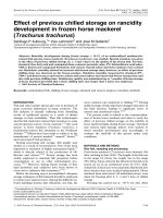
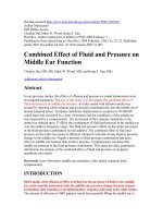
concentration, and FM showing the lowest one. 1H NMR spectra of FF
and FM exhibited α-anomeric protons (5–5.6 ppm), ring protons
(3.4–4.4), O-acetyl groups (~2.2 ppm) and methyl protons (1–1.3 ppm),
whereas O-acetyl groups were not identified for FU spectrum (Fig. 1A).
The highest values for the signal at the high-field region, which are
characteristic for α-L-fucopyranoside residues, were identified for FM,
followed by FF and FU. Similar regions were reportedly found in 1H
NMR spectra of other fucoidans from different brown seaweeds (Mon
sur, Jaswir, Simsek, Amid, & Alam, 2017; Rasin et al., 2021). It should
be noteworthy that all tested fucoidans presented a similar molar mass
distribution with a molecular weight above of >786 kDa (Fig. 1B).
Since we previously failed to observe a patent anti-inflammatory
effect of fucoidans on synoviocytes, in the present study we evaluated
if these sulfated polysaccharides were acting on fibrosis rather than
inflammation. In order to explore this possibility, we established an in
vitro model of profibrotic activation of FLS by stimulation with TGF-β
(Ciregia et al., 2021; Vaamonde-Garcia et al., 2019). As shown in Fig. 2,
TGF-β elicited a phenotypic change in FLS, promoting cell proliferation
and inducing the expression of a classic myofibroblast marker, the
The Griess reaction was used to determine the effects of fucoidans on
NO production in FLS after 48-hour treatment with TGF-β alone or
together with 30 μg/mL FF, FM or FU. Briefly, 50 μL of culture super
natant were collected and mixed with 50 μL of Griess reagent for nitrite
measurementsm and sodium nitrite was used as standard. A Nanoquant
Infinite M200 spectrophotometer was employed to measure absorbance
at 570 nm.
2.13. Apoptosis assay
The Annexin V method was used for the detection and measurement
of apoptosis. FLS were treated for 48 h with TGF-β alone or together with
30 μg/mL FF, FM or FU. Staurosporine was employed as positive control
of apoptosis. After that time, apoptosis was monitored by Annexin V/
Propidium iodide (PI) assay (Immunostep, Salamanca, Spain), following
the manufacturer’s instructions, employing a FACSCalibur flow cytom
eter (Becton Dickinson, Mountain View, CA, USA). Briefly, Annexin V
labeled with FITC was identified by green fluorescence and used to
quantify apoptotic cells. Simultaneous staining with non-vital dye PI
(which shows red fluorescence) allows the discrimination of intact cells
(Annexin V-FITC negative, PI negative), early apoptotic cells (Annexin
V-FITC positive, PI negative), late apoptotic cells (Annexin V-FITC
positive, PI positive), and necrotic cells (Annexin V-FITC negative, PI
positive). Apoptosis was calculated as percentage of apoptotic cells
(including both early and late apoptosis) for each condition.
2.14. Statistical analysis
All data in the graphs are reported as points representing one single
experiment with FLS obtained from one single patient, with standard
Fig. 1. Profiles of (A) 1H NMR and (B) HPSEC of the fucoidans from Fucus vesiculosus (FF), Undaria pinnatifida (FU), and Macrocystis pyrifera (FM). Vertical dashed
line indicates the weight of the pattern in dalton (Da). Symbols: FF (green line), FM (grey line) and FU (blue line). (For interpretation of the references to colour in
this figure legend, the reader is referred to the web version of this article.)
4
M. Pi˜
neiro-Ramil et al.
Carbohydrate Polymers 282 (2022) 119134
hydroxylated lysine residues within the telopeptides, contributing to
increase pyridinoline cross-links in collagen deposits (Yamauchi & Sri
cholpech, 2012). This change in cross-linking is related to irreversible
accumulation of collagen in fibrotic tissues (van der Slot et al., 2003).
Likewise, previous studies indicated that upregulated expression of
plod2b, and subsequent increase in pyridinoline cross-links, is the cause
of the persistent fibrosis both in synoviocytes stimulated with TGF-β in
vitro and in in vivo models of experimental OA (Remst et al., 2013;
Remst, Blaney Davidson, & van der Kraan, 2015).
In our study, co-incubation of FLS with fucoidans reduced the gene
expression of collagen I and III and fibronectin, the highest dose being
generally more effective (Fig. 3A-C). Accordingly, treatment with
similar concentrations of fucoidans reduced the expression of collagen I
and α-sma in a model of radiation-induced fibrosis in fibroblasts, where
modest inhibition of fibronectin expression was also detected (Wu,
Chen, Tsai, Hsu, & Hwang, 2020). Moreover, different studies reported
that fucoidans downregulate α-sma, col1a1, fibronectin and vimentin
protein levels in TGFβ-induced cells (H. Y. Chen et al., 2018; J. Chen
et al., 2015). However, in our study, these sulfated polysaccharides
failed to modulate the expression of plod2b (Fig. 3D).
These results were confirmed at the protein level by intracellular
collagen staining and measurement of type I collagen released to cell
supernatant (Fig. 4). FLS co-treated with TGF-β and either FF or FM
showed a reduction of intracellular collagen levels, which are raised by
TGF-β, as indicated by PSR staining (Fig. 4A). Besides, the amount of
type I pro-collagen in the culture media was significantly increased in
those FLS stimulated with TGF-β, but co-incubation with FF, FU and FM
strongly diminished its release (Fig. 4B). Accordingly, oligo-fucoidan
treatment significantly reduced collagen accumulation in a model of
renal tubulointerstitial fibrosis in mice (C. H. Chen et al., 2017). Inter
estingly, fucoidan produced by Laminaria japonica, which presents a
percentage of fucose and sulfate similar to those of FF, FU and FM, but a
lower molecular weight, attenuated interstitial and perivascular fibrosis,
reducing collagen content and expression of ECM components in an
animal model of diabetic cardiopathy (Yu et al., 2014). Taken together,
these findings suggest that fucoidans’ chemical composition plays a
pivotal role in their antifibrotic effects.
Relative levels of pr olifer ation
A.
Proliferation
4
*
3
#
2
#
1
0
#
l
0
0
0
5
5
5
sa FF F3 FU U3 M M3 GF
F F T
F
F
Ba
#
#
#
0
0
0
5
5
5
FF FF3 FU FU3 FM M3
F
TGF-
B.
Relative expression
20
-SMA
*
15
10
α-sma
~42 kDa
#
#
#
Tubulin
~50 kDa
1
1,72
1,17
1,41
1,31
5
FM
30
FF
30
FU
30
TG
F-
FM
30
FU
30
Ba
sa
l
FF
30
0
TGF-
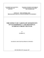
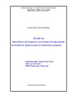
Fig. 2. Effect of fucoidans on FLS proliferation and differentiation into myo
fibroblast. FLS were incubated for 24 h with 5 or 30 μg/mL FF, FM, and FU,
with or without TFG-β. Then, (A) cell proliferation was determined by BrdU
assay (n = 5). (B) Alpha-smooth muscle actin 2 (α-sma) protein expression was
assayed by immunohistochemistry and confirmed by western blot. Upper panel,
representative images of cells stimulated as indicated and stained with α-sma.
Left-lower panel, quantitative analysis of α-sma staining (n = 3). Original
magnification: 100×. Right-lower panel, representative image of protein levels
of α-sma analyzed by western blot and quantification shown on the bottom.
Graphs represent means ± SEM. *, statistically different vs. basal condition (p <
0.05); #, statistically different vs. stimulated with TFG-β alone (p < 0.05); FLS,
fibroblast-like synoviocytes; FF, fucoidan from Fucus vesiculosus; FM, fucoidan
from Macrocystis pyrifera; FU, fucoidan from Undaria pinnatifida; TFG-β, trans
forming growth factor-beta 1.
3.3. Fucoidans attenuate TFG-β-elicited cell migration and invasion
Following TGF-β-induced activation, FLS acquire tumor cell-like
characteristics such as impaired apoptosis, unlimited proliferation,
and migration and invasion abilities. Regarding the latter processes, FLS
can directly migrate and invade into the articular tissues surrounded by
the synovium, so that a progressive degradation of articular cartilage
and bone take place, exacerbating joint damage (Shu, Shi, Nie, & Guan,
2015; Yan et al., 2016). As shown in Fig. 5, TFG-β promoted FLS
migration and invasion. Treatment with FF, FU or FM attenuated,
although not significantly, the migration capacity of TFG-β-activated
FLS, being FM the fucoidan which showed the clearest effect (Fig. 5A).
All three fucoidans also diminished cell invasion induced by TFG-β, and
again significant differences were found between FLS treated with FM
and those treated with TGF-β alone (Fig. 5B). Similarly, a previous study
using FLS derived from rheumatoid arthritis patients showed that
fucoidans reduced invasiveness of IL-1β-treated cells (Shu et al., 2015).
In a similar way, fucoidan from Fucus vesiculosus inhibited cell migration
and invasion activity from human lung cancer cells in vitro (Lee, Kim, &
Kim, 2012).
protein α-sma. Interestingly, fucoidans reduced the proliferation
induced by TGF-β in a dose-dependent manner (Fig. 2A). Likewise, the
expression of α-sma was attenuated in those cells co-treated with
fucoidans (Fig. 2B). Accordingly, previous studies showed that fucoi
dans reversed TGF-β-induced differentiation of fibroblasts into myofi
broblasts, diminishing the expression of α-sma and reducing cell
proliferation (H. Y. Chen et al., 2018; Y. Zhang, Du, Yu, & Zhu, 2020; Y.
Zhang et al., 2018).
3.2. Fucoidans diminish the synthesis and release of ECM proteins
induced by TFG-β
One of the events that characterize fibrosis is the accumulation of
ECM proteins, mainly collagen, in the synovial tissue (Frangogiannis,
2020; Henderson et al., 2020). Likewise, α-sma-positive FLS are the
main responsible for the production and secretion of ECM proteins,
including fibrillar collagen (Frangogiannis, 2020; Henderson et al.,
2020; Schuster et al., 2021). As expected, TGF-β-activated FLS showed
increased expression of collagen I, collagen III, fibronectin, and
procollagen-lysine, also known as 2-oxoglutarate 5-dioxygenase 2b
(Plod2b) (Fig. 3). Plod2b is a gene involved in enzymatic increment of
3.4. Fucoidans induce apoptosis and stimulate NO production
Evasion or impairment of apoptosis activation favors persistent fi
broblasts differentiation into cells with a profibrotic phenotype, thus
preventing fibrosis resolution (Hinz & Lagares, 2020). Interestingly,
when apoptosis was evaluated in our in vitro model of synovial fibrosis,
we observed that TFG-β downregulated cell apoptosis (Fig. 6A). Co5
M. Pi˜
neiro-Ramil et al.
Carbohydrate Polymers 282 (2022) 119134
Fig. 3. Effect of fucoidans on gene expression of extracellular matrix proteins induced by TFG-β. FLS were stimulated for 48 h with FF, FM or FU, with or without
TGF-β. Then, gene expression of collagen I (col1a1), collagen III (col3a1), fibronectin 1 (fn1), and procollagen-lysine 2-oxoglutarate 5-dioxygenase 2b (plod2b) was
analyzed by RT-qPCR. Reference gene glyceraldehyde-3-phosphate dehydrogenase (gapdh) was used for normalizing gene expression (n = 5). Graphs represent means
± SEM. *, statistically different vs. basal condition (p < 0.05); #, statistically different vs. stimulated with TFG-β alone (p < 0.05); FLS, fibroblast-like synoviocytes;
FF, fucoidan from Fucus vesiculosus; FM, fucoidan from Macrocystis pyrifera; FU, fucoidan from Undaria pinnatifida; TFG-β, transforming growth factor-beta 1.
incubation of FLS with FF, FU and FM attenuated this effect and allowed
the recovering of basal levels of apoptosis, whose values were far lower
from those obtained with positive control staurosporine. In a similar
way, fucoidans have been described to induce apoptosis in different
types of cancer cells (Lin et al., 2020). For instance, fucoidans from Fucus
vesiculosus induce apoptosis and inhibit epithelial-mesenchymal transi
tion (EMT) in breast cancer cells (He et al., 2019). Besides, in a previous
study, Shu et al. (2015) observed that fucoidans activate apoptosis in
rheumatoid arthritis FLS stimulated with IL-1β, thus exerting antisurvival activities on pathological synoviocytes (Shu et al., 2015).
Evidence indicates that the pro-apoptotic effects of these sulfated
polysaccharides could be mediated by NO release (Jin, Song, Kim, Park,
& Kwak, 2010; Takeda et al., 2012). Since it is known that NO induces
apoptosis in OA synoviocytes (Borderie et al., 1999), we evaluated if FF,
FU and FM could activate the production of NO in our model. As shown
in Fig. 6B, the treatment with all three fucoidans reversed the inhibition
of NO release induced by TFG-β. These findings suggest that the in
duction of NO production by fucoidans could participate in their proapoptotic effects and therefore in the amelioration of fibrosis. Like
wise, Li et al. (2016) observed that fucoidans protect against liver
fibrosis through inhibition of autophagy (Li et al., 2016), a physiological
process in which damaged or unnecessary organelles are destroyed, and
which is now considered a target for the prevention of synovial fibrosis
(Maglaviceanu, Wu, & Kapoor, 2021). Since it is widely recognized that
there is a crosstalk between apoptosis and autophagy (Fairlie, Tran, &
Lee, 2020), the role of autophagy in the antifibrotic effect of fucoidans
should not be discarded and future investigation are warranted.
3.5. Fucoidans modulate TFG-β-induced smad signaling
Smad signaling plays a central role in downstream pathways
involved in TGF-β-induced activation of fibrosis (Hu et al., 2018). Two
receptors are involved in these intracellular pathways: ALK5, which
mediates phosphorylation of Smad2/3; and ALK1, which mediates
phosphorylation of Smad1/5/8. In this study, we analyzed the levels of
pSmad2/3 and pSmad1/5 as a measure of the activation of these path
ways. As expected, TFG-β-stimulated cells showed phosphorylation of
smad 2/3 and smad 1/5 (Fig. 7). Interestingly, all three fucoidans
modulated these TFG-β signaling pathways in a different way. Fucoidan
from Fucus vesiculosus significantly reduced pSmad 2/3 levels, whereas
FM and FU failed to exert a consistent inhibitory effect (Fig. 7A-B).
Conversely, phosphorylation of smad 1/5 was only significantly upre
gulated by fucoidan from Macrocystis pyrifera (Fig. 7A-C).
In a previous study, a fucoidan with a similar fucose:sulfate ratio
(29% fucose: 30% sulfate) ameliorated fibrosis both in vivo and in vitro,
at least partially through inhibiting smad3 phosphorylation and TGF-β
signaling (J. Chen et al., 2015). Similarly, fucoidan from Fucus ves
iculosus has been shown to suppress the upregulation of phosphorylated
Smad2/3 induced by TGF-β, protecting retinal epithelial cells and he
patic stellate cells against EMT (Y. Zhang et al., 2018) in an in vivo model
of liver fibrosis (Li et al., 2016). Additionally, beneficial effects of
fucoidans have been also associated to upregulation of pSmad 1/5 levels
and smad1-dependent smad 7 expression (Chale-Dzul, P´
erez-Cabeza de
Vaca, Quintal-Novelo, Olivera-Castillo, & Moo-Puc, 2020; Kim, Kang,
Park, & Lee, 2015). Nonetheless, fucoidan from seaweed Nemacystus
decipiens disrupts angiogenesis by blocking pSmad 1/5/8 signaling (W.
Wang et al., 2016). Therefore, future studies will be needed in order to
elucidate the precise role of smads in the protective effects of fucoidans.
6
M. Pi˜
neiro-Ramil et al.
Carbohydrate Polymers 282 (2022) 119134
Fig. 4. Fucoidan modulation of protein collagen expression induced by TFG-β
in FLS. FLS were stimulated for 48 h with FF, FM or FU, with or without TGF-β.
Then, (A) levels of total intracellular collagens were analyzed by picrosirius red
(PSR) stanning (n = 5). (B) Pro-collagen I α1 released in the culture supernatant
was quantified by ELISA (n = 5). Graphs represent means ± SEM. *, statistically
different vs. basal condition (p < 0.05); #, statistically different vs. stimulated
with TFG-β alone (p < 0.05); FLS, fibroblast-like synoviocytes; FF, fucoidan
from Fucus vesiculosus; FM, fucoidan from Macrocystis pyrifera; FU, fucoidan
from Undaria pinnatifida; TFG-β, transforming growth factor-beta 1. (For
interpretation of the references to colour in this figure legend, the reader is
referred to the web version of this article.)
Fig. 5. Effect of fucoidans on cell migration and invasion in TFG-β-activated
FLS. FLS were stimulated for 48 h with FF, FM or FU, with or without TGF-β.
Then, (A) wound healing assays were performed. Upper panel, representative
images showing cell monolayers just after wounding (day 0) and 24 h later (day
1). Original magnification: 40×. Lower panel, quantitative analysis of wound
closure. Complete wound closure = 100% (n = 5). (B) Invasion assays were
performed. Upper panel, representative images of crystal violet-stained invasive
FLS after different treatments. Original magnification: 6.7×. Lower panel,
quantitative analysis of crystal violet staining after invasion assay (n = 4).
Graphs represent means ± SEM. *, statistically different vs. basal condition (p <
0.05); #, statistically different vs. stimulated with TFG-β alone (p < 0.05); FLS,
fibroblast-like synoviocytes; FF, fucoidan from Fucus vesiculosus; FM, fucoidan
from Macrocystis pyrifera; FU, fucoidan from Undaria pinnatifida; TFG-β, trans
forming growth factor-beta 1. (For interpretation of the references to colour in
this figure legend, the reader is referred to the web version of this article.)
Taken together, these results suggest that these sulfated poly
saccharides exert their antifibrotic actions in different ways, at least in
terms of modulation of TFG-β/Smad signaling. Distinct chemical
composition and structure among fucoidans could explain this phe
nomenon and thereby their different capacities to attenuate fibrosisrelated processes such as cell invasion and ECM overproduction.
3. Conclusions and future perspectives
Overall, these findings along with those from our previous work
(Vaamonde-García et al., 2021) and from other authors support the use
of these marine polysaccharides as therapeutic agents in the treatment of
rheumatic pathologies. Additionally, the findings here presented
encourage the development of further studies targeting the composition
and structure of fucoidans, as well as their relation with their biological
properties. Hereby, clinical trials should also be developed in the pursuit
of therapeutic applications of fucoidans which have already shown
promising effects in in vitro approaches.
Collectively, our results indicate the antifibrotic effect of fucoidans
on activated synoviocytes. This effect is mediated by the inhibition of
fibroblast-to-myofibroblast transition, which leads to the upregulation
of ECM production and the acquisition of tumor cell-like characteristics
such as unlimited proliferation, migration and invasion abilities, and
impaired apoptosis. Regarding the latest, fucoidans induce NO produc
tion and release, which is likely to activate the programmed cell death of
pathological fibroblasts, thereby attenuating synovial fibrosis. Further
more, the protective action of these sulfated polysaccharides is differ
ently mediated by the modulation of TGF-β1/Smad pathways.
7
M. Pi˜
neiro-Ramil et al.
Carbohydrate Polymers 282 (2022) 119134
Fig. 6. Fucoidan recovery of apoptosis and NO production in TFG-β-activated FLS. FLS were stimulated for 48 h with TGF-β alone or together with FF, FM or FU.
Staurosporine was employed as positive control. Then, (A) apoptosis was evaluated by flow cytometry. Upper panel, representative histograms from one experiment
showing distribution of cell population and percentage of apoptotic cells (Annexin V and FITC positive). Lower panel, quantitative analysis of percentage of cells
under apoptosis per condition (n = 4). (B) NO production was evaluated by Griess reaction (n = 4). Graphs represent means ± SEM. #, statistically different vs.
stimulated with TFG-β alone (p < 0.05); FLS, fibroblast-like synoviocytes; FF, fucoidan from Fucus vesiculosus; FM, fucoidan from Macrocystis pyrifera; FU, fucoidan
from Undaria pinnatifida; TFG-β, transforming growth factor-beta 1; Stau, staurosporine.
CRediT authorship contribution statement
Francisco J Blanco: Writing – review & editing. Herminia Domí
nguez: Writing – review & editing. Rosa Meijide-Faílde: Writing –
review & editing. Carlos Vaamonde-García: Conceptualization, Formal
analysis and Investigation, Supervision, Writing – review & editing.
˜ eiro-Ramil: Formal analysis and Investigation, Writing –
María Pin
´ rez-Ferna
´ndez: Formal analysis and
review & editing. Noelia Flo
´ mez: Formal
Investigation, Writing – review & editing. Olalla Ramil-Go
analysis and Investigation, Writing– review & editing. María Dolores
Torres: Formal analysis and Investigation, Writing– review & editing.
8
M. Pi˜
neiro-Ramil et al.
Carbohydrate Polymers 282 (2022) 119134
acknowledged (Grupos con Potencial de Crecemento 2020, grant num
´n de Galicia
ber ED431B 2020/55; Centro Singular de Investigacio
2019–2022, grant number ED431G2019/06; Rede Galega de Terapia
Celular 2016, grant number R2016/036). N.F.-F. thanks Xunta de
Galicia for her postdoctoral contract [grant number ED481B 2018/071].
M.D.T. thanks Ministry of Economy and Competitiveness of Spain for
her postdoctoral grant [grant number RYC2018-024454-I]. C.V.-G.
thanks Xunta de Galicia for his postdoctoral contract [grant number
˜ a/CISUG
ED481D 2017/023]. Authors also thank Universidade da Corun
for funding open access charge.
References
Bae, M., Park, Y. K., & Lee, J. Y. (2018). Food components with antifibrotic activity and
implications in prevention of liver disease. The Journal of Nutritional Biochemistry, 55,
1–11.
Bierke, S., Abdelativ, Y., Hees, T., Karpinksi, K., Hă
aner, M., Park, H., & Petersen, W.
(2021). Risk of arthrofibrosis in anatomical anterior cruciate ligament
reconstruction: The role of timing and meniscus suture. Archives of Orthopaedic and
Trauma Surgery, 141(5), 743750.
Bittkau, K. S., Dă
orschmann, P., Blỹmel, M., Tasdemir, D., Roider, J., Klettner, A., &
Alban, S. (2019). Comparison of the effects of fucoidans on the cell viability of tumor
and non-tumor cell lines. Marine Drugs, 17(8).
Borderie, D., Hilliquin, P., Hernvann, A., Lemarechal, H., Menkes, C. J., &
Ekindjian, O. G. (1999). Apoptosis induced by nitric oxide is associated with nuclear
p53 protein expression in cultured osteoarthritic synoviocytes. Osteoarthritis and
Cartilage, 7(2), 203–213.
Chale-Dzul, J., P´erez-Cabeza de Vaca, R., Quintal-Novelo, C., Olivera-Castillo, L., & MooPuc, R. (2020). Hepatoprotective effect of a fucoidan extract from Sargassum fluitans
borgesen against CCl. International Journal of Biological Macromolecules, 145,
500–509.
Chen, C. H., Sue, Y. M., Cheng, C. Y., Chen, Y. C., Liu, C. T., Hsu, Y. H., … Chen, T. H.
(2017). Oligo-fucoidan prevents renal tubulointerstitial fibrosis by inhibiting the
CD44 signal pathway. Scientific Reports, 7, 40183.
Chen, H. Y., Huang, T. C., Lin, L. C., Shieh, T. M., Wu, C. H., Wang, K. L., … Hsia, S. M.
(2018). Fucoidan inhibits the proliferation of leiomyoma cells and decreases
extracellular matrix-associated protein expression. Cellular Physiology and
Biochemistry, 49(5), 1970–1986.
Chen, J., Cui, W., Zhang, Q., Jia, Y., Sun, Y., Weng, L., … Yang, B. (2015). Low molecular
weight fucoidan ameliorates diabetic nephropathy via inhibiting epithelialmesenchymal transition and fibrotic processes. American Journal of Translational
Research, 7(9), 1553–1563.
Ciregia, F., Deroyer, C., Cobraiville, G., Plener, Z., Malaise, O., Gillet, P., … de Seny, D.
(2021). Modulation of α. Experimental & Molecular Medicine, 53(2), 210–222.
Fairlie, W. D., Tran, S., & Lee, E. F. (2020). Crosstalk between apoptosis and autophagy
signaling pathways. International Review of Cell and Molecular Biology, 352, 115–158.
Ferreira, S. S., Passos, C. P., Madureira, P., Vilanova, M., & Coimbra, M. A. (2015).
Structure-function relationships of immunostimulatory polysaccharides: A review.
Carbohydrate Polymers, 132, 378–396.
Frangogiannis, N. (2020). Transforming growth factor-β in tissue fibrosis. The Journal of
Experimental Medicine, 217(3), Article e20190103.
He, X., Xue, M., Jiang, S., Li, W., Yu, J., & Xiang, S. (2019). Fucoidan promotes apoptosis
and inhibits EMT of breast cancer cells. Biological & Pharmaceutical Bulletin, 42(3),
442–447.
Henderson, N. C., Rieder, F., & Wynn, T. A. (2020). Fibrosis: From mechanisms to
medicines. Nature, 587(7835), 555–566.
Hill, C. L., Hunter, D. J., Niu, J., Clancy, M., Guermazi, A., Genant, H., … Felson, D. T.
(2007). Synovitis detected on magnetic resonance imaging and its relation to pain
and cartilage loss in knee osteoarthritis. Annals of the Rheumatic Diseases, 66(12),
1599–1603.
Hinz, B., & Lagares, D. (2020). Evasion of apoptosis by myofibroblasts: A hallmark of
fibrotic diseases. Nature Reviews Rheumatology, 16(1), 11–31.
Hu, H. H., Chen, D. Q., Wang, Y. N., Feng, Y. L., Cao, G., Vaziri, N. D., & Zhao, Y. Y.
(2018). New insights into TGF-β/Smad signaling in tissue fibrosis. Chemico-Biological
Interactions, 292, 76–83.
Jin, J. O., Song, M. G., Kim, Y. N., Park, J. I., & Kwak, J. Y. (2010). The mechanism of
fucoidan-induced apoptosis in leukemic cells: Involvement of ERK1/2, JNK,
glutathione, and nitric oxide. Molecular Carcinogenesis, 49(8), 771–782.
Junquiera, L. C., Junqueira, L. C., & Brentani, R. R. (1979). A simple and sensitive
method for the quantitative estimation of collagen. Analytical Biochemistry, 94(1),
96–99.
Kasperkovitz, P. V., Timmer, T. C., Smeets, T. J., Verbeet, N. L., Tak, P. P., van
Baarsen, L. G., … Verweij, C. L. (2005). Fibroblast-like synoviocytes derived from
patients with rheumatoid arthritis show the imprint of synovial tissue heterogeneity:
Evidence of a link between an increased myofibroblast-like phenotype and highinflammation synovitis. Arthritis and Rheumatism, 52(2), 430–441.
Kim, B. S., Kang, H. J., Park, J. Y., & Lee, J. (2015). Fucoidan promotes osteoblast
differentiation via JNK- and ERK-dependent BMP2-Smad 1/5/8 signaling in human
mesenchymal stem cells. Experimental & Molecular Medicine, 47, Article e128.
Lee, H., Kim, J. S., & Kim, E. (2012). Fucoidan from seaweed Fucus vesiculosus inhibits
migration and invasion of human lung cancer cell via PI3K-akt-mTOR pathways.
PLoS One, 7(11), Article e50624.
Fig. 7. Effect of fucoidans on phosphorylated levels of Smad 2/3 and Smad
1/5 in TFG-β-activated FLS. Cells were stimulated for 1 h with or without TGFβ alone or together with FF, FM or FU. (A) Representative images of western
blot analysis from one experiment showing expression of pSmad 2/3, pSmad 1/
5, and GAPDH. (B) Quantitative analysis of pSmad 2,3 levels (n = 3). (C)
Quantitative analysis of pSmad 1,5 levels (n = 3). Graphs represent means ±
SEM. #, statistically different vs. stimulated with TFG-β alone (p < 0.05); FLS,
fibroblast-like synoviocytes; FF, fucoidan from Fucus vesiculosus; FM, fucoidan
from Macrocystis pyrifera; FU, fucoidan from Undaria pinnatifida; TFG-β, trans
forming growth factor-beta 1.
Declaration of competing interest
The authors declare that they have no known competing financial
interests or personal relationships that could have appeared to influence
the work reported in this paper.
Acknowledgements
We are would like to thank the donors, medical staff and colleagues
from CHUAC for providing the clinical samples. We are also grateful for
the support and assistance from the laboratory CICA-INIBIC laboratory
staff. Graphical abstract was created using images from SMART Servier
Medical Art (smart.servier.com).
Funding sources
Financial support from the Xunta de Galicia and the European Union
(European Regional Development Fund - ERDF) is gratefully
9
M. Pi˜
neiro-Ramil et al.
Carbohydrate Polymers 282 (2022) 119134
Vaamonde-García, C., & L´
opez-Armada, M. J. (2019). Role of mitochondrial dysfunction
on rheumatic diseases. Biochemical Pharmacology, Jul 165, 181–195. />10.1016/j.bcp.2019.03.008
Vaamonde-Garcia, C., Malaise, O., Charlier, E., Deroyer, C., Neuville, S., Gillet, P., … de
Seny, D. (2019). 15-deoxy-Δ-12, 14-prostaglandin J2 acts cooperatively with
prednisolone to reduce TGF-β-induced pro-fibrotic pathways in human osteoarthritis
fibroblasts. Biochemical Pharmacology, 165, 66–78.
van der Slot, A. J., Zuurmond, A. M., Bardoel, A. F., Wijmenga, C., Pruijs, H. E.,
Sillence, D. O., … Bank, R. A. (2003). Identification of PLOD2 as telopeptide lysyl
hydroxylase, an important enzyme in fibrosis. The Journal of Biological Chemistry, 278
(42), 40967–40972.
Varone, F., Gibiino, G., Gasbarrini, A., & Richeldi, L. (2019). Evaluation of the lung
microbiome as a therapeutic target in the management of idiopathic pulmonary
fibrosis: Role of antioxidant/antibiotic combination therapy. European Review for
Medical and Pharmacological Sciences, 23(14), 6379–6386.
Walton, K. L., Johnson, K. E., & Harrison, C. A. (2017). Targeting TGF-β mediated SMAD
signaling for the prevention of fibrosis. Frontiers in Pharmacology, 8, 461.
Wang, L., Zhang, P., Li, X., Zhang, Y., Zhan, Q., & Wang, C. (2019). Low-molecularweight fucoidan attenuates bleomycin-induced pulmonary fibrosis: Possible role in
inhibiting TGF-β1-induced epithelial-mesenchymal transition through ERK pathway.
American Journal of Translational Research, 11(4), 2590–2602.
Wang, W., Chen, H., Zhang, L., Qin, Y., Cong, Q., Wang, P., & Ding, K. (2016). A fucoidan
from Nemacystus decipiens disrupts angiogenesis through targeting bone
morphogenetic protein 4. Carbohydrate Polymers, 144, 305–314.
Watt, F. E., Hamid, B., Garriga, C., Judge, A., Hrusecka, R., Custers, R. J. H., …
Vincent, T. L. (2020). The molecular profile of synovial fluid changes upon joint
distraction and is associated with clinical response in knee osteoarthritis.
Osteoarthritis and Cartilage, 28(3), 324–333.
Wu, S. Y., Chen, Y. T., Tsai, G. Y., Hsu, F. Y., & Hwang, P. A. (2020). Protective effect of
low-molecular-weight fucoidan on radiation-induced fibrosis through TGF-β1/Smad
pathway-mediated inhibition of collagen I accumulation. Marine Drugs, 18(3).
Wu, S. Y., Yang, W. Y., Cheng, C. C., Hsiao, M. C., Tsai, S. L., Lin, H. K., … Yuh, C. H.
(2020). Low molecular weight fucoidan prevents radiation-induced fibrosis and
secondary tumors in a zebrafish model. Cancers (Basel), 12(6).
Yamauchi, M., & Sricholpech, M. (2012). Lysine post-translational modifications of
collagen. Essays in Biochemistry, 52, 113–133.
Yan, S., Yang, B., Shang, C., Ma, Z., Tang, Z., Liu, G., … Zhang, Y. (2016). Platelet-rich
plasma promotes the migration and invasion of synovial fibroblasts in patients with
rheumatoid arthritis. Molecular Medicine Reports, 14(3), 2269–2275.
Yu, X., Zhang, Q., Cui, W., Zeng, Z., Yang, W., Zhang, C., … Luo, D. (2014). Low
molecular weight fucoidan alleviates cardiac dysfunction in diabetic goto-kakizaki
rats by reducing oxidative stress and cardiomyocyte apoptosis. Journal Diabetes
Research, 2014, Article 420929.
Zayed, A., El-Aasr, M., Ibrahim, A. S., & Ulber, R. (2020). Fucoidan characterization:
Determination of purity and physicochemical and chemical properties. Marine Drugs,
18(11).
Zayed, A., & Ulber, R. (2019). Fucoidan production: Approval key challenges and
opportunities. Carbohydrate Polymers, 211, 289–297.
Zhang, L., Xing, R., Huang, Z., Ding, L., Li, M., Li, X., … Mao, J. (2021). Synovial fibrosis
involvement in osteoarthritis. Frontiers in Medicine (Lausanne), 8, Article 684389.
Zhang, Y., Du, H., Yu, X., & Zhu, J. (2020). Fucoidan attenuates hyperoxia-induced lung
injury in newborn rats by mediating lung fibroblasts differentiate into
myofibroblasts. Annals of Translational Medicine, 8(22), 1501.
Zhang, Y., Zhao, D., Yang, S., Yao, H., Li, M., Zhao, C., … Wang, F. (2018). Protective
effects of fucoidan on epithelial-mesenchymal transition of retinal pigment epithelial
cells and progression of proliferative vitreoretinopathy. Cellular Physiology and
Biochemistry, 46(4), 1704–1715.
Li, J., Chen, K., Li, S., Feng, J., Liu, T., Wang, F., … Guo, C. (2016). Protective effect of
fucoidan from Fucus vesiculosus on liver fibrosis via the TGF-β1/Smad pathwaymediated inhibition of extracellular matrix and autophagy. Drug Design, Development
and Therapy, 10, 619–630.
Lin, Y., Qi, X., Liu, H., Xue, K., Xu, S., & Tian, Z. (2020). The anti-cancer effects of
fucoidan: A review of both in vivo and in vitro investigations. Cancer Cell
International, 20, 154.
Luangmonkong, T., Suriguga, S., Mutsaers, H. A. M., Groothuis, G. M. M., Olinga, P., &
Boersema, M. (2018). Targeting oxidative stress for the treatment of liver fibrosis.
Reviews of Physiology, Biochemistry and Pharmacology, 175, 71–102.
Maglaviceanu, A., Wu, B., & Kapoor, M. (2021). Fibroblast-like synoviocytes: Role in
synovial fibrosis associated with osteoarthritis. Wound Repair and Regeneration, 29
(4), 642–649.
Meng, X. M., Nikolic-Paterson, D. J., & Lan, H. Y. (2016). TGF-β: The master regulator of
fibrosis. Nature Reviews. Nephrology, 12(6), 325–338.
Monsur, H. A., Jaswir, I., Simsek, S., Amid, A., & Alam, Z. (2017). Chemical structure of
sulfated polysaccharides from brown seaweed (Turbinaria turbinata). International
Journal of Food Properties, 20, 1457–1469.
Mu˜
noz-F´elix, J. M., Gonz´
alez-Nú˜
nez, M., & L´
opez-Novoa, J. M. (2013). ALK1-Smad1/5
signaling pathway in fibrosis development: Friend or foe? Cytokine & Growth Factor
Reviews, 24(6), 523–537.
Pradhan, B., Patra, S., Nayak, R., Behera, C., Dash, S. R., Nayak, S., … Jena, M. (2020).
Multifunctional role of fucoidan, sulfated polysaccharides in human health and
disease: A journey under the sea in pursuit of potent therapeutic agents. International
Journal of Biological Macromolecules, 164, 4263–4278.
Rasin, A. B., Shevchenko, N. M., Silchenko, A. S., Kusaykin, M. I., Likhatskaya, G. N.,
Zvyagintsevа, T. N., & Ermakova, S. P. (2021). Relationship between the structure of
a highly regular fucoidan from Fucus evanescens and its ability to form
nanoparticles. International Journal of Biological Macromolecules, 185, 679–687.
Remst, D. F., Blaney Davidson, E. N., & van der Kraan, P. M. (2015). Unravelling
osteoarthritis-related synovial fibrosis: A step closer to solving joint stiffness.
Rheumatology (Oxford), 54(11), 1954–1963.
Remst, D. F., Blaney Davidson, E. N., Vitters, E. L., Blom, A. B., Stoop, R., Snabel, J. M.,
… van der Kraan, P. M. (2013). Osteoarthritis-related fibrosis is associated with both
elevated pyridinoline cross-link formation and lysyl hydroxylase 2b expression.
Osteoarthritis and Cartilage, 21(1), 157–164.
Ryu, M. J., & Chung, H. S. (2016). Fucoidan reduces oxidative stress by regulating the
gene expression of HO-1 and SOD-1 through the Nrf2/ERK signaling pathway in
HaCaT cells. Molecular Medicine Reports, 14(4), 3255–3260.
Sagel, S. D., Khan, U., Jain, R., Graff, G., Daines, C. L., Dunitz, J. M., … Shaffer, M. L.
(2018). Effects of an antioxidant-enriched multivitamin in cystic fibrosis. A
randomized, controlled, multicenter clinical trial. American Journal of Respiratory
and Critical Care Medicine, 198(5), 639–647.
Schuster, R., Rockel, J. S., Kapoor, M., & Hinz, B. (2021). The inflammatory speech of
fibroblasts. Immunological Reviews, 302(1), 126–146.
Shu, Z., Shi, X., Nie, D., & Guan, B. (2015). Low-molecular-weight fucoidan inhibits the
viability and invasiveness and triggers apoptosis in IL-1β-treated human rheumatoid
arthritis fibroblast synoviocytes. Inflammation, 38(5), 1777–1786.
Steenvoorden, M. M., Tolboom, T. C., van der Pluijm, G., Lă
owik, C., Visser, C. P.,
DeGroot, J., Toes, R. E. (2006). Transition of healthy to diseased synovial tissue in
rheumatoid arthritis is associated with gain of mesenchymal/fibrotic characteristics.
Arthritis Research & Therapy, 8(6), R165.
Takeda, K., Tomimori, K., Kimura, R., Ishikawa, C., Nowling, T. K., & Mori, N. (2012).
Anti-tumor activity of fucoidan is mediated by nitric oxide released from
macrophages. International Journal of Oncology, 40(1), 251–260.
Vaamonde-García, C., Fl´
orez-Fern´
andez, N., Torres, M. D., Lamas-V´
azquez, M. J.,
Blanco, F. J., Domínguez, H., & Meijide-Faílde, R. (2021). Study of fucoidans as
natural biomolecules for therapeutical applications in osteoarthritis. Carbohydrate
Polymers, 258, Article 117692.
10