Bacterial nanocellulose enables auxetic supporting implants
Bạn đang xem bản rút gọn của tài liệu. Xem và tải ngay bản đầy đủ của tài liệu tại đây (11.72 MB, 10 trang )
Carbohydrate Polymers 284 (2022) 119198
Contents lists available at ScienceDirect
Carbohydrate Polymers
journal homepage: www.elsevier.com/locate/carbpol
Bacterial nanocellulose enables auxetic supporting implants
Rubina Ajdary a, e, Roozbeh Abidnejad a, Janika Lehtonen a, Jani Kuula b, Eija Raussi-Lehto b, c,
Esko Kankuri d, Blaise Tardy a, Orlando J. Rojas a, e, *
a
Department of Bioproducts and Biosystems, School of Chemical Engineering, Aalto University, P.O. Box 16300, FI-00076 Aalto, Espoo, Finland
Department of Neuroscience and Biomedical Engineering, School of Science, Aalto University, P.O. Box 16300, FI-00076 Aalto, Espoo, Finland
c
R&D Development Services, Metropolia University of Applied Sciences, PL 4000, 00079 Metropolia, Helsinki, Finland
d
Department of Pharmacology, Faculty of Medicine, University of Helsinki, 00290 Helsinki, Finland
e
Bioproducts Institute, Department of Chemical & Biological Engineering, Department of Chemistry and Department of Wood Science, The University of British Columbia,
2360 East Mall, Vancouver, BC V6T 1Z3, Canada
b
A R T I C L E I N F O
A B S T R A C T
Keywords:
Bacteria nanocellulose
3D printing
Molding
Auxetic
Owing to its purity and exceptional mechanical performance, bacterial nanocellulose (BNC) is well suited for
tissue engineering applications. BNC assembles as a network that features similarities with the extracellular
matrix (ECM) while exhibiting excellent integrity in the wet state, suitable for suturing and sterilization. The
development of complex 3D forms is shown by taking advantage of the aerobic process involved in the biogenesis
of BNC at the air/culture medium interphase. Hence, solid supports are used to guide the formation of BNC
biofilms that easily form auxetic structures. Such biomaterials are demonstrated as implantable meshes with
prescribed opening size and infill density. The measured mechanical strength is easily adjustable (48–456 MPa
tensile strength) while ensuring shape stability (>87% shape retention after 100 burst loading/unloading cycles).
We further study the cytotoxicity, monocyte/macrophage pro-inflammatory activation, and phenotype to
demonstrate the prospective use of BNC as supportive implants with long-term comfort and minimal biomaterial
fatigue.
1. Introduction
In addition to plants and trees, cellulose can be biosynthesized by
algae (Valonia) and some bacteria strains, such as Komagataeibacter,
Sarina, and Agrobacterium (Ross, Mayer, & Benziman, 1991). Cellulose
produced by different resources shares the same molecular formula.
However, they are used for different purposes, considering their struc
tural and morphological differences. For instance, bacteria-derived
cellulose is highly pure and is produced at remarkably high rates and
low energy (Gorgieva & Trˇcek, 2019; Naomi, Idrus, & Fauzi, 2020). In
addition to purity, bacterial nanocellulose (BNC) has outstanding tensile
strength, resulting from nanofibrillar entanglement and the web-like
networks it forms. Compared to plant-based cellulose, BNC has a
higher degree of polymerization, crystallinity, and water holding ca
pacity. The high porosity of BNC networks, combined with their high
surface area, afford materials that display strong interactions with active
compounds and therapeutics (Ajdary, Tardy, Mattos, Bai, & Rojas,
2020). Given its biocompatibility (Helenius et al., 2006), BNC is used in
˜ as-Gutie´rrez, Martinez-Correa, Sua
´reztissue engineering (bone (Can
˜ o, Arboleda-Toro, & Castro-Herazo, 2020; Oliveira Barud et al.,
Avendan
2020; Pang et al., 2020), skin (Fonseca et al., 2021; Pang et al., 2020),
conduits and vascular grafts (Bao, Tang, Hong, Lu, & Chen, 2020; Lee &
Park, 2017), drug delivery (Fey et al., 2020), and wound dressings
´s et al., 2021)). In
(Anton-Sales et al., 2020; Naomi et al., 2020; Queiro
addition to biomedical applications, BNC has been studied as a platform
for immobilization (Cai et al., 2018; Yuan, Chen, Hong, & Zhu, 2018),
and membrane filtration (Lehtonen et al., 2021; Xu et al., 2018).
Various methods have been used for BNC production, including
static and agitated culturing in bioreactors. The static method was fol
lowed in this research and involved the formation of gelatinous mem
branes (biofilms) on the surface of a support that provided access to
oxygen (aerotaxis) and nutrition from the culture medium. The bacteria
strain and culture conditions (pH, nutrition, oxygen delivery, tempera
ture) have a determining impact on BNC's properties. Komagataeibacter,
also known as G. xylinum, has a higher BNC production rate than other
bacteria types (Wang, Tavakoli, & Tang, 2019). Such non-
* Corresponding author at: Department of Bioproducts and Biosystems, School of Chemical Engineering, Aalto University, P.O. Box 16300, FI-00076 Aalto, Espoo,
Finland.
E-mail addresses: , (O.J. Rojas).
/>Received 3 October 2021; Received in revised form 26 January 2022; Accepted 27 January 2022
Available online 31 January 2022
0144-8617/© 2022 The Author(s). Published by Elsevier Ltd. This is an open access article under the CC BY license ( />
R. Ajdary et al.
Carbohydrate Polymers 284 (2022) 119198
photosynthetic, aerobic bacteria strain converts glucose and other
organic compounds into cellulose within a few days. Despite BNC's
inherent characteristics, most of the material developments have
focused on full-infill planar structures and obtaining precise geometries.
Recently, Greca et al. (Greca et al., 2020; Greca, Lehtonen, Tardy, Guo,
& Rojas, 2018) proposed a facile and customizable approach to fine-tune
the morphology of BNC, in all directions of (x, y, z), by superhydrophobization of the solid support, and altering the hydrostatic
pressure and accessibility to nutrients. Rühs et al. (2020) developed a
universal method to grow a BNC coating in situ on the external surface of
complex 3D objects. The BNC demonstrated enhanced lubrication and
functioned as a load-bearing network with high energy dissipation
under shear and compression.
Molding and silicone templating allow the development of custom
ized structures. However, the latter have not evolved further since their
introduction, more than three decades ago (Bungay & Serafica, 1986).
Bottan et al. (2015) investigated molding techniques for bio-lithography
guided-assembly and introduced texture on BNC surfaces. Geisel et al.
(2016) developed a process to guide the neural stem cells through
controlling the surface topography of BNC and by growing it on
patterned multi-level polydimethylsiloxane (PDMS) substrates. Yang
et al. (Yang et al., 2018) applied micropatterning in PDMS to manu
facture bacterial cellulose-based intervertebral disc implants that
demonstrated excellent tissue integration and shape stability (at least 3
months after implantation in rats).
Herein, we produced BNC structures with superior structural integ
rity and auxetic behavior, as defined by the Poisson's ratio, the relative
change in the natural dimension under directional load (Lakes, 2017).
The term auxetic refers to materials with negative Poisson's ratio, which
counterintuitively expand in a direction normal to that of the tension or
load (Knight, Moalli, & Abramowitch, 2018; Papadopoulou, Laucks, &
Tibbits, 2017; Prawoto, 2012). While auxetic assemblies are rare in
nature, they are found in iron pyrites, pyrolytic graphite, cadmium,
zeolite, and in some tissues (cat skin, cancellous bone) (Lakes, 1987; Liu
& Hu, 2010). Remarkable auxetic geometries have been studied for
biomimicry to develop a wide range of materials, for example, by using
additive manufacturing (Cheng et al., 2020; Jiang & Li, 2018). Ac
cording to molecular mechanics, crystalline cellulose Iβ demonstrates an
auxetic effect by unfolding the crystalline chains along the loading di
rection (Yao, Alderson, & Alderson, 2016). On a larger scale, some
commonly used cellulosic papers exhibit auxetic response depending on
the structure of the fiber network and processing conditions, which
impact the interwoven fiber organizations and hydrogen bonds at
junction points (Verma, Shofner, & Griffin, 2014).
Synthetic plastic meshes (those made with polypropylene or PP) are
commonly implanted in the body to treat gynecological pelvic disorders
and hernias in clinical practices. The implanted structures support, lift,
or hold any weakened tissue in the desired position. Although PP im
´n et al.,
plants have been applied in clinical practice since 1958 (Baylo
2017), overall statistics reveal the challenges associated with the
remarkable chemo-mechanical downgrade after implantation, suggest
ing that the implanted PP used for surgical treatments is not inert in the
human body (Ajdary et al., 2021; Iakovlev, Guelcher, & Bendavid, 2015;
Sternschuss, Ostergard, & Patel, 2012). According to the literature, PP
undergoes various degrees of degradation (e.g., oxidative degradation,
depolymerization, additive leaching), stress cracking, shrinkage, and
cause infection and inflammation (Sternschuss et al., 2012). The
disadvantageous associations and patient-reported complications were
reasons for the U.S. Food and Drug Administration to ban some PP mesh
products available in the market in 2019 (U.S. Food and Drug Admin
istration, 2021).
Controlling structural changes under load is an essential feature in
biomedical structures. The openings in mesh implants are often designed
and reported under no loading. However, when implanted, the opening
geometry considerably changes according to the applied load. For
example, the void openings of polypropylene meshes, used as a typical
implant material, are easily collapsed under load (for example, in the
pelvic floor). Unfortunately, the shrinkage of the void openings (<1
mm) challenges tissue ingrowth and possibly leads to inflammation,
pain, and an increased risk of bridging fibrosis (Knight et al., 2018).
Previous reports emphasize the importance of maintaining the void
openings to better integrate the structure with the host tissue and
minimize the challenges caused by geometrical changes in the structure.
Auxetic structures could be designed to facilitate larger openings that
efficiently and geometrically comply with the anatomical changes dur
ing movements and dynamic deformations (Stavric & Wiltsche, 2019).
As described herein, the combination of additive manufacturing and
mold templating techniques enabled the formation of auxetic BNC
structures for biomedical uses and with controlled structural patterns,
openings, and mechanical performance. BNC, with high similarity to the
extracellular matrix, excellent porosity (from mesoporous to macro
porous range), and exceptional wet strength, is a promising alternative
to replace PP meshes.
2. Experimental section
2.1. Materials
The strain used for BNC production (Komagataeibacter medellinensis)
was supplied by the School of Engineering, Universidad Pontificia
Bolivariana, Colombia (Castro et al., 2013). D-(+)-Glucose, yeast extract,
peptone, sodium phosphate dibasic, and citric acid were purchased from
Sigma Aldrich (St. Louis, MO, USA). Phosphate buffer saline (pH 7.4)
and acetate buffer solution (pH 5) were used in the characterizations.
Milli-Q water (purified with a Millipore Synergy UV unit, Burlington,
MA, USA) was used throughout the experiments (18.2 MΩ cm). Other
solvents included ethanol (ETAX Aa 99.5%, Aldrich, Steinheim, Ger
many) and acetone (AnalaR NORMAPUR 99.8%, VWR Chemicals).
GYNECARE TVT EXACT® polypropylene mesh was purchased from
Johnson & Johnson (New Brunswick, NJ, USA) and was utilized as a
control sample for in vitro tests. The THP-1 human monocyte/macro
phage cell line was obtained from the European Collection of Authen
ticated Cell Cultures (ECACC, cat#88081201, Salisbury, UK).
2.2. Design and fabrication of auxetic molds
Three auxetic 3D models (triangle, round, star) were designed by
using Solidworks, and the files (STL format) were sliced by an Ultimaker
Cura software. The polylactic acid filament was processed by additive
manufacturing using Fused Filament Fabrication (Ultimaker 2) to 3D
print triangle, round, and star molds (10 cm × 10 cm × 4 cm), used as
positive molds. Then, Mold Star TM 30 silicone rubber (components A
and B) was used to fabricate the respective negative molds. Firstly, an
equal mass ratio of component A and part B were added together and
mixed. Then, the 3D printed models were placed in a square-shaped
container, and the mixed silicone rubber filled the 3D printed models
and the container. The full curing time of the silicone rubber was about
6 h. However, partial hardening was initiated after about 30 min after
mixing the components A and B. After full curing in room condition, the
3D printed models were taken out, yielding soft silicone molds. The steps
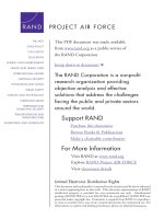
used in preparing the silicone molds from the 3D models are displayed in
Fig. 1.
2.3. Synthesis of BNC structures
First, glucose, yeast extract, peptone, and Na2HPO4 were mixed
using a dry mass ratio of (8:2:2:1). Then MilliQ-water was added (1 L
final volume containing 20 g glucose, 5 g yeast extract, 5 g of peptone,
2.5 g of Na2HPO4). All the components were dissolved, and the pH of the
medium was adjusted to 4.5 with citric acid. The container was then
sterilized in an autoclave for 15 min at 121 ◦ C, then cooled to room
temperature. Later, the bacterial strain was added to the bottle and was
2
R. Ajdary et al.
Carbohydrate Polymers 284 (2022) 119198
Fig. 1. From auxetic 3D-printed models to cast silicone molds. From left to right, triangle, round, and star auxetic shapes. Top — 3D models. Middle — 3D printed
molds produced by fused filament fabrication of PLA filaments. Bottom — cast silicone auxetic molds.
shaken gently to distribute it homogeneously. Then, the BNC culture
medium containing the bacteria was poured into the sterilized silicone
molds (previously autoclaved at 121 ◦ C, 1 bar), covered, and sealed with
parafilm. The medium was left still for 7 to 14 days at 28 ◦ C in the
incubator. After the given time, the produced bacterial cellulose biofilm
was washed several times with deionized water and left in deionized
water for one day. The water was changed several times within the day
to remove the components used in the growing medium. Then, the
bacterial cellulose pellicle was purified with 0.1 M NaOH at 60 ◦ C for 4 h
and, thereafter, washed several times in hot deionized water (6 h).
a metal stub using double-sided carbon tape and sputtered with a 3–4
nm layer of gold‑palladium alloy using a LEICA EM ACE600 (Leica
Camera AG, Wetzlar, Germany) sputter coater.
2.4.3. Weight loss
The dried samples were cut into pieces of similar size (using an 8-mm
round biopsy punch) and weighted (W0) separately. Then, the samples
were immersed in 3 ml buffer (acetate buffer solution, pH 5) and
phosphate buffer saline (PBS, pH 7.4) for 28 days at 37 ◦ C. At each time
point (1, 7, 14, 21, 28 days), the samples were taken from the buffer,
washed with deionized water, re-dried at 37 ◦ C for 24 h, and weighed
again in the dry state (Wd). Finally, the weight loss was calculated using
the equation below.
2.4. Characterization of mold-guided auxetic BNC structures
2.4.1. Surface area
The surface area and the average pore size of freeze-dried BNC were
measured by N2 adsorption at given relative pressures (Micromeritics
Tristar II), following the Brunauer–Emmett–Teller (BET) and Bar
rett–Joyner–Halenda (BJH) models. Before the measurements, the BNC
samples were frozen at − 18 ◦ C, followed by freeze-drying under vacuum
(− 49 ◦ C for at least 48 h). Prior to BET analysis, a degassing step was
performed for 4 h at 120 ◦ C.
Weight loss (%) =
W0 − Wd
× 100.
W0
2.4.4. Mechanical properties
Ball burst strength or burst resistance of the wet BNC films was
measured with a TA.XTPlus Texture Analyzer (Stable Micro Systems
Ltd., Surrey, UK) equipped with a spherical SMS P/0.25S probe. To
perform the test, a BNC film (disc-shaped, 5-cm diameter) was placed
and fixed between two discs containing a 1-cm diameter opening in their
centers. Sandpaper was attached to the internal section of the discs to
avoid slippage of the BNC films (Fig. S1). An Exponent Connect software
was used to analyze the data after the measurement. To determine the
performance stability, measurements were repeated during 100 cycles at
3% strain and at a 120 mm/min measurement speed.
The tensile measurements of wet BNC ribbons were conducted by
2.4.2. Structure
A Java-based image analysis program, ImageJ, was used to deter
mine the size of the openings and the overall structural infill density
(Rueden et al., 2017). The microstructure of the surface and crosssection of BNC was observed with a Zeiss Sigma VP scanning electron
microscope (Carl Zeiss AG, Oberkochen, Germany) operated at an
accelerated voltage of 2 kV. Prior to imaging, the samples were fixed on
3
R. Ajdary et al.
Carbohydrate Polymers 284 (2022) 119198
using TA.XTPlus Texture Analyzer (Stable Micro Systems Ltd., Surrey,
UK) equipped with a miniature tensile grip. The BNC specimens tested
(cross-head speed of 5 mm/min) had a gauge length of 2.5 cm (S. Wang
et al., 2018), and a thickness varying from 0.8 mm to 2.7 mm (measured
by a digital caliper after a culturing time of 7, 10, 14 days). The texture
in the internal section of the grip inhibited the slippage of the BNC
ribbon (Fig. S2).
The Poisson's ratio of the BNC structure was calculated by analyzing
a recorded video of the structure under manual stretching through
MATLAB 9.7 R2019b (MathWorks Inc., Natick, MA, USA) image pro
cessing toolbox. The images extracted from the videos were further
analyzed by ImageJ software to determine the transverse and the lon
gitudinal strains. Finally, Poisson's ratio was calculated following the
equation below at a single strain value (Lai & Yu, 2020).
′
Poisson s ratio = −
stopped, and spectrophotometric analysis was performed using a
microplate reader. The optical density results at 492 nm were corrected
with those at 620 nm. Levels of basal LDH activity measured from naïve
culture medium samples were subtracted from the obtained values prior
to analysis.
Concentrations of interleukin-8 (IL-8) in the supernatant of the cul
ture medium samples were measured using a human IL-8 specific
enzyme-linked immunosorbent quantitative assay (ELISA, 88-8086;
Invitrogen, Thermo Fisher Scientific) according to the manufacturer's
instructions. If needed at re-analysis, samples initially measuring with
too high IL-8 concentrations (not within the assay linear range) were
pre-diluted in naïve culture medium. Briefly, 96-well ELISA plates
(NUNC maxisorp, 442404, Thermo Fisher Scientific) were coated with
the capture antibody overnight at +4 ◦ C. The wells were aspirated and
washed four times with the kit-supplied wash buffer using an automated
programmable plate washer (Wallac 1296-026 Delfia Platewash, Per
kinElmer Inc., Waltham, MA, USA). Non-specific binding was blocked
with assay diluent for 1 h at room temperature, and wells were washed
with wash buffer. Samples (100 μl) were then pipetted into the wells,
and the plate was incubated for 2 h at RT. After four wash cycles, 100 μl
detection antibody was added to each well, incubated for 1 h, and fol
lowed by four wash cycles. The Avidin-HRP conjugate was added to the
wells, incubated for 30 min, followed by four wash cycles. The tetra
methylbenzidine substrate solution was added to each well, the plate
was incubated for 15 min, and a stop solution was added to end the
reaction. The optical density was measured using a microplate reader at
450 nm wavelength and 570 nm correction wavelength. Naïve culture
medium served as a baseline-control sample. Samples for the standard
curve were included in each assay run. The standard curves for each
assay run were generated using non-linear regression sigmoidal 4parameter-logistic curve-fitting, and sample concentrations were inter
polated from the curves in GraphPad Prism 9 software (version 9.0.1,
GraphPad Software LLC, San Diego, CA, USA). Statistical analysis was
performed in GraphPad Prism using the nonparametric Mann-Whitney
test where p values less than 0.05 were considered significant.
Transverse strain (εT )
.
Longitudinal strain (εL )
2.4.5. Cell culturing and incubation
The human monocyte/macrophage cell line, THP-1, was cultured in
Roswell Park Memorial Institute (RPMI)-1640 medium (GiBNCo 31870025, Thermo Fisher Scientific, Waltham, MA, USA) supplemented with
10% heat-inactivated fetal bovine serum (GiBNCo 10500-064), 2 mM Lglutamine (GiBNCo A2916801) and antibiotics (GiBNCo 15140-122,
penicillin G 100 U/ml, streptomycin 100 μg/ml and GiBNCo 15290026, amphotericin B 250 ng/ml) under regular cell culture conditions
(+37 ◦ C humidified atmosphere supplemented with 5% CO2). Before
experimentation, the materials were incubated overnight in 70%
ethanol and washed meticulously with phosphate-buffered saline
without calcium or magnesium (Lonza Bio Whittaker, 17-516F, Basel,
Switzerland), followed by washes in a culture medium. At the beginning
of the experiments, a 6-mm diameter biopsy punch (BP-60F, kai Europe
GmbH, Solingen, Germany) was used to cut round, equal-size samples of
the materials that were placed individually into wells of a non-adherent
96-well plate. THP-1 cells were counted using an automated cell counter
(Countess II, Applied Biosystems, Thermo Fisher Scientific), and 80,000
cells/well suspended in culture medium were pipetted to each well with
material as well as to the empty control wells without material. 12-OTetradecanoylphorbol-13-acetate (TPA, Sigma P8139, Merck KGaA,
Darmstadt, Germany) at a final concentration of 300 nM was used to
induce macrophage differentiation of the THP-1 cells, as reported preư
ăa
ăn ă
ă, Hukkanen, Rauhala, & Kankuri, 2006).
viously (Va
anen, Salmenpera
3. Result and discussion
3.1. Auxetic designs and culture time
Additive manufacturing techniques, such as fused filament fabrica
tion, allow the production of complex structures challenging and
arduous to form with other methods. Still, the slow production rate
during fused filament fabrication does not allow the rapid
manufacturing of multiple copies. Combining the fused filament fabri
cation technique and silicone molding accelerated the process to form
functional molds for the growth of mechanically robust BNC networks.
Three auxetic designs, namely, “triangle”, “round”, and “star” shapes,
were evaluated to demonstrate the fabrication of BNC biomaterials.
These designs facilitated the formation of structures of a tunable open
ing size to fulfill the minimum 1-mm opening width required for
biomedical structures. As shown in Fig. 1, all three mold designs were
cast with silicone to produce molds with a high level of detail and ac
curacy. Beyond the three designs evaluated herein, this method offers
potential for any planar auxetic designs.
Initially, the positive and negative molds were developed with a tilt
angle of 0 degrees between the base of the molds and the templating
pillars (Figs. S3, S4). However, according to the experimental observa
tions, a tilt angle of about 5◦ in the positive 3D model facilitated the
silicone molding process (Fig. 2a–b, Fig. S5). Also, compared with the
structure developed in the 0-degree molds, the slight tilt angle in the
mold simplified the removal of the BNC network, which was obtained
after culturing for 7, 10, and 14 days. The tilted angle in the silicone
mold may also function as additional support for the BNC network in the
air/culture medium interface. Importantly, the total volume of the cul
ture medium in the tilted negative silicone molds impacted the height of
2.4.6. Microscopy
Phase-contrast microscopy of THP-1 cells incubated with or without
the materials was carried out using an Olympus CKX-41 inverted mi
croscope and 20× objective (Olympus Corporation, Tokyo, Japan).
Black and white images of cell morphology on the bottom of the wells
were obtained using a digital microscope camera (DC 300, Leica
Microsystems GmbH, Wetzlar, Germany) fitted with a 0.5× adapter (UTV0.5XC-3; Olympus). Leica IM 500 software (version 1.20 release 19,
Leica Microsystems) was used for image documentation.
2.4.7. Measurements of cytotoxicity and pro-inflammatory cell activation
After a 3-day incubation of undifferentiated or TPA-differentiated
THP-1 monocyte/macrophages with or without the materials, the cul
ture medium was collected for analysis. Immediately after collection,
the samples were centrifuged at 20,000 rpm for 10 min to remove any
cell debris or particulates; supernatants were divided into aliquots and
transferred to new tubes for storage at − 20 ◦ C until analysis. Cytotox
icity was measured from the culture medium supernatant using the
colorimetric LDH cytotoxicity detection kit plus (Roche 04744926001,
Merck) according to the manufacturer's instructions and as described
earlier (Den Hollander et al., 2015). Briefly, 100 μl of the assay reagent
was mixed with 100 μl of the cell culture sample supernatant. After a 30min incubation in the dark at room temperature, the reaction was
4
R. Ajdary et al.
Carbohydrate Polymers 284 (2022) 119198
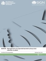
Fig. 2. The tilt angle (a) α = 0 and (b) α =
5◦ in the positive mold and its impact on the
openings of the structures. Due to the angle,
α = 5◦ , the higher the level reached by the
BNC culture medium, the larger BNC infill is
obtained in the final mesh. (c) The triangle
auxetic unit cell and (d) the maximum infill
(the smallest opening) obtained under com
plete filling of the mold with BNC culture
medium. (e, f) Round auxetic unit cell, and
(g, h) star auxetic unit cell. (i) The triangle
mold filled partially, and (j) fully with BNC
culture medium.
the BNC structures formed, thus the opening size and the infill density in
each design. For instance, the higher structure infill density was asso
ciated with smaller openings as the level of culture medium increased in
the silicone mold (Fig. S5). The minimum opening size (largest infill
density) was obtained when the culture medium completely filled the
mold, as indicated in Fig. 2 for each design. Based on the image analysis
of the obtained BNC structure, the maximum infill density in the triangle
design was 70 ± 3.1% (about 30% openings), while this value was 59 ±
2.1 and 62 ± 2.6% for the round and star shapes, respectively. Note that
proper covering and sealing of the culture medium containing molds
with parafilm minimized the evaporation of the liquid.
of the plastic-based mesh implants and supports the dynamic and
repeated body movements.
3.2.2. Porosity, surface area, and microstructure
The static BNC culturing method resulted in cellulose hydrogel
(BNC) membranes that form at the air/culture medium interface.
Herein, the bacteria access to air at the interface between the liquid
culture medium and the silicon surface, in a process called aerotaxis. The
aerobic bacteria strain transforms glucose, and other organic nutrients
into cellulose, within a few days. These bacteria, like other living or
ganisms, have an optimum growth environment with controlled factors
such as oxygen, temperature, culture time, and pH. The physicochemical
properties of BNC structures were highly dependent on the environ
mental factors, and the variables could be narrowed down to study the
BNC formation. Aerobes such as K. medellinensis, require oxygen for
energy conversion and cellular respiration. These requirements for an
optimum growth environment (pH, temperature, oxygen) were met by
adjusting the pH to 4.5 and maintaining the static culture in an incu
bator at 28 ◦ C. As indicated in Fig. 4a, the BNC thickness increased with
the culture time since the bacteria had more time to generate and
accumulate more nanofibrils. The substantial water content in the hy
drophilic BNC slowed down the drying at room conditions. As shown in
Fig. S8, it took about 72 h for the structure to lose 95% of the initial
water. As the thickness of BNC increased with the culture time, struc
tures with larger BET surface areas were obtained. A BET surface area of
about 25 m2 g− 1 to almost 60 m2 g− 1 resulted from 7 to 14 days of
culturing (Fig. 4b, Fig. S9). Importantly, increasing thickness due to
increased incubation also resulted in high cross-linking of the network,
and thus in smaller mesopores (Fig. 4b), as was also previously inferred
from measuring the flow across BNC membranes (Lehtonen et al., 2021).
However, the never-dried BNC was expected to have a larger surface
area compared with the freeze-dried ones since the hydrophilic BNC
3.2. Characterization of auxetic BNC structures formed by aerotaxis
3.2.1. Mold-guided auxetic BNC materials
Auxetic geometries exhibit exceptional structural properties (e.g.,
controllable expansion under tension and geometrical compliance to the
anatomical changes during movements and dynamic deformation),
going beyond their material composition (Stavric & Wiltsche, 2019).
Herein, we combined 3D printing and molding techniques to develop
mold-guided BNC structures via hybrid manufacturing. The full process,
from the 3D model to the final extracted mesh, is shown in Fig. S6. Each
of the developed systems demonstrated anisotropic mechanical response
and negative Poisson's ratio. The calculated Poisson's ratio deviated
according to the auxetic unit (opening shape and size) and the extent of
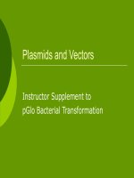
applied stress, as well as its directionality. Never-dried purified BNC
structures cultured for 10 days displayed a negligible change in thick
ness upon stretching and exhibited auxetic properties, as shown in
Figs. 3 and S7. The triangle unit cell had a negative Poisson's ratio of ν =
− 0.19, while the value for the round and star auxetic unit cells were ν =
− 0.36 and ν = − 0.13. The reversible structural expansion under tension
can minimize tissue damage, which commonly occurs during shrinkage
5
R. Ajdary et al.
Carbohydrate Polymers 284 (2022) 119198
Fig. 3. Auxetic BNC meshes obtained after 10 days of culturing (triangle, round, and star patterns). The structures exhibit reversible expansion under tension.
degradation, which otherwise would take place through enzymatic ac
tivities or a combination of hydrolytic mechanisms and autocatalytic
oxidation. The higher degree of polymerization and crystallinity
(~90%) of BNC compared with the values for the plant-based nano
cellulose (crystallinity of 44–65%) has a substantial effect on the more
challenging structural degradation (Amorim et al., 2020). After 28 days
of exposure to pH 7.4 and pH 5, the pellicles exhibited mass loss of less
than 2% and 4%, respectively, mostly due to the fibrils' detachment
when handling the material during the washing step and characteriza
tion (Fig. S11). This suggests that the BNC structures are potential
candidates to facilitate long-term tissue support.
undergoes irreversible hydrogen bonding when dehydrated for the
sample preparation step in BET characterization. The culture time had
no evident effect on the surface and cross-section of the microstructures.
As shown in Fig. 4, the interfacially grown, physically entangled BNC
showed a relatively dense network, while the cross-section of the
lyophilized BNC was highly porous (macroporous range and voids, not
measured by BET). Although the solid content in the BNC structure was
less than 0.5 wt%, the physically entangled nanofibrils retained the
porous microstructure after freeze-drying and provided mesoporous
structure, ideal for the nutrition and oxygen transport in the wet BNC. As
shown in Fig. S10, the solid morphology of PP does not provide a
microenvironment similar to the ECM, and the interstices between the
multi-filaments could host bacteria and immune cells and lead to
inflammation and infection in the tissue (Dă
allenbach, 2015).
3.2.4. Mechanical performance
Biomaterials, especially those used to support organs, are expected to
be strong, flexible, stable, and biocompatible with the target tissue, i.e.,
to be able to lift and hold the intended weight when used in-vivo. Both
burst and tensile strength are viable approaches to examine the pressure
retention of materials and implants under load. Our biofabricated BNC
meshes exhibited high flexibility due to the large water content (>99.5
wt%) and long aspect ratio of the nanofibrils. The dense physical
entanglement of high aspect ratio nanofibrils translated into robust
mechanical performance, as shown in Fig. 5. In tensile mode (Fig. 5a),
the wet BNC ribbon load-elongation curve resembled that of Achilles
tendons (Barfod, 2014), where the toe region occurs at below 1% strain.
As the load increased, the crimped nanofibrils straightened until
microscopic and macroscopic failure occurred and the BNC ribbon
ruptured, after 2–4% elongation, depending on the BNC culture time.
The wet BNC structure underwent slightly higher elongation under ball
burst testing with the load reaching 15 N (1.53 kg) and 26 N (2.65 kg)
3.2.3. Weight loss
The pH during incubation is a critical factor in BNC growth; an acidic
environment has been demonstrated to enhance the bacterium activity
to produce thicker BNC pellicles. A deeper investigation to assess the
importance of pH showed that although more BNC was formed at lower
pH (pH 5 to pH 3.5), pellicles were hardly formed at pH 3 and below
(Aramwit & Bang, 2014). However, this might vary depending on the
BNC strain. In this work, the growing pH was fixed at the beginning at
4.5, for all samples. Additionally, we aimed at studying the BNC weight
loss at pH 7.4 (normal body condition) and pH 5 (pH associated with
some body parts such as the areas in the pelvis, duodenum, small in
testine, and colon) (Fallingborg, 1999; Savchenko, 2021). According to
previous degradation studies (Ajdary et al., 2020; Lin & Dufresne,
2014), highly crystalline structure of nanocellulose prevents
6
R. Ajdary et al.
Carbohydrate Polymers 284 (2022) 119198
Fig. 4. (a) The BNC structure thickness and solid content produced after 7, 10, and 14 days incubation. (b) The BET surface area and the average pore diameter for
BNC samples cultured for 7, 10, 14 days. The SEM images of (c) BNC surface at 30,000× magnification, and (d) BNC cross-section at 10,000× magnification.
Fig. 5. (a) The load-elongation curve for BNC samples subjected to tensile tests, and (b) load-elongation profiles for BNC ball burst strength tests for BNC samples
after 7-, 10-, and 14-days culture time. (c) The cyclic burst strength during 100 cycles at 3% strain.
7
R. Ajdary et al.
Carbohydrate Polymers 284 (2022) 119198
for BNC samples cultured for 10 and 14 days, respectively, before
samples rupture. The mild NaOH purification treatment had a negligible
effect on the modulus and tensile strength properties of the BNC.
However, some reports demonstrated a decrease in the entanglement
density and porosity of the nanofibrils due to the microstructural
swelling after strong alkali post-treatments (McKenna, Mikkelsen, Wehr,
Gidley, & Menzies, 2009; Tang, Jia, Jia, & Yang, 2010). The values of
ultimate tensile strength of wet BNC increased ten-fold, from 48.9 ± 6.3
MPa to 456.2 ± 33 MPa by prolonging the incubation time, from 7 to 14
days, respectively. These values are comparable with that of human
muscle tissue (Barnes, Przybyla, & Weaver, 2017), and the exceptionally
high water content in BNC provided a lubricious surface that tended to
reduce the soft tissue friction during mobility and activity. A purified
BNC film (10 days) was studied further to examine the effect of cyclic
loading on the performance. While 9% reduction occurred in the first 15
cycles, 87% of the performance was sustained after 100 cyclic loadings
(Fig. 5c). Other reports have shown that the exceptional mechanical
performance of BNC equips the microstructure with suture retention
capabilities, which is an important factor in implants (Hong, Wei, &
Chen, 2015).
medium was evaluated as a marker of cell membrane damage and
material-induced cytotoxicity, and concentrations of IL-8 were quanti
fied to evaluate monocyte/macrophage pro-inflammatory activation
and phenotype (Fig. 6). In undifferentiated THP-1 monocyte cultures,
both polypropylene control and BNC demonstrated a cell-flattening ef
fect suggesting minor to moderate differentiation into a macrophagelike phenotype (Fig. 6a–c). Induction of macrophage differentiation
using TPA was manifested in all cultures as dominant cell flattening on
the non-adherent culture surface (Fig. 6d–f). A shift towards a
macrophage-like phenotype was further indicated by the increased
release of LDH (Fig. 6g) and an increased secretion of IL-8 from the
undifferentiated THP-1 cells incubated with BNC (Fig. 6h). When a
dominant macrophage-like phenotype was induced on the THP-1 cells
by TPA, a more than 400-fold increase in the release of IL-8 was
observed. Incubation of these differentiated cells with BNC suppressed
the pro-inflammatory macrophage phenotype as evidenced by a signif
icantly decreased secretion of IL-8 into the culture medium (Fig. 6h).
Incubation with BNC also suppressed the THP-1 cells pro-inflammatory
macrophage differentiation-associated increase in LDH release (Fig. 6g).
Taken together, BNC drove monocyte activation but suppressed the
TPA-induced pro-inflammatory macrophage-like phenotype. The
observed activities of BNC on human monocyte/macrophages deserve
further attention. Further research focusing on the type of macrophage
differentiation activated by the BNC can be expected to provide a deeper
3.2.5. Cytotoxicity and interleukin-8 release from THP-1 cells
Cellular gross morphology was evaluated after 3-day incubation. The
release of lactate dehydrogenase (LDH) from the cells to the culture
Fig. 6. (a–c) The digital phase-contrast microscopy images (20× objective) from cultures of THP-1 cells without TPA stimulation. (a) Control without material, (b)
polypropylene (PP) mesh, (c) BNC. (d–f) Digital phase-contrast microscopy images (20× objective) from cultures of TPA (300 nM)-differentiated THP-1 macro
phages. (d) Control without material, (e) polypropylene (PP) mesh, (f) BNC. The scale bar from a to f is 100 μm. (g) Lactate dehydrogenase (LDH)-release from cells to
culture medium after 3-day incubation of THP-1 cells with the materials, without other external cell stimulation, and with TPA (300 nM)-differentiated THP-1
macrophages (*p < 0.01 as compared to Control). (h) Concentrations of interleukin-8 (CXCL8) in culture media after a 3-day incubation of THP-1 cells with the
materials, without other external cell stimulation, and with TPA (300 nM)-differentiated THP-1 macrophages (*p < 0.05 as compared to Control).
8
R. Ajdary et al.
Carbohydrate Polymers 284 (2022) 119198
mechanistic understanding of its ability to suppress the TPA-induced
pro-inflammatory macrophage-like phenotype as observed here by a
decreased secretion of IL-8 after exposure to the BNC.
Declaration of competing interest
4. Conclusion
Acknowledgements
Bacterial nanocellulose outperforms many commonly used thermo
plastics in biomedicine due to its similarity to the Extra Cellular Matrix,
outstanding mechanical performance in wet conditions, ease of suturing
and sterilization, high porosity (mesoporous, macroporous and macro
scopic voids), and large surface area. To advance the realm of applica
tions of BNC in biomedical devices, herein, all-nanocellulose BNC
meshes with negative Poisson's ratio were produced by following a
hybrid manufacturing protocol. Three auxetic cell units, namely, trian
gle-, round-, and star-shaped were examined to develop structures with
overall infill density of 70 ± 3.1% (about 30% openings), 59 ± 2.1, and
62 ± 2.6%, respectively. Depending on the culture time, BNC exhibited
tensile strengths of 48–456 MPa (7 to 14 days of incubation), with over
87% stability after 100 burst load/unload cycles at 3% strain. The
developed BNC meshes exhibited a negative Poisson's ratio (v = − 0.36
to − 0.13) via hybrid manufacturing. The reversible structural expansion
under tension minimizes the tissue damage that commonly occurs by
shrinkage of plastic-based mesh implants. Furthermore, the cytotoxicity
and interleukin-8 release from THP-1 cells in interaction with BNC was
investigated, and it is concluded that BNC drove monocyte activation
but suppressed the TPA-induced pro-inflammatory macrophage-like
phenotype. The approach presented herein indicates a green method
of producing biomaterials for in-vivo applications, which are expected to
maximize comfort, minimize material fatigue, and thus improve the
overall success of future long-term mesh implants. Furthermore, given
the range of research streams on bacterial cellulose, where new func
tionalities can be incorporated, one can expect additional bioactive
molecules to be incorporated into such designs, for instance, to facilitate
growth or to decrease the risks of infection post-implantation.
The supporting information includes 8 figures. Fig. S1: Setup used to
prevent wet BNC structures from slippage during bursting strength tests.
Fig. S2: The tensile mechanical tests for wet BNC. Fig. S3: The positive
PLA and negative silicon molds with no tilt angle. Fig. S4: Illustration of
the challenge of BNC removal from the molds with no tilt angle. Fig. S5:
The structure opening, structure infill, and the infill density of the BNC,
and the tilt angle in the silicon molds. Fig. S6: The structural develop
ment by mold guiding. Fig. S7: Auxetic BNC meshes obtained after
culturing for 10 days. Fig. S8: The wet and air-dried BNC structures.
Fig. S9: Nitrogen adsorption isotherms. Fig. S10. Polypropylene knitted
mesh and the SEM images at 40×, 200×, 1000×, and 15,000×. Fig. S11:
The weight loss of BNC in pH 7.4 and pH 5 for 28 days. Supplementary
data to this article can be found online at doi: />carbpol.2022.119198.
The authors acknowledge the fund from the Business Finland TUTLI
fund (“Solving the Mesh”, Project number 211795, BF 6108/31/2019).
R.A. also acknowledges funding from the Finnish Foundation for Tech
nology Promotion (TES) and FinnCERES GoGlobal mobility fund. O.J.R.
is grateful for the support received from the ERC Advanced Grant
Agreement No. 788489 (“BioElCell”), the Canada Excellence Research
Chair initiative (CERC-2018-00006), and Canada Foundation for Inno
vation (Project number 38623). The authors are grateful for the kind
help of Aki Laakso in the design of the auxetic structures and Dr. Alp
Karakoc for his insightful comments. We would also like to show our
ăinen for the valuable
gratitude to Dr. Tomi S. Mikkola and Ilkka Hyytia
discussions throughout the project. We are also immensely thankful to
Lahja Eurajoki for the expert technical assistance in cell culture exper
iments. This work made use of the facilities of Aalto University's
Nanomicroscopy Center.
None.
References
Ajdary, R., Ezazi, N. Z., Correia, A., Kemell, M., Huan, S., Ruskoaho, H. J., Hirvonen, J.,
Santos, H. A., & Rojas, O. J. (2020). Multifunctional 3D-printed patches for longterm drug release therapies after myocardial infarction. Advanced Functional
Materials, 30(34), 1–10. />Ajdary, R., Reyes, G., Kuula, J., Raussi-Lehto, E., Mikkola, T. S., Kankuri, E., &
Rojas, O. J. (2021). Direct ink writing of biocompatible nanocellulose and chitosan
hydrogels for implant mesh matrices. ACS Polymers Au. />acspolymersau.1c00045
Ajdary, R., Tardy, B. L., Mattos, B. D., Bai, L., & Rojas, O. J. (2020). Plant nanomaterials
and inspiration from nature: Water interactions and hierarchically structured
hydrogels. Advanced Materials, 2001085. />Amorim, J. D. P., de Souza, K. C., Duarte, C. R., da Silva Duarte, I., de Assis Sales
Ribeiro, F., Silva, G. S., de Farias, P. M. A., Stingl, A., Costa, A. F. S., Vinhas, G. M., &
Sarubbo, L. A. (2020). Plant and bacterial nanocellulose: Production, properties and
applications in medicine, food, cosmetics, electronics and engineering. A review.
Environmental Chemistry Letters, 18(3), 851–869. />Aramwit, P., & Bang, N. (2014). In The characteristics of bacterial nanocellulose gel releasing
silk sericin for facial treatment (pp. 1–11).
Bao, L., Tang, J., Hong, F. F., Lu, X., & Chen, L. (2020). Physicochemical properties and
in vitro biocompatibility of three bacterial nanocellulose conduits for blood vessel
applications. Carbohydrate Polymers, 239(March), Article 116246. />10.1016/j.carbpol.2020.116246
Barfod, K. W. (2014). Achilles tendon rupture; assessment of nonoperative treatment.
Danish Medical Journal, 61(4), Article B4837.
Barnes, J. M., Przybyla, L., & Weaver, V. M. (2017). Tissue mechanics regulate brain
development, homeostasis and disease. Journal of Cell Science, 130, 71–82. https://
doi.org/10.1242/jcs.191742
Bayl´
on, K., Rodríguez-Camarillo, P., Elías-Zú˜
niga, A., Díaz-Elizondo, J. A., Gilkerson, R.,
& Lozano, K. (2017). Past, present and future of surgical meshes: A review.
Membranes, 7(3), 1–23. />Bottan, S., Robotti, F., Jayathissa, P., Hegglin, A., Bahamonde, N., HerediaGuerrero, J. A., Bayer, I. S., Scarpellini, A., Merker, H., Lindenblatt, N.,
Poulikakos, D., & Ferrari, A. (2015). Surface-structured bacterial cellulose with
guided assembly-based biolithography (GAB). ACS Nano, 9(1), 206–219. https://doi.
org/10.1021/nn5036125
Bungay, H. R., & Serafica, G. C. (1986). Production of microbial cellulose (Patent No.
6071727).
Cai, Q., Hu, C., Yang, N., Wang, Q., Wang, J., Pan, H., Hu, Y., & Ruan, C. (2018).
Enhanced activity and stability of industrial lipases immobilized onto spherelike
bacterial cellulose. International Journal of Biological Macromolecules, 109,
1174–1181. />Ca˜
nas-Guti´errez, A., Martinez-Correa, E., Su´
arez-Avenda˜
no, D., Arboleda-Toro, D., &
Castro-Herazo, C. (2020). Influence of bacterial nanocellulose surface modification
on calcium phosphates precipitation for bone tissue engineering. Cellulose, 27(18),
10747–10763. />Castro, C., Cleenwerck, I., Trˇcek, J., Zuluaga, R., de Vos, P., Caro, G., Aguirre, R.,
Putaux, J. L., & Ga˜
n´
an, P. (2013). Gluconacetobacter medellinensis sp. nov.,
cellulose- and non-cellulose-producing acetic acid bacteria isolated from vinegar.
International Journal of Systematic and Evolutionary Microbiology, 63(PART3),
1119–1125. />Cheng, Q., Liu, Y., Lyu, J., Lu, Q., Zhang, X., & Song, W. (2020). 3D printing-directed
auxetic kevlar aerogel architectures with multiple functionalization options. Journal
of Materials Chemistry A, 8(28), 1423–14253. />
CRediT authorship contribution statement
Rubina Ajdary: Conceptualization, Methodology, Formal analysis,
Investigation, Writing – original draft, Writing – review & editing,
Visualization. Roozbeh Abidnejad: Methodology, Investigation,
Writing – review & editing, Visualization. Janika Lehtonen: Method
ology, Investigation, Writing – review & editing, Visualization. Jani
Kuula: Methodology, Investigation, Writing – review & editing, Visu
alization. Eija Raussi-Lehto: Methodology, Investigation, Writing –
review & editing, Visualization. Esko Kankuri: Methodology, Formal
analysis, Investigation, Writing – original draft, Writing – review &
editing, Visualization. Blaise Tardy: Methodology, Investigation,
Writing – review & editing, Visualization. Orlando J. Rojas: Supervi
sion, Funding acquisition, Conceptualization, Writing – review &
editing.
9
R. Ajdary et al.
Carbohydrate Polymers 284 (2022) 119198
Naomi, R., Idrus, R. B. H., & Fauzi, M. B. (2020). Plant-vs. bacterial-derived cellulose for
wound healing: A review. International Journal of Environmental Research and Public
Health, 17(18), 1–25. />Oliveira Barud, H. G., da Silva, R. R., Borges, M. A. C., Castro, G. R., Ribeiro, S. J. L., & da
Silva Barud, H. (2020). Bacterial nanocellulose in dentistry: Perspectives and
challenges. Molecules (Basel, Switzerland), 26(1). />molecules26010049
Pang, M., Huang, Y., Meng, F., Zhuang, Y., Liu, H., Du, M., Ma, Q., Wang, Q., Chen, Z.,
Chen, L., Cai, T., & Cai, Y. (2020). Application of bacterial cellulose in skin and bone
tissue engineering. European Polymer Journal, 122, Article 109365. />10.1016/j.eurpolymj.2019.109365 (July 2019).
Papadopoulou, A., Laucks, J., & Tibbits, S. (2017). Auxetic materials in design and
architecture. Nature Reviews Materials, 2, 1–3. />natrevmats.2017.78
Prawoto, Y. (2012). Seeing auxetic materials from the mechanics point of view: A
structural review on the negative Poisson’s ratio. Computational Materials Science, 58,
140–153. (June 2012).
Queir´
os, E. C., Pinheiro, S. P., Pereira, J. E., Prada, J., Pires, I., Dourado, F., Parpot, P., &
Gama, M. (2021). Hemostatic dressings made of oxidized bacterial nanocellulose
membranes. Polysaccharides, 2(1), 80–99. />polysaccharides2010006
Ross, P., Mayer, R., & Benziman, M. (1991). Cellulose biosynthesis and function in
bacteria. Microbiological Reviews, 55(1), 35–58.
Rueden, C. T., Schindelin, J., Hiner, M. C., DeZonia, B. E., Walter, A. E., Arena, E. T., &
Eliceiri, K. W. (2017). Image J2: ImageJ for the next generation of scientific image
data. Microscopy and Microanalysis, 18(1), 529. />Rühs, P. A., Malollari, K. G., Binelli, M. R., Crockett, R., Balkenende, D. W. R.,
Studart, A. R., & Messersmith, P. B. (2020). Conformal bacterial cellulose coatings as
lubricious surfaces. ACS Nano, 14(4), 3885–3895. />acsnano.9b09956
Savchenko, M. (2021). Normal vaginal pH: How to test, balance, and restore vaginal pH.
lth/menstrual-cycle/health/symptoms-and-diseases/normal-vaginal
-ph-balance.
Stavric, M., & Wiltsche, A. (2019). Geometrical elaboration of auxetic structures. Nexus
Network Journal, 21(1), 79–90. />Sternschuss, G., Ostergard, D., & Patel, H. (2012). Post-implantation alterations of
polypropylene in the human. The Journal of Urology, 188(1), 27–31. />10.1016/j.juro.2012.02.2559
Tang, W., Jia, S., Jia, Y., & Yang, H. (2010). The influence of fermentation conditions and
post-treatment methods on porosity of bacterial cellulose membrane. World Journal
of Microbiology and Biotechnology, 26(1), 125–131. />U.S. Food, & Drug Administration. (2021). FDA takes action to protect women’s health,
orders manufacturers of surgical mesh intended for transvaginal repair of pelvic
organ prolapse to stop selling all devices accessed July />vents/press-announcements/fda-takes-action-protect-womens-health-orders-man
ufacturers-surgical-mesh-intended-transvaginal.
ănă
Vă
aa
anen, A. J., Salmenperă
a, P., Hukkanen, M., Rauhala, P., & Kankuri, E. (2006).
Cathepsin B is a differentiation-resistant target for nitroxyl (HNO) in THP-1
monocyte/macrophages. Free Radical Biology and Medicine, 41(1), 120–131. https://
doi.org/10.1016/j.freeradbiomed.2006.03.016
Verma, P., Shofner, M. L., & Griffin, A. C. (2014). Deconstructing the auxetic behavior of
paper. Physica Status Solidi (B) Basic Research, 251(2), 289–296. />10.1002/pssb.201384243
Wang, J., Tavakoli, J., & Tang, Y. (2019). Bacterial cellulose production, properties and
applications with different culture methods – A review. Carbohydrate Polymers, 219
(May), 63–76. />Wang, S., Li, T., Chen, C., Kong, W., Zhu, S., Dai, J., Diaz, A. J., Hitz, E., Solares, S. D.,
Li, T., & Hu, L. (2018). Transparent, anisotropic biofilm with aligned bacterial
cellulose nanofibers. Advanced Functional Materials, 28(24), 1707491. https://doi.
org/10.1002/adfm.201707491
Xu, T., Jiang, Q., Ghim, D., Liu, K. K., Sun, H., Derami, H. G., Wang, Z., Tadepalli, S.,
Jun, Y. S., Zhang, Q., & Singamaneni, S. (2018). Catalytically active bacterial
nanocellulose-based ultrafiltration membrane. Small, 14(15), 1–8. />10.1002/smll.201704006
Yang, J., Wang, L., Zhang, W., Sun, Z., Li, Y., Yang, M., Zeng, D., Peng, B., Zheng, W.,
Jiang, X., & Yang, G. (2018). Reverse reconstruction and bioprinting of bacterial
cellulose-based functional total intervertebral disc for therapeutic implantation.
Small, 14(7), Article 1702582. />Yao, Y. T., Alderson, K. L., & Alderson, A. (2016). Modeling of negative Poisson’s ratio
(auxetic) crystallie cellulose. Cellulose, 23, 3429–3448. />s10570-016-1069-9
Yuan, H., Chen, L., Hong, F. F., & Zhu, M. (2018). Evaluation of nanocellulose carriers
produced by four different bacterial strains for laccase immobilization. Carbohydrate
Polymers, 196(May), 457464. />
Dă
allenbach, P. (2015). To mesh or not to mesh: A review of pelvic organ reconstructive
surgery. International Journal of Women’s Health, 7, 331343. />10.2147/IJWH.S71236
Den Hollander, B., Sundstră
om, M., Pelander, A., Siltanen, A., Ojanperă
a, I., Mervaala, E.,
Korpi, E. R., & Kankuri, E. (2015). Mitochondrial respiratory dysfunction due to the
conversion of substituted cathinones to methylbenzamides in SH-SY5Y cells.
Scientific Reports, 5(5), 14294. />Fallingborg, J. (1999). Intraluminal pH of the human gastrointestinal tract. Danish
Medical Bulletin, 43(6), 183–196.
Fey, C., Betz, J., Rosenbaum, C., Kralisch, D., Vielreicher, M., Friedrich, O., Metzger, M.,
& Zdzieblo, D. (2020). Bacterial nanocellulose as novel carrier for intestinal
epithelial cells in drug delivery studies. Materials Science and Engineering C, 109,
Article 110613. (December 2019).
Fonseca, D. F. S., Vilela, C., Pinto, R. J. B., Bastos, V., Oliveira, H., Catarino, J., Faísca, P.,
Rosado, C., Silvestre, A. J. D., & Freire, C. S. R. (2021). Bacterial nanocellulosehyaluronic acid microneedle patches for skin applications: In vitro and in vivo
evaluation. Materials Science and Engineering C, 118, Article 111350. />10.1016/j.msec.2020.111350 (March 2020).
Geisel, N., Clasohm, J., Shi, X., Lamboni, L., Yang, J., Mattern, K., Yang, G.,
Schă
afer, K. H., & Saumer, M. (2016). Microstructured multilevel bacterial cellulose
allows the guided growth of neural stem cells. Small, 12(39), 5407–5413. https://
doi.org/10.1002/smll.201601679
Gorgieva, S., & Trˇcek, J. (2019). Bacterial cellulose: Production, modification and
perspectives in biomedical applications. Nanomaterials, 9(10), 1–20. />10.3390/nano9101352
Greca, L. G., Lehtonen, J., Tardy, B. L., Guo, J., & Rojas, O. J. (2018). Biofabrication of
multifunctional nanocellulosic 3D structures: A facile and customizable route.
Materials Horizons, 5(3), 408–415. />Greca, L. G., Rafiee, M., Karakoỗ, A., Lehtonen, J., Mattos, B. D., Tardy, B. L., &
Rojas, O. J. (2020). Guiding bacterial activity for biofabrication of complex materials
via controlled wetting of superhydrophobic surfaces. ACS Nano, 14(10),
1292912937. />Helenius, G., Bă
ackdahl, H., Bodin, A., Nannmark, U., Gatenholm, P., & Risberg, B.
(2006). In vivo biocompatibility of bacterial cellulose. Journal of Biomedical Materials
Research - Part A, 76A(2), 431–438. />Hong, F., Wei, B., & Chen, L. (2015). Preliminary study on biosynthesis of bacterial
nanocellulose tubes in a novel double-silicone-tube bioreactor for potential vascular
prosthesis. BioMed Research International, 560365. />560365
Iakovlev, V. V., Guelcher, S. A., & Bendavid, R. (2015). Degradation of polypropylene in
vivo: A microscopic analysis of meshes explanted from patients. Journal of Biomedical
Materials Research - Part B Applied Biomaterials, 105B(2), 237–248. />10.1002/jbm.b.33502
Jiang, Y., & Li, Y. (2018). 3D printed auxetic mechanical metamaterial with chiral cells
and re-entrant cores. Scientific Reports, 8, 2397. />Knight, K. M., Moalli, P. A., & Abramowitch, S. D. (2018). Preventing mesh pore collapse
by designing mesh pores with auxetic geometries: A comprehensive evaluation via
computational modeling. Journal of Biomechanical Engineering, 140(5), 1–8. https://
doi.org/10.1115/1.4039058
Lai, C. W., & Yu, S. S. (2020). 3D printable strain sensors from deep eutectic solvents and
cellulose nanocrystals. ACS Applied Materials and Interfaces, 12(30), 34235–34244.
/>Lakes, R. (1987). Foam structures with a negative poisson’s ratio. Science, 235,
1038–1040. />Lakes, R. S. (2017). Negative-Poisson’s-ratio materials: Auxetic solids. Annual Review of
Materials Research, 47, 63–81. />Lee, S. E., & Park, Y. S. (2017). The role of bacterial cellulose in artificial blood vessels.
Molecular and Cellular Toxicology, 13(3), 257–261. />Lehtonen, J., Chen, X., Beaumont, M., Hassinen, J., Orelma, H., Dum´ee, L. F.,
Tardy, B. L., & Rojas, O. J. (2021). Impact of incubation conditions and posttreatment on the properties of bacterial cellulose membranes for pressure-driven
filtration. Carbohydrate Polymers, 251(January), Article 117073. />10.1016/j.carbpol.2020.117073
Lin, N., & Dufresne, A. (2014). Nanocellulose in biomedicine: Current status and future
prospect. European Polymer Journal, 59(October), 302–325. />10.1016/j.eurpolymj.2014.07.025
Liu, Y., & Hu, H. (2010). A review on auxetic structures and polymeric materials.
Scientific Research and Essays, 5, 1052–1063.
McKenna, B. A., Mikkelsen, D., Wehr, J. B., Gidley, M. J., & Menzies, N. W. (2009).
Mechanical and structural properties of native and alkali-treated bacterial cellulose
produced by gluconacetobacter xylinus strain ATCC 53524. Cellulose, 16(6),
1047–1055. />
10