Self-assembly of ferria – nanocellulose composite fibres
Bạn đang xem bản rút gọn của tài liệu. Xem và tải ngay bản đầy đủ của tài liệu tại đây (4.44 MB, 9 trang )
Carbohydrate Polymers 291 (2022) 119560
Contents lists available at ScienceDirect
Carbohydrate Polymers
journal homepage: www.elsevier.com/locate/carbpol
Self-assembly of ferria – nanocellulose composite fibres
T.C. Breijaert a, G. Daniel b, D. Hedlund c, P. Svedlindh c, V.G. Kessler a, H. Granberg d,
K. Håkansson d, G.A. Seisenbaeva a, *
a
Department of Molecular Sciences, Biocentrum, Swedish University of Agricultural Sciences, Almas All´e 5, SE-756 51 Uppsala, Sweden
Department of Forest Biomaterials and Technology, Wood Science, Swedish University of Agricultural Sciences, Vallvă
agen 9C-D, 756 51 Uppsala, Sweden
c
Department of Materials Science and Engineering, Uppsala University, Box 35, 751 03 Uppsala, Sweden
d
Department of Material and Surface Design, Smart Materials, Research Institutes of Sweden (RISE), Drottning Kristinas vă
ag 61, 114 28 Stockholm, Sweden
b
A R T I C L E I N F O
A B S T R A C T
Keywords:
Nanocellulose
Magnetite
Magnetic composites
Hybrid materials
Photo-induced drug delivery
An environmentally benign synthesis of a magnetically responsive carboxymethylated cellulose nanofibril-based
material is reported. Applied experimental conditions lead to the in-situ formation of magnetite nanoparticles
with primary particle sizes of 2.0–4.0 nm or secondary particles of 3.6–16.4 nm depending on whether nucle
ation occurred between individual carboxymethylated cellulose nanofibrils, or on exposed fibril surfaces. The
increase in magnetite particle size on the cellulose fibril surfaces was attributed to Ostwald ripening, while the
small particles formed within the carboxymethyl cellulose aggregates were presumably due to steric interactions.
The magnetite nanoparticles were capable of coordinating to carboxymethylated cellulose nanofibrils to form
large “fibre-like” assemblies. The confinement of small particles within aggregates of reductive cellulose mole
cules was most likely responsible for excellent conservation of magnetic characteristics on storage of this ma
terial. The possibility for using the material in drug delivery applications with release rate controlled by daylight
illumination is presented.
1. Introduction
The UN 2030 agenda for sustainable development, highlights key
areas where the development of sustainably produced materials is ex
pected to play a crucial role (Transforming Our World: The 2030 Agenda
for Sustainable Development | Department of Economic and Social Affairs, n.
d.). It emphasizes the need for development of innovative solid materials
for key applications, such as smart packaging, advanced adsorbents,
wound healing and tissue engineering scaffolds using environmentally
sustainable raw materials. Major focus today is therefore set on natural
bio-based polymers.
Cellulose is the most abundant renewable polymer on the planet
accounting for multiple teratons of annual biomass production (Klemm
et al., 2005). Cellulose is found in all plant forms where it often forms the
major constituent (e.g. cotton, wood). Historically and currently, these
plant derived forms of cellulose have been used for everyday applica
tions in the form of fabrics, pulp and paper and wood constructions
(Hon, 1994).
Potentially industrially important forms of cellulose can also be
derived from higher order structures, which have been physically and/
or chemically treated to produce nano-sized cellulose nanofibrils (CNFs,
nanocellulose) or cellulose nanocrystals (CNCs). The latter can be used
in polymer matrices (Favier et al., 1995; Grunert & Winter, 2002; Oun &
Rhim, 2017) as actuators (Hartings et al., 2018; Kim et al., 2006) and
transistors (Lim et al., 2009), etc. (Arantes et al., 2017; Hu et al., 2009;
Khalilzadeh et al., 2020; Wu et al., 2018). Due to its bio-availability,
biocompatibility, and chemical functionality, cellulose is an attractive
material for use in environmentally benign applications (Klemm et al.,
2005).
One of the major challenges in the development and adaptation of
cellulose-based materials is its relative inertness. In order to expand its
usage beyond that of simple fibres or crystals, cellulose must be chem
ically modified to not only increase its solubility but also to diversify and
increase the range of possible applications. The development of car
boxymethyl cellulose (CMC) for example has led to its use in food as well
as more technical applications such as protein purification (Hao et al.,
2021; He et al., 2021) and coatings (Dimic-Misic et al., 2013; Souza
et al., 2019). Properties of nanocellulose-derived materials are related to
size, morphology and surface chemistry of the particles. The nano
particles can be cellulose nanocrystals (CNC), cellulose nanofibres (CNF)
* Corresponding author.
E-mail address: (G.A. Seisenbaeva).
/>Received 18 February 2022; Received in revised form 12 April 2022; Accepted 28 April 2022
Available online 3 May 2022
0144-8617/© 2022 The Author(s). Published by Elsevier Ltd. This is an open access article under the CC BY license ( />
T.C. Breijaert et al.
Carbohydrate Polymers 291 (2022) 119560
carboxymethylated (Wågberg et al., 2008). After carboxymethylation of
ă Dissolving Plus), the celư
a softwood sulphite dissolving pulp (Domsjo
lulose material was passed through a homogeniser (Microfluidizer M110EH, Microfluidics Corp., USA) at 1700 bar with two serial chambers
of 200- and 100 μm, respectively. The carboxylate content of the
nanocellulose was determined via conductometric titration. The C-CNF
applied here differs in its characteristics from TEMPO oxidized nano
cellulose in that it has a combination of both carboxyl- and carbox
ymethyl surface functional groups, while TEMPO-CNF has aldehyde and
carboxyl groups (Aaen et al., 2019). In addition, the crystal structure is a
slightly different between the two grades. Furthermore, the mechanical
treatment to delaminate the pulp fibres into fibrils induces variations
and commonly the C-CNF has more residual fibre fragments, unless
extra cleaning and separation steps are performed.
Iron(II) sulphate heptahydrate (pro analysis), anhydrous iron(III)
chloride, and ammonia (25% based on NH3) were obtained from SigmaAldrich, Sweden AB. All chemicals were used without further purifica
tion. Water was purified using a Millipore system and purged with ni
trogen for several hours prior to use. Ammonia solution was prepared
using nitrogen purged water and stored under nitrogen.
or bacterial cellulose fibres (BCF) (Sacui et al., 2014).
A significant number of studies to date involving the use of natural
biopolymers for advanced technical solutions have focussed on the
incorporation of responsive metal oxide materials into, or onto a
biopolymer matrix. By including the spinel-type iron oxides such as
magnetite or iron(II)-deficient maghemite into biopolymer matrices,
composite materials may be obtained with favourable magnetic and
catalytic properties. These characteristics may be exploited in the
development of materials suitable for applications as Magnetic Reso
nance Image contrasting agents (Abbasi Pour et al., 2017; Biliuta et al.,
2017), antibacterial agents (Biliuta et al., 2017), in magneto-optical
applications (Chen et al., 2020; Li et al., 2013), protein adsorption
membranes (Wu et al., 2018), metal ion removal (Yu et al., 2012, 2014),
electrochemical sensors (Khalilzadeh et al., 2020) and for medical ap
plications (Abbasi Pour et al., 2017; Chaabane et al., 2020).
With magnetically responsive cellulose-iron oxide composite mate
rials, work has focussed on the production of materials either by
incorporating pre-synthesized iron oxide particles into a fibril-matrix or
via the in-situ growth of particles onto pre-formed biopolymer fibrils/
fibres surfaces. Numerous techniques have been developed for produc
tion of nano-ferria in a broad range of sizes and morphologies. They
include solvothermal synthesis starting from organic precursors or iron
carbonyl, resulting in small uniform well-crystallized particles that are
often rendered hydrophobic by the conditions of synthesis. An alterna
tive approach is based on co-precipitation in aqueous media. Its draw
back lies, however, in relatively appreciable solubility of ferria in polar
aqueous media that can result in considerable size variation because of
the Ostwald ripening (Thanh, 2012). The challenge in use of pre-formed
particles lies in the difficulty of their uniform distribution. With the
synthesis of composites, attention has generally been towards in-situ
particle growth on a matrix. For efficient distribution of the inorganic
content, the matrix should be activated via surface oxidation or esteri
fication. The reactivity of cellulose resembles in this case that of gra
phene oxide with oxidized surface groups, for example by carboxylation
(Dimic-Misic et al., 2019). To our knowledge, no reports have so far been
made on cellulose based nanocomposite materials where magnetic iron
oxides and nanocellulose self-assemble into large fibre-like structures. In
earlier studies, focus was on the in-situ formation of magnetite nano
particles on relatively large, unmodified, cellulose nanofibres, resulting
in the formation of magnetite particle decorated nanofibres with metal
oxide particles greater than 10 nm (Galland et al., 2013).
Our hypothesis was that producing magnetic iron oxide in-situ in the
presence of highly functionalized nano cellulose would result in a dense
self-assembled material with:
2.2. Characterization
Samples were characterized using a Bruker Dimension FastScan
Atomic Force Microscope (AFM) with a Nanoscope V controller in Sca
nAsyst mode using a Fastscan-B AFM probe (silicon tip, f0: 400 kHz, k:4
N/m, tip radius: 5 nm nominally) and a scan rate of 1-3 Hz. Samples
were prepared on freshly exfoliated mica. Data was processed using
Gwyddion 2.56 with align rows-median to remove skipping lines.
Scanning Electron Microscopy (SEM) observations were conducted
using a Hitachi FlexSEM 1000 at an acceleration voltage of 5 kV, spot
size 20, and 5 mm working distance. Samples were prepared on Cu foil
from aqueous suspensions.
For Transmission Electron Microscopy (TEM), ethanol exchanged
oxides were deposited on holey carbon grids (Pelco® 50 mesh grids:
Pitch 508 μm; hole width 425 μm; bar width 83 μm; transmission 70%)
and observed using a Philips CM/12 microscope (Thermo Fisher Inc.)
fitted with LaB6 and operated at 80 or 100 kV. Oxide treated C-CNFs
were also embedded in LR White resin (London Resin Co., Basingstoke,
UK) following dehydration in ethanol (20–100%, 20% steps, 5 min
each). Ultrathin sections (70–100 nm thickness) were cut using a
Reichert Ultracut E ultramicrotome and collected on copper grids.
Negative TEM films were scanned using an Epson Perfection Pro 750
film scanner. Sections were observed unstained or after post staining
with 2% w/v aq. uranyl acetate (10 min) at 80–100 kV.
Powder X-ray Diffraction (PXRD) data was obtained using lyophi
lized samples on a Bruker APEX-II diffractometer equipped with an AXS
Smart APEX CCD Area Detector and graphite-monochromated Mo-Kα (λ
= 0.71073 Å) radiation source. Data was processed with the EVA-12
software package.
Fourier Transform Infrared (FTIR) analysis was done on a Perkin
Elmer Spectrum 100 FT-IR Spectrometer using KBr pellets. Thermog
ravimetric Analysis (TGA) was done using a Perkin Elmer Pyris 1 TGA at
a heating rate of 5 ◦ C/min.
Magnetic measurements were performed using a Lake Shore Cryo
tronics Series 7400 vibrating sample magnetometer (VSM). Measure
ments were performed at 300 K (26,85 ◦ C) in the magnetic field range
±10 kOe with the magnetic moments normalized using the weight of
iron oxide solid phase.
1) Potentially interesting morphologies;
2) Stable magnetic characteristics (through protection of magnetic
particles within a dense composite through encapsulation in a
reductive matrix); and
3) Capacity for visible light controlled release of adsorbed pharma
ceuticals, exploiting photo magnetic properties of the obtained
composite material.
2. Materials and methods
2.1. Materials
Carboxymethylated nanocellulose fibrils (C-CNF, derived from
wood, Degree of Substitution (DS) of cellulose surface hydroxyl groups
0.098) was prepared at RISE Bioeconomy and Health according to the
method of Wågberg et al. (2008) as a hydrogel with solid concentration
2.26% by weight. DS is the (average) number of substituent groups
attached per base unit (in the case of condensation polymers) or per
monomeric unit (in the case of addition polymers). The term has been
mainly used in cellulose chemistry. The DS value indicates that
approximately 10% of all hydroxyl groups have been
2.3. Synthesis
22.089 g of 2.26 wt% C-CNF (499 mg solid C-CNF) was transferred to
a 250 mL round bottom flask equipped with a Teflon coated stirring
bean and nitrogen inlet. C-CNF suspended in 200 mL nitrogen purged
ultrapure water was added and vigorously stirred for 30 min. Then, 327
2
T.C. Breijaert et al.
Carbohydrate Polymers 291 (2022) 119560
mg FeCl3 (2.02 mmol) and 328.5 mg (1.18 mmol) FeSO4⋅7H2O was
added, forming a 1.7:1 stoichiometry between Fe3+/Fe2+ and concen
tration of 9.10 mM Fe3+/5.33 mM Fe2+ respectively. The pH was
adjusted to pH 5 (according to litmus), using 0.5 mM HCl and the sus
pension allowed to stir for 30 min at room temperature under a constant
flow of bubbling nitrogen. Then, 1.5 M NH4OH was added dropwise
using a syringe until pH 9 (according to litmus). Stirring was stopped
and the particles allowed to settle for 5 min, before decanting into 50 mL
falcon tubes and washing 4× with N2-purged ultrapure water and
collection via centrifugation (10 min, 5000 RPM).
both samples placed on an orbital shaker under a daylight lamp. Ali
quots were periodically removed, the composite collected, and the
tetracycline content in the supernatant determined via UV–VIS at 357
nm. Experiments were repeated in triplicate for reproducibility.
3. Results and discussion
3.1. Production of iron oxide in the presence of carboxymethylated
nanocellulose fibrils
Magnetite is an easily produced magnetic metal oxide with an in
verse spinel structured metal oxide consisting of iron(III) and iron(II) in
a 1:2 stoichiometry which may be produced by co-precipitation in the
presence of ammonia in the following reaction:
2.4. Adsorption experiments
An aliquot equating to ca. 15 mg composite material was removed
and mixed with ca. 3 mg tetracycline, placed in an aluminium-wrapped
falcon tube and diluted to a final volume of 40 mL. This was placed on an
orbital shaker and allowed to shake for several days. Periodically, ali
quots were taken and the composite collected via a strong magnet. The
supernatant was measured using UV–VIS at 357 nm. Experiments were
repeated in triplicate for reproducibility.
2FeIII Cl3 + FeII SO4 + 8 NH 4 OH→Fe3 O4 + 6NH 4 Cl + NH 4 SO4 + 4H2 O
Dissolution of Fe(II) and Fe(III) salts in the presence of C-CNFs fol
lowed by the slow addition of ammonia lead to the formation of a slight
orange hued suspension, with the suspension darkening to a reddishbrown and then black as the pH increased. When the final pH was
achieved, a black precipitate was present which slowly turned reddishbrown over time in the presence of ambient air due to the oxidation of
Fe2+ to Fe3+. Thermogravimetric analysis (SI Fig. S3) of the resulting
sample showed a thermal decomposition temperature of 256–257 ◦ C
with a residual mass of 31.8 wt%.
2.5. Desorption experiments
An aliquot equating to ca. 15 mg composite material was taken and
mixed with ca. 3 mg tetracycline in a 50 mL pointed flask fitted with a
stirring bar and diluted to 30 mL. The flask was heated overnight at
50 ◦ C in a darkened fume hood with the setup wrapped in aluminium to
avoid incident light. After stirring overnight, the product was cooled to
room temperature and divided equally into two aluminium wrapped
falcon tubes. The composite material was collected using a strong
magnet, and the supernatant removed and diluted to 17 mL with 0.02 M
citrate buffer (pH 6.0). One of the aluminium jackets was removed and
3.2. Characterization of (bulk) composite material
3.2.1. XRD patterns of nanocomposite material
To determine which iron oxide phase is formed during the coprecipitation reaction of iron(II/III) in the presence of C-CNF, the
powder X-ray pattern was measured and compared with certified
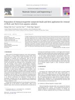
Fig. 1. A) PXRD pattern of a synthesized iron oxide – carboxymethylated cellulose nanofibril composite and iron oxide, measured using a Mo kα X-ray source.
Maghemite (00-039-1346) and magnetite (00-019-0629) reference patterns overlaid. B) and D) Measured FTIR spectra of synthesized iron oxide–carboxymethylated
cellulose nanofibril composite, magnetite and (sodium) carboxymethylated cellulose nanofibrils, NIST is the reference spectra. C) Magnetization vs magnetic field for
powder and liquid samples.
3
T.C. Breijaert et al.
Carbohydrate Polymers 291 (2022) 119560
patterns for both magnetite and the iron(II) deficient maghemite
(Fig. 1A and SI Fig. S2). It was proven that the primary phase of the iron
oxide formed in the presence of C-CNFs nanofibrils was magnetite,
maghemite or a mixture of the two oxides. However, with the current
setup it was impossible to differentiate between the two iron oxides.
Scherrer analysis of the crystallite size was made applying the formula τ
= Kλ / (β ⋅ cosƟ) and showed that the average size of freshly produced
pure ferria under the applied conditions was 3.7 nm, while for the
composite it was 3.4 nm. This shows that composite formation
contributed to the preservation of smaller particles.
AFM where it was apparent that the reaction is capable of producing a
composite consisting of “fibre-like” structures (Fig. 2). Observations
along the axis of these fibre-like structures showed random increases in
surface height, likely attributed to the presence of iron oxide formed
during the co-precipitation reaction.
The fibre-like structures observed had an average width of 18.55 ±
1.66 nm with lengths ranging from 70 nm to nearly a micrometer in
longitudinal direction. However, the measured widths do not take tip
convolution into account, which increases the observed widths
compared to actual widths. By decreasing the measuring area to 200 ×
200 nm (Fig. 2C, E), spherical to ellipsoidal particles were observed
which appear surrounded by a C-CNF network.
The presence of multiple spherical to ellipsoidal particles across the
longitudinal direction of the fibre-like structures (Fig. 2C, E) indicate
that the iron oxide particles formed during the precipitation of iron(II)
and iron(III) interact with the C-CNFs to form a composite material that
self-assembled into fibre-like domains. This interesting property that
should be exploited in future applications. However, it was impossible to
determine from measured AFM data whether the particles were
distributed on the actual surface of the network or within the C-CNF
network itself without more detailed examination.
3.2.2. Fourier Transform Infrared spectroscopy (FTIR)
In an attempt to distinguish between the magnetite and maghemite
iron oxides, the product was examined using FTIR. It should be noted
however, that infrared spectroscopic results of magnetite/maghemite
mixtures are not absolute due to a strong overlap of the most charac
teristics bands of iron oxides (Ellid et al., 2003). The iron oxide produced
via in-situ precipitation of iron(III) chloride and iron(II) sulphate by
ammonia is shown in Fig. 1B. The product obtained exhibits strong
absorption bands at 627 and 576 cm− 1, with a minor adsorption band in
the region of 446 cm− 1. Ishii et al. (1972) assigned the IR band at 565
cm− 1 to the ν1(F1u) vibration mode in magnetite, while a small shift to a
higher wave number may be attributed to sub-stoichiometric magnetite
(Ellid et al., 2003). The peak at 627 cm− 1 can be assigned to the Fe–O
vibration in the iron(II) deficient maghemite, which has formed due to
oxidation (Klotz et al., 1999). The additional peaks at 1128, 1043, 975
cm− 1 may be attributed to the presence of bound sulphate groups pre
sent in the sample (Musi´c et al., 2000). Finally, the peak observed at
1624 cm− 1 and the broad peak at 3400 cm− 1 can be attributed to
moisture.
The produced iron oxide-C-CNF composite material exhibited similar
absorption bands in the range 580–620 cm− 1 with a smaller, less wellpronounced peak at 665 cm− 1, which is slightly shifted, compared to
the synthesized magnetite sample. This may be attributed to the
magnetite formed in the sample, being coordinated to carboxylate
groups present in C-CNF with some maghemite having formed due to
oxidation in air. Additional absorption bands appear at 2890, 1597,
1426, 1373, 1318, 1200, 1160, 1110, 1060, 1022, 897 cm− 1 which are
primarily attributed to the various absorption bands present within CCNF.
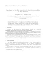
3.3.2. Electron microscopy
To supplement the AFM data, samples were prepared for SEM and
TEM as described in the method section (Fig. 3). SEM confirmed that the
fibre-like structures were not completely homogeneous showing aggre
gates along the surface of the individual fibre networks (Fig. 3A).
Energy-dispersive X-ray analysis across the aggregates showed iron and
oxygen, indicating the metal oxide was distributed homogeneously
within the aggregate structure (Fig. 3A). Cross sections of these aggre
gates shown with TEM further suggest a homogeneous structure (Fig. 5).
Examination of the composite by TEM without negative staining
(Fig. 4, SI Fig. S1) showed the iron-C-CNF network was composed of
single fibrils having a cross section of 3–4.5 nm or double fibrils in the
range of 5.5–8 nm (SI Fig. S1) and as well as strongly scattering elements
in the order of 5.5–8 nm, distributed along the lateral dimensions of the
network. Under normal circumstances, cellulose-based samples require
staining with uranyl acetate or similar heavy metal stains to be visible.
However, in the present case, the composite fibre structure was visible
due to the presence of iron oxide nanoparticles bound to individual
carboxymethyl cellulose fibrils.
Based on the presence of iron oxide in both the aggregates and fibrillike structures within the sample, it would seem feasible that initially the
metal salts hydrolyse to form hydrated species, which then coordinate
with partially deprotonated C-CNFs. The addition of base leads to
further deprotonation of the C-CNFs and production of iron hydroxide
species that nucleate to form magnetite at high pH (Seisenbaeva and
Kessler, 2014), with the surface remaining iron coordinated to the car
boxymethylated cellulose nanofibrils. However, this does not explain
the formation of both the “fibre-like” assemblies and larger aggregates.
Using electron microscopy and AFM, our investigation revealed three
distinct composite structures. This included self-assembled fibril struc
tures containing surface-bound iron oxide nanoparticles, larger C-CNF
aggregates with iron oxide nanoparticles in the range of a few nano
meters, and large iron oxide particles that formed on the surface of fibril
aggregates, the latter stimulated by Ostwald ripening. The larger parti
cles may result from both aggregation and Ostwald ripening, although
the larger crystal domain size for the composite indicates domination of
the Ostwald ripening phenomenon. It is assumed however, that the
source of C-CNFs will play a significant role in the formation of the
observed structures. In this study, the C-CNFs were derived from wood
and had uniform particle sizes in the range of 3–4.5 nm for single fibrils
according to TEM.
To determine whether the iron oxides are precipitated along the
outer regions of the large C-CNFs network, or were incorporated within
the bulk of the aggregates, samples were embedded, sectioned and
3.2.3. Magnetic characterization
Fig. 1C shows the magnetization versus magnetic field for the pow
der and liquid samples. The measured magnetizations at an applied field
of 10 kOe are 63 emu/g and 53 emu/g for the powder and liquid sam
ples, respectively. Both values are somewhat smaller than expected for
magnetite and maghemite, which may be explained by spin disorder and
spin canting for surface spins in iron oxide nanoparticles. Moreover, as
expected, the nanoparticles exhibit superparamagnetic behaviour at
300 K (26,85 ◦ C) and hence zero remanence and coercivity. A plausible
explanation for the minor drop in magnetization of the sample dispersed
in ethanol is that partial oxidation has occurred with time, but only to
rather low extent. The samples were stored for 10 months before mea
surements and thus revealed considerable stability against oxidation.
Bulk magnetic properties are demonstrated in SI Fig. S4 and in a Sup
plementary video. The major volume of ferria was kept in the form of
non-aggregated primary particles bound within the formed selfassembly fibres. This precludes both diffusion of oxygen and release of
ions from the coordination-saturated surface of the particles. As a result
– no apparent oxidation occurs on storage.
3.3. Morphological investigation of iron oxide composite materials
3.3.1. Atomic force microscopy
To examine the nanoscale structures formed when iron oxide is
precipitated in the presence of C-CNFs, the sample was examined using
4
T.C. Breijaert et al.
Carbohydrate Polymers 291 (2022) 119560
Fig. 2. Scanasyst AFM images of an Iron-oxide carboxymethyl cellulose composite. Top down view at 512 px resolution and A) 1 × 1 μm, B) 500 × 500 nm, C) 200 ×
200 nm scan sizes. 3D view a composite at D) 1 × 1 μm, E) 500 × 500 nm and F) 200 × 200 nm.
Fig. 3. A) SEM-EDS image of a large aggregate showing the presence and distribution of carbon, oxygen and iron, attributed to carboxymethylated cellulose
nanofibrils and iron oxide respectively. B) SEM image of the iron oxide – C-CNF. C) TEM image without staining of iron oxide-C-CNF.
examined by TEM to provide additional information on the ultrastruc
tural nature of the composite material.
As shown in Figs. 3–5 and S1, Fig. S1, the iron oxides were present in
three major forms, including, iron oxide aggregates, inclusion of iron
oxides within the C-CNF aggregate structure, and adsorption onto the CCNF fibril surfaces. While the surface adsorbed iron oxide nanoparticles
grew into large sizes due to Ostwald ripening (i.e. ca. 5,5–8 nm), the iron
oxide nanoparticles encapsulated within the C-CNF structure remained
in the order of 1,5–2,5 nm. This observation is in good agreement with
calculations of the average size of crystalline domain applying the
Scherrer formula. It indicates that carboxymethylated cellulose nano
fibrils not only allow for the formation of C-CNF-iron oxide network
clusters, it is also effective in retaining the iron oxide particle size range
to a few nanometers, so long as the particles are encapsulated by
5
T.C. Breijaert et al.
Carbohydrate Polymers 291 (2022) 119560
Fig. 4. TEM Images of iron-oxide carboxymethyl cellulose composites without staining and at varying magnification.
Fig. 5. TEM images of a cross-sectioned resin embedded iron oxide – C-CNFs. Large aggregates are visible on the surface (inset shows the particle size distribution in
nm) and open structure of the iron-cellulose composite, while smaller particles appear aligned with individual fibrils (inset shows the particle size distribution in nm).
6
T.C. Breijaert et al.
Carbohydrate Polymers 291 (2022) 119560
cellulose fibrils and not present on the surface where Ostwald ripening
can occur. Ostwald ripening otherwise sometimes called isothermal
distillation is a phenomenon associated with minimization of the surface
energy in a precipitate, which results in dissolution of smaller particles
and simultaneous growth of the larger ones in the system (Voorhees,
1985). Whether the formation of these large composite aggregates is
caused by the formation of small iron oxide particles after bonding of
ionic iron to C-CNF or by the intercalation of the iron oxide particles
within the C-CNF after metal oxide formation remains unknown. TEM
suggests the iron oxides associate with the outer regions of the nanosized C-CNFs, following the orientation of the individual cellulose fi
brils (Figs. 5, S, S1). The statistic distribution of sizes for single-domain
particles according to TEM (see Fig. 5) shows that it is clearly smaller
inside the fibres 2.0–4.0 nm compared to that on their surface 3.6–16.4
nm, indicating that it was in the first hand Ostwald ripening that pro
duced the larger particles on the surface.
4. Conclusions
In this work, we demonstrated the synthesis of a magnetically
responsive composite material based on carboxymethyl cellulose and insitu synthesized magnetite, that self-assembled into fibre-like nano
structures which were characterized by AFM, SEM, TEM, FTIR, TGA and
PXRD. The material displayed stable magnetic characteristics on stor
age, both in solid state and in solution. In addition, the novel material
was studied in solution state as a potential drug vehicle for the delivery
of tetracycline. Thus, the main hypothesis of this work was proved valid.
Carboxymethylated cellulose nanofibrils derived from wood were
successfully decorated with iron oxide particles in an in situ process so
that magnetic iron oxide particles were of relative uniform size and
assembled into larger composite structures together with cellulose.
These structures could be divided into three broad categories: i) Large CCNF aggregates where the iron oxide nanoparticle size was small with
growth limited by the C-CNF structure; ii) large iron oxide particles that
form on the surface of the fibre aggregates, where particle growth is
stimulated by Ostwald ripening, and iii) cellulose-iron oxides forming
long fibre networks comprising iron oxide and cellulose with longitu
dinal dimensions far exceeding that of the initial components. For the
fibre-like networks, it is highly likely that the morphology and pH
response of both the metal oxide and C-CNF play a crucial role in its
formation. Variations in cellulose source and synthetic conditions may
have significant influence on the overall structures formed. The phase of
iron oxide synthesized in this method is the magnetically responsive
magnetite, which will oxidize to iron(II)-deficient maghemite with time
in the presence of oxygen. This method is simple and cost-effective,
which can lead to the development of further magnetically relevant
materials. However, the synthesis of the “fibre-like” structures remains
difficult with subtle changes in synthetic conditions having a profound
effect on the structures obtained.
Electronic supporting information includes additional details on
TEM, XRD and TGA studies, and demonstration of magnetic properties
of obtained materials (as photo and video evidence). Supplementary
data to this article can be found online at />carbpol.2022.119560.
3.4. Drug adsorption and desorption
Fe3O4 NPs are of interest not only for their magnetic properties but
also for their optical properties, as they are known to display photo
thermal conversion which may be exploited for therapy and drug de
livery (Estelrich & Busquets, 2018; Johnson et al., 2018; Sadat et al.,
2014; Wang et al., 2014). In order to examine the potential of the
composite material as a drug delivery vehicle, we tested the adsorption
and desorption of tetracycline, a broad-spectrum antibiotic. After 72
hour contact time, tetracycline showed a maximum drug adsorption of
62 μg/mg (79%) at room temperature with up to 27 μg/mg (35%) within
the first 240 min indicating that the initial adsorption is fast and
thereafter slows down (Fig. 6A).
To test the viability of the composite for drug delivery and the in
fluence daylight plays on the release of tetracycline, samples were pre
pared in batches and split equally. One part was exposed to a daylight
lamp during desorption, and the other kept dark by wrapping in foil.
Citrate buffer was added and release of tetracycline followed by UV–VIS
(Fig. 6B). Initial drug release was relatively slow with an approximate
11% release in the absence of light and 20% release in light after 3 h.
This increases to 33 and 85% with- and without light respectively, after
2 days indicating that the release desorption rate of tetracycline was
strongly influenced by daylight.
CRediT authorship contribution statement
TB has performed all the synthetic work and adsorption and
desorption experiments and wrote the draft of the manuscript, GD has
performed the TEM characterization and contributed to formulation and
Fig. 6. Tetracycline adsorption and desorption. A) Tetracycline adsorption in mg/g and B) tetracycline desorption in mg/g as a function of time.
7
T.C. Breijaert et al.
Carbohydrate Polymers 291 (2022) 119560
language editing of the manuscript, DH and PS have performed the
magnetic measurements and helped with their interpretation, VK
contributed with XRD measurements, HG and KH provided the C-CNF
material and helped with interpretation of data, GS contributed with the
project idea, TGA, FTIR, AFM and ESEM measurements and performed
the final editing of the manuscript. All authors participated actively in
discussion of results and contributed to editing of the manuscript.
Hao, J., Zhang, W., Wang, H., Ziya, N., Luo, Y., Jia, P., Zhang, G., & Ng, T. (2021).
Purification and properties of a laccase from the mushroom Agaricus sinodeliciosus.
Biotechnology and Applied Biochemistry, 68(2), 297–306. />bab.1926
Hartings, M., Douglass, K. O., Neice, C., & Ahmed, Z. (2018). Humidity responsive
photonic sensor based on a carboxymethyl cellulose mechanical actuator. Sensors
and Actuators B: Chemical, 265, 335–338. />snb.2018.03.065
He, X., Lu, W., Sun, C., Khalesi, H., Mata, A., Andaleeb, R., & Fang, Y. (2021). Cellulose
and cellulose derivatives: Different colloidal states and food-related applications.
Carbohydrate Polymers, 255, Article 117334. />carbpol.2020.117334
Hon, D. N.-S. (1994). Cellulose: A random walk along its historical path. Cellulose, 1(1),
1–25. />Hu, L., Choi, J. W., Yang, Y., Jeong, S., Mantia, F. L., Cui, L.-F., & Cui, Y. (2009). Highly
conductive paper for energy-storage devices. Proceedings of the National Academy of
Sciences, 106(51), 21490–21494. />Ishii, M., Nakahira, M., & Yamanaka, T. (1972). Infrared absorption spectra and cation
distributions in (Mn, Fe)3O4. Solid State Communications, 11(1), 209–212. https://
doi.org/10.1016/0038-1098(72)91162-3
Johnson, R. J. G., Schultz, J. D., & Lear, B. J. (2018). Photothermal effectiveness of
magnetite nanoparticles: Dependence upon particle size probed by experiment and
simulation. Molecules, 23(5), 1234. />Khalilzadeh, M. A., Tajik, S., Beitollahi, H., & Venditti, R. A. (2020). Green synthesis of
magnetic nanocomposite with iron oxide deposited on cellulose nanocrystals with
copper (Fe3O4@CNC/Cu): Investigation of catalytic activity for the development of
a venlafaxine electrochemical sensor. Industrial & Engineering Chemistry Research, 59
(10), 4219–4228. />Kim, J., Yun, S., & Ounaies, Z. (2006). Discovery of cellulose as a smart material.
Macromolecules, 39(12), 4202–4206. />Klemm, D., Heublein, B., Fink, H.-P., & Bohn, A. (2005). Cellulose: Fascinating
biopolymer and sustainable raw material. Angewandte Chemie International Edition,
44(22), 3358–3393. />Klotz, M., Ayral, A., Guizard, C., M´enager, C., & Cabuil, V. (1999). Silica coating on
colloidal maghemite particles. Journal of Colloid and Interface Science, 220(2),
357–361. />Li, Y., Zhu, H., Gu, H., Dai, H., Fang, Z., Weadock, N. J., Guo, Z., & Hu, L. (2013). Strong
transparent magnetic nanopaper prepared by immobilization of Fe3O4 nanoparticles
in a nanofibrillated cellulose network. Journal of Materials Chemistry A, 1(48),
15278–15283. />Lim, W., Douglas, E. A., Kim, S.-H., Norton, D. P., Pearton, S. J., Ren, F., Shen, H., &
Chang, W. H. (2009). High mobility InGaZnO4 thin-film transistors on paper. Applied
Physics Letters, 94(7), Article 072103. />ˇ c, A., Popovi´c, S., Nomura, K., & Sawada, T. (2000). Forced hydrolysis of
Musi´c, S., Sari´
Fe3+ ions in NH4Fe(SO4)2 solutions containing urotropin. Croatica Chemica Acta, 73
(2), 541–567.
Oun, A. A., & Rhim, J.-W. (2017). Characterization of carboxymethyl cellulose-based
nanocomposite films reinforced with oxidized nanocellulose isolated using
ammonium persulfate method. Carbohydrate Polymers, 174, 484–492. https://doi.
org/10.1016/j.carbpol.2017.06.121
Sacui, I. A., Nieuwendaal, R. C., Burnett, D. J., Jorfi, M., Weder, C., Foster, E. J.,
Olsson, R. T., Gilman, J. W., & Stranick, S. J. (2014). Comparison of the properties of
cellulose nanocrystals and cellulose nanofibrils isolated from bacteria, tunicate, and
wood processed using acid, enzymatic, mechanical, and oxidative methods. ACS
Appl. Mater. Interfaces, 6, 6127–6138. />Sadat, M. E., Kaveh Baghbador, M., Dunn, A. W., Wagner, H. P., Ewing, R. C., Zhang, J.,
Xu, H., Pauletti, G. M., Mast, D. B., & Shi, D. (2014). Photoluminescence and
photothermal effect of Fe3O4 nanoparticles for medical imaging and therapy.
Applied Physics Letters, 105(9), Article 091903. />Seisenbaeva, G. A., & Kessler, V. G. (2014). Precursor directed synthesis – “molecular”
mechanisms in the soft chemistry approaches and their use for template-free
synthesis of metal, metal oxide and metal chalcogenide nanoparticles and
nanostructures. Nanoscale, 6, 6229–6244. />Souza, S. F., Mariano, M., De Farias, M. A., & Bernardes, J. S. (2019). Effect of depletion
forces on the morphological structure of carboxymethyl cellulose and micro/nano
cellulose fiber suspensions. Journal of Colloid and Interface Science, 538, 228–236.
/>Thanh, N. T. H. (Ed.). (2012). Magnetic Nanoparticles: From Fabrication to Clinical
Applications (1st ed.). CRC Press. ISBN-10: 1439869324.
Transforming our world: The 2030 Agenda for Sustainable Development, n.d.
Transforming our world: The 2030 agenda for sustainable development |
Department of Economic and Social Affairs. (n.d.). Retrieved November 22, 2021,
from />Voorhees, P. W. (1985). The theory of ostwald ripening. Journal of Statistical Physics, 38,
231–252.
Wågberg, L., Decher, G., Norgren, M., Lindstră
om, T., Ankerfors, M., & Axnă
as, K. (2008).
The build-up of polyelectrolyte multilayers of microfibrillated cellulose and cationic
polyelectrolytes. Langmuir, 24(3), 784–795. />Wang, H., Shen, J., Li, Y., Wei, Z., Cao, G., Gai, Z., Hong, K., Banerjee, P., & Zhou, S.
(2014). Magnetic iron oxide–fluorescent carbon dots integrated nanoparticles for
dual-modal imaging, near-infrared light-responsive drug carrier and photothermal
therapy. Biomaterials Science, 2(6), 915–923. />
Declaration of competing interest
The authors declare that they have no affiliations with or involve
ment in any organization or entity with any financial interest or nonfinancial interest in the subject matter or material discussed in this
manuscript.
Acknowledgements
The authors are grateful to Professor Sidney Ribeiro for valuable
discussions.
Funding
The authors express their gratitude to the Swedish Research Council
STINT for support of the grant Nanocellulose Based Materials for Envi
ronmental and Theranostic Applications and to the Faculty of Natural
Resources and Agricultural Sciences, SLU for support of TB PhD position.
References
Aaen, R., Simon, S., Wernersson Brodin, F., & Syverud, K. (2019). The potential of
TEMPO-oxidized cellulose nanofibrils as rheology modifiers in food systems.
Cellulose, 26, 5483–5496.
Abbasi Pour, S., Shaterian, H. R., Afradi, M., & Yazdani-Elah-Abadi, A. (2017).
Carboxymethyl cellulose (CMC)-loaded co-cu doped manganese ferrite nanorods as a
new dual-modal simultaneous contrast agent for magnetic resonance imaging and
nanocarrier for drug delivery system. Journal of Magnetism and Magnetic Materials,
438, 85–94. />Arantes, A. C. C., Dauzacker, L. C. L., Bianchi, M. L., Wood, D. F., Williams, T. G.,
Orts, W. J., … Almeida, C.d. G. (2017). Renewable hybrid nanocatalyst from
magnetite and cellulose for treatment of textile effluents. Carbohydrate Polymers,
163, 101–107. />Biliuta, G., Sacarescu, L., Socoliuc, V., Iacob, M., Gheorghe, L., Negru, D., & Coseri, S.
(2017). Carboxylated polysaccharides decorated with ultrasmall magnetic
nanoparticles with antibacterial and MRI properties. Macromolecular Chemistry and
Physics, 218(10), 1700062. />Chaabane, L., Chahdoura, H., Mehdaoui, R., Snoussi, M., Beyou, E., Lahcini, M., &
Baouab, M. H. V. (2020). Functionalization of developed bacterial cellulose with
magnetite nanoparticles for nanobiotechnology and nanomedicine applications.
Carbohydrate Polymers, 247, Article 116707. />carbpol.2020.116707
Chen, X., Ye, Z., Yang, F., Feng, J., Li, Z., Huang, C., Ke, Q., & Yin, Y. (2020). Magnetic
cellulose microcrystals with tunable magneto-optical responses. Applied Materials
Today, 20, Article 100749. />Dimic-Misic, K., Gane, P. A. C., & Paltakari, J. (2013). Micro- and nanofibrillated
cellulose as a rheology modifier additive in CMC-containing pigment-coating
formulations. Industrial & Engineering Chemistry Research, 52(45), 16066–16083.
/>Dimic-Misic, K., Phiri, J., Nieminen, K., Maloney, T., & Gane, P. (2019). Characterising
exfoliated few-layer graphene interactions in co-processed nanofibrillated cellulose
suspension via water retention and dispersion rheology. Materials Science and
Engineering: B, 242, 37–51. />Ellid, M. S., Murayed, Y. S., Zoto, M. S., Musi´c, S., & Popovi´c, S. (2003). Chemical
reduction of hematite with starch. Journal of Radioanalytical and Nuclear Chemistry,
258(2), 299–305. />Estelrich, J., & Busquets, M. A. (2018). Iron oxide nanoparticles in photothermal therapy.
Molecules, 23(7), 1567. />Favier, V., Chanzy, H., & Cavaille, J. Y. (1995). Polymer nanocomposites reinforced by
cellulose whiskers. Macromolecules, 28(18), 6365–6367. />ma00122a053
Galland, S., Andersson, R. L., Salajkov´
a, M., Stră
om, V., Olsson, R. T., & Berglund, L. A.
(2013). Cellulose nanofibers decorated with magnetic nanoparticles – synthesis,
structure and use in magnetized high toughness membranes for a prototype
loudspeaker. Journal of Materials Chemistry C, 1(47), 7963–7972. />10.1039/C3TC31748J
Grunert, M., & Winter, W. T. (2002). Nanocomposites of cellulose acetate butyrate
reinforced with cellulose nanocrystals. Journal of Polymers and the Environment, 10
(1), 27–30. />
8
T.C. Breijaert et al.
Carbohydrate Polymers 291 (2022) 119560
Wu, H., Teng, C., Tian, H., Li, Y., & Wang, J. (2018). Fabrication of functional magnetic
cellulose nanocomposite membranes for controlled adsorption of protein. Cellulose,
25(5), 2977–2986. />Yu, X., Kang, D., Hu, Y., Tong, S., Ge, M., Cao, C., & Song, W. (2014). One-pot synthesis
of porous magnetic cellulose beads for the removal of metal ions. RSC Advances, 4
(59), 31362–31369. />
Yu, X., Tong, S., Ge, M., Zuo, J., Cao, C., & Song, W. (2012). One-step synthesis of
magnetic composites of cellulose@iron oxide nanoparticles for arsenic removal.
Journal of Materials Chemistry A, 1(3), 959–965. />C2TA00315E
9