Chitosan sulfate-lysozyme hybrid hydrogels as platforms with fine-tuned degradability and sustained inherent antibiotic and antioxidant activities
Bạn đang xem bản rút gọn của tài liệu. Xem và tải ngay bản đầy đủ của tài liệu tại đây (2.59 MB, 14 trang )
Carbohydrate Polymers 291 (2022) 119611
Contents lists available at ScienceDirect
Carbohydrate Polymers
journal homepage: www.elsevier.com/locate/carbpol
Chitosan sulfate-lysozyme hybrid hydrogels as platforms with fine-tuned
degradability and sustained inherent antibiotic and antioxidant activities
Antonio Aguanell , María Luisa del Pozo , Carlos P´erez-Martín 1, Gabriela Pontes ,
´ndez-Mayoralas , Eduardo García-Junceda *, Julia Revuelta *
Agatha Bastida , Alfonso Ferna
BioGlycoChem Group, Departamento de Química Bio-Org´
anica, Instituto de Química Org´
anica General, CSIC (IQOG-CSIC), Juan de la Cierva 3, 28006 Madrid, Spain
A R T I C L E I N F O
A B S T R A C T
Keywords:
Chitosan sulfate
Lysozyme
Polymers
Physicochemical parameters
Antibiotic activity
Antioxidant activity
The control of the properties and biological activities of chitosan-lysozyme hybrid hydrogels to exploit their
interesting biomedical applications depends largely on the chitosan acetylation pattern, a difficult parameter to
control. Herein, we have prepared sulfated chitosan-lysozyme hydrogels as versatile platforms with fine-tuned
degradability and persistent bactericidal and antioxidant properties. The use of chitosan sulfates instead of
chitosan has the advantage that the rate and mechanisms of lysozyme release, as well as antibacterial and
antioxidant activities, depend on the sulfation profile, a structural parameter that is easily controlled by simple
chemical modifications. Thus, while 6-O-sulfated chitosan hydrogels allow the release of loaded lysozyme in a
short time (60% in 24 h), due to a high rate of degradation that allows rapid antibiotic and antioxidant activities,
in 3-O-sulfated systems there is a slow release of lysozyme (80% in 21 days), resulting in long-lasting antibiotic
and antioxidant activities.
1. Introduction
Chitosan hydrogels are three-dimensional (3D) networks formed by
physical or chemical cross-linking of this sustainable polymer derived
from abundant renewable resources (Domalik-Pyzik et al., 2019). The
diverse biological activities of chitosan (analgesic, antitumor, antiinflammatory, antimicrobial, etc.) combined with various bioactive
properties such as non-toxicity, biodegradability, absorbability and
others, as well as its excellent ability to form hydrogels, have led to the
use of this polymer for the preparation of hydrogels for biomedical ap
plications (Eivazzadeh-Keihan et al., 2022), including drug delivery
´pez et al., 2021), wound
(Peers et al., 2020), tissue engineering (Pita-Lo
dressing (Liu et al., 2018a), and so on. Several studies have shown that
chitosan-based hydrogels further improve their properties when chem
ically modified by covalent conjugation and/or combined with small
molecules, other polymers, proteins, nanocomposites, or cells (Nicolle
et al., 2021; Sanchez-Salvadoret al., 2021; Torkaman et al., 2021).
Lysozyme, a glycoside hydrolase with high enzymatic specificity for
the hydrolysis of the glycosidic bonds of chitosan (Tomihata & Ikada,
1997), is widely used to modulate the properties of chitosan-based
biomaterials, such as degradation (Lonˇcarevi´c et al., 2017) and to
improve the profiles of controlled-release drugs (Herdiana et al., 2022).
In addition, antibacterial films prepared by incorporating lysozyme into
chitosan were reported not only to retain lysozyme activity but also to
enhance the antimicrobial ability of lysozyme (Li et al., 2017). This
enhancement of antibacterial activity was attributed not only to the
release of lysozyme, but also to a possible synergistic effect between
chitooligomers and lysozyme obtained after chitosan hydrolysis (Kim
et al., 2020; Saito et al., 2019). Finally, chitosan and lysozyme represent
a versatile combination to create porous structures by degrading
hydrogels. These spaces promote cell proliferation and migration and
contribute to osteogenic differentiation when mesenchymal stem cells
are encapsulated in chitosan-lysozyme hydrogels (Kim et al., 2018).
These results suggest that the strategy of combining lysozyme with
chitosan may be a promising approach to improve not only the func
tionalities of chitosan-based hydrogels but also their biomedical appli
cations. However, despite the above advantages, the combination of
chitosan and lysozyme in these systems also has important drawbacks.
On the one hand, the interaction between chitosan and lysozyme
strongly depends on the degree of acetylation of the chitosan (DA), and
* Corresponding authors.
E-mail addresses: (A. Aguanell), (M.L. del Pozo), (C. P´
erez-Martín),
(A. Bastida), (A. Fern´
andez-Mayoralas), (E. García-Junceda), (J. Revuelta).
1
Present address: Departamento de Química Org´
anica e Inorg´
anica, Universidad de Oviedo, Juli´
an Clavería 8, 33006 Oviedo, Spain.
/>Received 23 February 2022; Received in revised form 6 May 2022; Accepted 9 May 2022
Available online 12 May 2022
0144-8617/© 2022 The Authors. Published by Elsevier Ltd. This is an open access article under the CC BY-NC-ND license ( />
A. Aguanell et al.
Carbohydrate Polymers 291 (2022) 119611
low degrees of acetylation have been associated with low affinities be
tween lysozyme and the polysaccharide (Nordtveit et al., 1996). How
ever, a high degree of acetylation negatively affects the solubility of
chitosan, a crucial property not only for handling in the manufacture of
materials but also for use in biomedical applications (Pillai et al., 2009).
Moreover, the solubility properties of chitosan depend not only on its
average degree of acetylation but also on the distribution of acetyl
groups along the chain, and a block distribution of acetylation residues
significantly reduces the solubility of the polymer (Kurita et al., 1991).
Nevertheless, commercial chitosan is mainly prepared by chemical
deacetylation of chitin under heterogeneous conditions, resulting in
polymers in which the acetyl groups are distributed in blocks with a
random acetylation pattern (Weinhold et al., 2009).
On the other hand, it has been described that the substrate specificity
of lysozyme with respect to chitosan is related to specific acetylation
sequences. Lysozyme has a binding site that can accommodate a hex
asaccharide sequence with three or more acetylated units, whereas it
does not act on sequences characterized by a lower proportion of acet
ylated residues (Song et al., 1994). In addition, it is known that chitosan
with a low degree of deacetylation can act as an inhibitor of lysozyme
(Vårum et al., 1996). Although better defined, less dispersed chitosan
with non-random acetylation patterns is already obtained at laboratory
scale (Cord-Landwehr et al., 2020; Wattjes et al., 2019, 2020), further
research is needed to develop high-yield- and cost-effective protocols for
tailoring polymers with specific acetylation sequences.
Chemical modification of chitosan offers a great opportunity to
develop solutions for a wide range of biomedical and technological
applications (Nicolle et al., 2021). In this sense, the modification of
chitosan with sulfate groups has attracted increasing attention in recent
decades, as it confers new and attractive physicochemical properties to
polymers compared to the starting chitosan, as well as interesting
pharmacological properties and biological activities (Revuelta et al.,
2021). Advances in chemo- and/or regioselective chitosan sulfonation
and physicochemical characterization (Bedini et al., 2017) have paved
the way for the development of sulfated chitosan-based entities with a
wide range of possibilities. Nevertheless, successful process optimization
and development of these entities is currently only possible by under
standing how the specific structural properties of chitosan sulfates,
especially the sulfation profile, determine their functionalities and bio
logical activities. In this context, one of the most important challenges is
to identify the role of chemistry, structure, and the understanding and
use of these roles in biomedical applications. Recent advances in this
field have focused mainly on deciphering the structural determinants of
the so-called heparanized chitosans, a very interesting family of poly
saccharides that have shown the ability to mimic heparan sulfates and
heparin as ligands of various proteins, thereby exerting their biological
activity by mimicking the function of these glycosaminoglycans (Don
cel-P´erez et al., 2018; Revuelta et al., 2020). Morever, some progress has
been made in the last decade in the binding of lysozyme to chitosan
sulfates. In particular, regioselectively sulfated chitosans have been
described to have differential effects not only on their protein binding
affinity and specificity, but also on lysozyme activity (Wang et al., 2012;
Yuan et al., 2009).
Based on the above, we hypothesize that the preparation of hydro
gels based on chitosan sulfates and lysozyme can be a versatile alter
native to chitosan-lysozyme backbones. Our hydrogels offer versatile
platforms with fine-tuned degradability and persistent bactericidal and
antioxidant properties. The use of chitosan sulfates instead of chitosan
has the advantage that the rate and mechanisms of lysozyme release, as
well as antibacterial and antioxidant activities, depend on the profile of
sulfation along the chains, a structural parameter that, unlike the degree
of acetylation and the presence of specific acetylation sequences, can be
easily controlled by simple chemical modifications (Bedini et al., 2017).
Finally, our study also addresses the question of how the chitosan sulfate
structures control the behaviour of the hydrogels upon addition of
lysozyme.
2. Materials and methods
2.1. Materials
Chitosan (CS) (degree of deacetylation 85%; molecular weight
50–150 kDa) was purchased from IDEBIO, S.L. (Spain) and purified
before use (Nakal-Chidiac et al., 2020). Briefly, CS (5.0 g) was dissolved
in a 0.5 M solution of acetic acid in water (1 L), and the solution was
stirred for 24 h, keeping the pH between 4.0 and 4.5 by adding acetic
acid as needed. The solution was then filtered to remove undissolved
particles, and CS was precipitated again with an aqueous NaOH solution
(10% w/v) until the pH = 8. The resulting suspension was centrifuged
(15 min, 3900 rpm) and the supernatant was removed, with the
remaining solid washed with an EtOH/H2O mixture (70:30 v/v → 50:50
v/v → 30:70 v/v → 0:100) (400 mL). The resulting solid was finally
resuspended in H2O and lyophilized. All reagents were commercially
available and were used without further purification. For statistical
analysis, an unpaired t-test was performed.
2.2. Synthesis of chitosan sulfates
We synthesized 2-N-sulfated (2S-CS), 3-O-sulfated (3S-CS), 6-Osulfated (6S-CS), and 3,6-O-disulfated (3,6S-CS) chitosan according to
previously described procedures (Han et al., 2016; Holme & Perlin,
1997; Kariya et al., 2000; Zhang et al., 2010). Detailed procedures are
described in the Supplementary Information.
2.3. Characterization of chitosan sulfate samples
1
H NMR, 13C NMR and 2D (1H–13C HSQC) spectra were registered
on a Varian Unity Inova 500 MHz spectrometer.
The degree of acetylation (DA) was calculated from 1H NMR ac
cording to the method described by Jiang et al. (2017), using Eq. 1:
DA (%) =
3 × A2
× 100
6 × A1
(1)
where A1 are the protons integral values of positions C2–C6 on the sugar
ring and A2 are the protons integral values of the three N-acetyl protons
of the N-acetyl-D-glucosamine units.
The total degree of sulfation (DS) was determined from the sulfur (%
S) and nitrogen (%N) content determined by elemental analysis using a
Heraus CHN-O analyzer (Doncel-P´erez et al., 2018), and the calculation
was performed according to Eq. 2:
DS =
S%/32.06
N%/14.01
(2)
ζ-Potentials determinations were performed using a Malvern Zeta
sizer Nanoseries Nano ZS instrument. Chitosan sulfate samples were
dissolved at 1 mg/mL in 1 mM NaCl. Three replicates of each sample
were performed.
2.4. Preparation of hydrogels
Hydrogels were prepared according to Akakuru and Isiuku (2017)
procedure with modifications. Briefly, chitosan sulfate samples (≈1.2
mmol of repeating unit) were dissolved in 10 mL of 0.5% (v/v) aqueous
acetic acid at room temperature with constant stirring for 24 h to obtain
pale yellow viscous solutions. The solutions were then filtered using a
sintered glass crucible and a 4% (v/v) aqueous glutaraldehyde solution
was added (1 mL for 6S-CS, 3S-CS and 2S-CS or 2.5 mL for 3,6S-CS). The
obtained solutions were then poured into Petri dishes and dried over
night at room temperature to form the crosslinked hydrogels. When the
hydrogels were semi-dried, they were first washed with an aqueous 1.0
M NaOH solution and then with H2O until the supernatant had a neutral
pH. The hydrogels were then cut into small disks with a diameter of 20
2
A. Aguanell et al.
Carbohydrate Polymers 291 (2022) 119611
mm and a height of 2 mm and dried in an oven at 35 ◦ C for 48 h to
completely remove the remaining solvent and obtain xerogel films
´n et al., 2007) with a thickness between 30 and 45 μm, depending
(Alema
on the polysaccharide used (see Fig. S1).
F = Ktn
where F is the drug release fraction at time t (F = Mt / M∞) in which Mt
is the drug-released percentage at time t and M∞ is the total drugrelease percentage. Time has been normalized as t/t∞ where t∞ is the
total experiment time. The exponent “n” is known as “diffusional
exponent” and is related to the release mechanism, being obtained from
the plot of ln (F) versus ln (t).
2.5. Swelling behaviour
The swelling ratio of the hydrogel was determined by a gravimetric
method (Kim et al., 2020). The stored hydrogel disks were weighed (Wd)
and then immersed in 10 mL solutions with different pH values (3.5, 7.2
and 9.0) for 48 h at 25 ◦ C, and then weighed again (Ws). Finally, the
swelling ratio was quantified using Eq. 3:
(
)
Ws − Wd
Swelling ratio (S) (%) =
× 100
(3)
Wd
2.11. Lysozyme binding to sulfated chitosans by surface plasmon
resonance (SPR)
The surface of a CM5 sensor chip (Biacore Inc., GEHealthcare, Bos
ton, MA, USA) was activated with a freshly mixture of N-hydrox
ysuccimide (NHS; 100 mM) and 1-(3-(dimethylamino) propyl)ethylcarbodiimide (EDC; 400 mM) (1/1, v/v) in water. Lysozyme (50
μg/mL) in aqueous NaOAc (10 mM, pH 5.0) was then passed over the
surface until a ligand density of 7000 RUs was reached. Quenching of the
remaining active esters was achieved by passing aqueous ethanolamine
(1.0 M, pH 8.5) over the surface of the chip. The control flow cell was
activated with NHS and EDC and then treated with ethanolamine. HBSEP buffer (0.01 M HEPES, 150 mM NaCl, 3 mM EDTA, 0.05% poly
sorbate 20; pH 7.4) was used as the running buffer for immobilization,
binding, and affinity analysis. A concentration of 1 mg/mL of each
compound in HBS-EP buffer at a flow rate of 30 μL/min and a temper
ature of 25 ◦ C was used for the experiments. A 30 s injection of aqueous
NaCl (2.0 M) at a flow rate of 30 μL/min was used for regeneration to
reach the initial condition. Analysis was performed using BIAcore X100
analysis software (Biacore Inc., GE Healthcare, Boston, MA, USA).
2.6. Lysozyme absorption into hydrogels
Xerogel disks (ø = 2 cm) were transferred to a vial containing 2.5 mL
of lysozyme solution (10 mg/mL) in Tris-HCl 200 mM buffer (pH = 3.5)
and allowed to adsorb protein for 72 h in a shaker (37 ◦ C, 50 rpm). The
protein solution was removed from the vial and analysed using a
NanoDrop™ One C microvolume UV-VIS spectrophotometer equipped
with a Protein A280 application for lysozyme determination which as
sumes that the molar extinction coefficient of the protein at 280 nm is
36,000 M− 1 cm− 1. Finally charged-disks were vacuum-dried for 4 h.
2.7. Lysozyme binding activity of polysaccharides
The lysozyme binding activity of CS and chitosan sulfates (3,6S-CS,
2S-CS and 6S-CS) was measured based on the lysozyme–polysaccharides
flocculation formation activity according to a previously described
procedure (Yuan et al., 2009). A detailed description of the procedure
can be found in the Supporting Information.
2.12. Measurement of lysozyme activity by determination of reducing
sugars using the 3,5-dinitrosalicylic acid (DNS) method
Solutions of chitosan sulfates (4% w/v) in H2O (0.5 mL) were mixed
with 0.5 mL of a lysozyme solution (2% w/v) (both solutions were pre
heated at 50 ◦ C for 5 min before mixing). After 2, 4, 6, or 24 h of incu
bation at 50 ◦ C, an aliquot of the mixtures (10 μL) was taken and heated
at 100 ◦ C for 8 min to stop the reaction. The mixture was then centri
fuged and the supernatant was analysed by DNS-assay (Fig. S2) (Gusa
kov et al., 2011). Briefly, 30 μL of DNS reagent (1 g of 3,5-dinitrosalicylic
acid, 3 g of sodium/potassium tartrate in 80 mL of 0.5 M NaOH by
heating and stirring at 70 ◦ C) was added to the test aliquot and the
mixture was incubated in a boiling water bath for 5 min. After cooling to
room temperature, the absorbance of the supernatant was measured at
540 nm. The A540 values for the substrate and enzyme blank values were
subtracted from the A540 value for the analysed sample. The substrate
and enzyme blanks were prepared in the same manner as the analysed
sample except that 0.5 mL of the acetate buffer was added to the sub
strate (enzyme) solution instead of the enzyme (substrate) solution.
2.8. Hydrogels degradation
The degradation of the hydrogels was analysed using a gravimetric
method, in which the change in dry weight was measured 7 and 14 days
after incubation in distilled water. The change in dry weight was
quantified using Eq. 4:
Hydrogel degradation (%) =
(Wi − Wt )
× 100
Wt
(5)
(4)
where Wi and Wt indicate the dry weight at the beginning and at the
respective time points.
2.9. Morphological observation of hydrogels
The morphological changes of hydrogels after contact with lysozyme
were observed by scanning electron microscopy using a Hitachi S-8000
(Tokyo, Japan) operating in transmission mode at 100 kV on dry
samples.
2.13. Antimicrobial activity
Fresh cultures of E. coli were grown by suspending one colony from
the LB -agar culture in 5 mL of sterile LB medium and incubating for 24 h
at 37 ◦ C with constant shaking (136 rpm). Four falcons (50 mL) were
then inoculated with 5 mL of sterile LB medium with the amount of
bacterial culture required for an initial OD600 of 0.05. One falcon served
as a control and was used to determine the total number of colonies in
the culture. To each of the other three falcons, a lysozyme solution (33
μL, 0.3 μg/mL) and disks (ø = 2 cm) of xerogel without or with lysozyme
were added. After incubation at 37 ◦ C with constant shaking (90 rpm),
the growth of the cultures was monitored until the exponential growth
phase (OD600 of 0.3–0.4) was reached. The obtained bacterial suspen
sions were serially diluted and different dilutions (10− 4, 10− 5 and 10− 6
cfu mL− 1) were seeded on nutrient agar to determine the number of
2.10. Releasing of lysozyme from chitosan sulfate hydrogels
Loaded xerogels were washed with Tris-HCl 200 mM buffer (pH =
7.0) for 5 min and then transferred to a vial containing 2.5 mL of this
same buffer. The vial was kept in a shaker (37 ◦ C, 50 rpm) throughout
the experiment. The experiments were also performed in water with
different pH values (3.5 and 9.0). To measure the lysozyme concentra
tion, 5 μL of the supernatant were taken at different times. The amount
of lysozyme was determined using the Protein A280 application of the
NanoDrop™ One C microvolume UV-VIS spectrophotometer.
The values were fitted to the Korsmeyer-Peppas model according to
Eq. 5:
3
A. Aguanell et al.
Carbohydrate Polymers 291 (2022) 119611
viable bacteria and quantify the number of colony forming units (cfu
mL− 1). Inhibition of colony formation (%) was determined using Eq. 6:
Inhibition of colony formation (%) =
cfuexp
× 100
cfucont
Table 1
Sulfation of chitosans.
(6)
where cfuexp and cfucont indicate cfu mL− 1 of the experimental and
control groups, respectively.
The hydrogels were then removed from the falcon tubes and the
cultures centrifuged at 4000 rpm for 10 min, discarding the pellet. The
hydrogels and a new lysozyme solution (33 μL, 0.3 μg/mL) were
returned to the falcons, and the amount of bacterial cultures required for
an initial OD600 of 0.05 was added, and the procedure described above
was repeated to determine the number of colony-forming units (cfu
mL− 1). The same protocol was repeated for 3 consecutive days.
Polysaccharides
6S-CS
3,6S-CS
3S-CS
2S-CS
R2
H or Ac
H or Ac
H or Ac
H or Ac or SO3-
Polysaccharides
R2
6S-CS
3,6S-CS
3S-CS
2S-CS
H
H
H
H
or
or
or
or
R3
H
SO3SO3H
Ac
Ac
Ac
Ac or SO−3
R6
SO3SO3H
H
Yield
80%
88%
57%
79%
DA[a]
8.0
7.2
9.0
10.5
DS[b]
0.8
1.7
0.7
0.7
R3
R6
Yield
DA[a]
DS[b]
H
SO−3
SO−3
H
SO−3
SO−3
H
H
80%
88%
57%
79%
8.0
7.2
9.0
10.5
0.8
1.7
0.7
0.7
a
Degree of acetylation. Calculated according with reference (Jiang et al.,
2017).
b
Total DSS was determined using elemental analysis.
2.14. Antioxidant activity: DPPH-radical scavenging ability assay
Disks (ø = 2 cm) of each xerogel without lysozyme were immersed in
4 mL of 0.1 mM DPPH (2,2-diphenyl-1-picryl-1-hydrazyl-hydrate)
methanol solution. A 0.1 mM DPPH methanol solution (4 mL) without
xerogel was used as control. The solutions were kept in the dark and the
absorbance of the solution at 517 nm was determined at intervals of 1 h
to 24 h.
In addition, disks (ø = 2 cm) of each lysozyme-incorporated xerogel
were immersed in 5 mL Tris-HCl buffer (200 mM; pH = 7.0) and kept in
a shaker (37 ◦ C, 50 rpm) for 72 h. Aliquots of the supernatant solution
(0.5 mL) were taken at 24 to 72 h intervals and incubated with water
(0.5 mL) and DPPH (2 mL) at 25 ◦ C for 30 min. The concentration of
DPPH was 120 μM in the test solution. Then, the absorbance of the
remaining DPPH radical was measured at 517 nm against a blank.
The scavenging effect was calculated according to Eq. 7:
[
]
Asample 517 nm − Acontrol 517 nm
Scavenging effect (%) = 1 −
× 100
(7)
Ablanck 517 nm
the experimental value did not cause a significant deviation in the in
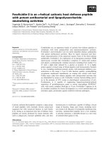
tegrated peak volumes (Guerrini et al., 2005). For example, in 3,6-CS,
the ratio between 6S/6H was determined by integrating the O-6 meth
ylene signals (δH,C = 4.23/66.6 and 3.86/60.2), sulfated and non
sulfated glucosamine residues, whereas the ratio between 3S/3H
(75:25) was calculated by integrating the signals corresponding to the 3sulfated and nonsulfated CH at position 3 (δH,C = 4.28/80.82 and 3.78/
72.8) (Fig. 1b).
4. Preparation and characterization of lysozyme-chitosan
sulfate hydrogels
Hydrogels were prepared by the Schiff base method using glutaral
dehyde as a cross-linking agent (Fig. 2a), and then freeze-dried xerogels
were loaded with lysozyme samples. To optimize the preparation pro
cedure, the effects of different parameters (concentrations of chitosan
sulfate and GA solutions, pH, and temperature) were analysed. The best
experimental conditions (see Section 2.3) were determined based on the
swelling ratio, the stability of the hydrogel and the amount of protein
absorbed. The appearance of the films of chitosan sulfate hydrogels is
shown in Fig. 2b.
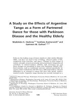
The swelling capacity of the hydrogels was evaluated by the degree
of swelling (S). Fig. 2c shows the water absorption behaviour of the
xerogels at different pH values (3.5, 7.2 and 9.0). The chitosan sulfatebased hydrogels described in this manuscript are polyampholitic sys
tems, due to the presence of amino and sulfate groups, and therefore
form networks with oppositely charged structures that can change the
charge state of the ionic groups as a function of pH. Since the swelling
properties of polyampholite hydrogels are always closely related to the
overall charge density and its distribution, we selected two pH values to
observe the response of the hydrogels when the amino groups are in the
ionized form (NH+
3 ) (pH = 3.5) or when the amino groups are depro
tonated (pH = 9.0).
For the CS hydrogel, the highest degree of swelling was obtained at
an acidic pH. The easy uptake of the solution in this hydrogel was
attributed to the protonated chitosan amine under these conditions.
Thus, when the pH is lower than the pKa of chitosan (pKa ≈ 6.20) (Strand
et al., 2001), the amino groups in the chitosan structure are in the
ionized form (NH+
3 ), which leads to the dissociation of secondary in
teractions such as intramolecular hydrogen bonds, allowing more water
to enter the gel network. This effect is not observed when pH is
increased, as amino groups are deprotonated and repulsion in the
polymer chains decreases, allowing shrinkage. An opposite effect is
observed when chitosan sulfate xerogels are swollen. In this case, the
amino groups, when in ionized form, interact strongly with the sulfonic
groups (–SO−3 ), whose pKa is nearly 2.60 (Larsson et al., 1981), keeping
where Asample 517nm represents the absorbance of the sample at 517 nm,
Ablank 517nm represents the absorbance of the blank at 517 nm and
Acontrol 517nm represents the absorbance of the control (distilled water
instead of DPPH) at 517 nm.
3. Results and discussion
3.1. Synthesis and characterization of chitosan sulfates
We prepared 2-N-sulfated (2S-CS) (Holme and Perlin, 1997), 3-Osulfated (3S-CS) (Kariya et al., 2000), 6-O-sulfated (6S-CS) (Han et al.,
2016) and 3,6-O-di-sulfated (3,6S-CS) (Zhang et al., 2010) chitosan
according to previously published procedures. Elemental analysis
showed that the degree of sulfation (DS) ranged from 0.7 to 1.7
(Table 1).
The regioselectivity of the sulfations was analysed by 13C NMR ex
periments (Fig. 1a and Table 2). After 6-sulfation, the 59.3 ppm signal of
C6(OH) in chitosan was shifted down to 66.5 ppm in sulfated chitosan,
representing the 13C signal of C6(SO−3 ) in 6S-CS. On the other hand, the
appearance of the 73.9 ppm signal C3(SO−3 ) and the partial disappear
ance of the 69.9 ppm signal C3(OH) indicate that the hydroxyl group at
C3 in the 3,6S-CS was sulfated. In addition, the complete disappearance
of the 67.7 ppm signal and the appearance of the 61.4 ppm signal
C6(OH) indicated that position 6 of 3,6S-CS in the 3S-CS was completely
6-O-desulfated. Finally, the data shown in Fig. 1a indicated that position
2 of chitosan in 2S-CS was regioselectively sulfated.
The ratio of sulfated to non-sulfated residues was determined by
integrating each array/body of signals with respect to the CH-2 density
of DEPT-HSQC spectra to estimate the degree of sulfation.
In doing so, we assumed that the compared signals had similar values
of the 1JCH coupling constant and that differences of about 5–8 Hz from
4
A. Aguanell et al.
Carbohydrate Polymers 291 (2022) 119611
Fig. 1. Characterization of chitosan sulfates. (a) Key regions of the 13C NMR spectra of the polysaccharides 6S-CS, 3,6S-CS, 3S-CS, and 2S-CS (b) Essential region of
the DEPT-HSQC spectra of 3,6S-CS. The densities in the colour boxes were integrated to estimate the degree of sulfation: 6-position (dashed red line) and 3-position
(solid green line).
Table 2
Key signals of 13C NMR spectra of chitosan and chitosan sulfates.
Polysaccharides
CS
6S-CS
3,6S-CS
3S-CS
2S-CS
Positions
C2(NH2)
C2(NHSO−3 )
C3(OH)
C3(SO−3 )
C6(OH)
C6(SO−3 )
55.2
55.9
57.2
57.0
56.7
–
–
–
–
63.5
69.8
69.9
–
–
74.5
–
–
73.9
71.3
–
59.3
60.2
–
61.4
61.8
–
66.5
67.7
–
–
the polymer network shrunk and reducing water uptake. When the pH of
the medium is increased, the electronic repulsion between the charged
sulfonic groups causes macromolecular expansion and consequently the
hydrogels tend to swell more (Durmaz & Okay, 2000; Singh et al., 2011).
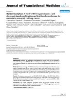
Lysozyme was taken up by static absorption at 10 mg/mL in 1.0 mM
Tris-HCl buffer (pH = 3.5) until absorption equilibrium was reached
(≈72 h), and the concentrations of free lysozyme in the supernatant
were measured (Fig. 3a). Although the amount of sulfate groups appears
to contribute to the absorption process, the results obtained suggest that
other parameters may influence the differences in absorption. Previous
results have shown that the lysozyme/chitosan sulfate binding ratios are
significantly different depending on the sulfation profile of the poly
saccharides (Yuan et al., 2009). To address this question, the binding
behaviour of lysozyme with chitosan and its sulfated derivatives in so
lution was measured in solution. As shown in Fig. 3b, the 3,6S-CS
polysaccharide shows the highest binding activity with lysozyme, while
almost half of the lysozyme binds with 6S-CS. In the case of 2S-CS, it was
observed that mixing the solutions of polysaccharide and lysozyme does
not lead to significant flocculation. Although some turbidity is observed,
the low values of lysozyme binding with 2S-CS could be due to the
presence of soluble complexes of the polysaccharide with lysozyme,
which were not identified in the experiment. A low binding value was
observed with 3S-CS and CS. The latter was attributed to the low acet
ylation degree of the chitosan used, a crucial parameter for the binding
of lysozyme to chitosan (Nordtveit et al., 1996). Finally, although the
polysaccharide with the highest degree of sulfation (3,6S-CS; DS = 1.7)
showed the highest binding capacity with lysozyme, the different
binding capacities observed for the different monosulfated derivatives
(with similar degrees of sulfation) suggest that DS is not the key factor
involved in the binding of polysaccharides with lysozyme such as the
sulfation profile along the chain.
The mass loss (%) of the hydrogels over time was determined as a
measure of degradation (Fig. 3c). Measurable differences in mass were
observed depending on the sulfation profile of the polysaccharides used
to prepare the hydrogels. For example, the presence of sulfate groups at
positions 6 or 2 significantly accelerated the rate of degradation, and
after 7 days, approximately 60% and 40% of the mass was lost for the
6S-CS and 2S-CS hydrogels, respectively, and at the end of the study (14
days), 80% and 60% of the gel mass was lost for both hydrogels. In
contrast, the hydrogels CS, 3,6S-CS and 3S-CS retained 85%, 75%, and
60%, of their weight respectively, by day 14. The degradation of the
hydrogels was examined using cryo-SEM. As shown in Fig. 3d, different
pores form in the hydrogel scaffold during lysozyme-mediated degra
dation. On day 0, both hydrogels (3S-CS and 2S-CS) had comparable
pore sizes and size distributions. However, on day 7, although the
average pore sizes and size distributions increased for both hydrogels,
the 2S-CS hydrogel showed a greater increase in pore size than the 3S-CS
hydrogel, which was attributed to the greater degradation of the first
hydrogel due to the increase in the amount of lysozyme in the hydrogel.
5. In vitro lysozyme release
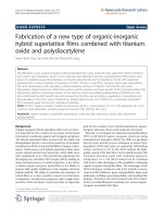
Fig. 4a shows the cumulative total release of lysozyme as a function
of time under neutral conditions (pH = 7.4) for chitosan and chitosan
sulfate hydrogels. As can be observed, lysozyme release varies depend
ing on the hydrogel used. There are many mechanisms by which drug
5
A. Aguanell et al.
Carbohydrate Polymers 291 (2022) 119611
Fig. 2. (a) Molecular structure of cross-linked chitosan sulfate molecules (left) and schematic representation of chitosan sulfate hydrogel networks formed by
chemical cross-linking (right). (b) Overall view of chitosan sulfate hydrogels. (c) Swelling ratio of hydrogels calculated by the ratio of wet and dry weights of
hydrogels for 48 h at different pH values (3.5, 7.2, and 9.0). Swelling ratios are the average of three replicates and standard deviation are shown. (d) Macroscopic
observation of hydrogel swelling over 48 h.
release can be controlled in a system: Dissolution, diffusion, osmosis,
partitioning, swelling, degradation, and binding affinity (Bruschi,
2015).
Since our hydrogels were designed with specific ligands for lysozyme
recognition, their binding affinities, which depend on the molecular
structure of the polysaccharide, could determine the release rate of
lysozyme (Yuan et al., 2009). In addition, the incorporated lysozyme
could trigger the hydrolysis of the chitosan sulfate, leading to the
degradation of the hydrogel and consequent release of the protein
(Wang et al., 2012). Finally, it is important to consider that the release of
the entrapped lysozyme largely depends on the degree of swelling of the
hydrogel. These mechanisms are illustrated in Fig. 4b.
Incubation of the hydrogel 6S-CS resulted in a biphasic release of
lysozyme. Thus, a relatively slow release was observed during the first
hours, while a sharp increase in the released lysozyme was observed
after this period. This result could be attributed to an increase in the
hydrolytic activity of lysozyme after this period. To clarify this behav
iour, lysozyme release was analysed at different pH values. When the
hydrogel was incubated at a pH of 3.5, only about 15% release was
observed after 6 h, whereas at a pH of 9.2, about 82% release was
observed after 4 h (Fig. 4c). Considering that chicken egg white lyso
zyme, the enzyme used in the manuscript, is active in a pH range of
6.0–10.0 and that maximum activity is observed at pH 9.2, it seems clear
that the release of lysozyme in 6S-CS hydrogels could be regulated by
the degradation of the hydrogel chains and, consequently, a
degradation-controlled release would be the main mechanism for lyso
zyme release from these hydrogels.
A biphasic release was also observed for the hydrogel 2S-CS. This
hydrogel showed a burst release of about 10% after 6 h, followed by a
slow release of about 31% on day 6. After this period, an increase in the
6
A. Aguanell et al.
Carbohydrate Polymers 291 (2022) 119611
Fig. 3. (a) Quantification of lysozyme loaded in the hydrogels. (b) Binding curves of chitosan sulfates (3,6S-CS, 2S-CS, 6S-CS, and 3S-CS) and CS with lysozyme. (c)
Degradation kinetics of hydrogels for 7 and 14 days by measuring dry weight. (d) Morphological observations of 2S-CS (left) and 3S-CS (right) hydrogels by cryo-SEM
at days 0 (top) and 7 (bottom). In Fig. 3a, b and c the shown values are the average of three replicates and standard deviations are shown.
amount of lysozyme released is observed. This behaviour could be
related to the intrinsic structural properties of the 2S-CS poly
saccharides. While the other polysaccharides have a N substitution de
gree of about 15%, this degree reaches values of 85% for 2S-CS. As a
result, the available free amino groups are much lower, leading to a
lower crosslink density in the formed network. Considering that
hydrogels with a higher degree of crosslinking degrade more slowly than
hydrogels with a lower degree of crosslinking (Jeon et al., 2007),
possible erosion/degradation of the hydrogel over time could be the
reason for the observed behaviour.
In contrast, in 3,6S-CS hydrogels, less than 2% of the encapsulated
lysozyme was released within 10 days, indicating that the lysozyme is
almost completely entrapped in the hydrogel matrix. This suggests that
the release of lysozyme in this case is mainly due to a reaction-diffusion
mechanism in which the concentrations of free and bound lysozyme are
determined by the equilibrium binding affinity between lysozyme and
3,6S-CS. Finally, for the hydrogels 3S-CS and CS, after a burst release of
about 10% and 7%, respectively, at 3 h, a slow release of 41% and 15%
of the total charge was observed after 11 days.
After this period, lysozyme continued to be released (data not
shown). After 21 days of incubation, more than 80% of the loaded
lysozyme was released in the 2S-CS and 3S-CS hydrogels, whereas in the
CS and 3,6S-CS hydrogels the cumulative drug release was approxi
mately 20% and 5%, respectively.
To further elucidate the mechanisms hypothesised for each hydrogel,
additional experiments were performed. First, the binding affinity be
tween the polysaccharides and lysozyme was analysed by surface plas
mon resonance (SPR) (Fig. 5a), and second, the hydrolytic activity of the
enzyme towards different polysaccharides was measured (Fig. 5b). The
highest binding affinity was observed for 3,6S-CS, which was about 1.2
and 1.5 times greater than that for 6S-CS and 2S-CS, respectively, while
the binding affinity for 3S-CS and CS was only about 16% and 4%,
respectively, of that of 6S-CS. In addition, all lysozyme samples bound to
chitosan and its sulfated derivatives appeared to show lytic activity after
incubation, although the results varied greatly depending on the poly
saccharide used. Thus, the lytic activities of the lysozyme bound to 6SCS and 3S-CS were much higher than those bound to the poly
saccharides 3,6S-CS and 2S-CS, based on the increase in reducing ends
observed after 24 h of incubation (1000% and 750% increase for 6S-CS
and for 3S-CS versus 180% and 350% for 3,6S-CS and 2S-CS). The
analysis of reducing sugars by DNS-assay was used as an indirect method
for the determination of lysozyme activity, because these reducing
sugars are formed by the enzymatic cleavage of the glycosidic bond
between two glucosamine-chitosan units (McKee, 2017). In this method,
the aldehyde functional group of the reducing end of the polysaccharide
is oxidized to a carboxyl group, and in the process the yellow 3,5-dintro
salicylic acid compound is reduced to 3-amino, 5-nitrosalicylic acid,
which has a reddish-brown colour and can be detected by measuring
UV-absorbance of the solution.
These results suggest that although lysozyme recognizes all sulfated
polysaccharides, only 6- and 3-sulfation allows a productive binding
mode, whereas nonproductive binding occurs when 3,6S-CS and 2S-CS
are combined with lysozyme. Previous studies have suggested that
although the net electrical charge density of the surface (estimated by
measuring the ζ-potential) drives the initial interaction between chito
san sulfates and proteins (Doncel-P´
erez et al., 2018; Yuan et al., 2009),
the unique properties of each protein-chitosan sulfate complex are
determined by other polysaccharide features, such as the conforma
tional fit of the polysaccharide to the protein active site (Revuelta et al.,
2020). Thus, the ability of 3,6S-CS and 2S-CS to bind lysozyme could be
explained by the fact that both have the highest net charge on the sur
face, as shown by their ζ-potential values (Fig. 5c). However, the
observed low lysozyme activity suggests that these polysaccharides
(3,6S-CS and 2S-CS), unlike 6S-CS and 3S-CS, would not allow the
molecular conformational adjustment required after the initial ionic
interaction. Finally, it is important to note that the sulfate group at
7
A. Aguanell et al.
Carbohydrate Polymers 291 (2022) 119611
Fig. 4. (a) Lysozyme release profile for chitosan and chitosan sulfate hydrogels; (b) proposed lysozyme release mechanisms for the hydrogels prepared here; (c)
lysozyme release profile for 6S-CS hydrogels at different pH values; (d) macroscopic observation of hydrogels degradation with lysozyme modification for 7 days.
Scale bar is 5 mm. Release data are the average of three replicates and standard deviation are shown.
position 3 of chitosan (3S-CS) significantly decreases the binding affinity
(Fig. 5a) but has little effect on the activity of the bound lysozyme
(Fig. 5b). Thus, it appears that lysozyme bound to any of the poly
saccharides exhibits high hydrolytic activity regardless of how strong or
weak the interaction of lysozyme with 6S-CS and 3S-CS polysaccharides
is. Finally, the results show no correlation between the activity of
lysozyme and the degree of sulfation, since no differences in activity are
observed between the most sulfated derivative (3,6S-CS) and the
unsulfated CS. Moreover, the monosulfated derivatives exhibit different
activities despite their similar degree of sulfation. These results are
consistent with observations previously made by other authors (Wang
et al., 2012).
These results correlated well with the release behaviour of lysozyme
observed with different hydrogels (see Fig. 4a). Consistent with the high
hydrolytic activity observed for lysozyme after binding to 6S-CS, it is
plausible to assume that the network structure retains the shape of the
native polysaccharide and allows lysozyme to efficiently hydrolyze the
hydrogel chains after productive binding, consistent with the previously
proposed degradation-controlled release mechanism. A similar mecha
nism could be attributed to protein release in hydrogel based on 3S-CS.
In contrast, for hydrogels based on 3,6S-CS and in agreement with the
low hydrolytic activity observed for the di-sulfated chitosan-lysozyme
complex, it is reasonable to assume that the release mechanism of
lysozyme could be controlled by the equilibrium binding affinity be
tween lysozyme and 3,6S-CS. Since the concentration gradient of the
protein is directly determined by its free state, the strong binding re
action between the polysaccharide and lysozyme means that the amount
of protein released is very small because it is almost completely
entrapped in the hydrogel matrix. A similar release mechanism was
proposed for the hydrogel 2S-CS. However, in this hydrogel, protein
release could be more efficient due to the lower affinity for lysozyme-2SCS binding and the high amount of free protein in binding equilibrium.
In both cases, the addition of a high concentration of NaCl promoted the
release of lysozyme by disrupting the ionic interactions. As shown in
Fig. 5d, complete removal of lysozyme from 3,6S-CS was observed only
when a 1.0 M NaCl solution was used, whereas in the 2S-CS hydrogel,
removal was observed when a 0.5 M NaCl solution was used, which
could be due to differences in the strength of ionic interactions in each
case.
The results described above suggest that the process of release of
lysozyme from the developed hydrogels is the result of a combination of
different mechanisms due to the presence of various physicochemical
8
A. Aguanell et al.
Carbohydrate Polymers 291 (2022) 119611
Fig. 5. (a) Binding affinity between polysaccharides and lysozyme analysed by SPR; (b) lytic activity of lysozyme against chitosan and chitosan sulfates determined
by measuring the reducing ends; (c) ζ-potential values. Values for the degree of sulfation are shown below. (d) Release of lysozyme from hydrogels in NaCl solutions.
In Fig. 5b, c and d the shown values are the average of three replicates and standard deviations are shown.
phenomena (diffusion, swelling, and/or erosion/degradation of the
matrix). Although it is difficult to find a mathematical model that de
scribes all the processes that occur, the Korsmeyer-Peppas model has
been widely used for systems in which different release mechanisms
interact (Korsmeyer et al., 1983; Ilgin et al., 2019). Table 3 shows the
estimated parameters after fitting the Korsmeyer-Peppas model to the
experimental data. This model uses the value of the release exponent (n),
which is the slope of a plot of ln cumulative release versus ln time. When
n is 0.5 or less, the release mechanism is theoretically assumed to follow
Fick's diffusion for thin films such as the hydrogels prepared here, where
drug release occurs by the usual molecular diffusion of a concentration
gradient. Higher values of n between 0.5 and 0.1 indicate non-Fickian or
anomalous transport, where release is controlled by a combination of
diffusion and erosion/degradation of the hydrogel. When n reaches a
value of 1.0 or more, the mechanism of release is mainly due to erosion/
degradation of the hydrogel (Lao et al., 2011).
As shown in Table 3, application of the lysozyme release data to the
Korsmeyer-Peppas model and regression analysis resulted in good fit
with coefficients of determination (r2) greater than 0.94 in all cases. The
values for the release exponent (n) were 0.105, 0.258, and 0.392 for CS,
3S-CS, and 3,6S-CS hydrogels, respectively. This indicates that the
release of lysozyme from each hydrogel after the initial burst (estimated
in 6 h) was controlled by Fick's diffusion through the hydrated matrix.
However, for the hydrogel 2S-CS, the value of n was 0.66, indicating that
hydrogel degradation cannot be disregarded, although Fick's diffusion is
still important. Finally, in the case of the hydrogel 6S-CS, the value of n
was 2.50, indicating that the release is completely controlled by the
degradation of the network. These results, on the one hand, confirm the
existence of different release mechanisms depending on the sulfation
profile of the chitosan and, on the other hand, are consistent with the
proposed mechanism for each hydrogel based on the experimental data.
6. Antimicrobial activity
The antimicrobial activities of the hydrogels against E. coli strain K12
were evaluated by quantifying the number of colony-forming units (cfu
mL− 1) of a culture after treatment with the different hydrogels (Fig. S3).
As shown in Fig. 6a, all hydrogels without lysozyme showed activity
against E. coli. After 24 h of incubation, the inhibition of bacterial
growth for the hydrogels based on CS was 32%. This inhibition value
increased to 47% and 35% when 3,6- and 6-sulfated chitosan hydrogels
were analysed, whereas lower inhibition values (25% and 5%, respec
tively) were obtained for hydrogels based on 3S-CS and 2S-CS).
Inhibition of bacterial growth in CS based hydrogels can be explained
by their cationic nature. The interaction of cationic polysaccharides such
as chitosan with the negatively charged cell wall of bacteria has been
described, resulting in increased cell permeability, decreased cell wall
integrity, and subsequent leakage of intracellular proteases and other
components (Matica et al., 2019). For chitosan sulfates, it seems clear
that anionic polysaccharides are unlikely to bind to the negatively
charged surface of microorganisms through electrostatic interactions. In
recent decades, it has been proposed that bacteria utilize heparan sulfate
proteoglycans present on the extracellular matrix to facilitate cell
adherence, attachment, and invasion and to evade defense mechanisms
(Rostand & Esko, 1997). In particular, heparan sulfates appear to bind
bacteria via adhesins, macromolecular components of the bacterial cell
Table 3
Values for lysozyme-release profile according to Korsmeyer-Peppas kinetic
model.
Hydrogel
na
r2
Kp(h− 1)b
a
b
2S-CS
3,6S-CS
3S-CS
CS
6-CS
0.66
0.948
1.66 ×
10− 2
0.39
0.956
1.69 ×
10− 2
0.25
0.962
1.62 ×
10− 2
0.10
0.943
1.72 ×
10− 2
2.50
0.97
7.8 ×
10− 2
Release exponent describing the transport mechanism.
Constant describing the drug-sample interaction.
9
A. Aguanell et al.
Carbohydrate Polymers 291 (2022) 119611
Fig. 6. (a) Percent cfu inhibition of hydrogels without lysozyme; (b) comparison of percent cfu inhibition of hydrogels without lysozyme (shown in light) and
lysozyme-incorporated hydrogels (shown in dark). The increase in inhibition after lysozyme incorporation is shown to the left of each bar. Kanamycin A (50 μg/mL)
was used as positive control; (c) proposed mechanisms of antibiotic action of hydrogels; (d) percentage cfu inhibition between 48 h and 72 h. *P < 0.001 (n = 3); **P
< 0.05 (n = 3).
mechanisms for the antibacterial effect of hydrogels have been proposed
based on the results obtained (Fig. 6c).
Previous studies have reported that binding of chitosan sulfates to
lysozyme can significantly alter the specific hydrolytic activity of the
enzyme with bacterial cell wall components (Wang et al., 2012). The
increase in activity observed for 3,6-disulfated chitosan-lysozyme
complexes may be the origin of the behaviour observed for 3,6S-CS
lysozyme-incorporated hydrogel. Although the estimated release of
lysozyme in 24 h was 10 fold lower than that of the control (0.2 mg
versus 2.0 mg), a higher inhibitory effect was observed (15% for the
hydrogel versus 12% for the control). In this context, lysozyme could
specifically bind to 3,6S-CS on the hydrogel surface, leading to the
formation of a polysaccharide-lysozyme complex with higher specific
hydrolytic activity with bacterial cell wall components than free lyso
zyme (Tan et al., 2014).
The stronger effect of lysozyme was shown in 6S-CS, 2S-CS and 3SCS hydrogels. In these, lysozyme cleaves the polysaccharide chains,
leading not only to degradation of the gel network (see Fig. 3c), but also
to the release of significant amounts of lysozyme (see Fig. 4a), which
could be the cause of inhibition of bacterial growth. However, the
observed antibacterial activities for these hydrogels did not correspond
in every case to the superposition effect stimulated by the hydrogels
without enzyme and the released lysozyme, with the exception of the 2SCS hydrogel. For example, for the 3S-CS hydrogel the inhibitory effect
was more than twice that of the lysozyme control (26% and 12%,
respectively), although the estimated amount of lysozyme released into
the hydrogel within 24 h was the same that used as the control (2 mg). In
contrast, for the hydrogel 6S-CS, the increase in observed activity was
relatively small despite the large amount of lysozyme released. One
possible explanation could be that lysozyme-mediated hydrogel degra
dation leads to the formation of lysozyme-chitosan-sulfate complexes,
surface that interact with specific target receptors on the host cell
(García et al., 2014). On this basis, sulfated polysaccharides in general
and chitosan sulfates in particular could target bacterial surface proteins
and inhibit the infection process (Liu et al., 2020; Tziveleka et al., 2018).
Although further studies are needed, this mechanism could explain the
different behaviour observed depending on the sulfation profile of the
polysaccharide used to prepare the hydrogel, considering that the sul
fation profile could be particularly relevant for the ionic binding be
tween the chitosan sulfates and the bacterial surface proteins, as is the
case when these polysaccharides are used as heparanized chitosans
mimicking the natural heparan sulfates (Doncel-P´erez et al., 2018;
Revuelta et al., 2020, 2021).
All lysozyme-incorporated hydrogels were significantly more effec
tive than hydrogels without lysozyme (Fig. 6b). This increase in anti
biotic activity can be attributed to several causes, such as the release of
lysozyme, the degradation of the hydrogel by the incorporation of
lysozyme, or the change in antibacterial properties of lysozyme when
conjugated to the polysaccharides.
Lysozyme (2.0 mg) used as a control inhibited bacterial growth by
approximately 12%. The synergistic effect of lysozyme on chitosanbased hydrogels on antimicrobial activity has been described previ
ously and is attributed to a strong surfactant activity of the lysozymechitosan conjugate, causing outer membrane disruption and subse
quent lysis of the peptidoglycan layer of Gram-negative bacteria (Song
et al., 2002; Tan et al., 2014). Thus, one explanation for the observed
effect of the lysozyme-incorporated CS hydrogel could be that the strong
surfactant activity of the lysozyme-chitosan conjugate on the hydrogel
surface causes destruction of the outer membrane and subsequent lysis
of the peptidoglycan.
Although the exact mechanism of the observed antibacterial effect of
chitosan sulfate-based hydrogels is not fully understood, two alternative
10
A. Aguanell et al.
Carbohydrate Polymers 291 (2022) 119611
which have different antibacterial properties depending on the effects of
the different sulfated chitosans on lysozyme activity (Aminlari et al.,
2014; Saito et al., 2019; Wang et al., 2012).
For the 6S-CS, 2S-CS, and 3S-CS hydrogels, antibiotic activity could
be attributed to the enzymatic activity of lysozyme bound to the prod
ucts of lysozyme-mediated degradation of the hydrogel. For 2S-CS, this
activity remains almost similar to the native protein after lysozyme
binding to 2-O-sulfated chains, whereas for 6S-CS, it decreases signifi
cantly after binding to 6-O-sulfated chains. In contrast, a synergistic
antibacterial effect is observed with 3S-CS. Binding of 3S-CS chains to
lysozyme not only maintains but even enhances the catalytic activity of
lysozyme, allowing efficient digestion of bacterial cell walls.
Finally, the antibacterial efficacy of hydrogels was investigated over
a longer period of time. For this purpose, the hydrogels were incubated
for 48 h. After this incubation, the hydrogels were incubated again for
24 h in a new bacterial culture. As can be seen in Fig. 6d, all hydrogels
retained their efficacy after three days, with different behaviors
depending on the hydrogel analysed. The best sustained antibacterial
activities were observed for the hydrogels 2S-CS, 3S-CS and 6S-CS
(45%, 29%, and 26%, respectively) and could be due to the progressive
lysozyme-mediated hydrogel degradation and subsequent release of
lysozyme-chitosan sulfate complexes.
In this way, our systems provide not only versatile platforms with
tunable properties, such as the rate and mechanism of lysozyme release,
but also a potential strategy to enhance the antibiotic activity of lyso
zyme against Gram-negative bacteria, such as E. coli, bacteria in which
lysozyme is less active due to the different structure of their cell wall
compared to Gram-positive bacteria (Liu et al., 2018b).
CS to donate hydrogen compared with the other hydrogels. As can be
seen in Fig. 7a, the activity of the hydrogels in scavenging DPPH radicals
increased with time. The longest time could lead to more active groups
hiding inside the hydrogel being exposed to DPPH, which facilitates
DPPH radical scavenging (Zhang et al., 2020).
When hydrogels with lysozyme were analysed, the antioxidant ac
tivity of CS and 3,6S-CS was similar to that of hydrogels without lyso
zyme. However, a different behaviour was observed when the
antioxidant activities of 3S-CS, 2S-CS, and 6S-CS were measured. In all
cases, a significant decrease was observed as time progressed, possibly
due to lysozyme-mediated degradation of the hydrogel (results not
shown).
To gain insight into this behaviour, the antioxidant activity of the
supernatants released from the gel was analysed. As can be seen in
Fig. 7b, a small scavenging effect for DPPH was observed for CS and
3,6S-CS, while for 6S-CS, 2S-CS, and 3S-CS the supernatants released
from the hydrogels showed greater antioxidant activity compared to the
hydrogels. Previous studies have shown that the DPPH radical scav
enging activity of chitosan and its derivatives increases with decreasing
molecular weight (Avelelas et al., 2019; Kim & Thomas, 2007; Yen et al.,
2008). Among the chitosan sulfate derivatives, those with low molecular
weight are generally described as more potent antioxidants, which may
be due to the ability of these polysaccharides to adopt more ordered and
extended structures, as we have previously described (Revuelta et al.,
2020).
Based on these previous results, it is reasonable to assume that
lysozyme-mediated degradation of the hydrogel resulted in the leaching
of smaller polysaccharide fragments, whose antioxidant activity is more
pronounced because of the greater accessibility of the reactive groups
compared with the less accessible reactive groups inside the hydrogel.
The presence of these fragments in the leachate was confirmed by the
DMMB assay (Fig. S4). Finally, the sulfation site seems to be of great
importance for the antioxidant activity of chitosan sulfate (Xing et al.,
2005). On the one hand, and considering that the antioxidant activity of
chitooligomers and their derivatives is related to the amount and ac
tivity of the hydroxyl group at C-6 and even more to the amino group at
C2 of the chitosan molecule (Xie et al., 2001), the substitution of these
functional groups in 6S-CS and 2S-CS by sulfate groups may decrease
the amount of active amino and hydroxyl groups in the polymer chains.
In contrast, sulfation of the hydroxyl group at C-3 can partially destroy
the inter- and intramolecular interactions of chitosan, resulting in a
more ordered and extended structure that could exert the observed high
activity. A similar correlation between antioxidant activities and sulfa
tion site has already been observed by other authors (Seedevi et al.,
7. Antioxidant activity
The antioxidant activity of the hydrogels was analysed using a DPPH
radical scavenging assay (Chen et al., 2021). Sulfated CS hydrogels
showed greater antioxidant activity than CS hydrogel (Fig. 7a). Previous
studies have shown that the degree of sulfation is an important param
eter for the antioxidant activity of polysaccharides (Chen & Huang,
2019; Zhong et al., 2019). Moreover, in regioselective sulfated de
rivatives, the best antioxidant effects were observed when 3,6-disulfated
chitosan was used (Seedevi et al., 2017; Xing et al., 2005).
Based on the generally accepted notion that the DPPH free radical
scavenging by antioxidants is due to their ability as hydrogen donating
(Chen & Ho, 1995) and although the mechanism of sulfated chitosans on
DPPH should be further investigated, a possible explanation for the
differences observed here could be the strong ability of hydrogel 3,6S-
Fig. 7. (a) Scavenging activity of hydrogels without lysozyme after 1, 8 and 24 h; (b) scavenging activity of supernatants after release of lysozyme after 24 and 72 h.
Galleic acid (100 mg/mL) and MeOH (50% v/v) have been employed as positive and negative controls respectively. *P < 0.001 (n = 3); **P < 0.05 (n = 3).
11
A. Aguanell et al.
Carbohydrate Polymers 291 (2022) 119611
2017; Xing et al., 2005).
Acknowledgment
8. Conclusions
The authors gratefully acknowledge the financial support provided
by the grant PID2019-105337RB-C21 (MICIN/FEDER).
In summary, hydrogels have been prepared based on 2-O-sulfated, 3O-sulfated, 6-O-sulfated, and 3,6-O-disulfated chitosans (CS) and lyso
zyme. Our study has shown that in these hydrogels, sulfate position
along the chitosan chain is the key factor that modulates the behaviour
of the hydrogels and provides a versatile platform with fine-tuned de
gradability and sustained antibiotic and antioxidant activities. Thus, the
release of lysozyme in 6S-CS hydrogels could be regulated by the
degradation of the hydrogel chains, but in this case of 3,6S-CS hydrogels
— which have the highest affinity for lysozyme — the release of lyso
zyme is mainly controlled by a reaction-diffusion mechanism. On the
other hand, the lytic activity of the lysozyme bound on 6S-CS and 3S-CS
were much higher than that on the polysaccharides 3,6S-CS and 2S-CS.
As for the antioxidant activity, CS and 3,6S-CS hydrogels with lysozyme
showed similar activity to that of hydrogels without lysozyme and
significantly higher than the antioxidant activity of 3S-CS, 2S-CS and 6SCS hydrogels with lysozyme.
Therefore, in the hydrogels we developed, both the rate and mech
anism of lysozyme release and the antibacterial and antioxidant activ
ities depend only on the positioning of sulfate groups along the chitosan
chains, a structural parameter that, unlike the degree and pattern of
acetylation, is easily controlled by rapid, inexpensive, simple, and pre
cise chemical modifications. The presented results indicate that the
strategy of combining lysozyme with chitosan sulfates is a promising
approach that greatly improves the versatility of current chitosanlysozyme scaffolds.
Finally, and given the structural and functional similarities of chi
tosan sulfate with heparan sulfates that allow them to affect and
modulate both cell morphology and function, thus controlling their
proliferation and/or differentiation (Doncel-P´
erez et al., 2018; Revuelta
et al., 2021), the scaffolds prepared in this manuscript are promising for
a range of tissue engineering applications (Zeng et al., 2019; Dinoro
et al., 2019). However, studies still need to be conducted to determine
the safety of the new hydrogels and evaluate their mechanical proper
ties, among other things. Nevertheless, it is worth noting that previous
studies have shown that chitosan-lysozyme hybrid hydrogels crosslinked
with glutaraldehyde are not cytotoxic materials (Kim et al., 2018).
Together with the non-cytotoxic effects observed for chitosan sulfates
(Revuelta et al., 2020), this suggests good safety of our systems. More
over, the chitosan sulfate-based hydrogels prepared in this manuscript
exhibited elastic modulus values that are in the range of other hydrogels
that have demonstrated their applicability in the development of scaf
folds for tissue engineering (Chen et al., 2013; Markert et al., 2013).
Appendix A. Supplementary data
Supplementary data to this article can be found online at https://doi.
org/10.1016/j.carbpol.2022.119611.
References
Akakuru, O., & Isiuku, B. (2017). Chitosan hydrogels and their glutaraldehydecrosslinked counterparts as potential drug release and tissue engineering systems Synthesis, characterization, swelling kinetics and mechanism. Journal of Physical
Chemistry & Biophysics, 7(3), 1000256.
Alem´
an, J. V., Chadwick, A. V., He, J., Hess, M., Horie, K., Jones, R. G., Kratochvíl, P.,
Meisel, I., Mita, I., Moad, G., Penczek, S., & Stepto, R. F. T. (2007). Definitions of
terms relating to the structure and processing of sols, gels, networks, and inorganicorganic hybrid materials (IUPAC Recommendations 2007). Pure and Applied
Chemistry, 79(10), 1801–1829. />Aminlari, L., Hashemi, M. M., & Aminlari, M. (2014). Modified lysozymes as novel broad
spectrum natural antimicrobial agents in foods. Journal of Food Science, 79(6),
R1077–R1090. />Avelelas, F., Horta, A., Pinto, L. F. V., Cotrim Marques, S., Marques Nunes, P.,
Pedrosa, R., & Leandro, S. M. (2019). Antifungal and antioxidant properties of
chitosan polymers obtained from nontraditional Polybius henslowii sources. Marine
Drugs, 17(4), 239. />Bedini, E., Laezza, A., Parrilli, M., & Iadonisi, A. (2017). A review of chemical methods
for the selective sulfation and desulfation of polysaccharides. Carbohydrate Polymers,
174, 1224–1239. />Bruschi, M. L. (2015). Main mechanisms to control the drug release. In Strategies to
Modify the Drug Release from Pharmaceutical Systems (pp. 37–62). Woodhead
Publishing.
Chen, C.-W., & Ho, C.-T. (1995). Antioxidant properties of polyphenols extracted from
green and black teas. Journal of Food Lipids, 2(1), 35–46. />j.1745-4522.1995.tb00028.x
Chen, L., & Huang, G. (2019). Antioxidant activities of sulfated pumpkin
polysaccharides. International Journal of Biological Macromolecules, 126, 743–746.
/>Chen, S., Wei, X., Sui, Z., Guo, M., Geng, J., Xiao, J., & Huang, D. (2021). Preparation of
antioxidant and antibacterial chitosan film from Periplaneta Americana. In , Vol. 12.
Insects. Issue 1.
Chen, Z., Wang, W., Guo, L., Yu, Y., & Yuan, Z. (2013). Preparation of enzymatically
cross-linked sulfated chitosan hydrogel and its potential application in thick tissue
engineering. Science China Chemistry, 56(12), 1701–1709. />s11426-013-4887-8
Cord-Landwehr, S., Richter, C., Wattjes, J., Sreekumar, S., Singh, R., Basa, S., El
Gueddari, N. E., & Moerschbacher, B. M. (2020). Patterns matter part 2: Chitosan
oligomers with defined patterns of acetylation. Reactive and Functional Polymers, 151,
Article 104577. />Dinoro, J., Maher, M., Talebian, S., Jafarkhani, M., Mehrali, M., Orive, G., Foroughi, J.,
Lord, M. S., & Dolatshahi-Pirouz, A. (2019). Sulfated polysaccharide-based scaffolds
for orthopaedic tissue engineering. Biomaterials, 214(December 2018), Article
119214. />Domalik-Pyzik, P., Chłopek, J., & Pielichowska, K. (2019). In M. I. H. Mondal (Ed.),
Chitosan-based hydrogels: Preparation, properties, and applications BT - Cellulose-based
superabsorbent hydrogels (pp. 1665–1693). Springer International Publishing. https://
doi.org/10.1007/978-3-319-77830-3_55.
Doncel-P´
erez, E., Aranaz, I., Bastida, A., Revuelta, J., Camacho, C., Acosta, N.,
Garrido, L., Civera, C., García-Junceda, E., Heras, A., & Fern´
andez-Mayoralas, A.
(2018). Synthesis, physicochemical characterization and biological evaluation of
chitosan sulfate as heparan sulfate mimics. Carbohydrate Polymers, 191, 225–233.
/>Durmaz, S., & Okay, O. (2000). Acrylamide/2-acrylamido-2-methylpropane sulfonic acid
sodium salt-based hydrogels: Synthesis and characterization. Polymer, 41(10),
3693–3704. />Eivazzadeh-Keihan, R., Noruzi, E. B., Mehrban, S. F., Aliabadi, H. A. M., Karimi, M.,
Mohammadi, A., Maleki, A., Mahdavi, M., Larijani, B., & Shalan, A. E. (2022).
Review: The latest advances in biomedical applications of chitosan hydrogel as a
powerful natural structure with eye-catching biological properties. Journal of
Materials Science, 57(6), 3855–3891. />´n De La Torre, S., & Quir´
García, B., Fern´
andez-Vega, I., Garcia-suarez, O., Casta˜
no
os, L.
(2014). The role of heparan sulfate proteoglycans in bacterial infections. Journal of
Medical Microbiology & Diagnosis, 3. />Guerrini, M., Naggi, A., Guglieri, S., Santarsiero, R., & Torri, G. (2005). Complex
glycosaminoglycans: Profiling substitution patterns by two-dimensional nuclear
magnetic resonance spectroscopy. Analytical Biochemistry, 337(1), 35–47. https://
doi.org/10.1016/j.ab.2004.10.012
Gusakov, A. V., Kondratyeva, E. G., & Sinitsyn, A. P. (2011). Comparison of two methods
for assaying reducing sugars in the determination of carbohydrase activities.
CRediT authorship contribution statement
Antonio Aguanell: Investigation, Methodology, Formal analysis.
María Luisa del Pozo: Investigation, Methodology, Formal analysis.
´rez-Martín: Investigation. Gabriela Pontes: Investigation.
Carlos Pe
´ndezAgatha Bastida: Writing – review & editing. Alfonso Ferna
Mayoralas: Writing – review & editing, Funding acquisition. Eduardo
García-Junceda: Conceptualization, Supervision, Visualization,
Writing – review & editing, Validation. Julia Revuelta: Conceptuali
zation, Supervision, Visualization, Writing – original draft, Writing –
review & editing, Project administration, Funding acquisition,
Validation.
Declaration of competing interest
The authors declare that they have no known competing financial
interests or personal relationships that could have appeared to influence
the work reported in this paper.
12
A. Aguanell et al.
Carbohydrate Polymers 291 (2022) 119611
International Journal of Analytical Chemistry, 2011, Article 283658. />10.1155/2011/283658
Han, Z., Zeng, Y., Zhang, M., Zhang, Y., & Zhang, L. (2016a). Monosaccharide
compositions of sulfated chitosans obtained by analysis of nitrous acid degraded and
pyrazolone-labeled products. Carbohydrate Polymers, 136, 376–383. />10.1016/j.carbpol.2015.07.087
Herdiana, Y., Wathoni, N., Shamsuddin, S., & Muchtaridi, M. (2022). Drug release study
of the chitosan-based nanoparticles. Heliyon, 8(1), Article e08674. />10.1016/j.heliyon.2021.e08674
Holme, K. R., & Perlin, A. S. (1997a). Chitosan N-sulfate.A water-soluble polyelectrolyte.
Carbohydrate Research, 302(1–2), 7–12. />00117-1
Ilgin, P., Ozay, H., & Ozay, O. (2019). A new dual stimuli responsive hydrogel: Modeling
approaches for the prediction of drug loading and release profile. European Polymer
Journal, 113, 244–253. />Jeon, O., Song, S. J., Lee, K.-J., Park, M. H., Lee, S.-H., Hahn, S. K., Kim, S., & Kim, B.-S.
(2007). Mechanical properties and degradation behaviors of hyaluronic acid
hydrogels cross-linked at various cross-linking densities. Carbohydrate Polymers, 70
(3), 251–257. />Jiang, Y., Fu, C., Wu, S., Liu, G., Guo, J., & Su, Z. (2017). Determination of the
deacetylation degree of chitooligosaccharides. Marine Drugs, 15(11), 332. https://
doi.org/10.3390/md15110332
Kariya, Y., Kyogashima, M., Suzuki, K., Isomura, T., Sakamoto, T., Horie, K., Ishihara, M.,
Takano, R., Kamei, K., & Hara, S. (2000). Preparation of completely 6-O-desulfated
heparin and its ability to enhance activity of basic fibroblast growth factor. The
Journal of Biological Chemistry, 275(34), 25949–25958. />M004140200
Kim, K. W., & Thomas, R. L. (2007). Antioxidative activity of chitosans with varying
molecular weights. Food Chemistry, 101(1), 308–313. />foodchem.2006.01.038
Kim, S., Cui, Z. K., Koo, B., Zheng, J. W., Aghaloo, T., & Lee, M. (2018). Chitosan
lysozyme conjugates for enzyme-triggered hydrogel degradation in tissue
engineering applications. ACS Applied Materials & Interfaces, 10(48), 41138–41145.
/>Kim, S., Fan, J. B., Lee, C. S., & Lee, M. (2020). Dual functional lysozyme-chitosan
conjugate for tunable degradation and antibacterial activity. ACS Applied Bio
Materials, 3(4), 2334–2343. />Korsmeyer, R. W., Gurny, R., Doelker, E., Buri, P., & Peppas, N. A. (1983). Mechanisms of
solute release from porous hydrophilic polymers. International Journal of
Pharmaceutics, 15(1), 25–35. />Kurita, K., Kamiya, M., & Nishimura, S.-I. (1991). Solubilization of a rigid
polysaccharide: Controlled partial N-acetylation of chitosan to develop solubility.
Carbohydrate Polymers, 16(1), 83–92. />90072-K
Lao, L. L., Peppas, N. A., Boey, F. Y. C., & Venkatraman, S. S. (2011). Modeling of drug
release from bulk-degrading polymers. International Journal of Pharmaceutics, 418(1),
2841. />Larsson, B., Nilsson, M., & Tjă
alve, H. (1981). The binding of inorganic and organic
cations and H+ to cartilage in vitro. Biochemical Pharmacology, 30(21), 2963–2970.
/>Li, X., Tu, H., Huang, M., Chen, J., Shi, X., Deng, H., Wang, S., & Du, Y. (2017).
Incorporation of lysozyme-rectorite composites into chitosan films for antibacterial
properties enhancement. International Journal of Biological Macromolecules, 102,
789–795. />Liu, H., Wang, C., Li, C., Qin, Y., Wang, Z., Yang, F., Li, Z., & Wang, J. (2018).
A functional chitosan-based hydrogel as a wound dressing and drug delivery system
in the treatment of wound healing. RSC Advances, 8(14), 7533–7549. https://doi.
org/10.1039/C7RA13510F
Liu, J., Wang, N., Liu, Y., Jin, Y., & Ma, M. (2018). The antimicrobial spectrum of
lysozyme broadened by reductive modification. Poultry Science, 97(11), 3992–3999.
/>Liu, Y., Ma, Y., Chen, Z., Li, D., Liu, W., Huang, L., Zou, C., Cao, M.-J., Liu, G.-M., &
Wang, Y. (2020). Antibacterial activity of sulfated galactans from Eucheuma serra
and Gracilari verrucosa against diarrheagenic Escherichia coli via the disruption of
the cell membrane structure. Marine Drugs, 18(8), 397. />md18080397
Lonˇcarevi´c, A., Ivankovi´c, M., & Rogina, A. (2017). Lysozyme-induced degradation of
chitosan: The characterisation of degraded chitosan scaffolds. Journal of Tissue Repair
and Regeneration, 1(1), 12–22. />Markert, C. D., Guo, X., Skardal, A., Wang, Z., Bharadwaj, S., Zhang, Y., Bonin, K., &
Guthold, M. (2013). Characterizing the micro-scale elastic modulus of hydrogels for
use in regenerative medicine. Journal of the Mechanical Behavior of Biomedical
Materials, 27, 115–127. />Matica, M. A., Aachmann, F. L., Tøndervik, A., Sletta, H., & Ostafe, V. (2019). Chitosan as
a wound dressing starting material: Antimicrobial properties and mode of action. In.
International Journal of Molecular Sciences, 20(23). />ijms20235889
McKee, L. S. (2017). In D. W. Abbott, & A. L. van Bueren (Eds.), Measuring enzyme kinetics
of glycoside hydrolases using the 3,5-dinitrosalicylic acid assay BT - Protein-carbohydrate
interactions: Methods and protocols (pp. 27–36). Springer New York. />10.1007/978-1-4939-6899-2_3.
Nakal-Chidiac, A., García, O., García-Fern´
andez, L., Martín-Saavedra, F. M., S´
anchezCasanova, S., Escudero-Duch, C., San Rom´
an, J., Vilaboa, N., & Aguilar, M. R.
(2020). Chitosan-stabilized silver nanoclusters with luminescent, photothermal and
antibacterial properties. Carbohydrate Polymers, 250, Article 116973. https://doi.
org/10.1016/j.carbpol.2020.116973
Nicolle, L., Journot, C. M. A., & Gerber-Lemaire, S. (2021). Chitosan functionalization:
Covalent and non-covalent interactions and their characterization. Polymers, 13(23),
4118. />Nordtveit, R. J., Vårum, K. M., & Smidsrød, O. (1996). Degradation of partially Nacetylated chitosans with hen egg white and human lysozyme. Carbohydrate
Polymers, 29(2), 163–167. />Peers, S., Montembault, A., & Ladavi`
ere, C. (2020). Chitosan hydrogels for sustained
drug delivery. Journal of Controlled Release, 326, 150–163. />j.jconrel.2020.06.012
Pillai, C. K. S., Paul, W., & Sharma, C. P. (2009). Chitin and chitosan polymers:
Chemistry, solubility and fiber formation. Progress in Polymer Science, 34(7),
641–678. />Pita-L´
opez, M. L., Fletes-Vargas, G., Espinosa-Andrews, H., & Rodríguez-Rodríguez, R.
(2021). Physically cross-linked chitosan-based hydrogels for tissue engineering
applications: A state-of-the-art review. European Polymer Journal, 145, Article
110176. />Revuelta, J., Aranaz, I., Acosta, N., Civera, C., Bastida, A., Pe˜
na, N., Monterrey, D. T.,
´ García-Junceda, E., & Fern´
Doncel-P´
erez, E., Garrido, L., Heras, A.,
andezMayoralas, A. (2020a). Unraveling the structural landscape of chitosan-based
heparan sulfate mimics binding to growth factors: Deciphering structural
determinants for optimal activity. ACS Applied Materials & Interfaces, 12(23),
25534–25545. />Revuelta, J., Fraile, I., Monterrey, D. T., Pe˜
na, N., Benito-Arenas, R., Bastida, A.,
Fern´
andez-Mayoralas, A., & García-Junceda, E. (2021a). Heparanized chitosans:
Towards the third generation of chitinous biomaterials. Materials Horizons, 8(10),
2596–2614. />Revuelta, J., Fraile, I., Monterrey, D. T., Pe˜
na, N., Benito-Arenas, R., Bastida, A.,
Fern´
andez-Mayoralas, A., & García-Junceda, E. (2021b). Heparanized chitosans:
Towards the third generation of chitinous biomaterials. Materials Horizons, 8(10),
2596–2614. />Rostand, K. S., & Esko, J. D. (1997). Microbial adherence to and invasion through
proteoglycans. Infection and Immunity, 65(1), 1–8. />iai.65.1.1-8.1997
Saito, H., Sakakibara, Y., Sakata, A., Kurashige, R., Murakami, D., Kageshima, H.,
Saito, A., & Miyazaki, Y. (2019). Antibacterial activity of lysozyme-chitosan
oligosaccharide conjugates (LYZOX) against Pseudomonas aeruginosa,
Acinetobacter baumannii and methicillin-resistant Staphylococcus aureus. PLOS
ONE, 14(5), Article e0217504. />Sanchez-Salvador, J. L., Balea, A., Monte, M. C., Negro, C., & Blanco, A. (2021). Chitosan
grafted/cross-linked with biodegradable polymers: A review. International Journal of
Biological Macromolecules, 178, 325–343. />ijbiomac.2021.02.200
Seedevi, P., Moovendhan, M., Vairamani, S., & Shanmugam, A. (2017). Evaluation of
antioxidant activities and chemical analysis of sulfated chitosan from Sepia prashadi.
International Journal of Biological Macromolecules, 99, 519–529. />10.1016/j.ijbiomac.2017.03.012
Singh, A., Sarkar, D. J., Singh, A. K., Parsad, R., Kumar, A., & Parmar, B. S. (2011).
Studies on novel nanosuperabsorbent composites: Swelling behavior in different
environments and effect on water absorption and retention properties of sandy loam
soil and soil-less medium. Journal of Applied Polymer Science, 120(3), 1448–1458.
/>Song, H., Inaka, K., Maenaka, K., & Matsushima, M. (1994). Structural changes of active
site cleft and different saccharide binding modes in human lysozyme co-crystallized
with hexa-N-acetyl-chitohexaose at pH 4.0. Journal of Molecular Biology, 244(5),
522–540. />Song, Y., Babiker, E. E., Usui, M., Saito, A., & Kato, A. (2002). Emulsifying properties and
bactericidal action of chitosan–lysozyme conjugates. Food Research International, 35
(5), 459–466. />Strand, S. P., Tømmeraas, K., Vårum, K. M., & Østgaard, K. (2001). Electrophoretic light
scattering studies of chitosans with different degrees of N-acetylation.
Biomacromolecules, 2(4), 1310–1314. />Tan, M., Wang, H., Wang, Y., Chen, G., Yuan, L., & Chen, H. (2014). Recyclable
antibacterial material: Silicon grafted with 3,6-O-sulfated chitosan and specifically
bound by lysozyme. Journal of Materials Chemistry B, 2(5), 569–576. />10.1039/C3TB21358G
Tomihata, K., & Ikada, Y. (1997). In vitro and in vivo degradation of films of chitin and
its deacetylated derivatives. Biomaterials, 18(7), 567–575. />S0142-9612(96)00167-6
Torkaman, S., Rahmani, H., Ashori, A., & Najafi, S. H. M. (2021). Modification of
chitosan using amino acids for wound healing purposes: A review. Carbohydrate
Polymers, 258, Article 117675. />Tziveleka, L.-A., Pippa, N., Georgantea, P., Ioannou, E., Demetzos, C., & Roussis, V.
(2018). Marine sulfated polysaccharides as versatile polyelectrolytes for the
development of drug delivery nanoplatforms: Complexation of ulvan with lysozyme.
International Journal of Biological Macromolecules, 118, 69–75. />10.1016/j.ijbiomac.2018.06.050
Vårum, K. M., Kristiansen Holme, H., Izume, M., Torger Stokke, B., & Smidsrød, O.
(1996). Determination of enzymatic hydrolysis specificity of partially N-acetylated
chitosans. Biochimica et Biophysica Acta (BBA) - General Subjects, 1291(1), 5–15.
/>Wang, H. W., Yuan, L., Zhao, T. L., Huang, H., Chen, H., & Wu, D. (2012). Altered
enzymatic activity of lysozymes bound to variously sulfated chitosans. Chinese
Journal of Polymer Science, 30(6), 893–899. />
13
A. Aguanell et al.
Carbohydrate Polymers 291 (2022) 119611
Yuan, L., Yue, Z., Chen, H., Huang, H., & Zhao, T. (2009). Biomacromolecular affinity:
Interactions between lysozyme and regioselectively sulfated chitosan. Colloids and
Surfaces B: Biointerfaces, 73(2), 346–350. />colsurfb.2009.06.003
Zeng, K., Groth, T., & Zhang, K. (2019). Recent advances in artificially sulfated
polysaccharides for applications in cell growth and differentiation, drug delivery,
and tissue engineering. ChemBioChem, 20(6), 737–746. />cbic.201800569
Zhang, K., Helm, J., Peschel, D., Gruner, M., Groth, T., & Fischer, S. (2010a). NMR and FT
Raman characterisation of regioselectively sulfated chitosan regarding the
distribution of sulfate groups and the degree of substitution. Polymer, 51(21),
4698–4705. />Zhang, W., Li, X., & Jiang, W. (2020). Development of antioxidant chitosan film with
banana peels extract and its application as coating in maintaining the storage quality
of apple. International Journal of Biological Macromolecules, 154, 1205–1214. https://
doi.org/10.1016/j.ijbiomac.2019.10.275
Zhong, Q., Wei, B., Wang, S., Ke, S., Chen, J., Zhang, H., & Wang, H. (2019). The
antioxidant activity of polysaccharides derived from marine organisms: An
overview. Marine Drugs, 17(12), 674. />
Wattjes, J., Niehues, A., Cord-Landwehr, S., Hoßbach, J., David, L., Delair, T., &
Moerschbacher, B. M. (2019). Enzymatic production and enzymatic-mass
spectrometric fingerprinting analysis of chitosan polymers with different nonrandom
patterns of acetylation. Journal of the American Chemical Society, 141(7), 3137–3145.
/>Wattjes, J., Sreekumar, S., Richter, C., Cord-Landwehr, S., Singh, R., El Gueddari, N. E., &
Moerschbacher, B. M. (2020). Patterns matter part 1: Chitosan polymers with nonrandom patterns of acetylation. Reactive and Functional Polymers, 151, Article
104583. />Weinhold, M. X., Sauvageau, J. C. M., Kumirska, J., & Thă
oming, J. (2009). Studies on
acetylation patterns of different chitosan preparations. Carbohydrate Polymers, 78(4),
678–684. />Xie, W., Xu, P., & Liu, Q. (2001). Antioxidant activity of water-soluble chitosan
derivatives. Bioorganic & Medicinal Chemistry Letters, 11(13), 1699–1701. https://
doi.org/10.1016/S0960-894X(01)00285-2
Xing, R., Yu, H., Liu, S., Zhang, W., Zhang, Q., Li, Z., & Li, P. (2005a). Antioxidant
activity of differently regioselective chitosan sulfates in vitro. Bioorganic and
Medicinal Chemistry, 13(4), 1387–1392. />Yen, M.-T., Yang, J.-H., & Mau, J.-L. (2008). Antioxidant properties of chitosan from crab
shells. Carbohydrate Polymers, 74(4), 840–844. />carbpol.2008.05.003
14