Osteoclast-mediated acidic hydrolysis of thermally gelled curdlan component of the bone scaffolds: Is it possible?
Bạn đang xem bản rút gọn của tài liệu. Xem và tải ngay bản đầy đủ của tài liệu tại đây (7.2 MB, 12 trang )
Carbohydrate Polymers 295 (2022) 119914
Contents lists available at ScienceDirect
Carbohydrate Polymers
journal homepage: www.elsevier.com/locate/carbpol
Osteoclast-mediated acidic hydrolysis of thermally gelled curdlan
component of the bone scaffolds: Is it possible?
Agata Przekora a, *, Letizia Penolazzi b, Grzegorz Kalisz c, Paulina Kazimierczak a,
Cristina Canal d, e, f, Michal Wojcik a, Roberta Piva b, Anna Sroka-Bartnicka c
a
Independent Unit of Tissue Engineering and Regenerative Medicine, Medical University of Lublin, Chodzki 1 Street, 20-093 Lublin, Poland
Department of Neuroscience and Rehabilitation, University of Ferrara, via Fossato di Mortara 74, 44121 Ferrara, Italy
Independent Unit of Spectroscopy and Chemical Imaging, Medical University of Lublin, Chodzki 4a Street, 20-093 Lublin, Poland
d
Biomaterials, Biomechanics and Tissue Engineering Group, Materials Science and Engineering Department, Research Center for Biomedical Engineering, Technical
University of Catalonia (UPC), Escola d'Enginyeria Barcelona Est (EEBE), C/Eduard Maristany 14, 08019 Barcelona, Spain
e
Barcelona Research Center in Multiscale Science and Engineering, UPC, 08019 Barcelona, Spain
f
Institut de Recerca Sant Joan de D´eu, Santa Rosa 39-57, 08950 Esplugues de Llobregat, Spain
b
c
A R T I C L E I N F O
A B S T R A C T
Keywords:
Glucan
SEM imaging
AFM
Raman spectroscopy
Degradation test
Biomaterials
ROS
Many biomaterials for bone regeneration have recently been produced using thermally gelled curdlan (1,3-β-Dglucan) as a binder for bioceramics. As the human organism does not produce enzymes having the ability to
degrade curdlan, it is not clear what is the fate of curdlan gel after its implantation in the bone. To clarify this
point, in this research osteoclasts were cultured on the curdlan gel to show its degradation by acidic hydrolysis.
The studies clearly demonstrated microstructural (AFM and SEM imaging) and chemical changes (Raman
spectroscopy) on the curdlan surface caused by osteoclast culture. Moreover, degradation test in a cell-free
system using HCl solution (pH = 4.5), mimicking environment in the resorption lacuna, showed great weight
loss of the sample, release of glucose, and chemical changes typical of curdlan degradation. Thus, the presented
research for the first time provides a strong evidence of osteoclast-mediated acidic hydrolysis of thermally ob
tained curdlan gel.
1. Introduction
Curdlan, a linear 1,3-β-D-glucan, is an exopolysaccharide character
ized by high molecular weight which is between 2.06 × 10⁴ and 5.0 ×
106 Da (Chaudhari et al., 2021). This homopolymer of D-glucose con
nected by β-1,3-glycosidic bonds was isolated for the first time in 1962
from Alcaligenes faecalis var. myxogenes 10C3 (Aquinas et al., 2021).
Nowadays it is known that curdlan may be obtained by microbial syn
thesis using various soil bacteria belonging to species of Genus Alcali
genes, Agrobacterium, Rhizobium, Bacillus, and Cellulomonas (Martinez
et al., 2015). Among them, the non-pathogenic Agrobacterium sp., a
gram-negative bacterium, is the most frequently used for curdlan syn
thesis (Aquinas et al., 2021). Agrobacterium fabrum, commonly known
curdlan-producing strain, was isolated from the nodules of groundnut
and pea plant (Laxmi et al., 2018). On an industrial scale, curdlan is
produced by using two bacterial strains that are commercially available
in American Type Culture Collection (ATCC): Agrobacterium sp. ATCC
31749 and Agrobacterium sp. ATCC 31750 (Chaudhari et al., 2021; Yu
et al., 2015).
Curdlan possesses some important features that make this poly
saccharide a promising candidate to be used in a multitude of applica
tions. It was proven to be biodegradable, non-toxic to eukaryotic cells
and the environment, and to have the ability to form stable gels by
heating of aqueous curdlan suspension or dialysis of alkaline curdlan
solution against calcium salt (Klimek et al., 2017; Zhang & Edgar, 2014).
Importantly, curdlan was approved by the U.S. Food and Drug Admin
istration (FDA) in 1996 (Mangolim et al., 2017). So far, it was used in
food industry as water-holding agent or as a stabilizer of physical
properties of some products, e.g. fish pastes and noodles (Chaudhari
et al., 2021; Przekora & Ginalska, 2014). Recently, a growing interest in
the biomedical and pharmaceutical applications of curdlan is observed.
Curdlan was used as effective drug carriers (Tukulula et al., 2015),
antibacterial curdlan/chitosan blending membranes (Sun et al., 2011),
wound dressings (Michalicha et al., 2021; Wojcik, Kazimierczak, Benko,
* Corresponding author.
E-mail address: (A. Przekora).
/>Received 16 March 2022; Received in revised form 18 July 2022; Accepted 19 July 2022
Available online 22 July 2022
0144-8617/© 2022 The Authors. Published by Elsevier Ltd. This is an open access article under the CC BY-NC-ND license ( />
A. Przekora et al.
Carbohydrate Polymers 295 (2022) 119914
et al., 2021; Wojcik, Kazimierczak, Vivcharenko, Koziol, & Przekora,
2021), and bone scaffolds/implants for regenerative medicine applica
tions (Borkowski et al., 2021; Klimek et al., 2017; Przekora & Ginalska,
2014, 2016).
In the case of scaffolds for bone regeneration, it is important that the
biomaterial is bioabsorbable, allowing for good osseointegration and
gradual replacement of the implant by newly-formed bone (Przekora,
2019). Bioabsorbability is the process by which the bone implant is
absorbed in the body after implantation, either by cells (osteoclastmediated resorption), dissolution or biodegradation. As bone tissue
consists of an organic matrix and a mineral part consisting of hydroxy
apatite (HA), a vast majority of bone scaffolds is made of calcium
phosphate ceramics such as HA or α-/β-tricalcium phosphate (TCP), and
other components in the form of biopolymers (e.g. collagen, alginate,
amylopectin, chondroitin sulphate, chitosan) or synthetic polymers (e.g.
poly(glycolic acid) (PGA), polylactic acid (PLA)) which mimic the
organic part of the bone tissue (Przekora, 2019; Przekora & Ginalska,
2014). While calcium phosphate ceramics are known to be resorbed by
osteoclasts during the bone remodeling process (Diez-Escudero et al.,
2017, 2019), degradation of polymer matrix of novel biomaterials must
be experimentally investigated. Some of the polymers, which are
commonly used for bone scaffold fabrication, were proven to be
degraded via either enzymatic (e.g. chitosan, collagen) or hydrolytic (e.
g. PGA, PLA) mechanism (Leong et al., 2008).
Since curdlan is non-toxic and has the unique ability to form stable
gel after heating its aqueous suspension, many biomaterials for bone
regeneration have been recently produced using this polysaccharide as a
binder for calcium phosphate ceramics. Osteoclasts produce proteolytic
enzymes that degrade the bone extracellular matrix (ECM) and some
polymer components of the biomaterials (Everts et al., 2006), whereas
bone mineral and bioceramics components are dissolved by acidification
occurring in the resorption lacuna (Henriksen et al., 2008; Low &
Kopeˇcek, 2012). However, human organism does not produce enzymes
capable of curdlan degradation, thus curdlan belongs to the polymers
with unknown degradation mechanism after implantation within the
bone in vivo and its fate in the living organism is unidentified. Therefore,
it is not clear whether curdlan-based bone implants may be fully
replaced with newly formed tissue. Importantly, this bacterial 1,3-β-Dglucan was proven to be degraded only by some glucanase and gluco
sidase enzymes produced by fungi, yeast or bacteria. It may also undergo
degradation via acidic hydrolysis, usually at high temperature of
80–100 ◦ C (Gidley & Nishinari, 2009; Zhang & Edgar, 2014). Although
curdlan solution was proven to undergo acidic hydrolysis, thermally
gelled curdlan (obtained from its water suspension) was demonstrated
to be quite resistant to acidic hydrolysis (Gidley & Nishinari, 2009).
Taking into account that the human organism does not produce
appropriate enzymes required for degradation of the curdlan matrix
after its implantation, it is very important to determine degradation
mechanism of thermally gelled curdlan, which is the component of
many bone scaffolds. As osteoclasts degrade bone by secretion of pro
teolytic enzymes (Everts et al., 2006) and primarily by acidification of
the surrounding environment (Henriksen et al., 2008; Low & Kopeˇcek,
2012), it was hypothesized that curdlan gel may undergo osteoclastmediated degradation via acidic hydrolysis due to significantly low
ered pH (4.0–4.5) in the resorption lacuna. To test our hypothesis, we
conducted comprehensive degradation studies on the thermally gelled
curdlan matrix with the use of osteoclast culture and advanced spec
troscopic and microscopy methods (e.g. Raman spectroscopy, AFM,
CLSM, SEM). Considering that not only osteoclasts produce reactive
oxygen species (ROS) and reactive nitrogen species (RNS) during bone
resorption, but also biomaterial may activate immune cells to produce
elevated ROS/RNS leading to oxidative damage of the implant, potential
ROS/RNS-mediated degradation of the curdlan gel was also determined.
This approach allowed to get an answer to the persistent question
whether curdlan gel, which is not prone to enzymatic degradation in the
human body, may undergo osteoclast-mediated acidic hydrolysis during
bone resorption process.
2. Materials and methods
2.1. Fabrication of thermally gelled curdlan matrix
The thermally irreversible curdlan gel in the form of a thin matrix
was prepared using curdlan powder purchased from Wako Chemicals
(Japan). The curdlan (cat. No. 281-80531; DP 6790; molecular formula:
◦
◦
(-C6H10O5-)n; specific rotation: [α]20
D = + 30 ~ + 35 ; gel stability: pH
2.0– 9.5 with max. gel strength: pH 2.0– 3.0) was produced by microbial
synthesis using Alcaligenes faecalis var. myxogenes. Curdlan suspension
(8 % w/v) was prepared in a sterile deionized water and then it was
spread on the 13 mm diameter round glass coverslip. The thermally
gelled curdlan matrix was obtained by 20 min heating in a waterbath at
90 ◦ C. Curdlan samples were air-dried at room temperature and sub
jected to sterilization using ethylene oxide. The thickness of the dried
curdlan matrix was estimated to be 95 μm ± 8.3 μm using electronic
micrometer with accuracy 0.001 mm (Schut Geometrical Metrology,
Groningen, The Netherlands).
2.2. Osteoclast culture on curdlan gel
Human osteoclasts were prepared as reported by Matsuzaki et al.
(1999) with slight modification. Briefly, peripheral blood (PB) was
collected from healthy normal volunteers after informed consent. PB
mononuclear cells (PBMCs) were prepared from diluted PB (1:2 in
Hanks Balanced Salt Solution) which was layered over Histopaque 1077
(Sigma Aldrich-Chemicals, USA) solution, centrifuged (400 g), then
washed and resuspended in D-Minimum Essential Medium (MEM)
(Euroclone, S.p.A., Italy)/10 % FBS (Euroclone, S.p.A., Italy). Curdlan
matrices were placed in agarose-coated 24-multiwell plates and pre
incubated in complete culture medium prior to cell seeding. 1× 10⁶
PBMCs were seeded on curdlan matrices and allowed to settled for 16 h;
wells were then rinsed to remove non-adherent cells. Monocytes were
then cultured in Dulbecco's MEM supplemented with 10 % FCS, 100 U/
mL penicillin and 10 U/mL streptomycin for 14 days in presence of 25
ng/mL human macrophage colony-stimulating factor (M-CSF) and 30
ng/mL receptor activator for nuclear factor κB ligand (RANKL) (Sigma
Aldrich-Chemicals, Poland). Culture media were replenished with fresh
media every 3–4 days until osteoclast maturation. Curdlan matrices
unseeded with osteoclasts were maintained in the complete medium
through the experiment and served as control samples.
2.2.1. Actin belt fluorescent staining
Active osteoclasts on the curdlan matrix were also observed by
confocal laser scanning microscope (CLSM, Olympus Fluoview equipped
with FV1000, Japan) upon fluorescent staining of actin belt. For this
purpose, samples after osteoclasts culture were fixed in 4 % formalde
hyde, permeabilized with 0.2 % TritonX-100 (both reagents from SigmaAldrich Chemicals, Poland), and stained using AlexaFluor635Phalloidin (Invitrogen, USA) and DAPI (Sigma-Aldrich Chemicals,
Poland) to visualize F-actin filaments and cell nuclei, respectively. The
staining procedure was described previously (Vivcharenko et al., 2020).
Additionally, vinculin was immunostained using human specific antivinculin primary antibody and secondary antibody conjugated to Alex
aFluor488 (both antibodies purchased from Abcam, UK). Imunno
fluorescent staining was described earlier (Przekora et al., 2017).
2.2.2. TRAP immunohistochemistry staining
To visualize mature osteoclasts, tartrate-resistant acid phosphatase
(TRAP) immunohistochemistry staining was performed. Cells were fixed
in 3 % para-formaldehyde with 0.1 M cacodilic buffer, pH 7.2 (0.1 M
Sodium cacodilate, 0.0025 % CaCl2) for 15 min, extensively washed in
the same buffer, and stained for TRAP (Acid Phosphatase Kit no. 386 –
Sigma, St. Louis, MO, USA). After washing with distilled water and
2
A. Przekora et al.
Carbohydrate Polymers 295 (2022) 119914
drying, mature TRAP positive multinucleated cells containing more than
three nuclei were considered as osteoclasts.
were put into the cell culture incubator (37 ◦ C, 5 % CO2, 95 % humidity)
for 14 days. Every 4–5 days (when degradation solution dried out form
the surface) another droplet of degradation solution or PBS was placed.
After 14 days of incubation, samples were air-dried and subjected to
SEM imaging and Raman spectroscopy analysis as described in sections
above.
2.3. Analysis of microstructural and chemical changes
The samples after osteoclast culture were subjected to osmotic lysis
in distilled water to remove the cells from the surface of the curdlan
matrix. Then, the samples were air-dried and analysed using spectro
scopic and microscopic techniques. To check whether osmotic lysis was
efficient and there were no cellular debris on the surface of curdlan gel
that could have affected gel topography, protein staining using 20 ng/
mL Texas Red C2-maleimide dye (Thermo Fisher Scientific, USA) fol
lowed by CLSM observation was performed. Obtained CLSM images
clearly showed that osmotic lysis was efficient since there were no
cellular debris (red fluorescence) on the surface of the curdlan gel
(Supplementary Material 1a). Moreover, SEM imaging (conducted as
described in Section 2.3.2) carried out for curdlan gel after incubation in
distilled water confirmed that osmotic lysis did not affect topography of
the sample (Supplementary Material 1b).
2.4.2. Quantitative analysis of curdlan degradation
The curdlan samples weighting 20 ± 2 mg were placed in the 1.5 mL
Eppendorf tubes containing 300 μL of degradation solution (HCl, pH =
4.5) or PBS (control). The tubes were placed in the cell culture incubator
for 14 days. At determined time intervals (5, 7, 9, 11, and 14 days), the
20 μL of degradation solution or PBS were collected to estimate the
concentration of glucose (degradation product) by colorimetric GODPOD method using commercially available kit (Cormay, Poland).
Additionally total carbohydrates in the solutions were detected by
colorimetric Total Carbohydrate Assay Kit (Sigma-Aldrich Chemicals,
Poland). After 14 days of incubation, the curdlan samples were air-dried
and their weight loss was assessed using analytical balance. The
degradation test was performed for three independent samples. An un
paired t-test was performed to evaluate statistically significant differ
ences (p < 0.05) between HCl-treated curdlan matrices and control PBStreated samples (GraphPad Prism 8.0.0 Software, USA).
2.3.1. Atomic force microscope imaging
Changes in topography of curdlan samples were monitored by AFM
(Dimension 3100, Veeco Digital Instruments, Bruker, Germany). Height
and amplitude images were recorded simultaneously in tapping mode in
air using a silicon Tap150al-G cantilever (NanoWorld Group, Neuchˆ
atel,
Switzerland) at a scan rate of 1 Hz. Peak and valley areas of 1 × 1 μm2
were analysed for CTR and patterned curdlan surfaces to obtain Sanano-peak and Sa-nano-valley, respectively.
2.5. ROS-mediated degradation of curdlan matrix
2.5.1. ROS/RNS generation by osteoclasts and immune cells
Neutrophils were isolated from human peripheral blood (informed
consent was obtained from the volunteers) according to the previously
described method (Wessely-Szponder et al., 2020; Zdziennicka et al.,
2021). Red blood cell lysis was performed with 0.83 % ammonium
chloride (Sigma-Aldrich Chemicals, Poland) followed by centrifugation
at 700g for 15 min at 4 ◦ C. The number and viability of neutrophils were
evaluated using an R1 Automated Cell Counter (Olympus, Warsaw,
Poland). The purity of isolated cells (estimated to be 85 % neutrophils)
was confirmed by May-Grunewald-Giemsa staining (Sigma-Aldrich
Chemicals, Poland). Then, the cells were suspended in PBS (SigmaAldrich Chemicals, Poland) and seeded at a density of 1 × 10⁶ onto the
curdlan gel placed in agarose-coated 24-multiwell plates. Monocytes
and monocyte-derived macrophages were isolated from PBMCs
collected from peripheral blood by gradient density centrifugation
method as described in Section 2.2. The cells were seeded at a density of
1 × 10⁶ onto the curdlan gel. Differentiation of monocytes towards
mature macrophages was induced by addition of 25 ng/mL M-CSF
(Sigma-Aldrich Chemicals, Poland) followed by 5-day culture at 37 ◦ C
with 5 % CO2. Differentiation of monocytes towards mature osteoclasts
was induced with M-CSF and RANKL as described in Section 2.2. Oste
oclasts and immune cells (neutrophils, monocytes, and macrophages)
seeded into the wells of 24-multiwell plate without curdlan matrix
served as controls.
ROS/RNS generation was assessed after 24-h culture of neutrophils
and monocytes on the curdlan gel, after 5-day culture in the case of
macrophages, and after 7-day culture of osteoclasts. Assessment of su
peroxide (O−2 ) and nitrite (NO−2 ) generation was conducted according to
the procedure reported in (Wessely-Szponder et al., 2020; Zdziennicka
et al., 2021). Briefly, nitric oxide (NO) production was measured using
the Griess reaction and calculated with a standard curve of different
concentration of NO−2 that is a stable product of NO in the medium.
Superoxide production was evaluated by colorimetric method. The cells
were incubated for 15 min with 0.1 % nitroblue tetrazolium solution
(NBT, Sigma-Aldrich Chemicals, Poland) at room temperature and the
absorbance was read at 545 nm. The generation of superoxide was
calculated using the extinction coefficient of NBT (21.1 nM). An un
paired t-test was performed to evaluate statistically significant differ
ences (p < 0.05) between control cells and immune cells seeded onto
curdlan gel (GraphPad Prism 8.0.0 Software, USA).
2.3.2. SEM imaging
For scanning electron microscope (SEM) imaging, the samples were
dehydrated in graded ethanol concentrations of 35 %, 50 %, 75 %, 95 %,
and 99.8 % and dried curdlan matrices were sputtered with a 8 nm gold
layer. The samples were then observed using SEM (JEOL JCM-6000Plus,
Japan) operated in a high vacuum environment at an accelerating
voltage of 5 kV.
2.3.3. Raman spectroscopy
Chemical changes on the surface of curdlan gel upon osteoclast
culture were analysed by Raman spectroscopy, using a DXR Raman
Microscope (Thermo Scientific, USA). The device was equipped with a
laser of 780 nm excitation wave and output power of 15 mW. For
obtaining the best Raman intensity of recorded spectra, parameters of
measurement were optimised in spectral range of 200–3000 cm− 1 with
10× objective and CCD camera (Sentech, Ebina, Japan) with 0.8-mega
pixel CCD sensor. A 50-pinhole aperture was used for single spectra
recording and mapping. Mapping consisted of 3 μm step size at total area
of measurement 0.02 mm2. Spectra and maps were recorded and the
data were analysed with dedicated software (Omnic ver. 8.2.0.387,
Thermo Fisher Scientific, USA). Measurements of peak heights were
analysed by an unpaired t-test to evaluate statistically significant dif
ferences (p < 0.05) between treated and control samples (Microsoft
Excel 2019, ver 2201).
2.4. Degradation test on curdlan matrix in a cell-free system
The test was performed using unseeded curdlan samples and
degradation solution (HCl), pH = 4.5 (Avantor Performance Materials,
Poland) mimicking conditions occurring in the resorption lacuna during
osteoclast-mediated bone resorption. Samples treated with phosphate
buffered saline (PBS, Sigma-Aldrich Chemicals, Poland) served as con
trol samples.
2.4.1. Microstructural and chemical changes
The 80 μL droplet of degradation solution (HCl, pH = 4.5) or PBS
(control) was placed on the surface of curdlan matrices and the samples
3
A. Przekora et al.
Carbohydrate Polymers 295 (2022) 119914
2.5.2. Hydrogen peroxide effect on curdlan degradation
The effect of hydrogen peroxide (H2O2) on curdlan degradation was
determined qualitatively by SEM imaging and quantitatively by evalu
ation of sugar release from the sample. The curdlan samples weighting
20 ± 2 mg were placed in the 1.5 mL Eppendorf tubes containing 300 μL
of degradation solutions: (1) 1 μM H2O2 in PBS; (2) 1 mM H2O2 in PBS;
(3) PBS (control). The tubes were placed in the cell culture incubator
(37 ◦ C, 5 % CO2, 95 % humidity) for 7 days. At determined time intervals
(2, 4, and 7 days), the 10 μL of degradation solution were collected to
estimate the concentration of total carbohydrates (degradation product)
by colorimetric Total Carbohydrate Assay Kit (Sigma-Aldrich Chemicals,
Poland). After 7 days of incubation, the curdlan samples were air-dried
and subjected to SEM imaging to observe microstructural changes. The
degradation test was performed for three independent samples. Oneway ANOVA followed by Tukey's test was used to calculate statisti
cally significant differences (p < 0.05) between H2O2-treated curdlan
matrices and control PBS-treated samples (GraphPad Prism 8.0.0 Soft
ware, USA).
3. Results and discussion
3.1.1. Osteoclast activity on curdlan gel
Biomaterials for bone regeneration are expected to be bioabsorbed
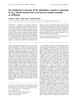
Fig. 1. CLSM images and 3D models presenting active osteoclasts grown on the surface of the curdlan gel: a – osteoclast multinucleation (sz – sealing zone); b –
actin belt.
4
A. Przekora et al.
Carbohydrate Polymers 295 (2022) 119914
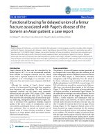
after osteoclast culture clearly showed the intact areas characterized by
smooth surface (similar to the surface of unseeded control sample
incubated in the culture medium) and rough topography that was
created by the osteoclast activity (Figs. 3 and 4a). Importantly, AFM
observation revealed that the activity of osteoclasts resulted in an
important roughening (Fig. 3b) of the surface, in contrast with the flat
and smooth of the control polymer surface (Fig. 3a). In fact, the average
roughness Ra increased from 1.47 nm in the control sample, to 20.50 nm
in the cell-treated curdlan due to the osteoclastic activity (measurements
parameters can be seen in Supplementary Material 2).
To confirm acidic hydrolysis of curdlan matrix, the degradation test
in the conditions mimicking that occurring in the resorption lacuna
during osteoclast-mediated degradation was performed using cell-free
system. After bone scaffold implantation, osteoclasts adhere to the
biomaterial surface and form a resorption lacuna. The pH within the
lacuna is lowered to about 4.0–4.5 by the release of protons (proton
pump and Na+–H+ exchanger) and chloride ions (chloride channels)
(Henriksen et al., 2008; Low & Kopeˇcek, 2012). Therefore, to simulate
acidic environment that is locally formed by osteoclasts, HCl degrada
tion solution with pH equal to 4.5 was prepared. Unseeded curdlan
samples were placed in the degradation solution and PBS (control) fol
lowed by incubation in the conditions mimicking physiological ones:
37 ◦ C, 5 % CO2, 95 % humidity, without agitation. SEM imaging per
formed after 14-day incubation showed similar results like for
osteoclast-seeded curdlan samples. HCl-treated sample was character
ized by rough surface, whereas control PBS-treated matrix exhibited
smooth and intact surface (Fig. 4b). Importantly, surface of both culture
medium- and PBS-treated control was smooth and similar to the surface
of untreated control sample (native curdlan gel, Fig. 4c), proving that
observed changes were related to acidic hydrolysis caused by either
osteoclasts (Fig. 4a) or HCl (Fig. 4b). Slight changes in topography of
control samples compared to native curdlan gel resulted from either
adsorption of the proteins (culture medium-treated sample) or salt
precipitation (PBS-treated sample).
Moreover, to quantitatively determine curdlan hydrolysis in the
environment mimicking resorption lacuna, the concentration of glucose
(degradation product) was assessed. The test clearly showed the increase
in glucose concentration in the degradation solution (HCl) with time
(Table 1). Curdlan gel incubated in PBS also released some glucose but
its level was constant through the full length of the experiment. In the
case of total carbohydrate assay, curdlan sample treated with HCl
released great amounts of sugars, whereas concentration of carbohy
drates in PBS was slightly higher than the concentration of glucose and it
was constant through the full length of the experiment. Thus, it was
after their implantation into the living organism. The main mechanism
responsible for gradual replacement of bone implant by newly formed
tissue is the resorption process that is mediated by osteoclasts. As os
teoclasts significantly lower the pH in the resorption lacuna, it may be
hypothesized that curdlan may be degraded after implantation by acidic
hydrolysis. According to the available literature osteoclast differentia
tion and bone-resorbing function highly depends on the substrate stiff
ness. Thus, it is not surprising that substrates having higher stiffness
(similar to the bone) promote osteoclast activity (Wang et al., 2022).
Since curdlan gel is characterized by high elasticity and low stiffness, the
primary aim of the study was to determine whether osteoclasts have the
ability to attach to the curdlan matrix and differentiate towards mature
bone-resorbing cells. CLSM observation revealed the presence of
multinucleated giant cells with the typical morphology of osteoclasts
(Fig. 1a). Multinucleation is a strong evidence of osteoclast maturation
(Kodama & Kaito, 2020). Moreover, both fluorescent staining of actin
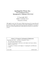
belt (Fig. 1b) and TRAP immunohistochemistry (Fig. 2) confirmed the
presence of mature TRAP-positive osteoclasts on the surface of curdlan
gel. It should be noted that TRAP, which is an enzyme having the ability
to degrade skeletal phosphoproteins (e.g. osteopontin), is considered to
be a histochemical marker of mature bone-resorbing cells (Hayman,
2008). Importantly, active osteoclasts form a resorption complex that is
made of an actin belt (or ring) that surrounds a so-called ruffled border
containing vacuolar H + -ATPase, which is responsible for lowering the
pH in the resorption lacuna. The actin belt is actually the area of tight
connection between the osteoclast plasma membrane and the bone
surface (Han et al., 2019). It was also proven that some focal adhesion
proteins, like vinculin and talin, participate in the formation of actin
belt. Lakkakorpi et al. demonstrated that actin belt in the active osteo
clasts is in fact formed by F-actin ring located between the double circle
of vinculin found in the periphery of the cell. They also proved that Factin/vinculin/talin zones correspond to the resorption lacuna edge and
are necessary for osteoclast attachment and bone-resorbing activity
(Lakkakorpi et al., 1989). Obtained CLSM images clearly showed the
presence of both F-actin and vinculin circles within the actin belt,
proving resorption activity of the osteoclasts grown on the curdlan gel
(Fig. 1b).
3.2. Determination of osteoclast-mediated curdlan hydrolysis
To prove the ability of osteoclasts to degrade thermally gelled cur
dlan, the samples upon osteoclast culture were subjected to osmotic lysis
to remove the cells followed by microscopy and spectroscopy analyses.
Microscopy observation with the use of AFM and SEM of curdlan surface
Fig. 2. Microscope images presenting mature TRAP-positive osteoclasts (brownish color) grown on the surface of curdlan gel (on the left – lower magnification image
showing curdlan gel covered by a number of active osteoclasts indicated by black arrows; on the right – higher magnification image presenting single TRAPpositive osteoclast).
5
A. Przekora et al.
Carbohydrate Polymers 295 (2022) 119914
Fig. 3. AFM images presenting surface of the curdlan samples: a – control of unseeded curdlan incubated in culture medium; b – curdlan after 14-day osteo
clast culture.
assumed that the presence of sugars in the PBS was not a result of cur
dlan degradation, but it was a contamination of the curdlan powder used
for the matrix production. It is worth noting that glucose and sucrose are
the main carbon sources used for microbial synthesis of curdlan (Aqui
nas et al., 2021). Greater amounts of total carbohydrates compared to
glucose in HCl degradation solution indicated either contamination of
the curdlan powder with sucrose or the presence of oligosaccharides due
to acidic hydrolysis of the sample. Substantial degradation of the cur
dlan during 14-day incubation in HCl solution was proven by significant
weight loss (by 58 ± 6 %) of the sample. Control PBS-treated sample
exhibited weight loss by only 11 ± 4 %.
Chemical changes on the surface of curdlan gel after osteoclast cul
ture and incubation in HCl degradation solution were analysed by
Raman spectroscopy. Initially, series of single spectra were collected
from rough and smooth areas after osteoclast culture as presented in
Fig. 4, alongside from HCl-treated and control samples. Recorded
spectra were averaged and normalized to 2905 cm− 1 band and pre
sented in Fig. 5a. Overlayed average Raman spectra of osteoclast- and
HCl-treated curdlan matrix may be seen in Supplementary Material 3.
Most distinguish shifts were identified in the range of 800–1500 cm− 1,
consisting of vibrations assigned to CC and CO stretching, vibrations of
C–O–C glycosidic bond, in plane ring deformation, OH and CH
bending and eventually CH2 in-plane bending in CH2OH group (Gieroba
et al., 2020). As mentioned above, hydrolysis of curdlan resulted in
higher concentration of glucose after breaking glycosidic bonds between
monomers (Prieto et al., 2011). Bands assigned to β-glycosidic bonds are
recognizable at 888 cm− 1 (HCC, HCO, CH deformational out-of-plane),
1093 with a shoulder band at 1148 cm− 1 (C–O–C stretching) and in
range 1200–1300 cm− 1. In Fig. 5b Raman shifts in samples exposed to
osteoclasts and HCl can be seen around 1460 and 1045 cm− 1 were
assigned to CH2 in-plane bending, CC, COH, CH deformation respec
tively. This suggests that during acidic hydrolysis the bands were broken
at random places in the polysaccharide chain, releasing maltodextrin
molecules, apart from glucose (de Veij et al., 2009). In both biological
(osteoclast-mediated) and chemical (cell-free) degradation tests, second
derivative revealed presence of the band at 1433 and 1448 cm− 1 that
can be assigned to rocking of CH and deformation of CH2 of carbohy
drate monomers (Wiercigroch et al., 2017). It was also noticed by shifts
around 1460 cm− 1 and appearing new bands at 1072 cm− 1 (osteoclasts)
or at 1085 and 1089 cm− 1 (HCl) derived from appearing mono
saccharides and disaccharides.
To properly describe the curdlan degradation process, bands at 888
and 2905 cm− 1 were chosen, assigned to β-glycosidic bond and CH2
stretching of aliphatic chain vibration, respectively. Raman intensity
ratio of these bands (Rh = I888:I2905) was calculated from spectra and
subjected to unpaired t-test, when statistical significance (p < 0.05) for
difference was confirmed. It was later implemented in analysis of Raman
maps, as a spatial visualization, presented in Fig. 6. To exclude the in
fluence of various manipulations during experiment, additional control
groups were analysed with Raman imaging. The culture medium-treated
and PBS-treated curdlan samples were controls for osteoclast-mediated
degradation test (Fig. 6a) and cell-free system experiment performed
in HCl (Fig. 6b), respectively. Raman image of untreated curdlan gel –
native sample (Fig. 6c) was considered as negative control revealing no
chemical changes. For visualization of microscopic image, β-glycosidic
bonds, CH2 of aliphatic chains content, Rh and carbohydrates were
chosen for HCl- and cell-treated curdlan samples. Carbohydrates were
presented with region I (200–800 cm− 1) (Wiercigroch et al., 2017), as
expected that hexoses and disaccharides composed of hexose were
present from the previous experiments data. In Fig. 6a, heatmap of Rh =
I888:I2905 revealed distinguishable area of lower ratio values, marked in
blue, assigned to rough surface areas (marked with letter ‘r’). The shape
appearing at the bottom of chemical map resulting from degradation of
curdlan matrix resembled the shape and size of osteoclasts shown in
Figs. 1 and 2. For better visualization of process semi-quantitative
evaluation of glucose concentration in resorption lacuna was per
formed. Similarly, to Rh the higher concentration of carbohydrates
resembled the lacuna edge, and varied in sample treated with cells,
comparing to HCl-treated one (Fig. 6b). In both control samples (culture
medium- and PBS-treated) no effects resembling those induced by os
teoclasts were observed. However, higher Raman intensity in carbohy
drates in PBS sample was observed due to overlapping bands of
phosphate buffer in ranges near ~206 cm− 1 and 462 cm− 1, which is
known phenomenon (Baranov et al., 2010).
The Raman map of HCl-treated curdlan matrix showed quite evenly
distributed carbohydrates and lack of outlining areas, as the whole
mapped sample was staying in contact with HCl, which also corresponds
with SEM images (Fig. 6b). Interestingly, comparison with the Raman
intensity of carbohydrates in maps obtained for osteoclast-treated
samples (Fig. 6a) and HCl-treated matrix (Fig. 6b) may suggest that
chemical hydrolysis of curdlan in the cell-free system was less efficient
than osteoclast-mediated process, but further, more quantitative ana
lyses are needed to confirm this assumption. Similarly to carbohydrates,
but in contrary to data shown in Fig. 6a, the distribution of Rh is even,
not showing any recognizable cell-shaped structures. It should be noted
that osteoclasts, similarly to immune cells, may generate ROS/RNS that
facilitate resorption of bone tissue during remodeling (Agidigbi & Kim,
2019). Thus, it may be assumed that more efficient curdlan hydrolysis
upon osteoclast culture compared to HCl-treated sample could have
resulted from enhanced degradation process due to ROS/RNS genera
tion by the cells.
3.3. Determination of ROS-mediated curdlan degradation
It is known that implanted biomaterials may exert inflammatory
response and activate immune cells to generate excessive amounts of
ROS/RNS. Consequently, prolonged inflammation may result in oxida
tive damage of the implant and its failure (Przekora, 2019). Within this
study, curdlan-induced ROS/RNS generation by immune cells (neutro
phils, monocytes, and macrophages) was determined. Neutrophils and
6
A. Przekora et al.
Carbohydrate Polymers 295 (2022) 119914
Fig. 4. SEM images presenting surface of the curdlan
samples: a – osteoclast-mediated degradation of cur
dlan sample (control – unseeded curdlan gel incu
bated in culture medium; osteoclasts – sample after
14-day culture of bone-resorbing cells: s – smooth
intact area; r – rough degraded area); b – cell-free
chemical degradation test in the environment
mimicking resorption lacuna (control – unseeded
sample incubated in PBS; HCl (pH = 4.5) – unseeded
sample incubated in HCl degradation solution); c –
untreated curdlan gel (native sample).
7
A. Przekora et al.
Carbohydrate Polymers 295 (2022) 119914
without curdlan matrix (Fig. 7a). It is not surprising since β-glucans
(including curdlan) are known to have immunomodulatory properties
with the ability to enhance activity of immune cells (Ali et al., 2015;
Kataoka et al., 2002; Ratitong et al., 2021). For instance, De Souza
Bonfim-Mendonca et al. demonstrated that β-glucan derived from
Laminaria digitata induced ROS production in human neutrophils (De
Souza Bonfim-Mendonỗa et al., 2014). Ulvestad et al. observed increased
production of superoxide and NO by macrophages stimulated with
˙
curdlan (Ulvestad et al., 2018). Similarly, Zelechowska
et al. found out
that curdlan not only acted as a chemoattractant for mast cells, but also
˙
stimulated those cells to produce elevated ROS (Zelechowska
et al.,
2020). Whereas Kataoka et al. detected increased expression of induc
ible nitric oxide synthase (iNOS – an enzyme producing NO) in curdlantreated mouse macrophages (Kataoka et al., 2002). Nevertheless, in our
studies thermally gelled curdlan was used – known to have reduced
immunomodulatory properties compared to curdlan solution (Kataoka
et al., 2002) – therefore production of ROS/RNS by neutrophils and
monocytes was only slightly promoted, however with statistical signif
icance. Macrophages cultured on the curdlan gel showed comparable
NO production and slightly lower generation of superoxide compared to
control macrophages incubated without curdlan sample.
Since it is known that osteoclasts release ROS/RNS enhancing bone
resorption (Agidigbi & Kim, 2019), production of superoxide and NO by
osteoclasts cultured on the curdlan gel was also determined. Osteoclasts
grown on the tested sample gave similar results to macrophages, i.e.
Table 1
Degradation of curdlan matrix in the environment mimicking resorption lacuna
determined by measurement of glucose and total carbohydrates concentration in
the degradation solution (HCl) and PBS (control).
Concentration of released glucose [μg/mL]
PBS
HCl
5 days
7 days
9 days
11 days
14 days
30.38 ±
7.71
59.95 ±
4.15a
34.32 ±
0.28
82.64 ±
12.58a
37.30 ±
4.04
80.64 ±
13.29a
38.09 ±
8.25
86.85 ±
4.07a
37.56 ±
8.13
94.41 ±
3.50a
Concentration of released total carbohydrates [μg/mL]
PBS
HCl
5 days
7 days
9 days
11 days
14 days
41.39 ±
0.23
71.74 ±
2.51a
42.67 ±
2.14
91.25 ±
2.40a
45.86 ± 1.13
45.65 ± 1.18
102.90 ±
3.99a
110.80 ±
0.90a
42.25 ±
2.17
117 ±
2.77a
a
Statistically significant results compared to the control sample incubated in
PBS (p < 0.05, unpaired t-test).
monocytes cultured on the curdlan sample produced significantly higher
amounts of ROS and RNS (on the basis of superoxide and NO generation,
respectively) in comparison with corresponding control cells cultured
Fig. 5. Raman spectra of curdlan matrix after osteoclast-mediated degradation (on the left) and cell-free chemical degradation in the environment mimicking
resorption lacuna (on the right): a – spectra in range 200–3000 cm− 1 normalized to 2905 cm− 1 band, b – range 800–1500 cm− 1 with ascribed maxima of bands, c –
second derivative spectra of ranges 1400–1500 cm− 1 and 1040–1140 cm− 1 with ascribed most distinguish differences between samples.
8
A. Przekora et al.
Carbohydrate Polymers 295 (2022) 119914
Fig. 6. Chemical Raman images of cur
dlan samples: a – osteoclast-mediated
degradation (control – unseeded curdlan
gel incubated in culture medium; osteo
clasts – sample after 14-day culture of
bone-resorbing cells: s – smooth intact
area; r – rough degraded area; white bars
represent 50 μm), b – cell-free chemical
degradation test in the environment
mimicking resorption lacuna (control –
unseeded sample incubated in PBS; HCl
(pH = 4.5) – unseeded sample incubated
in HCl degradation solution; white bars
represent 5 μm), c – untreated (native)
curdlan gel (white bars represent 50 μm).
9
A. Przekora et al.
Carbohydrate Polymers 295 (2022) 119914
Fig. 7. ROS-mediated degradation of curdlan gel: a – ROS/RNS generation by immune cells and osteoclasts (control – cells cultured without curdlan sample; curdlan
– cells cultured on the tested sample; * statistically significant results compared to corresponding control cells, p < 0.05, unpaired t-test); b – degradation of curdlan
matrix in H2O2 solutions determined by measurement of released total carbohydrates (PBS – control solution without H2O2); c – SEM images presenting surface of the
curdlan after incubation in H2O2 solutions (white bars represent 20 μm; surface of the control sample after incubation in PBS and untreated curdlan sample may be
seen in Fig. 4b and c, respectively).
they produced slightly lower amounts of ROS/RNS compared to the
control cells (Fig. 7a). Thus, thermally gelled curdlan did not have the
ability to enhance ROS/RNS generation by active osteoclasts.
Although differences in ROS/RNS production between cells cultured
on the curdlan gel and control cells were slight, even physiological level
of reactive oxygen species may contribute to curdlan degradation. To
check the ability of ROS to degrade thermally gelled curdlan, the sample
was exposed to hydrogen peroxide (H2O2). It should be noted that H2O2
was proven to be involved in the ROS-mediated degradation of betaăm, 2013). The experiment demon
glucans (Faure, Werder, & Nystro
strated that H2O2 did not participate in the degradation of thermally
gelled curdlan as tested sample did not release augmented levels of
carbohydrates (measured by Total Carbohydrate Assay kit) after incu
bation in H2O2 solutions compared to the control incubated in PBS
(Fig. 7b). Moreover, there were no differences in the amount of released
carbohydrates between low (1 μM) and high (1 mM) concentration of
H2O2 in the degradation solution. SEM imaging confirmed that exposure
of curdlan to H2O2 solutions did not lead to its degradation as no
changes in curdlan microstructure were observed (Fig. 7c). Thus, the
experiment clearly showed that H2O2 itself did not have the ability to
degrade curdlan gel. Therefore, immune cells most likely would not be
able to damage thermally gelled curdlan by ROS/RNS release upon
biomaterial implantation. However, combination of acidified microen
vironment in the resorption lacuna with ROS generated by osteoclasts
may potentially enhance chemical hydrolysis, which had the reflection
in Raman imaging that showed greater hydrolysis of curdlan after os
teoclasts culture compared to HCl-treated sample (Fig. 6a and b).
curdlan after its implantation into the bone. Within this research it was
clearly shown that osteoclasts may easily adhere to the surface of the
curdlan gel and acidify microenvironment leading to its degradation by
acidic hydrolysis. Osteoclast culture on the surface of curdlan gel
resulted in noticeably changed topography manifested by increased
roughness as demonstrated by SEM and AFM imaging. Moreover, cur
dlan degradation was proven by detection of chemical changes by
Raman spectroscopy. Both Raman spectra and chemical Raman images
obtained for osteoclast-treated samples clearly indicated acidic hydro
lysis of the curdlan. Moreover based on obtained results it may be
assumed that combination of ROS/RNS produced by osteoclasts with
acidified microenvironment in the resorption lacuna will most likely
boost curdlan degradation. Therefore presented studies for the first time
provide a strong evidence of osteoclast-mediated acidic hydrolysis of
thermally obtained curdlan gel, which is a very important issue taking
into account clinical applications of curdlan-based biomaterials. How
ever, it should be noted that many other cell types (including immune
cells such as macrophages, neutrophils, dendritic cells) are involved in
bone remodeling after biomaterial implantation. Thus, other factors like
oxidative stress caused by excessive ROS/RNS generation by immune
cells in response to biomaterial may also have impact on curdlan
degradation. Within this study it was proven that immune cells (but not
osteoclasts) produced slightly increased amounts of ROS/RNS in contact
with curdlan gel. However, even ROS/RNS production at physiological
level may potentially enhance curdlan degradation mediated by osteo
clasts. To reliably determine the effect of ROS/RNS release by immune
cells on osteoclast-mediated degradation of curdlan, more complex
cellular model is needed such as co-culture system. Nevertheless, both
macrophages and osteoclasts are derived from monocytes, thus it is huge
challenge to establish co-culture system in vitro to test osteoclastmediated degradation of curdlan in more complex microenvironment.
Supplementary data to this article can be found online at https://doi.
org/10.1016/j.carbpol.2022.119914.
4. Conclusions
Curdlan is the component of many recently developed bone im
plants, including commercial ones (FlexiOss®, Medical Inventi, Poland).
Since human organism does not produce enzymes having the ability to
degrade curdlan, it is not clear what is the fate of thermally gelled
10
A. Przekora et al.
Carbohydrate Polymers 295 (2022) 119914
CRediT authorship contribution statement
oxygen species production in human neutrophils to improve the killing of Candida
albicans and Candida glabrata isolates from vulvovaginal candidiasis. PLoS ONE, 9
(9), Article 9. />Diez-Escudero, A., Espanol, M., Montufar, E. B., Di Pompo, G., Ciapetti, G., Baldini, N., &
Ginebra, M. P. (2017). Focus ion beam/scanning electron microscopy
characterization of osteoclastic resorption of calcium phosphate substrates. Tissue
Engineering - Part C: Methods, 23(2), 118–124. />tec.2016.0361
Diez-Escudero, A., Torreggiani, E., Di Pompo, G., Espanol, M., Persson, C., Ciapetti, G.
Ginebra, M. P., … (2019). Effect of calcium phosphate heparinization on the in vitro
inflammatory response and osteoclastogenesis of human blood precursor cells.
Journal of Tissue Engineering and Regenerative Medicine, 13(7), 1217–1229. https://
doi.org/10.1002/term.2872
Everts, V., Korper, W., Hoeben, K. A., Jansen, I. D. C., Bromme, D., Cleutjens, K. B. J. M.
Beertsen, W., … (2006). Osteoclastic bone degradation and the role of different
cysteine proteinases and matrix metalloproteinases: Differences between calvaria
and long bone. Journal of Bone and Mineral Research, 21(9), 1399–1408. https://doi.
org/10.1359/jbmr.060614
Faure, A. M., Werder, J., & Nystră
om, L. (2013). Reactive oxygen species responsible for
beta-glucan degradation. Food Chemistry, 141, 589–596. />foodchem.2013.02.096
Gidley, M., & Nishinari, K. (2009). Physico-chemistry of (1,3)-β-glucans. In A. Bacic,
G. Fincher, & B. Stone (Eds.), Chemistry, biochemistry and biology of (1,3)-β-glucans
and related polysaccharides (pp. 47–118). New York: Academic Press (Elsevier).
/>Gieroba, B., Sroka-Bartnicka, A., Kazimierczak, P., Kalisz, G., Pieta, I. S.,
Nowakowski, R., & Przekora, A. (2020). Effect of gelation temperature on the
molecular structure and physicochemical properties of the curdlan matrix:
Spectroscopic and microscopic analyses. International Journal of Molecular Sciences,
21(17), 6154. />Han, G., Zuo, J., & Holliday, L. S. (2019). Specialized roles for actin in osteoclasts:
Unanswered questions and therapeutic opportunities. Biomolecules, 9(17). https://
doi.org/10.3390/biom9010017
Hayman, A. (2008). Tartrate-resistant acid phosphatase (TRAP) and the osteoclast/
immune cell dichotomy. Autoimmunity, 41(3), 218–223. />08916930701694667
Henriksen, K., Sørensen, M. G., Jensen, V. K., Dziegiel, M. H., Nosjean, O., &
Karsdal, M. A. (2008). Ion transporters involved in acidification of the resorption
lacuna in osteoclasts. Calcified Tissue International, 83(3), 230–242. />10.1007/s00223-008-9168-8
Kataoka, K., Muta, T., Yamazaki, S., & Takeshige, K. (2002). Activation of macrophages
by linear (1→3)-β-D-glucans. Implications for the recognition of fungi by innate
immunity. Journal of Biological Chemistry, 277(39), 36825–36831. />10.1074/jbc.M206756200
Klimek, K., Przekora, A., Benko, A., Niemiec, W., Blazewicz, M., & Ginalska, G. (2017).
The use of calcium ions instead of heat treatment for β-1,3-glucan gelation improves
biocompatibility of the β-1,3-glucan/HA bone scaffold. Carbohydrate Polymers, 164.
/>Kodama, J., & Kaito, T. (2020). Osteoclast multinucleation: Review of current literature.
International Journal of Molecular Sciences, 21(16), 5685. />ijms21165685
ănă
Lakkakorpi, P., Tuukkanen, J., Hentunen, T., Jă
arvelin, K., & Vă
aa
anen, K. (1989).
Organization of osteoclast microfilaments during the attachment to bone surface in
vitro. Journal of Bone and Mineral Research, 4(6), 817–825. />jbmr.5650040605
Laxmi, V. M., Latha, D., & Jayasree, A. S. (2018). Production and characterization of
curdlan from Agrobacterium sp. International Journal of Pharmaceutical Sciences and
Research, 9(11), 4871–4874. />(11).4871-74
Leong, K. F., Chua, C. K., Sudarmadji, N., & Yeong, W. Y. (2008). Engineering
functionally graded tissue engineering scaffolds. Journal of the Mechanical Behavior of
Biomedical Materials, 1(2), 140–152. />Low, S. A., & Kopeˇcek, J. (2012). Targeting polymer therapeutics to bone. Advanced Drug
Delivery Reviews, 64(12), 1189–1204. />Mangolim, C. S., Da Silva, T. T., Fenelon, V. C., Koga, L. N., De Ferreira, S. B. S.,
Bruschi, M. L., & Matioli, G. (2017). Description of recovery method used for curdlan
produced by Agrobacterium sp. IFO 13140 and its relation to the morphology and
physicochemical and technological properties of the polysaccharide. PLoS ONE, 12
(2), Article e0171469. />Martinez, C. O., Ruiz, S. P., Nogueira, M. T., Bona, E., Portilho, M., & Matioli, G. (2015).
Effective immobilization of Agrobacterium sp. IFO 13140 cells in loofa sponge for
curdlan biosynthesis. Molecules, 20(5), 7957–7973. />molecules20057957
Matsuzaki, K., Katayama, K., Takahashi, Y., Nakamura, I., Udagawa, N., Tsurukai, T.
Suda, T., … (1999). Human osteoclast-like cells are formed from peripheral blood
mononuclear cells in a coculture with SaOS-2 cells transfected with the parathyroid
hormone (PTH)/PTH-related protein receptor gene. Endocrinology, 140(2), 925–932.
/>Michalicha, A., Roguska, A., Przekora, A., Budzy´
nska, B., & Belcarz, A. (2021). Poly
(levodopa)-modified β-glucan as a candidate for wound dressings. Carbohydrate
Polymers, 272, Article 118485. />Prieto, M. A., V´
azquez, J. A., & Murado, M. A. (2011). Hydrolysis optimization of
mannan, curdlan and cell walls from endomyces fibuliger grown in mussel
processing wastewaters. Process Biochemistry, 46(8), 1579–1588. />10.1016/j.procbio.2011.04.014
The manuscript was written through contributions of all authors. All
authors have given approval to the final version of the manuscript.
AP: Conceptualization, Methodology, Investigation, Data analysis,
Resources, Project administration, Supervision, Writing – original draft;
LP: Investigation, Methodology, Data visualization, Data analysis,
Writing – review & editing; GK: Investigation, Methodology, Data
visualization, Data analysis, Writing – original draft; PK: Investigation,
Methodology, Data visualization, Data analysis, Writing – review &
editing; CC: Investigation, Data visualization, Writing – review & edit
ing; MW: Investigation, Data visualization; RP: Resources, Supervision,
Writing – review & editing; ASB: Resources, Supervision, Writing – re
view & editing.
Data availability
The raw/processed data required to reproduce these findings can be
obtained from the corresponding author ()
upon reasonable request.
Declaration of competing interest
The authors declare that they have no known competing financial
interests or personal relationships that could have appeared to influence
the work reported in this paper.
Data availability
Data will be made available on request.
Acknowledgment
The research was funded by the Ministry of Education and Science in
Poland within statutory activity of Medical University of Lublin (DS3/
2022 project). Paulina Kazimierczak has received annual support
(scholarship – START) from the Foundation for Polish Science (FNP) in
2021 for the most talented young scientists. Authors acknowledge the
support of Trifon Trifonov in performing the AFM measurements. Cris
tina Canal acknowledges MINECO for PID2019-103892RB-I00 project,
and Generalitat de Catalunya for SGR2017-1165 and the ICREA
Academia Award for Excellence in Research.
References
Agidigbi, T. S., & Kim, C. (2019). Reactive oxygen species in osteoclast differentiation
and possible pharmaceutical targets of ROS-mediated osteoclast diseases.
International Journal of Molecular Sciences, 20(14), 3576. />ijms20143576
Ali, M. F., Driscoll, C. B., Walters, P. R., Limper, A. H., & Carmona, E. M. (2015). Betaglucan-activated human B lymphocytes participate in innate immune responses by
releasing proinflammatory cytokines and stimulating neutrophil chemotaxis. The
Journal of Immunology, 195(11), 5318–5326. />jimmunol.1500559
Aquinas, N., Selvaraj, S., & Bhat, M. R. (2021). A review presenting production,
characterization, and applications of biopolymer curdlan in food and pharmaceutical
sectors. Polymer Bulletin. />Baranov, A. V., Bogdanov, K. V., Ushakova, E. V., Cherevkov, S. A., Fedorov, A. V., &
Tscharntke, S. (2010). Comparative analysis of Raman spectra of PbS macro- and
nanocrystals. Optics and Spectroscopy, 109(2), 268–271. />s0030400x10080199
Borkowski, L., Przekora, A., Belcarz, A., Palka, K., Jojczuk, M., Lukasiewicz, P.
Ginalska, G., … (2021). Highly porous fluorapatite/β-1,3-glucan composite for bone
tissue regeneration: Characterization and in vitro assessment of biomedical
potential. International Journal of Molecular Sciences, 22(19), 10414. />10.3390/ijms221910414
Chaudhari, V., Buttar, H. S., Bagwe-Parab, S., Tuli, H. S., Vora, A., & Kaur, G. (2021).
Therapeutic and industrial applications of curdlan with overview on its recent
patents. Frontiers in Nutrition, 28(8), Article 646988. />fnut.2021.646988
De Souza Bonfim-Mendonỗa, P., Ratti, B. A., Da Silva Ribeiro Godoy, J., Negri, M., De
Lima, N. C. A., Fiorini, A., & Svidzinski, T. I. E. (2014). β-Glucan induces reactive
11
A. Przekora et al.
Carbohydrate Polymers 295 (2022) 119914
Przekora, A. (2019). The summary of the most important cell-biomaterial interactions
that need to be considered during in vitro biocompatibility testing of bone scaffolds
for tissue engineering applications. Materials Science and Engineering C, 97. https://
doi.org/10.1016/j.msec.2019.01.061
Przekora, A., & Ginalska, G. (2014). Addition of 1,3-β-d-glucan to chitosan-based
composites enhances osteoblast adhesion, growth, and proliferation. International
Journal of Biological Macromolecules, 70, 474–481. />ijbiomac.2014.07.035
Przekora, A., & Ginalska, G. (2016). In vitro evaluation of the risk of inflammatory
response after chitosan/HA and chitosan/β-1,3-glucan/HA bone scaffold
implantation. Materials Science and Engineering C, 61, 355–361. />10.1016/j.msec.2015.12.066
Przekora, A., Vandrovcova, M., Travnickova, M., Pajorova, J., Molitor, M., Ginalska, G.,
& Bacakova, L. (2017). Evaluation of the potential of chitosan/β-1,3-glucan/
hydroxyapatite material as a scaffold for living bone graft production in vitro by
comparison of ADSC and BMDSC behaviour on its surface. Biomedical Materials
(Bristol), 12(1). />Ratitong, B., Marshall, M., & Pearlman, E. (2021). β-Glucan-stimulated neutrophil
secretion of IL-1α is independent of GSDMD and mediated through extracellular
vesicles. Cell Reports, 35(7), Article 109139. />celrep.2021.109139
Sun, Y., Liu, Y., Li, Y., Lv, M., Li, P., Xu, H., & Wang, L. (2011). Preparation and
characterization of novel curdlan/chitosan blending membranes for antibacterial
applications. Carbohydrate Polymers, 84(3), 952–959. />carbpol.2010.12.055
Tukulula, M., Hayeshi, R., Fonteh, P., Meyer, D., Ndamase, A., Madziva, M. T.Dube, A.,
… (2015). Curdlan-conjugated PLGA nanoparticles possess macrophage stimulant
activity and drug delivery capabilities. Pharmaceutical Research, 32(8), 2713–2726.
/>Ulvestad, J. S., Kumari, J., Seternes, T., Chi, H., & Dalmo, R. A. (2018). Studies on the
effects of LPS, ß-glucan and metabolic inhibitors on the respiratory burst and gene
expression in Atlantic salmon macrophages. Journal of Fish Diseases, 41(7),
1117–1127. />de Veij, M., Vandenabeele, P., De Beer, T., Remon, J. P., & Moens, L. (2009). Reference
database of Raman spectra of pharmaceutical excipients. Journal of Raman
Spectroscopy, 40(3), 297–307. />
Vivcharenko, V., Wojcik, M., & Przekora, A. (2020). Cellular response to vitamin Cenriched chitosan/agarose film with potential application as artificial skin substitute
for chronic wound treatment. Cells, 9(5). />Wang, Q., Xie, J., Zhou, C., & Lai, W. (2022). Substrate stiffness regulates the
differentiation profile and functions of osteoclasts via cytoskeletal arrangement. Cell
Proliferation, 55, Article e13172. />˙ nska, B., Tarczy´
Wessely-Szponder, J., Michalska, J., Szponder, T., Zyli´
nska, M., &
Szubstarski, M. (2020). The role of antimicrobial neutrophil extract in modification
of the inflammatory response during osteochondral autograft and allograft
transplantation in rabbits. Journal of Comparative Pathology, 175, 49–63. https://doi.
org/10.1016/j.jcpa.2019.12.007
Wiercigroch, E., Szafraniec, E., Czamara, K., Pacia, M. Z., Majzner, K., Kochan, K.
Malek, K., … (2017). Raman and infrared spectroscopy of carbohydrates: A review.
Spectrochimica ActaPart A, Molecular and Biomolecular Spectroscopy, 185, 317–335.
/>Wojcik, M., Kazimierczak, P., Benko, A., Palka, K., Vivcharenko, V., & Przekora, A.
(2021). Superabsorbent curdlan-based foam dressings with typical hydrocolloids
properties for highly exuding wound management. Materials Science and Engineering
C, 124, Article 112068. />Wojcik, M., Kazimierczak, P., Vivcharenko, V., Koziol, M., & Przekora, A. (2021). Effect
of vitamin c/hydrocortisone immobilization within curdlan-based wound dressings
on in vitro cellular response in context of the management of chronic and burn
wounds. International Journal of Molecular Sciences, 22(21), 11474. />10.3390/ijms222111474
Yu, X., Zhang, C., Yang, L., Zhao, L., Lin, C., Liu, Z., & Mao, Z. (2015). CrdR function in a
curdlan-producing Agrobacterium sp. ATCC31749 strain. BMC Microbiology, 15(1),
25. />Zdziennicka, J., Szponder, T., & Wessely-Szponder, J. (2021). Application of natural
neutrophil products for stimulation of monocyte-derived macrophages obtained
before and after osteochondral or bone injury. Microorganisms, 9(1), 124. https://
doi.org/10.3390/microorganisms9010124
˙
Zelechowska,
P., R´
oz˙ alska, S., Wiktorska, M., Brzezi´
nska-Błaszczyk, E., & Agier, J.
(2020). Curdlan stimulates tissue mast cells to synthesize pro-inflammatory
mediators, generate ROS, and migrate via Dectin-1 receptor. Cellular Immunology,
351, Article 104079. />Zhang, R., & Edgar, K. J. (2014). Properties, chemistry, and applications of the bioactive
polysaccharide curdlan. Biomacromolecules, 15(4), 1079–1096. />10.1021/bm500038g
12