Chitosan-sheath and chitin-core nanowhiskers
Bạn đang xem bản rút gọn của tài liệu. Xem và tải ngay bản đầy đủ của tài liệu tại đây (3.35 MB, 9 trang )
Carbohydrate Polymers 107 (2014) 158–166
Contents lists available at ScienceDirect
Carbohydrate Polymers
journal homepage: www.elsevier.com/locate/carbpol
Chitosan-sheath and chitin-core nanowhiskers
Antonio G.B. Pereira a,b , Edvani C. Muniz a , You-Lo Hsieh b,∗
a
Grupo de Materiais Poliméricos e Compósitos (GMPC) - Department of Chemistry, Maringá State University, Av. Colombo, 5790, Maringá, Paraná
87020-900, Brazil
b
Fiber and Polymer Science, University of California, Davis, One Shields Avenue, Davis, California 95616, USA
a r t i c l e
i n f o
Article history:
Received 3 August 2013
Received in revised form 12 February 2014
Accepted 14 February 2014
Available online 25 February 2014
Keywords:
Nanowhiskers
Chitin
Chitosan
Deacetylation
Surface modification
a b s t r a c t
Chitosan-sheath and ␣-chitin-core nanowhiskers (CsNWs) have been successfully generated by surface
deacetylation of chitin nanowhiskers (CtNWs) in the never-dried state. Acid hydrolysis (3N HCl, 30 mL/g,
104 ◦ C) of pure chitin derived from crab shell yielded 65% 4–10 nm thick, 16 nm wide and 214 nm long
chitin whiskers (CtNWs) that were 86% crystalline and 81% acetylated. Surface deacetylation of CtNWs
was robust in their never-dried state in 50% NaOH at a moderate 50 ◦ C for 6 h, yielding 92% CsNWs. All
deacetylated CsNWs retain the same ␣-chitin crystalline core at reduced 50% crystallinity and similar
dimensions (4–12 nm thick, 15 nm wide, 247 long) as CtNWs, but reduced 60% acetylation reflecting the
deacetylated surface layers. Progressive surface deacetylation was evident by the increased IP as well as
increased positive charges under acidic pH and reduced negative charges at alkaline pH with increasing
reaction time.
© 2014 Elsevier Ltd. All rights reserved.
1. Introduction
Chitin, a (1 −→ 4)-linked N-acetyl--d-glucosamine polysaccharide (Percot et al., 2002), is a structural component in the
exoskeletons or cell walls (Tharanathan & Kittur, 2003) of arthropods, nematodes and fungi (Rinaudo, 2006). Chitin is highly
crystalline and has been shown to exist in three polymorphic forms:
˛ (the most common with antiparallel chain alignment), ˇ (parallel
chain alignment) and (the least common of two parallel alternate
with one antiparallel chain) (Merzendorfer, 2006). In the native
form, chitin chains assemble spontaneously into 2–5 nm wide and
300 nm long nanofibrils, each comprising ca. 25 molecules, which
are organized into larger 50–300 nm wide fibrils, with some variations associated to the sources (Raabe et al., 2005). Acid hydrolysis
has yielded crystalline chitin nanorods, commonly referred as
nanowhiskers, by differential removal of amorphous domains of
chitin (Marchessault et al., 1959; Paillet & Dufresne, 2001), that
are a few tens of nanometer in diameters and 150 nm up to 2 m
in lengths, depending on sources and hydrolysis conditions (Zeng
et al., 2011).
Chitin nanowhiskers (CtNWs) have been used to improve properties of other polymers. Incorporating 30 wt% of CtNWs in gel
∗ Corresponding author. Tel.: +1 5307520843; fax: +1 5307527584.
E-mail address: (Y.-L. Hsieh).
/>0144-8617/© 2014 Elsevier Ltd. All rights reserved.
spun polyvinyl alcohol (PVA) fibers improved their Young’s modulus from 28 GPa to 50 GPa (Uddin et al., 2012). Coating a very
thin 0.1 m layer of CtNWs to 25 m thick poly(lactic acid) (PLA)
film could substantially decrease its O2 permeability by 180 times
(Fan et al., 2012). Adding 50 wt% CtNWs in sodium alginate (SA)
microspheres increased their swelling in pH 7.4 phosphate buffer
from 1815% to 2329% (Lin et al., 2011). CtNWs have also been
surface modified by reacting the hydroxyl groups with phenyl
isocyanate, alkenyl succinic anhydride and 3-isopropenyl-␣,␣ dimethylbenzyl isocyanate (Gopalan Nair et al., 2003), but these
modified surfaces did not enhance the mechanical properties of
CtNWs-rubber composites for which they were intended.
Chitin can be easily deacetylated into chitosan whose primary
amine groups exhibit cationic polyelectrolyte behavior in acidic
aqueous medium as well as mucoadhesivety, antibacterial and antifungal biological activities, desirable for many applications (Dash
et al., 2011; Rabea et al., 2003; Rinaudo, 2006; Shukla et al., 2013).
Efforts to deacetylate CtNWs to date have shown to destroy the
original crystalline structure or the nanowhisker form. Deacetylating CtNWs in 40 wt% NaOH solution under different lengths of
time (14–28 h) and temperatures (100–180 ◦ C) increase the degree
of deacetylation (DD) with increasing reaction times and temperatures, reaching a maximum of 98% DD after 14 h at 180 ◦ C or 18 h at
150 ◦ C (Phongying, Aiba, & Chirachanchai, 2006, 2007). Under these
conditions, the crystalline CtNWs were turned into amorphous
fibrillar porous structure, indicating deacetylation of the CtNWs
bulk. Microwave-assisted alkaline hydrolysis of CtNWs has shown
A.G.B. Pereira et al. / Carbohydrate Polymers 107 (2014) 158–166
to reach 80% and 95% deacetylation in 40 and 60 wt% NaOH, respectively, after for 6 h (Lertwattanaseri et al., 2009), also loosing the
nanowhisker morphology. It is clear that these deacetylation conditions are too harsh and have deacetylated their bulk, destroying
the desirable crystalline structure.
This study was aimed to produce isolated chitosan
nanowhiskers (CsNWs) by deacetylating only the surfaces of
CtNWs while retaining the crystalline core. Pure chitin was to
be isolated from crab shell chitin and hydrolyzed to CtNWs by
HCl. Deacetylation of CtNWs by NaOH was systematically studied under different conditions of temperatures and/or reaction
times to hydrolyze the nanowhisker surfaces while preserving
the crystalline core. Controlling reaction temperature and time
was deemed paramount to reach the balance between effective
deacetylation on the surfaces while retaining the highly crystalline
bulk. The chitosan amine functional surfaces and crystalline chitin
core of CsNWs present attractive surface-core combination of
nanowhiskers for potentially improved applications as reinforcement in polymeric nanocomposites and for formation of aerogels,
liquid crystal system, among others.
2. Materials and methods
2.1. Materials
Chitin from crab shells (CAS 1398-61-4) was purchased from
Sigma (St Louis, MO, USA). Potassium hydroxide (85%, EM Science,
CAS 1310-58-3), sodium hydroxide (97%, EMD, CAS 1310-73-2),
hydrochloric acid (36.5%, EMD, CAS 7647-01-0), pH 4 acetate buffer
(15% sodium acetate and 48% acetic acid, VWR), sodium chlorite (80%, Alfa Aesar, CAS 7758-19-2) were used without further
purification. Purified water (Millipore-Milli-QTM Water System)
was used in all experiments. All aqueous solutions were prepared
in % unless specified.
2.1.1. Chitin purification and whiskers
Crab shell chitin was purified by removing residual proteins and
color impurities following a previous procedure (Paillet & Dufresne,
2001). Proteins were removed by stirring 5 g of chitin in 150 mL of
5% KOH aqueous solution at boil for 6 h, then at room temperature
for another 12 h, filtered and washed with water. The solid was
bleached in 150 mL of 1.7% NaClO2 in pH 4 acetate buffer at 80 ◦ C
for 2 h, filtered, then bleached again under the same condition for
another 2 h, filtered and washed with water. The solid material was
re-suspended in 150 mL of 5% KOH for 48 h and centrifuged. The
purified chitin was washed, then oven dried (50 ◦ C) and weighed
to determine yield.
Chitin whiskers (CtNWs) were prepared by hydrolyzing the
purified chitin with 3N HCl at boil under stirring for 90 min. The
ratio HCl solution/chitin was ca. 30 mL/1 g. The suspension was
diluted by adding 50 mL of water and centrifuged at 3400 rpm
for 15 min. The precipitate was recovered and re-suspended in
200 mL of water and centrifuged. This process was performed three
times. The precipitate was recovered and re-suspended in water
and dialyzed until neutral pH. The suspension was adjusted to pH
3 with 1N HCl and sonicated (Misonix, ultrasonic liquid processors,
model CL5, USA) for 5 min four times with 5 min pause between
sonications followed by centrifugation at 3000 rpm for 10 min for
removing any remaining precipitate. The suspension was either
kept at 8 ◦ C or freeze-dried (Labconco, USA) at –50 ◦ C for 2 days
when necessary. The yield of chitin whiskers was determined gravimetrically.
2.1.2. Chitosan whiskers
Chitosan whiskers (CsNWs) were obtained by the deacetylation
of chitin nanocrystals with 50% NaOH aqueous solution at varying
159
Table 1
Deacetylation of CtNWs with 50% NaOH under different conditions.
Condition
Chitin/
Time
NaOH
(h)
(g/100 mL)
Temperature
(◦ C)
Yield
(%)
Chitin whiskers
condition
1
1.8
(0.5%
NaBH4 )
0.414
0.5
120
89
Freeze-dried
0.5
120
61
Suspension,
never-drieda
Suspension,
never-drieda
2
6
3
0.414
24
48
a
92
50
94
74
1.38 wt-%.
temperatures for different lengths of time (Table 1). Following
deacetylation, the suspension was diluted with water and centrifuged at 5000 rpm for 10 min, then the supernatant decanted.
This process was repeated three times. The samples were dialyzed
in water to reach neutral pH, adjusted to pH 3 by 1N HCl, then
homogenized by sonication.
2.2. Characterization
Characterization of CtNWs was performed on never-dried
suspension, freeze-dried or air-dried samples. Zeta potential
measurements were performed on suspensions of never-dried
nanowhiskers. FTIR, XRD, SEM, TGA and DSC were performed
on freeze-dried samples whereas suspensions were air-dried on
appropriate surfaces for AFM and TEM analysis. Freeze drying of
CtNWs or CsNWs suspensions was performed by freezing in liquid nitrogen (ca. –196 ◦ C) and freeze-dried for 3 days (−48 ◦ C and
0.12 mbar, Labconco Free zone dry system, USA).
2.2.1. Zeta potential
Zeta potential measurements were performed on a ZetaSizer
(nano ZS90, Malvern) coupled to a autotitrator device (MPT-2). Both
CtNW and CsNW suspensions were diluted to 0.1% and the pH was
adjusted to ca. 1 by 1N HCl. Zeta potential was measured over the
pH range from 1 to 12 using 0.5N NaOH as the titrant.
2.2.2. Fourier transform infra-red spectroscopy (FTIR)
The FTIR spectra of freeze-dried samples pressed with KBr were
collected over 64 scans in a Nicolet 6700 FTIR spectrophotometer (Thermo Electron Corporation) operated in the 4000–400 cm−1
region at 4 cm−1 resolution.
2.2.3. X-ray diffraction (XRD)
XRD spectra were collected on a Scintag XDS 2000 powder
diffractometer using a Ni-filtered Cu K␣ radiation ( = 1.5406 Å) at
an 45 kV anode voltage and a 40 mA current. Freeze-dried samples
were pressed between two glass slides into 1-mm thick flat sheets.
XRD patterns were recorded from 2Â = 5◦ to 2Â = 40◦ at a scan rate of
2◦ /min. The XRD data were smoothed using 10 points in a secondorder regression based on Savitzky–Golay filter. The crystallinity
Index (CrI) was calculated as:
CrI =
Ac
× 100
AT
(1)
where, Ac is the total crystalline diffraction peak area, and AT is the
amorphous area under the curve 2Â = 5◦ to 30◦ . The XRD patterns
were then deconvoluted based on Gaussian or Lorentzian functions in the Origin software release 8.5 to resolve the individual
160
A.G.B. Pereira et al. / Carbohydrate Polymers 107 (2014) 158–166
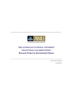
Fig. 1. Chitin purification and HCl hydrolysis: (a) crude (left) and purified (right) chitin; (b) 0.2% (left) and 0.7% (right) CtNWs suspensions (pH 3); (c) FTIR of freeze-dried
CtNWs.
peaks. The crystallite size was estimated using the Scherrer’s Eq.
(2) (Goodrich & Winter, 2006):
Dhkl = 0.9 ˇ1/2 cos Â
(2)
where, Dhkl is the crystal dimension perpendicular to the diffraction
plane with hkl Miller indices, is the X-ray radiation wavenumber
( = 0.154 nm), and ˇ1/2 is the full width at half maximum (FWHM)
of diffraction peak (Fan et al., 2012).
2.2.4. Scanning electron microscopy (SEM)
Freeze-dried samples were sputter gold coated and imaged
using a field emission scanning electron microscope (FE-SEM) (XL
30-SFEG, FEI/Philips, USA) at a 5 mm working distance and a 5-kV
accelerating voltage.
2.2.5. Thermogravimetric analysis (TGA) and differential
scanning calorimetry (DSC)
DSC and TGA of freeze-dried CtNWs and deacetylated CtNWs
(method 3) were performed from 30 ◦ C to 550 ◦ C at a 10 ◦ C/min
heating rate under flowing N2 (30 mL/min for DSC and 50 mL/min
for TGA) in a DSC-60 differential scanning calorimeter and TGA-50
thermogravimetry analyzer (Shimadzu), respectively.
2.2.6. Atomic force microscopy (AFM)
10 L of 0.001% CtNWs or CsNWs suspension pH 3 was
deposited into a freshly clipped mica substrate and air dried before
image acquisition using an Asylum Research Atomic Force Microscope.
2.2.7. Transmission electron microscopy (TEM)
CtNW and deacetylated CtNW samples were prepared
by depositing 8 L 0.01% suspensions onto glow-discharged
A.G.B. Pereira et al. / Carbohydrate Polymers 107 (2014) 158–166
161
Fig. 2. CtNWs: (a) TEM; (b) width and length distribution profile; (c) AFM height image (d); AFM height profile for line shown in c. (e) and (f) SEM of freeze-dried sample.
carbon-coated TEM grids (300-mesh copper, formvar-carbon, Ted
Pella Inc., Redding, CA), and any excess liquid was removed by
blotting with a filter paper. The specimens were then negatively
stained with 2% uranyl acetate solution for 5 min, blotted with
a filter paper to remove excess staining solution and allowed to
dry under the ambient condition. The samples were observed
using a Philips CM12 transmission electron microscope operating at 100 kV accelerating voltage. The widths and lengths of
nanocrystals were collected and the averages were calculated from
measurements over 200 samples using analySIS FIVE software.
162
A.G.B. Pereira et al. / Carbohydrate Polymers 107 (2014) 158–166
3. Results and discussion
Pure chitin was obtained by removing proteins (5% KOH, at boil
for 2 h, then ambient 12 h) and the yellow-brownish color (1.7%
NaClO2 , pH 4, 80 ◦ C, 4 h) from crude crab shell chitin to white powders at a 75% yield (Fig. 1a). Acid hydrolysis (3N HCl, 30 mL/g, at
boil for 90 min) of pure chitin yielded 65% chitin nanowhiskers
(CtNWs), or 48.8% of the original crude crab shell chitin. Hydrochloric acid accesses the amorphous regions to cleave chitin chains
to soluble fragments to isolate the crystalline domains. The 35%
mass loss from acid hydrolysis was slightly less than the 40% mass
loss reported under a similar condition but 30 min longer time (3N
HCl, 20 mL/g, 104 ◦ C, 2 h) (Revol & Marchessault, 1993). Aqueous
CtNWs suspensions at either 0.2 or 0.7% appeared slightly translucent (Fig. 1b), showing the hydroxyl and acetylamide groups on
the whisker surfaces to be sufficiently polar to keep the whiskers
separated and afloat. This appearance is similar to chitin suspensions at same pH (ca. 3) reported by others (Fan et al., 2010). FTIR
of CtNWs exhibited absorption peaks characteristic of chitin, i.e.,
O H stretching at 3450 cm−1 , N H stretching at 3264 cm−1 and
3105 cm−1 , C H vibrations in the 2900–2800 cm−1 range, amide I
C O at 1655 cm−1 , amide II (a combination of C N H stretching
and N H bending) at 1560 cm−1 and C N stretching at 1166 cm−1
(Fig. 1c). The shoulder at 1655 cm−1 is due to intermolecular
hydrogen bondings between acetylamide groups (C O· · ·H N) and
intramolecular hydrogen bonding between acetylamide C O and
C6 hydroxyl OH, reported to be associated only with ␣-chitin (Lavall
et al., 2007; Rinaudo, 2006), indicating CtNWs to be ␣-chitin. The
sharper peaks in the CtNWs spectrum than those of crude chitin are
attributed to its higher chitin purity and crystallinity. These FTIR
data confirm that the acid hydrolysis did not alter the chemical
nature nor ␣-chitin form of CtNWs from crude chitin, as expected.
TEM images of CtNWs show their widths to range from 5 nm
to 30 nm with an 16 nm average and their lengths from 100 nm to
350 nm with an 214 nm average, or a 13 length-to-width aspect
ratio (Fig. 2a,b). The AFM height profile shows CtNWs to vary from
4 nm to 10 nm, similar to the lower range for widths shown in
TEM (Fig. 2d). The AFM image also showed large size variations
among CtNWs (Fig. 2c). However, lateral dimensions observed in
AFM are not as accurate as those derived from TEM due to possible
tip broadening effect.
The SEM images (Fig. 2e,f) of rapidly frozen and freeze-dried
CtNWs show them to self-assemble into tens of micron thick
sheet-like layers with much smaller sub-micron fibrillated network
structures in between. Deacetylation conducted on freeze-dried
CtNWs (50% NaOH, 120 ◦ C, 0.5 h, condition 1 in Table 1) produced
clusters of larger particles rather than individual whiskers even at
an elevated temperature. In light of losing individual nanowhisker
form to this bulky assembled structure from freeze drying, deacetylation was performed on never-dried CtNW sample, shown as
deacetylation condition 2.
Indeed deacetylation (50% NaOH, 120 ◦ C, 0.5 h) of CtNWs in the
never-dried state, i.e., condition 2, produced individually isolated
rod-shape nanowhiskers (Fig. 3a). The AFM height profile showed
similar height distribution ranging from 4 nm to 12 nm as the original CtNWs. The yield was ca. 60% however, relatively low possibly
due to hydrolysis of smaller CtNWs into soluble oligomers.
To reduce possible cleavage of the chitin backbone during
deacetylation, the reaction was conducted at a much lower 50 ◦ C,
but longer time from 6 h to 48 h (condition 3 in Table 1). The yield
was clearly improved to ca. 74% for the 48 h deacetylation reaction
and to 92% and 94% yields for the 6 h and 24 h reactions, respectively. The appearance of deacetylated CtNWs suspensions is quite
similar to the original CtNWs suspension, suggesting a good dispersibility in aqueous media. In all cases, CtNWs deacetylated for
6, 24 and 48 h show the same rod-shape morphology that is independent of the lengths of deacetylation time and as the original
CtNWs (Fig. 3). Therefore, the optimal yield of deacetylated CtNWs
in the never-dried state was achieved for 6 h of reaction with 50%
NaOH at 50 ◦ C.
The AFM phase images show smaller and more numerous
CsNWs with increasing reaction times, but height profiles are similarly varied from 1 nm to 8 nm in thickness. From TEM images,
the average width of CsNWs 48 h derived is 15 ± 5.4 nm, comparable to CtNWs, while the average length is 247 ± 88 nm, broadly
distributed (Fig. 4). Most importantly, deacetylation at a moderate temperature of 50 ◦ C over significantly wide range of time did
not affect, statistically, neither the average width nor the average length of the nanowhiskers although the average aspect ratio
increased slightly to 16. The lower yield at 48 h reaction suggests the loss to more deacetylated chitosan chains and/or smaller
nanowhiskers while similar 92–94% yields from the 6 and 24 h
reactions suggest mainly surface reactions within 24 h.
FTIR spectra (Fig. 5a) for CsNWs deacetylated from CtNWs for
6 h, 24 h and 48 h are essentially the same as that of CtNWs, showing
no observable effects of deacetylation or reaction time. Commercial
chitosan, in which most C2 acetylamide groups are converted to
amine groups, presented similar vibrational modes of CtNWs with
exception of the amide II band at 1560 cm−1 . Deacetylation of chitin
in the bulk is expected to show some decreasing or disappearance
of bands associated with amide groups at 1560 cm−1 (C N H and
N H) and 1655 cm−1 (C O, primary amide). Using the degree of
Fig. 3. AFM of CsNWs (deacetylation condition 2, 50% NaOH, 120 ◦ C, 0.5 h): (a) height image; (b) height profile (line shown a).
A.G.B. Pereira et al. / Carbohydrate Polymers 107 (2014) 158–166
163
Fig. 4. AFM phase images (left) and height profiles (right) of CsNWs deacetylated (50% NaOH, 50 ◦ C) for: (a) 6 h; (b) 24 h and (c) 48 h; (d) TEM (d) and width (e) and length
(f) distribution of CsNWs 48 h.
164
A.G.B. Pereira et al. / Carbohydrate Polymers 107 (2014) 158–166
Fig. 5. CtNWs and CsNWs hydrolyzed in 50% NaOH, 50 ◦ C for 6, 24 and 48 h: (a) FTIR spectra; (b) Zeta potential at 0.1% concentration with isoelectric point (IP) indicated.
acetylation (%DA) proposed by Baxter et al. (Baxter et al., 1992), the
absorbance ratio at 1655 cm−1 (C=O) and 3450 cm−1 (A1655 /A3450 )
was calculated to derive %DA follows:
%DA = (
A1655
) × 115
A3450
(3)
The DA calculated by Eq. (3) is 84% for crude chitin, 81% for
CtNWs and ca. 60% for all three CsNWs, independent of the lengths
of deacetylation.
Crude chitin is expected to be less than 100% deacetyalted due
to the likely partial hydrolysis in NaOH used to extract proteins.
Fig. 6. CtNWs and CsNWs from deacetylation (50% NaOH, 50 ◦ C) for 6 h, 24 h and 48 h: (a) XRD patterns; (b) Deconvolution of CtNWs XRD; (c) DSC and (d) TGA.
A.G.B. Pereira et al. / Carbohydrate Polymers 107 (2014) 158–166
165
Table 2
Crystalline structure of CtNWs and CsNWs deacetylated by method 3.
Sample
Crystallinity index (%)
Crystallite dimension at (0 2 0) plane(nm)
Crystallite dimension at (1 1 0) plane(nm)
CtNWs
CsNWs 6 ha
CsNWs 24 ha
CsNWs 48 ha
86
47
51
54
9.2
9.1
9.7
10.3
6.5
6
5.8
5.5
a
Deacetylation reaction time.
The harsh acid hydrolysis (3N HCl at boil, 90 min) of pure chitin
to CtNWs only cause a very small 3% decrease in DA for CtNWs.
Deacetylation under a relatively mild condition (50% NaOH, 50 ◦ C),
however, cause a 21% drop in DA for all CsNWs, irrespective of
lengths of reaction time. This similarly decreased DA further confirms the reaction time-independent surface effect of deacetylation,
i.e., occurring on the surface layer leaving the chitin core unaffected.
This is another confirmation of surface deacetylation of CtNWs to
chitosan surface and chitin core nanowhiskers or CsNWs.
CtNWs as well as CsNWs deacetylated in 50% NaOH at 50 ◦ C at
varying lengths of time all have positive zeta potential values under
acidic conditions (Fig. 5b). At pH 3 to 6, CtNWs exhibited ca. 30 mV
positive charges and remain as stable aqueous suspensions. At pH
below 3, the positive charges reduced to around 20 mV and the suspensions became unstable and began to precipitate, possibly due
to the excess of counter-ions in the electrical double layer leading to neutralization of the electrostatic repulsion among CtNWs.
As pH increased to above 6, the zeta potential of CtNWs decreased
as the amino groups deprotonated. The isoelectric point (IP) where
the zeta potential is zero and the colloidal system is least stable was
observed at pH 7.6. At pH >7.6, the zeta potential became increasing
negative, reaching a maximum of ca. −40 mV at pH 12. This may be
due to the associated OH− from NaOH titrant in the slipping plane
of the colloid.
The zeta potential profiles over pH presented similar trends
among CsNWs deacetylated for different lengths of time. The most
deacetylated CsNWs 48 h are more highly positively charged under
acidic pHs but less negatively charged at more basic pHs. The IP
also raised to higher pH with longer deacetylation, i.e., from pH
7.6 for CtNWs to pH 8.2 for CsNWs 6 h and CsNWs 24 h then to
pH 8.8 for CsNWs 48 h, approaching the pKa of 9.5 of primary
amines (R − NH3 + + H2 O ↔ R − NH2 + H3 O+ ) (Lansalot et al., 2003).
Fully acetylated CtNWs would present a different zeta potential
profile in acidic media due to the absence of amine groups to be
protonated and generate positive charges. Therefore, the positive
zeta potential clearly indicates CtNWs to be partially deacetylated,
consistent with the 84% DA derived from FTIR. The zeta potential profiles, i.e., magnitude (in acidic medium) increasing with
reaction times gave clear evidences that acetylamide groups were
increasingly converted to amine. This confirms that longer 24 h
deacetylation gave rise to more extensive surface reaction while at
the similar over 90% yield as the 6 h reaction. Most importantly, this
surface charge characterization by zeta potential not only further
confirms the successful deacetylation of CsNWs, but also is more
sensitive to show surface changes from the extent of reaction. The
increasing IP as well as increasing positive charges under acidic
pH and reducing negative charges at alkaline pH show progressive
deacetylation on the surfaces with time.
The normalized XRD patterns over the 5–30◦ 2Â for CtNWs
and deacetylated CtNWs show refraction peaks located at 2Â = 9.3◦ ,
19.1◦ , 20.7◦ , 23.1◦ and 26.2◦ , associated with (0 2 0), (1 1 0), (1 2 0),
(1 3 0) and (0 1 3) crystallographic planes of ␣-chitin, respectively
(Fig. 6a), and are consistent to those reported by others (Goodrich &
Winter, 2006; Minke & Blackwell, 1978). Deacetylation reduced the
intensities of most signals and especially at 2Â = 9.3◦ . All XRD curves
were smoothed and deconvoluted to resolve the individual peaks
at correlation coefficients (R2 ) of 0.99 or higher as exemplified by
the deconvoluted curve of CtNWs shown in Fig. 6b. The crystallinity
decreases from 86% for CtNWs to ca. 50% for deacetylated CsNWs
(Table 2). The crystallite dimensions at (0 2 0) and (1 1 0) planes
for CtNWs are 9.2 nm and 6.5 nm, respectively, consistent with
those observed previously (Fan et al., 2010). The crystallite size, at
(0 2 0) planes, did not change by deacetylation for 6 h, but slightly
increased with longer deacetylation times of 24 h and 48 h. On the
other hand, at (1 1 0) plane the crystallite dimension decreased
slightly with longer reaction time. Such small variations in the crystallite dimensions corroborates with zeta potential and FTIR data
suggesting the deacetylation happens preferentially on the surfaces
rather than the bulk.
CtNWs show mainly one thermal transition associated to degradation beginning at above 200 ◦ C and peaked at 350 ◦ C (Fig. 6c).
All three CsNWs show small endothermic transition around 80 ◦ C,
related to the evaporation of bounded water. This small change is
consistent with both the more hydrophilic nature of CsNWs and the
surface effects of deacetylation. The degradation peaks also lower
to 343 ◦ C and 330 ◦ C for CsNW 24 h and CsNW 48H, respectively,
showing CtNWs to be slightly more thermally stable than CsNWs
and the longest deacetyaled CsNWs 48 h to be least stable (Fig. 6d).
It is clear that deacetylation did not significantly alter neither
the rod-like morphology nor nanowhisker dimensions, but induced
more surface changes with increasing reaction time in the form
of increasing IP points, higher positive charges at acidic condition and lower negative charges under basic conditions, decreased
crystallinity and lower thermal stability. These findings show successful surface deacetylation of CtNWs in 50% NaOH at a moderate
50 ◦ C to chitosan-surface and chitin-core CsNWs. The chitin core
remains similar while the chitosan surface varied by the length of
deacetylation reaction time.
4. Conclusion
This paper reports robust and efficient derivation of chitosansheath and ␣-chitin core nanowhiskers or CsNWs. Pure chitin was
isolated from crab shell chitin by removing proteins (5% KOH, at
boil, 18 h) and bleaching (1.7% NaClO2 , pH 4, 80 ◦ C, 4 h) with 84%
degree of acetylation (DA) and 75% yield. Acid hydrolysis (3N HCl,
30 mL/g, 104 ◦ C) of pure chitin yielded 65% crystalline (86% CrI)
and slightly deacetylated (81% DA) chitin whiskers (CtNWs) that
were 4–10 nm thick, 16 nm wide and 214 nm long on the average
with ca. 13 length-to-width aspect ratio. Surface deacetylation of
CtNWs was optimal in their never-dried state in 50% NaOH at a
moderate 50 ◦ C for 6 h, yielding 92% CsNWs. Irrespective of reaction time, all surface deacetylated CsNWs had similar dimensions
(4–12 nm thick, 15 ± 5.4 nm wide, 247 ± 88 nm long) with a 16
aspect ratio as well as reduced 50% CrI and 60% DA, confirming
surface deacetylation of CtNWs to chitosan surface and chitin core
nanowhiskers or CsNWs. While all CsNWs had similar extent of
␣-chitin cores, progressive deacetylation occurring in the surface
layers was evident by the increased IP as well as increased positive
charges under acidic pH and reduced negative charges at alkaline pH with increasing reaction time. These chitosan surface and
crystalline ␣-chitin core CsNWs successfully derived have uniquely
166
A.G.B. Pereira et al. / Carbohydrate Polymers 107 (2014) 158–166
pH-dependent charged and reactive surfaces, highly crystalline
core and nanometric dimensions to be highly versatile shealth-core
nanomaterials. These chitosan shealth and crystalline ␣-chitin core
CsNWs have the advantage of being derived from a greatly available natural polymer. Their unique combination of shealth and core
characteristics is versatile for broad applications, including surface
modifying agents, self-assembling nanomaterials, fillers for composites, among others.
Acknowledgment
A.G.B. Pereira is very grateful to Dr. F. Jiang for the assistance on
TEM, SEM and XRD and to CAPES for the doctorate’s fellowship (Process BEX 2394/11-1) as a visiting scholar at University of California,
Davis.
References
Baxter, A., Dillon, M., Anthony Taylor, K. D., & Roberts, G. A. F. (1992). Improved
method for i.r. determination of the degree of N-acetylation of chitosan. International Journal of Biological Macromolecules, 14(3), 166–169.
Dash, M., Chiellini, F., Ottenbrite, R. M., & Chiellini, E. (2011). Chitosan—A versatile
semi-synthetic polymer in biomedical applications. Progress in Polymer Science,
36(8), 981–1014.
Fan, Y., Fukuzumi, H., Saito, T., & Isogai, A. (2012). Comparative characterization of
aqueous dispersions and cast films of different chitin nanowhiskers/nanofibers.
International Journal of Biological Macromolecules, 50(1), 69–76.
Fan, Y., Saito, T., & Isogai, A. (2010). Individual chitin nano-whiskers prepared from
partially deacetylated ␣-chitin by fibril surface cationization. Carbohydrate Polymers, 79(4), 1046–1051.
Goodrich, J. D., & Winter, W. T. (2006). ␣-Chitin nanocrystals prepared from shrimp
shells and their specific surface area measurement. Biomacromolecules, 8(1),
252–257.
Gopalan Nair, K., Dufresne, A., Gandini, A., & Belgacem, M. N. (2003). Crab shell
chitin whiskers reinforced natural rubber nanocomposites. 3. Effect of chemical
modification of chitin whiskers. Biomacromolecules, 4(6), 1835–1842.
Lansalot, M., Elaissari, A., & Mondain-Monval, O. (2003). Polymer colloids. New York:
Marcel Dekker.
Lavall, R. L., Assis, O. B. G., & Campana-Filho, S. P. (2007). -Chitin from the pens
of Loligo sp.: Extraction and characterization. Bioresource Technology, 98(13),
2465–2472.
Lertwattanaseri, T., Ichikawa, N., Mizoguchi, T., Tanaka, Y., & Chirachanchai, S. (2009).
Microwave technique for efficient deacetylation of chitin nanowhiskers to a
chitosan nanoscaffold. Carbohydrate Research, 344(3), 331–335.
Lin, N., Huang, J., Chang, P. R., Feng, L., & Yu, J. (2011). Effect of polysaccharide
nanocrystals on structure, properties, and drug release kinetics of alginate-based
microspheres. Colloids and Surfaces B: Biointerfaces, 85(2), 270–279.
Marchessault, R. H., Morehead, F. F., & Walter, N. M. (1959). Liquid crystal systems
from Fibrillar Polysaccharides. Nature, 184, 632–633.
Merzendorfer, H. (2006). Insect chitin synthases: A review. Journal of Comparative
Physiology B, 176(1), 1–15.
Minke, R., & Blackwell, J. (1978). The structure of ␣-chitin. Journal of Molecular Biology, 120(2), 167–181.
Paillet, M., & Dufresne, A. (2001). Chitin whisker reinforced thermoplastic nanocomposites. Macromolecules, 34(19), 6527–6530.
Percot, A., Viton, C., & Domard, A. (2002). Optimization of chitin extraction from
shrimp shells. Biomacromolecules, 4(1), 12–18.
Phongying, S., Aiba, S.-i., & Chirachanchai, S. (2006). A novel soft and cotton-like
chitosan-sugar nanoscaffold. Biopolymers, 83(3), 280–288.
Phongying, S., Aiba, S.-i., & Chirachanchai, S. (2007). Direct chitosan nanoscaffold
formation via chitin whiskers. Polymer, 48(1), 393–400.
Raabe, D., Sachs, C., & Romano, P. (2005). The crustacean exoskeleton as an example
of a structurally and mechanically graded biological nanocomposite material.
Acta Materialia, 53(15), 4281–4292, ialia.
Rabea, E. I., Badawy, M. E. T., Stevens, C. V., Smagghe, G., & Steurbaut, W. (2003).
Chitosan as antimicrobial agent: Applications and mode of action. Biomacromolecules, 4(6), 1457–1465.
Revol, J. F., & Marchessault, R. H. (1993). In vitro chiral nematic ordering of chitin
crystallites. International Journal of Biological Macromolecules, 15(6), 329–335.
Rinaudo, M. (2006). Chitin and chitosan: Properties and applications. Progress in
Polymer Science, 31(7), 603–632.
Shukla, S. K., Mishra, A. K., Arotiba, O. A., & Mamba, B. B. (2013). Chitosan-based
nanomaterials: A state-of-the-art review. International Journal of Biological
Macromolecules, 59(0), 46–58.
Tharanathan, R. N., & Kittur, F. S. (2003). Chitin—The undisputed biomolecule of great
potential. Critical Reviews in Food Science and Nutrition, 43(1), 61–87.
Uddin, A. J., Fujie, M., Sembo, S., & Gotoh, Y. (2012). Outstanding reinforcing effect of
highly oriented chitin whiskers in PVA nanocomposites. Carbohydrate Polymers,
87(1), 799–805.
Zeng, J.-B., He, Y.-S., Li, S.-L., & Wang, Y.-Z. (2011). Chitin whiskers: An overview.
Biomacromolecules, 13(1), 1–11.