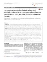
Artemisia absinthium and Artemisia vulgaris: A comparative study of infusion polysaccharides
Bạn đang xem bản rút gọn của tài liệu. Xem và tải ngay bản đầy đủ của tài liệu tại đây (1.1 MB, 8 trang )
Carbohydrate Polymers 102 (2014) 738–745
Contents lists available at ScienceDirect
Carbohydrate Polymers
journal homepage: www.elsevier.com/locate/carbpol
Artemisia absinthium and Artemisia vulgaris: A comparative study of
infusion polysaccharides
Marília Locatelli Corrêa-Ferreira, Guilhermina Rodrigues Noleto,
Carmen Lúcia Oliveira Petkowicz ∗
Universidade Federal do Paraná, Departamento de Bioquímica e Biologia Molecular, CP 19046, 81531-980 Curitiba, PR, Brazil
a r t i c l e
i n f o
Article history:
Received 22 July 2013
Received in revised form 28 October 2013
Accepted 30 October 2013
Available online 7 November 2013
Keywords:
Artemisia absinthium
Artemisia vulgaris
Infusion
Polysaccharides
Arabinogalactan
Fructan
a b s t r a c t
The aerial parts of Artemisia absinthium and Artemisia vulgaris are used in infusions for the treatment of
several diseases. Besides secondary metabolites, carbohydrates are also extracted with hot water and
are present in the infusions. The plant carbohydrates exhibit several of therapeutic properties and their
biological functions are related to chemical structure. In this study, the polysaccharides from infusions
of the aerial parts of A. absinthium and A. vulgaris were isolated and characterized. In the A. absinthium
infusion, a type II arabinogalactan was isolated. The polysaccharide had a Gal:Ara ratio of 2.3:1, and
most of the galactose was (1→3)- and (1→6)-linked, as typically found in type II arabinogalactans. In
the A. vulgaris infusion, an inulin-type fructan was the main polysaccharide. NMR analysis confirmed the
structure of the polymer, which is composed of a chain of fructosyl units -(2←1) linked to a starting
␣-d-glucose unit.
© 2013 Elsevier Ltd. All rights reserved.
1. Introduction
Artemisia absinthium and Artemisia vulgaris belong to the Asteraceae family and are known and marketed throughout the world for
their medicinal properties. The aerial parts of these plants are used
in traditional medicine as infusions, to which anthelmintic, antibacterial, antipyretic, cytostatic, stomachic and antitumor actions have
been attributed (Blagojevic, Radulovic, Palic, & Stojanovic, 2006;
Kordali, Cakir, Mavi, Kilic, & Yildirim, 2005; Lorenzi & Matos, 2008).
Several low molar mass compounds have been identified in
A. absinthium and A. vulgaris, such as sesquiterpene lactones, lignans, flavonoids and monoterpenes (Aberham, Cicek, Schneider, &
Stuppner, 2010; Govindaraj, Kumari, Cioni, & Flamini, 2008; LopesLutz, Alviano, Alviano, & Kolodziejczyk, 2008) which are considered
the main active compounds of these plants (Gilani & Janbaz, 1995;
Khan & Gilani, 2009; Lee et al., 2004). However, when an infusion is prepared, numerous types of low molar mass products
are extracted along with macromolecular compounds. Polysaccharides are one of the main macromolecular components of infusion
extracts because they are the predominant components of the plant
cell wall and are also present as reserve compounds in plant tissues
(Reid, 1997). The infusion is prepared with hot water which allows
the extraction of reserve and structural polysaccharides present in
∗ Corresponding author. Tel.: +55 41 3361 1661; fax: +55 41 3266 2042.
E-mail address: (C.L. Oliveira Petkowicz).
0144-8617/$ – see front matter © 2013 Elsevier Ltd. All rights reserved.
/>
the herbs. Structural polysaccharides from the cell wall are usually categorized as pectins, hemicelluloses and cellulose, based on
their extractability. Pectins and arabinogalactans-proteins (AGPs)
are water-soluble polysaccharides and can be extracted using aqueous solutions (Fincher, Stone, & Clarke, 1983; Reid, 1997).
Starch is the most abundant reserve carbohydrate in plants
(Jobling, 2004) and one of the most widespread alternatives to
starch as reserve carbohydrate are the oligomers and polymers
of fructose, the fructans (Hendry, 1993). These polymers are also
readily soluble in water (Edelman & Jefford, 1968), and can also be
extracted by the infusion process.
Recently, plant polysaccharides have attracted a great deal of
attention for their industrial and biological applications because
of their structural variability, broad spectrum of properties and
relatively low toxicity (Schepetkin & Quinn, 2006).
Artemisia species used in traditional medicine have shown
to contain water-soluble polysaccharides with a wide variety of
biological properties. It was demonstrated that a water-soluble carbohydrate fraction isolated from Artemisia iwayomogi can modulate
the functional differentiation of bone marrow-derived dendritic
cells (Lee et al., 2008), and showed immunomodulating and antitumor activities in mice (Koo et al., 1994). Polysaccharide fractions
from Artemisia tripartita exhibited potent phagocyte immunomodulatory activity, ROS scavenging and complement-fixing activity
(Xie et al., 2008).
No reports were found describing the composition of the
polysaccharides from the aerial parts of A. absinthium L. and
M.L. Corrêa-Ferreira et al. / Carbohydrate Polymers 102 (2014) 738–745
739
A. vulgaris L. In the present study, the polysaccharides present in
the infusions from aerial parts of A. absinthium and A. vulgaris were
isolated and characterized.
temperatures were 300 ◦ C and 250 ◦ C, respectively. The oven temperature was programmed from 100 to 220 ◦ C at a rate of 40 ◦ C/min
with helium as the carrier gas (1.0 ml/min).
2. Experimental
2.6. High-performance size-exclusion chromatography (HPSEC)
2.1. Materials
The isolated polysaccharides were analyzed by HPSEC using a
Waters unit coupled to a refractive index (RI), a Wyatt Technology
Dawn-F multi-angle laser light scattering (MALLS) detector and a
Pharmacia LKB Uvicord VW 2251 ultraviolet (UV) detector used
in 280 nm. Four Waters Ultrahydrogel columns (2000; 500; 250;
120) were connected in series and coupled to the multidetection
instrument. A solution of 0.1 mol/l NaNO2 and 0.02% NaN3 was used
as eluent at a flux of 0.6 ml/min. Prior to the analyses, the samples (1.0 mg/ml) were filtered through a 0.22 m cellulose acetate
membrane. The data were collected and analyzed by a Wyatt Technology ASTRA program. All the analyses were carried out at 25 ◦ C.
The refractive index increment of the solvent–solute solution with
respect to a change in solute concentration (dn/dc) was determined
using a Waters 2410 differential refractometer. The average molecular mass (Mw ) was calculated using Wyatt Technology ASTRA
software.
Dried aerial parts of A. vulgaris were kindly supplied by Laboratório Santos Flora Comércio de Ervas Ltda, São Paulo, Brazil (lot
number ARTER01/0310) and those of A. absinthium were purchased
(Hubert Comércio de Produtos Alimentícios Ltda, São Paulo, lot
number LOSNR01/0109). According to these companies, the plants
were cultivated in southern Brazil.
2.2. Isolation of polysaccharides
The infusion of aerial parts from A. vulgaris and A. absinthium was
prepared according to the traditional medicine method: one cup
(200 ml) of boiling water was added to one teaspoon of herb (1.0 g
for A. absinthium and 1.4 g for A. vulgaris) (Lorenzi & Matos, 2008).
The material was infused until it reached 40 ◦ C and then filtered.
Each extract was concentrated and treated with ethanol (4:1 v/v).
The material was kept at 4 ◦ C overnight and then the polysaccharide
pellets were isolated by centrifugation (8000 rpm, 20 min), washed
two times with ethanol and dried under vacuum. The A. absinthium
and A. vulgaris infusions were performed several times to obtain
enough material for chemical characterization.
2.7. Nuclear magnetic resonance spectroscopy (NMR)
The 13 C NMR spectra were obtained from samples in D2 O at 50 ◦ C
using a Bruker DRX 400 Avance spectrometer. Chemical shifts are
expressed in ı (ppm) relative to acetone ı (30.2).
2.3. Purification of polysaccharide from A. absinthium infusion
2.8. Fourier transform infrared (FT-IR)
The crude polysaccharide from A. absinthium infusion (named
AIP) was dissolved in an appropriate volume of distilled water and
dialyzed for two days against distilled water (cut-off Mw 12,000 Da).
The retained portion was concentrated and freeze-dried. The product was dissolved in water and submitted to freeze-thawing until
no more precipitate appeared. The soluble fraction was treated
with amylase, and the starch-free fraction was submitted to ultrafiltration using a 30 kDa membrane followed by filtration through
a 0.1 m membrane. The eluate yielded a purified polysaccharide
(AIP-F1).
The FT-IR spectra of purified fraction AIP-F1 was recorded on a
BOMEM MB-100 FT-IR spectrometer. The dried sample was ground
with potassium bromide powder and pressed into a pellet for spectrometric measurement in the frequency range of 4000–400 cm−1 .
2.4. Colorimetric methods
Total carbohydrate was assayed by the phenol–sulfuric acid
method (Dubois, Gilles, Hamilton, Rebers, & Smith 1956) using
galactose as standard and protein by the Bradford method (1976),
using BSA as standard. Uronic acid was estimated by the mhydroxydiphenyl method (Blumenkrantz & Asboe-Hansen, 1973)
using glucuronic acid as standard. The amounts of fructose and
fructose-yielding carbohydrates were estimated by a ketosespecific modification of the anthrone method described by Pollock
(1982) based on the method of Jermyn (1956) using inulin as a
standard.
2.5. Neutral monosaccharide composition
Polysaccharides were hydrolyzed with trifluoroacetic acid
(2 mol/l) in boiling water for 5 h. The hydrolyzate was evaporated
to dryness and the residues were reduced with NaBH4 (Wolfrom
& Thompson, 1963b) and acetylated with pyridine–acetic anhydride (1:1, v/v, 1 h, at 100 ◦ C) (Wolfrom & Thompson, 1963a). The
resulting alditol acetates were examined by gas chromatography
(GC) using a THERMO Trace GC Ultra gas chromatograph equipped
with a Ross injector and a DB-225 capillary column (0.25 mm internal diameter × 30 m). The flame ionization detector and injector
2.9. Carboxy-reduction of fraction AIP-F1
The carboxyl groups of the uronic acid residues of AIP-F1
were reduced to their corresponding alcohols by NaBH4 , using
the carboxydiimide method (Anderson and Stone, 1985; Taylor
& Conrad, 1972), to give a reduced polysaccharide fraction (APICX). The reduction of uronic acid residues was measured by a
colorimetric assay (Blumenkrantz & Asboe-Hansen, 1973). API-CX
was hydrolyzed in 2 mol/l trifluoroacetic acid in boiling water for
5 h. The solution was evaporated and the monosaccharides were
reduced and acetylated as above. The resulting alditol acetates were
analyzed by gas chromatography–mass spectrometry (GC–MS).
2.10. Methylation analysis
The AGP linkage analysis was done on carboxyl-reduced
polysaccharide (API-CX). The fraction was methylated according
to the method of Kvernheim (1987), using butyllithium (15% in
hexane) in DMSO-MeI, under a nitrogen atmosphere. The per-Omethylated product was first hydrolyzed with formic acid (90%)
in boiling water for 1 h and then with trifluoroacetic acid (2 mol/l)
for an additional hour. The hydrolyzate was evaporated to dryness,
and the residues were reduced and acetylated to give a mixture of
partially O-methylated alditol acetates.
2.11. Gas chromatography–mass spectrometry (GC–MS)
GC–MS was performed using a 3800 Varian gas chromatograph linked to a 2000 R-12 Varian Ion-Trap mass spectrometer,
with helium as carrier gas (1 ml/min). A capillary column
740
M.L. Corrêa-Ferreira et al. / Carbohydrate Polymers 102 (2014) 738–745
(30 m × 0.25 mm internal diameter) of DB-225 was held at 50 ◦ C
during injection and then programmed at 40 ◦ C/min to 210 ◦ C (constant temperature).
GC was performed using a THERMO Trace GC Ultra gas chromatograph equipped with a Ross injector and a DB-225 capillary
column (0.25 mm internal diameter × 30 m). The flame ionization
detector and injector temperatures were 300 ◦ C and 250 ◦ C, respectively. The oven temperature was programmed from 100 to 220 ◦ C
at a rate of 40 ◦ C/min with helium as the carrier gas (1.0 ml/min).
2.12. Periodate oxidation
AIP-F1 was oxidized in 0.05 mol/l NaIO4 at room temperature
(25 ◦ C) and in the dark for 72 h. The reaction was stopped with
1,2-ethanediol and then the solution was dialyzed for 48 h. The
resulting polyaldehydes were reduced with NaBH4 , neutralized
with acetic acid and dialyzed for 48 h. The polyalcohol was submitted to total acid hydrolysis, and the products were analyzed as
alditol acetates by GC as described above.
2.13. Fructose detection and chromatographic analyses of
fructans
To verify the presence of fructose in A. vulgaris infusion polysaccharides, the sample was hydrolyzed in 10 mmol/l H2 SO4 (pH 2.0)
in boiling water for 15 min. The hydrolyzate was neutralized with
BaCO3 and the insoluble material was filtered. The monosaccharides were analyzed by high performance liquid chromatography
(HPLC) and thin-layer-chromatography (TLC). After hydrolysis and
neutralization, the sample from A. vulgaris was applied to the origin
of a silica gel TLC plate (Macherey-Nagel). The plate was developed three times in 1-butanol/2-propanol/water (3:12:4, v/v/v) at
room temperature. The compounds were visualized by spraying
with urea-phosphoric acid reagent, a ketose-specific stain (Sims,
Cairns, & Furneaux, 2001; Wise, Dimler, Davis, & Rist, 1955). The
monosaccharides were also analyzed by HPLC using a Shimadzu
system (Japan) equipped with a CBM-10A interface module, CTO10A column oven, LC-10AD pump and with a RID-10A refractive
index detector. A Supelcogel Pb column (30 cm × 7.8 mm) (Supelco
e USA) and Supelcogel Pb pre-column (5 cm × 4.6 mm) were used.
The HPLC-column was eluted with water at a flow rate of 0.5 ml/min
at 80 ◦ C.
3. Results and discussion
3.1. Polysaccharides from the infusion of the aerial parts from A.
absinthium
Polysaccharides from the aerial parts of A. absinthium were
isolated from a traditionally prepared infusion (Lorenzi & Matos,
2008). The crude polysaccharide yield from the A. absinthium infusion (AIP) was 5.8%, which represents 58 mg of polysaccharide in
each cup of infusion. The sample displayed a polymodal elution profile by HPSEC (data not shown) and was submitted to several steps
of purification as shown in Fig. 1. Initially, the sample was dialyzed
(12 kDa cut off), and this was followed by a freeze-thawing step.
After the removal of the insoluble material, the soluble part was
subjected to treatment with ␣-amylase followed by ultrafiltration
using 30 kDa and then 0.1 m membranes. The fraction obtained
after ultrafiltration using the 0.1 m membrane was named AIP-F1
and had a yield of 6.0%, based on the crude polysaccharide obtained
from the A. absinthium infusion.
Fig. 1. Purification scheme of polysaccharides from the infusion of aerial parts from
A. absinthium.
Fig. 2. Elution profile of AIP-F1 obtained by HPSEC-MALLS/RI/UV.
3.2. Characterization of fraction AIP-F1
Fraction AIP-F1 showed a monomodal elution profile when analyzed by HPSEC-MALLS/RI/UV (Fig. 2) and was investigated by
chemical and spectroscopic methods. The monosaccharide composition of AIP-F1 is shown in Table 1. Galactose, followed by
arabinose were the main components of AIP-F1. Minor amounts
of rhamnose, xylose, mannose, glucose, uronic acids and fucose
were also found. The fraction AIP-F1 contained 10.4% uronic
acids which were reduced to their respective neutral sugars. Glucuronic acid was the predominant uronic acid in this fraction,
Table 1
Monosaccharide composition of fractions AIP, AIP-F1 and AIP-CX.
Monosaccharide composition (%)a
Fraction
AIP
AIP-F1
AIP-CX
Rha
Fuc
Ara
Xyl
Man
Gal
Glc
UA
5.3
10.9
10.3
tr
tr
3.0
14.9
16.0
11.7
4.7
3.9
3.4
12.8
7.4
10.5
29.8
36.5
38.4
28.2
14.9
21.6
4.3
10.4
1.1
tr, trace.
UA = uronic acid.
a
Neutral monosaccharide determined by GC and uronic acid determined by colorimetric method.
M.L. Corrêa-Ferreira et al. / Carbohydrate Polymers 102 (2014) 738–745
Fig. 3.
13
741
C NMR spectrum of AIP-F1. Solvent was D2 O at 70 ◦ C. Numerical values for ı are in ppm.
due to increased glucose in the carboxyl-reduced sample (AIPCX) compared with the native sample (AIP-F1), as shown in
Table 1.
The monosaccharide composition and the chemical shifts in the
13 C NMR spectrum (Fig. 3) indicated that AIP-F1 consists of a type
II arabinogalactan. Type II arabinogalactans encompasses a broad
group of short (1→3) and (1→6)--d-galactan chains connected
to each other by (1→3) and (1→6)-linked branch point residues.
Most of the remaining galactose units are substituted by a terminal
arabinofuranose (Fincher et al., 1983). Although the type II arabinogalactans side chains often terminate in ␣-L-Araf, other sugars
can be present, such as Fucp, Rhap and the uronic acids GlcpA and
GalpA, which are usually in terminal positions (Steinhorn, Sims,
Carnachan, Carr, & Schlothauer, 2011, Thude & Classen, 2005). The
type II arabinogalactans are frequently linked to protein moiety
(known as arabinogalactan-proteins or AGP) and the protein content is usually between 2 and 10% (Fincher et al., 1983).
In the 13 C NMR spectrum of AIP-F1, the signal at ı 103.4 was
attributed to C-1 of -d-Galp units and the signals at ı 81.3
and 68.5 ppm were attributed to the C-3-linked and C-6-linked
-d-Galp units, respectively (Baron Maurer et al., 2010; Capek,
Matulova, Navarini, & Liverani, 2009), which are typical of a type
II arabinogalactan. The chemical shifts at ı 107.9 and 107.0 were
attributed to C-1 of internal ␣-l-Araf residues, respectively and the
signal at 109.1 ppm was due to C-1 of terminal ␣-l-Araf (Baron
´ & Kubaˇcková, 1998;
Maurer et al., 2010; Karácsonyi, Pätoprsty,
Steinhorn et al., 2011). The signals at ı 62.3 and 61.2 which
appeared inverted in the DEPT experiment (data not shown), were
attributed to the non-substituted C-5 of ␣-l-Araf and the C-6 of
-d-Galp units, respectively.
The signal of -d-GlcpA can be overlapped with the signal of
the C-1 of -d-Galp at 103.4 ppm (Redgwell et al., 2011; Steinhorn
et al., 2011).
In addition to glucuronic acid, rhamnose was also found as a
constituent of the arabinogalactan present in AIP-F1. The signal at
100.7 ppm was attributed to C-1 of ␣-l-Rhap units and their CH3 -6
was found at a lower frequency, 16.7 ppm.
Infrared spectroscopy is quite extensively applied in plant
cell wall analysis (Kacurakova, Capek, Sasinkova, Wellner, &
Ebringerova, 2000). The FT-IR spectrum of AIP-F1 showed a prominent band near 3400 cm−1 , characteristic of polysaccharides, due
to hydroxyl group of monosaccharide units (Coimbra, Barros,
Rutledge, & Delgadillo, 1999). The -arabinogalactans have two
typical bands in FT-IR analysis, at 1074 and 1045 cm−1 , which are
attributed to galactopyranose in the backbone and arabinofuranose units in side branches, respectively (Kacurakova et al., 2000).
These two bands were identified in the FT-IR spectrum of AIP-F1,
confirming the presence of a type II arabinogalactan. The relative
IR absorption intensities of the bands of galactose and arabinose
vary in the sample according to their relative amounts (Kacurakova
et al., 2000). The ratio of Gal:Ara was 2.3:1 in AIP-F1 and in the
FT-IR spectrum; the galactose-related band was larger than the
arabinose-related band. The ratio of Gal:Ara in AIP-F1 was comparable to the arabinogalactan isolated from the stigmas and styles
of Nicotiana alata (2.1:1) (Gane et al., 1995), but lower than that
obtained for kanuka honey arabinogalactan (5.3:1) (Steinhorn et al.,
2011).
While type I arabinogalactans are usually found as neutral
side-chains on plant cell-wall pectic polysaccharides, type II arabinogalactans are often covalently linked to proteins being known
as arabinogalactan-proteins (AGP) (Steinhorn et al., 2011). The
FT-IR spectrum of AIP-F1 showed high absorbance at wavenumbers characteristic of protein: 1610 cm−1 (amide I) and 1400 cm−1
(amide III) (Boulet, Williams, & Doco, 2007). The fraction AIP-F1
contained 7% proteins, which are probably covalently linked to the
polysaccharide, suggesting that AIP-F1 is an AGP. This hypothesis
was also supported by the elution profile of AIP-F1 by HPSECMALLS/RI/UV (Fig. 2), which showed a single peak at 280 nm
detected simultaneously by RI, MALLS and UV. The protein percentage in AIP-F1 is in accordance with previously studied AGPs
which typically contains 1% to 10% (w/w) of proteins (Ellis, Egelund,
Schultz, & Bacic, 2010). The peak at 1610 cm−1 in FT-IR can also be
overlapped with the band of uronic acid (Gnanasambandam and
Proctor, 1999), which is also present in AIP-F1.
742
M.L. Corrêa-Ferreira et al. / Carbohydrate Polymers 102 (2014) 738–745
Fig. 4. HPLC chromatogram of VPI hydrolyzed sample and a fructose standard.
Table 2
Profile of partially O-methylated alditol acetates obtained by methylation analysis
of AIP-F1.
O-Me-alditol acetate
Linkages
2,3,4-Me3 -Rha
2,4-Me2 -Rha
3-Me-Rha
2,3,5-Me3 -Ara
3,5-Me2 -Ara
2-Me-Ara
2,3-Me2 -Ara
2,6-Me2 -Gal
2,4-Me2 -Gal
2-Me-Gal
2,4,6-Me3 -Gal
2,3,4-Me3 -Gal
2,3,4,6-Me4 -Gal
2,3,4,6-Me4 -Glc
2,3,6-Me3 -Glc
2,3,4,6-Me4 -Man
Terminal
→)3
→)2,4
Terminal
→)2
→)3,5
→)5
→)3,4
→)3,6
→)3,4,6
→)3
→)6
Terminal
Terminal
→)4
Terminal
Results from methylation analysis of the carboxy-reduced AIPCX sample are given in Table 2. The presence of 6-O-, 3-O- and
3,6-di-O substituted Galp units were consistent with the presence
of (1→3)-linked Galp backbone with side-chains of (1→6)-linked
Galp. Arabinose was found to be 2-O-, 3-O-, 5-O- and 3,5-di-Osubstituted and as nonreducing end-units, as described for type
II arabinogalactans found in kanuka honey (Steinhorn et al., 2011)
and Lycium barbarum (Redgwell et al., 2011).
Rhamnose, glucose and mannose were also found as nonreducing end-units in AIP-CX, consistent with other arabinogalactans
structures (Redgwell et al., 2011; Thude & Classen, 2005). It is
believed that these monosaccharides at the periphery of AGPs
might be important for their biological activities (Göllner, Ichinose,
Kaneko, Blaschek, & Classen, 2011). The presence of glucose as
nonreducing end-units can also be due to glucuronic acid because
the acidic units were reduced to their respective neutral sugars
prior to methylation analysis. The presence of glucuronic acid
as nonreducing end-units in type II arabinogalactans has been
described for L. barbarum (Redgwell et al., 2011), and in wheat flour
AGP, the nonreducing end-units of glucuronic acid was identified
as linked at O-6 to the side-chains of (1→6)-linked Galp (Tryfona
et al., 2010).
It has also been reported that some glucuronic acid units from
AGP can be substituted by terminal Rhap (1→4)-linked to glucuronic acid (Redgwell et al., 2011). The presence of the derivative
2,3,6-Me3 -Glc in the methylation products of AIP-CX suggest that
some glucuronic acid units can also be O-4 substituted.
Rhamnose was found to be 3-O-, 2,4-di-O substituted and also as
nonreducing end-units. The arabinogalactans from fruits of Lycium
ruthenicum also contain (1→2,4)-linked rhamnosyl residues, which
are likely in branches (Peng et al., 2012). Arabinogalactans from
Centella asiatica (Wang, Zheng, Zuo, & Fang, 2005), Echinacea pallida
(Thude & Classen, 2005) and L. barbarum (Redgwell et al., 2011) also
showed rhamnose in their composition.
Periodate oxidation of AIP-F1 was performed to confirm the
presence of 1,3-Galp in the backbone. After oxidation of 100 mg AIPF1, 50 mg of oxidation-resistant polysaccharide was obtained. The
ratio Gal:Ara of the oxidation-resistant polysaccharide was 5.6:1,
which is much higher than that of the native polysaccharide (2.3:1).
This increase in the galactose proportion suggests that most of the
galactose is (1→3)-linked and resistant to periodate oxidation, as
typically found in type II arabinogalactans (Fincher et al., 1983).
The AGPs are widely distributed in higher plants, where they
have been found in seeds, leaves, roots and fruits. Their molar
masses are very heterogeneous, presumably reflecting different extents of glycosylation (Clarke, Anderson, & Stone, 1979;
Showalter, 1993). The weight-average molar mass (Mw ) of AGP
from the A. absinthium infusion (AIP-F1) was calculated to be
84,160 g/mol, similar to that found for an AGII from Avena sativa
(83,000 g/mol; Göllner et al., 2011), but higher than those from L.
barbarum (50,000–60,000 g/mol; Redgwell et al., 2011) and lower
than those from Echinacea sp (1.1 × 10⁶ –1.2 × 10⁶ g/mol; Thude &
Classen, 2005).
The structure of protein and glycan moieties of AGPs is highly
diverse and several studies have implicated these molecules in
many important biological processes in plants, such as cell proliferation and survival, growth, resistance to stresses and plant
microbe interaction (Seifert & Roberts, 2007; Pickard, 2013). In
addition, some type II arabinogalactans have recently been investigated as potential immunomodulators of the human immune
system (Classen, Thude, Blaschek, Wack, & Bodinet, 2006; Nosal’ova
et al., 2011), and the fine structure of AGP influences its biological activity (Classen et al., 2006). Therefore, the investigation of
the structural and functional properties of arabinogalactans is an
opportunity for the discovery of novel therapeutic agents with
immunomodulatory properties.
3.3. Polysaccharides from the infusion of aerial parts from of A.
vulgaris
As described for A. absinthium, polysaccharides from the aerial
parts of A. vulgaris were isolated from boiling water infusions prepared according to the traditional method (Lorenzi & Matos, 2008).
The crude polysaccharide yield from A. vulgaris infusion (VPI) was
7.0%, representing 98 mg of polysaccharide in each cup of infusion. The anthrone method indicated that 85% of the carbohydrate
content in the VPI consisted of fructose. Rha, Ara, Man, Gal,
M.L. Corrêa-Ferreira et al. / Carbohydrate Polymers 102 (2014) 738–745
Fig. 5. Elution profile of VPI obtained by HPSEC/RI.
Glc, uronic acids and Xyl were also found in a ratio of
1.7:2.5:5.2:4.2:14.0:1.8:1.
Fructans are acid labile, therefore VPI was submitted to mild acid
hydrolysis (H2 SO4 , pH 2.0, 15 min), and the resulting monosaccharides were analyzed by HPLC and TLC. The presence of fructose in
this sample was confirmed by TLC using a ketose-specific stain (data
not shown) and by HPLC (Fig. 4). Therefore, VPI was suggested to
be a fructan-type polysaccharide.
A polymodal elution profile was observed by HPSEC for VPI using
the refractive index detector (Fig. 5). The sample heterogeneity
may reflect different degrees of fructan polymerization (de Oliveira
et al., 2011). The main peak in the chromatogram of VPI eluted after
55 min and was not detected by MALLS, indicating that low molar
mass components were predominant in this fraction. This result
is in agreement with other studies, where fructans are described
as polydispersed reserve carbohydrates that contain 1–70 units of
fructose (Choque Delgado, Tamashiro, & Pastore, 2010).
In the 13 C NMR spectrum of VPI (Fig. 6), the signals at ı
103.3 and 92.5 ppm were assigned to C-2 of (2→1)-linked -dfructofuranose and C-1 of the starting nonreducing end-units of
␣-d-glucopyranose, respectively. These signals are typical of inulintype fructans which mainly consist of -(2←1) fructosyl-fructose
linkages with a starting ␣-d-glucose unit (Roberfroid, 2007).
Fig. 6.
13
743
High intensity signals in the spectrum are in accordance with
those expected from the fructose ring carbons, being at ı in ppm: C1 (61.3), C-2 (103.3), C-3 (77.5), C-4 (74.9), C-5 (81.3) and C-6 (62.3).
A group of low intensity signals were attributed to the ␣-d-glucose
of the nonreducing end units of inulin: C-1 (92.5), C-3 (73.0), C-4
(69.7), C-5 (71.4) and C-6 (60.6). All of the assignments were based
on previous reports (Chandrashekar, Prashanth, & Venkatesh, 2011;
Fontana, Baron, Diniz, & Franco, 1994).
According to the results, the main water-soluble carbohydrate
present in A. vulgaris infusion is an inulin-type fructan. The presence of inulin-type fructans was also reported for the leaves of
Stevia rebaudiana and Matricaria maritima (Cerantola et al., 2004; de
Oliveira et al., 2011), both species of the Asteraceae family (order
Asterales) in which A. vulgaris is also included. It is known that
almost all families included in the order Asterales contain fructans,
at least in storage organs (Hendry, 1993).
It has been reported that A. vulgaris contains inulin as one
of its active components (Govindaraj et al., 2008), but oligofructosides were described only for the roots of plants in Artemisia
species (Kennedy, Stevenson, White, Lombard, & Buffa, 1988). To
our knowledge, this is the first time that inulin has been reported
in the leaves of an Artemisia specie. However, fructans were not
the major carbohydrate in the infusion of A. absinthium, which also
belongs to the Asteraceae family. Instead, apart from a starch, an
arabinogalactan was extracted from the leaves of A. absinthium.
Inulin-type fructans have attracted a great of attention, especially in the food industry, because fructans add nutritional value
to the product. Inulin is classified as a functional food because of its
chemical nature and physiological and nutritional effects. Fructooligosaccharides and inulin are described as having prebiotic
properties (Roberfroid, 2007). The regular intake of these carbohydrates modulates the composition of intestinal flora, enhances
resistance against intestinal pathogens and regulates the levels of
serum cholesterol and the absorption of calcium and other minerals. Fructans also seem to be involved in the positive modulation of
the immune system, as well as in the reduction of the risk of several
diseases, including cancer (Choque Delgado et al., 2010; Roberfroid,
2007; Taper & Roberfroid, 1999). According to the results from
the present work, every cup of A. vulgaris infusion contains 83 mg
of inulin-type fructans on average. It is possible that the fructans
C NMR spectrum of VPI. Solvent was D2 O at 70 ◦ C. Numerical values for ı are in ppm.
744
M.L. Corrêa-Ferreira et al. / Carbohydrate Polymers 102 (2014) 738–745
present in A. vulgaris infusion can contribute to the positive effects
on health attributed to the infusion.
4. Conclusion
The infusions from aerial parts A. absinthium and A. vulgaris,
which are used in traditional herbal medicine, contain polysaccharides. Although both species belong to the Asteraceae family, the
infusion of A. absinthium contains a type II arabinogalactan, whereas
the infusion of A. vulgaris contains an inulin-type fructan as the
main polysaccharide.
Acknowledgments
The authors thank the Brazilian agencies CNPq, CAPES and
Fundac¸ão Araucária for financial support and Laboratório Santos
Flora Comércio de Ervas Ltda for the aerial parts of A. vulgaris.
References
Aberham, A., Cicek, S. S., Schneider, P., & Stuppner, H. (2010). Analysis of sesquiterpene lactones, lignans, and flavonoids in wormwood (Artemisia absinthium L.)
using high-performance liquid chromatography (HPLC)–mass spectrometry,
reversed phase HPLC, and HPLC-solid phase extraction-nuclear magnetic resonance. Journal of Agricultural and Food Chemistry, 58(20), 10817–10823.
Anderson, M. A., & Stone, B. A. (1985). A radiochemical approach to the determination of carboxylic-acid groups in polysaccharides. Carbohydrate Polymers, 5(2),
115–129.
Baron Maurer, J. B., Bacic, A., Pereira-Netto, A. B., Donatti, L., Zawadzki-Baggio, S. F.,
& Pettolino, F. A. (2010). Arabinogalactan-proteins from cell suspension cultures
of Araucaria angustifolia. Phytochemistry, 71(11–12), 1400–1409.
Blagojevic, P., Radulovic, N., Palic, R., & Stojanovic, G. (2006). Chemical composition
of the essential oils of Serbian wild-growing Artemisia absinthium and Artemisia
vulgaris. Journal of Agricultural and Food Chemistry, 54(13), 4780–4789.
Blumenkrantz, N., & Asboe-Hansen, G. (1973). New method for quantitativedetermination of uronic acids. Analytical Biochemistry, 54(2), 484–489.
Boulet, J. C., Williams, P., & Doco, T. (2007). A Fourier transform infrared spectroscopy
study of wine polysaccharides. Carbohydrate Polymers, 69(1), 79–85.
Bradford, M. M. (1976). Rapid and sensitive method for quantitation of microgram
quantities of protein utilizing principle of protein–dye binding. Analytical Biochemistry, 72(1–2), 248–254.
Capek, P., Matulova, M., Navarini, L., & Liverani, F. S. (2009). A comparative study
of arabinogalactan-protein isolates from instant coffee powder of Coffea arabica
beans. Journal of Food and Nutrition Research, 48(2), 80–86.
Cerantola, S., Kervarec, N., Pichon, R., Magne, C., Bessieres, M.-A., & Deslandes,
E. (2004). NMR characterisation of inulin-type fructooligosaccharides as the
major water-soluble carbohydrates from Matricaria maritima (L.). Carbohydrate
Research, 339, 2445–2449.
Chandrashekar, P. M., Prashanth, K. V. H., & Venkatesh, Y. P. (2011). Isolation, structural elucidation and immunomodulatory activity of fructans from aged garlic
extract. Phytochemistry, 72(2–3), 255–264.
Choque Delgado, G. T., Tamashiro, W. M. S. C., & Pastore, G. M. (2010). Immunomodulatory effects of fructans. Food Research International, 43(5), 1231–1236.
Clarke, A. E., Anderson, R. L., & Stone, B. A. (1979). Form and function of
arabinogalactan-proteins. Phytochemistry, 18(4), 521–540.
Classen, B., Thude, S., Blaschek, W., Wack, M., & Bodinet, C. (2006). Immunomodulatory effects of arabinogalactan-proteins from Baptisia and Echinacea.
Phytomedicine, 13(9–10), 688–694.
Coimbra, M. A., Barros, A., Rutledge, D. N., & Delgadillo, I. (1999). FTIR spectroscopy
as a tool for the analysis of olive pulp cell-wall polysaccharide extracts. Carbohydrate Research, 317(1–4), 145–154.
de Oliveira, A. J. B., Gonc¸alves, R. A. C., Chierrito, T. P. C., dos Santos, M. M., de Souza,
L. M., Gorin, P. A. J., et al. (2011). Structure and degree of polymerisation of
fructooligosaccharides present in roots and leaves of Stevia rebaudiana (Bert.)
Bertoni. Food Chemistry, 129(2), 305–311.
Dubois, M., Gilles, K. A., Hamilton, J. K., Rebers, P. A., & Smith, F. (1956). Colorimetric
method for determination of sugars and related substances. Analytical Chemistry,
28(3), 350–356.
Edelman, J., & Jefford, T. G. (1968). Mechanism of fructosan metabolism in higher
plants as exemplified in Helianthus tuberosus. New Phytologist, 67(3), 517–531.
Ellis, M., Egelund, J., Schultz, C. J., & Bacic, A. (2010). Arabinogalactan-proteins: Key
regulators at the cell surface? Plant Physiology, 153, 403–419.
Fincher, G. B., Stone, B. A., & Clarke, A. E. (1983). Arabinogalactan-proteins – Structure, biosynthesis, and function. Annual Review of Plant Physiology and Plant
Molecular Biology, 34, 47–70.
Fontana, J. D., Baron, M., Diniz, A. C. P., & Franco, V. C. (1994). Microbial inulinase
secretion using chemically-modified inulins. Applied Biochemistry and Biotechnology, 45-6, 257–268.
Gane, A. M., Craik, D., Munro, S. L. A., Howlett, G. J., Clarke, A. E., & Bacic, A. (1995).
Structural-analysis of the carbohydrate moiety of arabinogalactan-proteins
from stigmas and styles of Nicotiana alata. Carbohydrate Research, 277(1),
67–85.
Gilani, A. U. H., & Janbaz, K. H. (1995). Preventive and curative effects of Artemisia
absinthium on acetaminophen and CCl4 -induced hepatotoxicity. General Pharmacology, 26(2), 309–315.
Gnanasambandam, R., & Proctor, A. (1999). Preparation of soy hull pectin. Food
Chemistry, 65(4), 461–467.
Göllner, E. M., Ichinose, H., Kaneko, S., Blaschek, W., & Classen, B. (2011). An
arabinogalactan-protein from whole grain of Avena sativa L. belongs to the
wattle-blossom type of arabinogalactan-proteins. Journal of Cereal Science, 53(2),
244–249.
Govindaraj, S., Kumari, B. D. R., Cioni, P. L., & Flamini, G. (2008). Mass propagation and essential oil analysis of Artemisia vulgaris. Journal of Bioscience and
Bioengineering, 105(3), 176–183.
Hendry, G. A. F. (1993). Evolutionary origins and natural functions of fructans – a climatological, biogeographic and mechanistic appraisal. New Phytologist, 123(1),
3–14.
Jermyn, M. A. (1956). New method for determining ketohexoses in the presence of
aldohexoses. Nature, 177(4497), 38–39.
Jobling, S. (2004). Improving starch for food and industrial applications. Current
Opinion in Plant Biology, 7(2), 210–218.
Kacurakova, M., Capek, P., Sasinkova, V., Wellner, N., & Ebringerova, A. (2000). FT-IR
study of plant cell wall model compounds: Pectic polysaccharides and hemicelluloses. Carbohydrate Polymers, 43(2), 195–203.
´ V. r., & Kubaˇcková, M. (1998). Structural study on
Karácsonyi, t., Pätoprsty,
arabinogalactan-proteins from Picea abies L Karst. Carbohydrate Research,
307(3–4), 271–279.
Kennedy, J. F., Stevenson, D. L., White, C. A., Lombard, A., & Buffa, M. (1988). Analysis
of the oligosaccharides from the roots of Arnica montana L, Artemisia absinthium
L, and Artemisia dracunculus L. Carbohydrate Polymers, 9(4), 277–285.
Khan, A. U., & Gilani, A. H. (2009). Antispasmodic and bronchodilator activities of
Artemisia vulgaris are mediated through dual blockade of muscarinic receptors
and calcium influx. Journal of Ethnopharmacology, 126(3), 480–486.
Koo, K., Kwak, J., Lee, K., Zee, O., Woo, E., Park, H., et al. (1994). Antitumor and
immunomodulating activities of the polysaccharide fractions from Artemisia
selengensis and Artemisia iwayomogi. Archives of Pharmacal Research, 17(5),
371–374.
Kordali, S., Cakir, A., Mavi, A., Kilic, H., & Yildirim, A. (2005). Screening of chemical
composition and antifungal and antioxidant activities of the essential oils from
three Turkish Artemisia species. Journal of Agricultural and Food Chemistry, 53(5),
1408–1416.
Kvernheim, A. L. (1987). Methylation analysis of polysaccharides with butyllithium
in dimethylsulfoxide. Acta Chemica Scandinavica Series B: Organic Chemistry and
Biochemistry, 41(2), 150–152.
Lee, H. G., Kim, H., Oh, W. K., Yu, K. A., Choe, Y. K., Ahn, J. S., et al. (2004). Tetramethoxy
hydroxyflavone p7F downregulates inflammatory mediators via the inhibition
of nuclear factor kappa B. Signal Transduction Pathways, Chromatin Structure and
Gene Expression Mechanisms as Therapeutic Targets, 1030, 555–568.
Lee, J.-A., Sung, H.-N., Jeon, C.-H., Gill, B.-C., Oh, G.-S., Youn, H.-J., et al. (2008).
AIP1 a carbohydrate fraction from Artemisia iwayomogi, modulate the functional differentiation of bone marrow-derived dendritic cells. International
Immunopharmacology, 8(4), 534–541.
Lopes-Lutz, D., Alviano, D. S., Alviano, C. S., & Kolodziejczyk, P. P. (2008). Screening
of chemical composition, antimicrobial and antioxidant activities of Artemisia
essential oils. Phytochemistry, 69(8), 1732–1738.
Lorenzi, H., & Matos, F. J. A. (2008). Plantas medicinais no Brasil: nativas e exóticas (2th
ed.). São Paulo: Instituto Plantarum (pp. 11–25, 118–121).
Nosal’ova, G., Prisenznakova, L., Paulovicova, E., Capek, P., Matulova, M., Navarini,
L., et al. (2011). Antitussive and immunomodulating activities of instant coffee arabinogalactan-protein. International Journal of Biological Macromolecules,
49(4), 493–497.
Peng, Q., Song, J., Lv, X., Wang, Z., Huang, L., & Du, Y. (2012). Structural characterization of an arabinogalactan-protein from the fruits of Lycium ruthenicum. Journal
of Agricultural and Food Chemistry, 60(37), 9424–9429.
Pickard, B. G. (2013). Arabinogalactan proteins – Becoming less mysterious. New
Phytologist, 197(1), 3–5.
Pollock, C. J. (1982). Patterns of turnover of fructans in leaves of Dactylis glomerata
L. New Phytologist, 90(4), 645–650.
Redgwell, R. J., Curti, D., Wang, J., Dobruchowska, J. M., Gerwig, G. J., Kamerling, J. P.,
et al. (2011). Cell wall polysaccharides of Chinese Wolfberry (Lycium barbarum).
Part 2. Characterisation of arabinogalactan-proteins. Carbohydrate Polymers,
84(3), 1075–1083.
Reid, J. S. G. (1997). Carbohydrate metabolism: Structural carbohydrate. In P. M. Dey,
& J. B. Harborne (Eds.), Plant biochemistry (pp. 205–236). United States: Academic
Press.
Roberfroid, M. B. (2007). Inulin-type fructans: Functional food ingredients. Journal
of Nutrition, 137(11), 2493S–2502S.
Schepetkin, I. A., & Quinn, M. T. (2006). Botanical polysaccharides: Macrophage
immunomodulation and therapeutic potential. International Immunopharmacology, 6(3), 317–333.
Seifert, G. J., & Roberts, K. (2007). The biology of arabinogalactan proteins. Annual
Review of Plant Biology, 58, 137–161.
Showalter, A. M. (1993). Structure and function of plant cell wall proteins. The Plant
Cell, 5, 9–23.
Sims, I. M., Cairns, A. J., & Furneaux, R. H. (2001). Structure of fructans from excised
leaves of New Zealand flax. Phytochemistry, 57(5), 661–668.
M.L. Corrêa-Ferreira et al. / Carbohydrate Polymers 102 (2014) 738–745
Steinhorn, G., Sims, I. M., Carnachan, S. M., Carr, A. J., & Schlothauer, R. (2011). Isolation and characterisation of arabinogalactan-proteins from New Zealand kanuka
honey. Food Chemistry, 128(4), 949–956.
Taper, H. S., & Roberfroid, M. (1999). Influence of inulin and oligofructose on breast
cancer and tumor growth. Journal of Nutrition, 129(7), 1488S–1491S.
Taylor, R. L., & Conrad, H. E. (1972). Stoichiometric depolymerization of polyuronides
and glycosaminoglycuronans to monosaccharides following reduction of their
carbodiimide-activated carboxyl groups. Biochemistry, 11(8), 1383–1388.
Thude, S., & Classen, B. (2005). High molecular weight constituents from roots of
Echinacea pallida: An arabinogalactan-protein and an arabinan. Phytochemistry,
66(9), 1026–1032.
Tryfona, T., Liang, H.-C., Kotake, T., Kaneko, S., Marsh, J., Ichinose, H., et al. (2010).
Carbohydrate structural analysis of wheat flour arabinogalactan protein. Carbohydrate Research, 345(18), 2648–2656.
745
Wang, X. S., Zheng, Y., Zuo, J. P., & Fang, J. N. (2005). Structural features of an
immunoactive acidic arabinogalactan from Centella asiatica. Carbohydrate Polymers, 59(3), 281–288.
Wise, C. S., Dimler, R. J., Davis, H. A., & Rist, C. E. (1955). Determination of easily
hydrolyzable fructose units in dextran preparations. Analytical Chemistry, 27(1),
33–36.
Wolfrom, M. L., & Thompson, A. (1963a). Acetylation. Methods in Carbohydrate Chemistry, 2, 21.
Wolfrom, M. L., & Thompson, A. (1963b). Reduction with sodium borohydride. Methods in Carbohydrate Chemistry, 2, 65.
Xie, G., Schepetkin, I. A., Siemsen, D. W., Kirpotina, L. N., Wiley, J. A., & Quinn, M.
T. (2008). Fractionation and characterization of biologically-active polysaccharides from Artemisia tripartita. Phytochemistry, 69(6), 1359–1371.