Water-soluble chitosan derivatives and pH-responsive hydrogels by selective C-6 oxidation mediated by TEMPO-laccase redox system
Bạn đang xem bản rút gọn của tài liệu. Xem và tải ngay bản đầy đủ của tài liệu tại đây (1.15 MB, 11 trang )
Carbohydrate Polymers 186 (2018) 299–309
Contents lists available at ScienceDirect
Carbohydrate Polymers
journal homepage: www.elsevier.com/locate/carbpol
Water-soluble chitosan derivatives and pH-responsive hydrogels by selective
C-6 oxidation mediated by TEMPO-laccase redox system
T
⁎
Suse Botelho da Silvaa,b, , Malgorzata Krolickaa,c, Lambertus A.M. van den Broeka,
⁎
August E. Frissena, Carmen Gabriela Boeriua,
a
Wageningen Food & Biobased Research, Department Biobased Products, Bornse Weilanden 9, 6708 WG Wageningen, The Netherlands
Polytechnic School, UNISINOS University, Av. Unisinos 950, 93022-000 São Leopoldo, RS, Brazil
c
Wageningen University, Bioprocess Engineering Group, Droevendaalsesteeg 1, 6708 PD, Wageningen, The Netherlands
b
A R T I C L E I N F O
A B S T R A C T
Keywords:
Chitosan
Oxidation
2,2,6,6-Tetramethylpiperidinoxyl radical
(TEMPO)
Laccase
pH-responsive hydrogel
Soluble chitosan
Chitosan is a polysaccharide with recognized antioxidant, antimicrobial and wound healing activities. However,
this polymer is soluble only in dilute acidic solutions, which restricts much of its applications. A usual strategy
for improving the functionality of polysaccharides is the selective oxidation mediated by 2,2,6,6-tetra-methyl-1piperidinidyloxy (TEMPO) using laccase as a co-oxidant. In this work, the TEMPO-laccase redox system was used
for the first time to selectively oxidize chitosan in order to produce tailored derivatives. The reaction was
performed at pH 4.5 under continuous air supply and the oxidized products were characterized structurally and
functionally. The TEMPO-laccase oxidation successfully added aldehyde and carboxylate groups to chitosan
structure resulting in derivatives with oxidation between 4 and 7%. These derivatives showed increased solubility and decreased viscosity in solution. If chitosan is dissolved in diluted hydrochloric acid prior to TEMPOlaccase oxidation, a crosslinked chitosan derivative was produced, which was able to form a pH-responsive
hydrogel.
1. Introduction
Polysaccharides obtained from renewable sources or agro-industrial
waste streams play an important role in the context of a biobased and
sustainable economy, or bio-economy (Persin et al., 2011). Chitin is the
most abundant polysaccharide in nature after cellulose, it is found
mainly in the exoskeleton of crustaceans and insects, but also in fungi
and algae. Industrially, chitin is obtained from crab and shrimp processing waste in countries such as USA, Japan and India (Kardas et al.,
2013), and also Brazil. The partial or fully deacetylation of N-acetylglucosamine moieties of chitin leads to formation of chitosan, also
considered as a natural biopolymer (Luo & Wang, 2013). Therefore,
chitin and chitosan are copolymers with similar structure, but in chitin
predominates 2-acetamido-2-deoxy-D-glucopyranose units and in chitosan, 2-amino-2-deoxy-D-glucopyranose units are more frequent. Chitin
is insoluble in most common solvents, while chitosan has a limited
solubility, being soluble for example in acidic aqueous solutions
(Mourya, Inamdar, & Choudhari, 2011).
Chitosan is recognized as a nontoxic, biodegradable and biocompatible polymer, whose properties are being exploited in industrial
and technological applications since the last century. These applications
⁎
cover areas such as food, pharmaceutical, textile, packaging, cosmetics
and agriculture, and are often justified by the antimicrobial activity of
this polymer, but also for its antioxidant activity (Kong, Chen, Xing, &
Park, 2010; Luo & Wang, 2013). The poor solubility of chitosan, however, restricts many of its potential applications. One of the strategies to
improve the solubility of chitosan and other polysaccharides is to
modify the structure of the molecule by the addition of hydrophilic
functional groups. Chitosan is a versatile molecule that can be modified
by different methods mainly due to its structure, which contains several
hydroxyls (primary hydroxyl at C-6 and secondary hydroxyl at C-3),
and highly reactive amino groups (C-2) (Luo & Wang, 2013; Prashanth
& Tharanathan, 2007). Direct oxidation of the hydroxyl groups is possible by the introduction of carboxyl and carbonyl groups at C-6 (Bragd,
Besemer, & van Bekkum, 2000). In addition to increasing solubility, the
insertion of these new functional groups could also convert chitosan
into a potential crosslinking structure, which newly inserted aldehyde
groups would hypothetically crosslink with existing animo groups. Tan,
Chu, Payne, and Marra (2009) reported a development of a composite
hydrogel derived from succinyl-chitosan and oxidized hyaluronic acid
stabilized with this kind of crosslinks.
In the last decades, the catalytic oxidation of carbohydrates using
Corresponding authors at: Wageningen Food & Biobased Research, Bornse Weilanden 9, 6708 WG Wageningen, The Netherlands.
E-mail addresses: (S. Botelho da Silva), (C.G. Boeriu).
/>Received 18 August 2017; Received in revised form 5 January 2018; Accepted 16 January 2018
Available online 03 February 2018
0144-8617/ © 2018 The Authors. Published by Elsevier Ltd. This is an open access article under the CC BY-NC-ND license ( />
Carbohydrate Polymers 186 (2018) 299–309
S. Botelho da Silva et al.
Table 1
Synthesis conditions of modified chitosan products, deacetylation and oxidation degree, and aldehyde and carboxylate contents.
Sample
Acid1
Product
P1
P2
P3
P4
HAc2
HAc
HAc
HCl
Product
Product
Product
Product
1
2
3
4
TEMPO (g/100 g
chitosan)
Laccase (U/g
chitosan)
Ratio Laccase/
TEMPO (U/g)
DD%3
1
10
10
10
7
70
7
7
700
700
70
70
83.6
79.8
83.4
82.7
a
a
a
a
OD%4
1.1
7.0
4.6
4.0
a
b
c
c
Aldehyde content (mmol/kg
chitosan)
Carboxylate content (mmol/kg
chitosan)
50.9 a
385 b
264 c
227 c
13.5
28.0
10.4
10.8
a
b
a
a
a, b, c: The same letter in each column are not significantly different at the 5% level (Tukey’s test).
1
0.1 M acid diluted solution used to dissolve chitosan prior to TEMPO-laccase oxidation.
2
HAc is acetic acid.
3
DD is deacetylation degree.
4
OD is oxidation degree.
using the Mark–Houwink–Sakurada equation (Kasaai, 2007) and 1H
NMR spectroscopy (Hirai, Odani, & Nakajima, 1991), respectively.
Laccase from Trametes versicolor, 2,2,6,6-tetramethylpiperidinoxyl radical (TEMPO), 3-ethylbenzothiazoline-6-sulphonic acid (ABTS), 2,5dihydroxybenzoic acid (DHB) and other chemicals were purchased
from Sigma-Aldrich. Endochitinase from Myceliophthora thermophila C1
was a kind gift from DuPont Industrial Biosciences (Wageningen, the
Netherlands) and was purified and characterized in our lab (Krolicka
et al., 2018).
the stable nitroxyl radical 2,2,6,6-tetramethylpiperidine-1-oxyl
(TEMPO) became one of the most promising methods for the conversion
processes of polysaccharides into polyuronic acids (Coseri et al., 2013).
This method is appropriate for the selective oxidation of primary alcohols to aldehydes or carboxylic acids groups. It is very effective for
the functionalization of high molecular weight polysaccharides, providing a material with increased solubility (Bragd, van Bekkum, &
Besemer, 2004). In classical TEMPO-mediated oxidations, well discussed by Bragd et al. (2004), sodium hypochlorite and bromide are
used as TEMPO regenerating agents in situ. The oxidation of TEMPO by
primary oxidants (NaOCl/NaBr) generates the nitrosonium ion, which
is the actual oxidation agent responsible for the conversion of alcohols
in aldehydes or carboxyl groups.
Although this process is widely applicable, there are some concerns
about the environmental impact because of the use of halogenated reagents (NaOCl and NaBr) and organic (co)-solvents (Sheldon & Arends,
2004). An alternative would be the use of oxidative enzymes to regenerate TEMPO, such as peroxidases and laccases, instead of NaOCl
and NaBr. In particular, laccases have been identified as promising ecofriendly oxidants, because they catalyze the oxidation of TEMPO and
other mediators via reduction of O2 producing only H2O (DíazRodríguez, Martínez-Montero, Lavandera, Gotor, & Gotor-Fernández,
2014). The chemo-enzymatic approach of TEMPO oxidation has been
previously used for cellulose and starch (Jaušovec, Vogrinčič, & Kokol,
2015; Kierulff, 2000; Mathew & Adlercreutz, 2009; Patel, Ludwig,
Haltrich, Rosenau, & Potthast, 2011; Xu, Song, Qian, & Shen, 2013).
Only recently, Pei, Yin, Shen, Bu, and Zhang (2016) used the same
approach to oxidize a chitooligomer, but to the best of our knowledge,
so far the TEMPO-laccase system had never been reported for the oxidation of the chitosan polymer.
Our hypothesis is that the TEMPO-laccase system can be used to
promote the regioselective oxidation of chitosan in order to produce
tailored derivatives with new enhanced properties. Therefore, the objective of this work was to evaluate the TEMPO-laccase oxidation applied to chitosan polymers and investigate the structural and functional
characteristics of the synthesized chitosan derivatives produced. These
derivatives were structurally characterized by FT-IR and NMR spectroscopy and matrix-assisted laser desorption ionization time-of-flight
mass spectrometry (MALDI-TOF MS) analysis. Furthermore, solubility,
rheological behavior, thermal stability, and gel morphology of chitosan
derivatives were also investigated in order to provide insights into the
reaction mechanism and to demonstrate the potential for new applications.
2.2. Laccase activity assay
Laccase activity was determined using ABTS as substrate at 20 °C.
The assay reaction contained 200 μL enzyme solution, 100 μL 1 mM
ABTS and 700 μL 100 mM sodium acetate buffer pH 4.5. The oxidation
of ABTS was monitored by the increase in absorbance at 436 nm
(ε436 = 2.9 × 10⁴ M−1 cm−1) (Niku-Paavola, Karhunen, Salola, &
Raunio, 1988). The enzyme activity was expressed in Units (U), defined
as 1 μmol of ABTS oxidized per min under the above conditions.
2.3. TEMPO-laccase oxidation of chitosan
Chitosan was dissolved overnight in 0.1 M acetic acid or 0.1 N HCl
under stirring at room temperature. After complete dissolution, the pH
was adjusted to 4.5 using a NaOH solution. Modification of chitosan
was performed using a procedure based on the TEMPO-laccase oxidation of alcohols (Arends, Li, & Sheldon, 2006) and cellulose (Mathew &
Adlercreutz, 2009; Xu et al., 2013). TEMPO was added into 0.1 M sodium acetate buffer (pH 4.5) and the mixture was stirred for a few
minutes until it was dissolved. The chitosan solution was added, and
compressed air was applied by a fritted glass sparger placed inside the
mixture. Finally, laccase in 0.1 M sodium acetate buffer (pH 4.5) was
added to the air-saturated mixture. The reaction mixture containing
1.5% (w/v) chitosan was treated with different TEMPO-laccase concentrations, namely TEMPO 1–10% (w/w of chitosan), and laccase
7 U–70 U/g chitosan, in different combinations (Table 1). The experiments were carried out at 20 °C for 18 h, under stirring at 750 rpm and
continuous air flow. The dissolved oxygen (DO) in the reaction mixture
was continuously measured by a DO Meter (WTW InoLab, Oxi 7310,
Germany). Initial reaction rates were calculated in terms of initial
consumption rate of dissolved oxygen, by linear regression of dissolved
oxygen data.
On completion of the reaction time, the pH of the reaction mixture
was adjusted to 7.0 with 6 M NaOH, and the modified chitosan product
was precipitated by adding ethanol (5:1 v/v) ratio to the volume of the
solution (Huang et al., 2013). The precipitated solid material was recovered by centrifugation (4000 × g, 6 min) and washed three times
with ethanol. Finally, the product was dried under vacuum at 40 °C for
48 h.
2. Material and methods
2.1. Materials
Commercial chitosan (Flonac C) was obtained from Nippon Suisan
Kaisha, Ltd. (Japan). The viscosity-average molecular weight (Mv) of
93 kDa and the deacetylation degree (DD) of 82.8 % were determined
300
Carbohydrate Polymers 186 (2018) 299–309
S. Botelho da Silva et al.
of absolute viscosity are the average of three replicate experiments.
The viscosity-average molecular weight (Mv) of untreated chitosan
was determined from the Mark–Houwink–Sakurada (MHS) Eq. (1)
using the rheological data of chitosan in solution. The intrinsic viscosity
(η) was obtained from the initial slope of the dependence between the
natural logarithm of relative viscosity and polymer concentration, following the approach developed by Wolf (2007). The constants of the
equation, k and a, were calculated considering the degree of acetylation
of chitosan, pH, and ionic strength of buffer solution according Kasaai
(2007).
2.4. Characterization of oxidized chitosan derivatives
2.4.1. FT-IR spectroscopy
Fourier Transform Infrared (FT-IR) spectroscopy of the native and
modified chitosan was carried out in attenuated total reflectance (ATR)
mode on a Varian Scimitar 1000 FT-IR spectrometer. Spectra were
collected over the range 4000–650 cm−1 with a resolution of 4 cm−1
and with 128 co-added scans.
2.4.2. NMR spectroscopy
NMR spectra of the native and modified chitosan were recorded on a
Bruker Avance II 400 MHz spectrometer. For 1H and 13C NMR spectra,
samples were dissolved in 0.1 M DCl. The deacetylation degree of
samples was determined by 1H NMR spectroscopy according to Hirai
et al. (1991).
η = kMv a
(1)
2.4.6. Aqueous solubility
The aqueous solubility was determined at pH 7.4 and therefore 1 mL
0.1 M sodium phosphate buffer (pH 7.4) was added to 10 mg sample.
The same procedure as described by Azevedo, Santhana Mariappan,
and Kumar (2012) was followed. The suspension was stirred for 48 h at
room temperature and subsequently, undissolved solids were separated
by centrifugation. Aliquots (0.8 mL) of the supernatant were dried
under vacuum at 50 °C for 24 h. The solubility was calculated considering the mass of solids in solution minus the mass of buffer solids
determined in the control.
2.4.3. Determination of carboxyl and aldehyde content and degree of
oxidation
The content of the aldehyde groups of the samples was determined
using the spectrophotometric method described by Jaušovec et al.
(2015). Briefly, chitosan samples reacted with 2,3,5-triphenyltetrazoliumchloride in the presence of KOH for 6 min at 80 °C. Methanol
was added to the reaction mixture to extract the reddish-crystals of
triphenyltetrazolium formazan that was recorded spectrophotometrically at 482 nm. The amount of aldehyde groups was calculated from the calibration curve prepared with D-glucosamine as
substrate.
The method for determination of uronic acids by Filisetti-Cozzi and
Carpita (1991) was used to quantify the amount of carboxyl groups. In
this procedure, samples were mixed with sulfamic/K-sulfamate and
hereafter treated with H2SO4 and sodium tetraborate, in order to generate products that react with m-hydroxybiphenyl. The absorbance was
measured at 525 nm and a calibration curve of galacturonic acid was
used as a standard to calculate the amount of carboxyl groups.
The oxidation degree (OD) was calculated from the sum of the
number of moles of aldehyde and carboxylate groups.
2.4.7. Hydrogel morphology
The hydrogel morphology was characterized by scanning electron
microscopy (SEM). Initially, the samples were prepared by dissolving
appropriate amounts of oxidized dried chitosan in 0.05 M potassium
biphthalate buffer (pH 4.0). To induce gel formation, the pH of the
solution was increased up to 7.0 using 0.025 M sodium bicarbonate/
carbonate buffer (pH 10.0), and the volume was adjusted with 0.04 M
sodium-potassium phosphate buffer (pH 7.0) to reach a concentration
of 3% (w/v). Hydrogels were frozen at −80 °C and subsequently lyophilized for 24 h. Samples were gold-coated prior to viewing at
15.00 KV in a Zeiss EVO MA15 scanning electron microscope (Carl Zeiss
SMT, Germany).
2.4.8. Thermal analysis
Thermal analysis (TGA-DTG) of untreated chitosan and modified
products was performed using a thermogravimetric analyzer model
TGA Discovery (TA Instruments, USA). The measurements were performed under nitrogen atmosphere from 25 to 600 °C at a heating rate
of 10 °C/min.
2.4.4. Fragment analysis from enzymatically depolymerized (modified)
chitosan using MALDI-TOF MS
To obtain low molecular mass fragments from (modified) chitosans,
the polymers were enzymatically hydrolyzed using an endo-chitinase
with chitosanase activity (Krolicka et al., 2018). Samples were incubated at 50 °C for 24 h under gentle stirring. The reaction was terminated by heating at 96 °C for 10 min, and samples were stored at 4 °C.
Fragments were identified using MALDI-TOF-MS. The mass spectra
were recorded on a Bruker UltraFlextreme (Bruker Daltonics, Germany)
in the reflective mode and positive ions were examined. The instrument
was calibrated using maltodextrins with known molecular masses and
the matrix solution consisted of 10 mg mL−1 DHB in 50% (v/v) acetonitrile in milliQ water. Prior to analysis, samples were desalted by
adding a small amount of Dowex AG50W-X8 resin in the hydrogen form
(Bio-Rad, Hercules, CA, USA) to 50 μL sample solution. Hereafter, the
suspension was centrifuged and 1 μL of the supernatant was transferred
to 9 μL matrix solution. 0.5 μL of the mixture was added to the matrix
plate and dried under a stream of dry air. The lowest laser intensity
required to obtain a good quality spectrum was used and 10 times 50
laser shots randomly obtained from the sample were accumulated.
Measurements were performed in the m/z 300–3000 range.
2.5. Statistical analysis
A one-way analysis of variance (ANOVA) was performed on experimental data. Significant differences between means were determined by Tukey’s test at 5% probability level.
3. Results and discussion
3.1. Oxidation of chitosan using the TEMPO-laccase catalytic redox system
In order to perform the oxidation of chitosan, we used the TEMPOlaccase catalytic redox system, which is an efficient catalytic system for
oxidation of aliphatic and aromatic alcohols (Arends et al., 2006) and
polyhydroxy polymers like cellulose and starch (Jaušovec et al., 2015;
Mathew & Adlercreutz, 2009). In this process (Scheme 1), the primary
hydroxyl of C-6 chitosan (R-CH2-OH) is selectively oxidized by TEMPO
radicals, leading mainly to the formation of carbonyl groups, but also to
the formation of carboxyl groups. TEMPO is continuously regenerated
in situ by laccase to form the oxoammonium ion, which oxidizes the
alcohol groups to aldehyde and is reduced by generating the N-hydroxy
TEMPO derivative. Finally, the reduced TEMPO is reoxidized by laccase
due to the reduction of O2 in H2O. The alcohol oxidation mediated by
2.4.5. Rheology and viscosity-average molecular weight
Rheological measurements of untreated and modified chitosan in
0.3 M CH3COOH/0.2 M CH3COONa aqueous solution (pH = 4.6) were
carried out at 25 ± 0.1 °C using a rotational rheometer in couette
geometry (Brookfield Model DV II+ Pro and Rheocalc software,
Brookfield Engineering Laboratories, Inc., USA). Rheological parameters were calculated from shear stress versus shear rate data. Values
301
Carbohydrate Polymers 186 (2018) 299–309
S. Botelho da Silva et al.
Scheme 1. Oxidation of chitosan mediated by TEMPO-laccase catalytic redox system in presence of oxygen, adapted from Bragd et al. (2004).
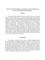
TEMPO-laccase. During the initial phase of the reaction, a fast decrease
in dissolved oxygen (DO) was observed, and consequently, the aldehyde
content rapidly increased in the first minutes (Fig. 1b). Hereafter, the
oxidation rate slightly decreased for the next 2 h until it reached a
plateau (Fig. 1a). The initial rate of oxygen consumption (measured for
the first 5 min) was 0.85 mmol kg−1 min−1. The decrease in DO is directly related to the demand of oxygen for TEMPO regeneration. As
more TEMPO is reduced for chitosan oxidation, laccase needs more
oxygen for the reoxidation of TEMPO (Scheme 1). The point of inflection in the curve (minimum DO) occurs around 45 min and is related to
the maximum number of active sites for the regeneration of TEMPO
(maximum oxygen demand). The subsequent increase in DO results
from a decrease in TEMPO regeneration. We hypothesize that the fast
replacement of the hydroxyl groups by carbonyl groups and the eventual formation of hemiacetals make the structure less accessible to
TEMPO, thereby reducing the formation rate of aldehydes. In addition,
it is possible that the DO can be decreased due to the degradation of
laccase in presence of oxoammonium salts, as reported by Arends et al.
(2006).
the TEMPO-laccase system is discussed by Arends et al. (2006).
In our design, chitosan was dissolved overnight in diluted acetic
acid or hydrochloric acid. Reactions were performed at different ratios
of TEMPO-chitosan and TEMPO-laccase, in order to optimise the conditions and to obtain products with different degrees of modification. In
Table 1, an overview of the different conditions studied is shown. The
combined use of laccase and TEMPO with continuously air supply
successfully catalyzed the oxidation of chitosan at 20 °C and pH 4.5.
Fig. 1 shows an example of the consumption of oxygen and formation of
aldehyde groups during the time course of oxidation mediated by
3.2. Structure and chemical characterization of oxidized chitosan
derivatives
3.2.1. Spectroscopic characterisation
FT-IR spectra from untreated chitosan and derived products are
shown in Fig. 2. The untreated chitosan reveals characteristics bands at
1645 cm−1 and 1583 cm−1 (Kumirska et al., 2010) for the stretching
and bending vibrations of amide I and amide II, respectively. The
spectrum of Product 1 (low TEMPO and low laccase) is quite similar to
the FT-IR spectrum of untreated chitosan, as expected due to the very
low level of substitution of this product. The band around 2873 cm−1
(CeH stretching, in eCH2 groups) was present for untreated chitosan
and was decreased for derivative products, particularly for Product 2.
This indicates that C-6 primary hydroxyls were converted to C-6 carbonyl and carboxylate groups.
The FT-IR spectrum of Product 2 indicates the presence of carboxyl
groups, as two new absorption bands appeared at 1593 cm−1 and
1404 cm−1, corresponding to symmetrical and asymmetrical stretching
vibration of COO−. A similar pattern was also reported by Pierre et al.
(2013) for chitosan oxidation using the TEMPO/NaOCl- NaBr system.
Fig. 1. Time course of dissolved oxygen (blue line) and formation of aldehyde groups (red
squares) during oxidation of chitosan (Product 3) mediated by the TEMPO-laccase
system. a) is full time course; b) Initial time course. (For interpretation of the references to
colour in this figure legend, the reader is referred to the web version of this article.)
302
Carbohydrate Polymers 186 (2018) 299–309
S. Botelho da Silva et al.
Fig. 2. FT-IR spectra of (a) chitosan, (b) Product 1, (c) Product 3, (d)
Product 4 and (e) Product 2.
1
H NMR results. In Fig. 3b, the resonance due to the aldehyde proton
(–CHO) appears around 9.1 ppm and the imine proton (eCH]Ne)
from Schiff’s base is clearly detected at 8.08 in the 1H NMR of Product
2. Azevedo et al. (2012) also reported the formation of Schiff’s base
detected by 1H NMR in aldehyde-functionalized chitosan obtained by
oxidation with nitrogen oxides.
To the best of our knowledge, this is the first time that the aldehydeand carboxylate-functionalization of chitosan by TEMPO-laccase oxidation is demonstrated. Previously, only the chemical approach using
TEMPO with halogenated reagents as primary oxidants has been reported (Bordenave, Grelier, & Coma, 2008; Kato, Kaminaga, Matsuo, &
Isogai, 2004; Pierre et al., 2013).
For Products 1, 3 and 4 these bands were hardly visible due to the low
concentration of COO− groups. These new bands can be also attributed
to acetates (CH3COO−) bonded to protonated amino groups, which
comes from the acetic acid used to dissolve chitosan prior to the oxidation reaction to obtain Products 1, 2 and 3. This hypothesis is supported by Mikhailov, Tuchkov, Lazarev, and Kulish (2014) and also by
Nunthanid et al. (2004) that reported a strong peak around
1550–1600 cm−1 and a weak peak near 1400 cm−1 in the FTIR spectrum of chitosan acetate.
The typical signal of aldehydic carbonyl groups around 1720 cm−1
was not detected since the aldehydes interact with neighboring alcohol
and amino groups. The new band at 791 cm−1 in Product 2 can be
attributed to hemiacetals formed by the interaction between aldehydes
and alcohol groups, while some changes in the wavenumber region of
1550–1650 cm−1 mainly present in Products 3 and 4 are related to the
formation of imine bonds between aldehyde and amino groups (Schiff
bases). DiFlavio et al. (2007) also reported the difficulty to detect the
aldehyde band in FT-IR spectra of regenerated cellulose treated with
TEMPO–NaBr–NaClO and cross-linked with polyvinylamine. This observation was explained by the interaction between aldehydes and alcohols and aldehydes and amines.
Further characterization of chitosan derivatives was performed by
1
H and 13C NMR (Fig. 3). The 13C NMR spectra of untreated chitosan as
a control (Fig. 3a) showed the typical signals of both GlcN and GlcNAc
residues, i.e. C2 (55.7 ppm), C6 (59.9 ppm), C3 (70.0 ppm), C5
(74.7 ppm), C4 (76.23 ppm), C1 (97.4 ppm) and the N-acetyl group of
GlcNAc (22.0 ppm), as reported previously by (Kumirska et al., 2010).
The spectrum of Product 2 (Fig. 3a) showed a reduction in the signal of
C6 and the appearance of additional peaks at ∼165–180 ppm suggesting the formation of a C6 oxidized chitosan derivative. The new
carbon resonance peaks appeared at 170.9 ppm and 174.9 ppm and are
attributed to the ion carboxylate carbon (eCOO−) and to the aldehyde
carbon (eCHO), respectively. The signal detected at 165.9 ppm is typical of imine carbon (eC]Ne) due to the formation of Schiff’s base
between the aldehyde and amine groups (Fig. 3a). The reduction in C6
resonance and the signal corresponding to (eCHO) were also observed
in Products 1, 3 and 4 (data not showed), however, the signal for
(eCOO) was not evident probably due to less drastic reaction conditions.
The aldehyde-functionalization of chitosan by the TEMPO-laccase
system as well as the formation of Schiff’s bases were also supported by
3.2.2. Degree of oxidation and aldehyde and carboxylate contents
The oxidation degree (OD) and the carboxylate and aldehyde contents of modified chitosan products are shown in Table 1 and support
the NMR and FT-IR analysis. The TEMPO-laccase oxidation introduced
predominantly carbonyl groups and low amounts of carboxyl functions
in chitosan. High carbonyl-carboxyl ratios are typical for TEMPO-laccase oxidation and it is related to the forming of hemiacetal linkages
between aldehydes and hydroxyl groups, which prevent further oxidation from carbonyl to carboxyl. Similar results were reported by
Jaušovec et al. (2015), Mathew & Adlercreutz (2009) and Patel et al.
(2011), that used TEMPO-laccase to oxidize cellulose and potato starch.
The concentration of TEMPO and laccase used in the reaction significantly affected the degree of oxidation and the content of aldehydes
and carboxyl groups. Comparing Product 1 with Product 2, a 10-fold
higher TEMPO and laccase (same laccase-TEMPO ratio) increased 7fold the aldehyde content and the oxidation degree whereas, the carboxylate content doubled (Table 1). If the TEMPO concentration was
increased 10-fold than the laccase-TEMPO ratio did not increase
(comparing P1 with P3 and P4), the aldehyde content increases only 4fold and the carboxylate content remained statistically the same
(Table 1). These results show the effect of fast TEMPO regeneration in
TEMPO-laccase oxidation, mainly on the production of carboxylate,
taken into account the range of concentrations tested. Higher TEMPO
concentration obviously demands more laccase for TEMPO regeneration. Products 3 and 4 were obtained with the same TEMPO concentration as Product 2 although with a lower laccase-TEMPO ratio, so
a lower number of TEMPO regeneration sites was available. As the
regeneration of TEMPO was slower for Products 3 and 4, less aldehyde
303
Carbohydrate Polymers 186 (2018) 299–309
S. Botelho da Silva et al.
Fig. 3.
13
C NMR (a) and 1H NMR (b) spectra from untreated chitosan (DD = 82.8%) and from modified chitosan (Product 2) obtained by TEMPO-laccase selective oxidation.
of C6-carbonyl and C6-carboxyl groups along the polymer chain, and
the random formation of aldehyde and carboxyl clusters, respectively.
Differences were observed between the type and substitution pattern of the enzyme-resistant oligomers obtained from the different
Products, which is mainly related to the concentration of TEMPO and
laccase used, as discussed above. These are:
has been produced and probably more hemiacetals were formed preventing the carboxylate groups formation. Similar effects of TEMPO and
laccase concentrations are also reported for cellulose by Jaušovec et al.
(2015) and Xu et al. (2013).
3.2.3. Substitution pattern along the polymer chain
To determine the distribution of the aldehyde and carboxyl groups
along the polysaccharide chain, the modified chitosan products were
subjected to enzymatic depolymerization and subsequently mass analysis of the degradation products. For degradation of chitosan and the
oxidized chitosan derivatives, a thermostable chitinase from
Myceliophthora thermophila C1 (Chi1), able to cleave the glycosidic
linkages GlcNAc-GlcNAc and GlcNAc-GlcN in chitin and chitosan, has
been used. The chitinase hydrolyzes the glycosidic linkages in a defined
way, but the introduction of carbonyl and carboxyl groups on the
polysaccharide chain sterically hinders the action of the enzyme
(Krolicka et al., 2018). Thus, in this way enzyme-resistant oligosaccharides are released. Identification of these enzyme-resistant oligosaccharides gives information about the substitution pattern of oxidized groups along the polymer chain.
MALDI-TOF-MS analysis of the chitosan and oxidized chitosan hydrolysates identified homo- and hetero-oligomers consisting of GlcNAc
and GlcN units with low polymerization degree (DP), namely dimers
(DP2) and trimers (DP3), as shown in Table 2. The main unsubstituted
chitosan oligosaccharides identified are (GlcN)2, (GlcNAc)2,
(GlcNAc,GlcN), (GlcNAc)3 and ((GlcNAc)2,GlcN). On the other hand,
the mass spectra of the digests of oxidized chitosan products contained
in addition a diversity of homo- and hetero-oligomers consisting of
GlcNAc and GlcN units with DP ranging from two to five, with aldehyde
and carboxyl substitution ranging from one to four (Table 2). Surprisingly, all oxidized oligosaccharides contained either carbonyl or carboxyl groups; oligomers containing both carbonyl and carboxyl substitution were not identified. The formation of larger oligosaccharides
with one or multiple substitutions suggests a heterogeneous distribution
• Low TEMPO, high enzyme (Product 1): low degree of oxidation,
short oligomers (DP2, DP3), low carbonyl/carboxyl ratio.
• High TEMPO (10% wt. vs chitosan) (Products 2, 3, 4): high degree of
•
oxidation, larger oligomers, high carbonyl/carboxyl ratio, random
clusters of CHO and COO− substituents.
In addition, for Product 4 low COO−, CHO clusters, but only one
short COO− oxidized oligomer (GlcNAc, GlcN) was identified, indicating a homogeneous distribution of COOH along the chain.
The differences in the substitution pattern between Products 3 and 4
seems also to be related to the acid used to dissolve chitosan before the
oxidation reaction, since the TEMPO and laccase concentration used in
both reactions were the same (Table 1). According to Thevarajah,
Bulanadi, Wagner, Gaborieau, and Castignolles (2016), the extent of
chitosan dissolution in HCl solution is higher than in acetic acid solution, however, HCl can induce additional deacetylation and a higher
depolymerization than acetic acid. The protonation of NH2 is also
higher with HCl than with acetic acid (Rinaudo, Pavlov, & Desbrières,
1999). These effects added to the formation of acetates in chitosan by
dissolution in acetic acid are sufficient to induce different changes in
the chain conformation depending on the used solvent. Therefore, the
regions susceptible to the attack of TEMPO during oxidation reaction
would be different for Products 3 and 4, as result of the different conformation of the chain in solution. This would explain the difference in
substitution pattern observed for the two products.
304
Carbohydrate Polymers 186 (2018) 299–309
S. Botelho da Silva et al.
Table 2
MALDI-TOF-MS identification of chitosan fragments obtained after enzymatic hydrolysis with chitinase Chi1 from Myceliophthora thermophila C1.
Ion composition
Type of adducts
Untreated Chitosan
Product 1
Product 2
(GlcN)2
GlcNAc,GlcN
(GlcNAc)2
(GlcNAc)2
(GlcNAc)2,GlcN
(GlcNAc)3
GlcNAc,GlcN
GlcNAc,GlcN
(GlcNAc)2
(GlcNAc)2,GlcN
GlcNAc, (GlcN)3
(GlcN)2
(GlcN)2
(GlcN)2
(GlcN)2
(GlcN)2
(GlcN)2
(GlcNAc)2,GlcN
(GlcNAc)2,GlcN
(GlcNAc)2,GlcN
(GlcNAc)2,GlcN
GlcNAc, (GlcN)2
(GlcNAc)3
(GlcNAc)3
(GlcN)4
GlcNAc, (GlcN)4
GlcNAc, (GlcN)4
[M+H]+
[M+Na]+
[M+H]+
[M+Na]+
[M+Na]+
[M+K]+
[M(COOH)+H]+
[M(COOH)2+K]+
[M(CHO)2+H]+
[M(COOH)+K]+
[M(CHO)4+H]+
[M(CHO)+H]+
[M(CHO)+Na]+
[M(CHO)2+K]+
[M(COOH)+K]+
[M(COOH)2+Na]+
[M(COOH)2+K]+
[M(CHO)+H]+
[M(CHO) + K]+
[M(COOH)+Na]+
[M(COOH)+K]+
[M(COOH)+Na]+
[M(CHO)3+Na]+
[M(COOH)3+Na]+
[M(CHO)4+K]+
[M(COOH)+Na]+
[M(CHO)4+Na]+
x
x
x
x
x
Product 3
Product 4
x
x
x
x
x
x
x
x
x
x
x
x
x
x
x
x
x
x
x
x
x
x
x
x
x
x
x
x
x
x
x
x
x
x
x
x
x
M (COOH), oxidized fragment: +14 Da mass shift, indicating a carboxyl group.
M (CHO), oxidized fragment: − 2 Da mass shift, indicating an aldehyde group.
3.3. Rheology of modified chitosan products in solution
3.4. Aqueous solubility and pH-responsive hydrogel
Fig. 4 shows the rheological characterization of untreated and
modified chitosan products with highest oxidation degree in 0.3 M
CH3COOH/0.2 M CH3COONa aqueous solution (pH = 4.6). In acid pH,
and at concentrations of less than 10 mg/mL, untreated and modified
chitosans solutions showed a Newtonian behavior. Modified products
showed an expressive decrease in dynamic viscosity compared with the
same concentration of untreated chitosan (Fig. 4). This change is related to the modification of the chitosan structure by the introduction of
hydrophilic carbonyl and carboxyl groups since the rheological parameters are strongly affected by the interaction between polymer and
solvent. Chitosans that produce low viscosity solutions are highly desirable for some biomedical applications such as blood thinning, cholesterol-lowering, and for application involving anti-oxidant, and antimicrobial properties (Lim, Lee, Israelachvili, Jho, & Hwang, 2015).
Chitosan has typically poor solubility at physiological pH, it is soluble in aqueous acid diluted solutions and this limits several of its
potential applications. The TEMPO-laccase oxidation overcomes this
problem since Products 2 and 3 show an increase in solubility in sodium
phosphate buffer at pH 7.4 (Fig. 5). As can be seen from the data in
Table 1 and Fig. 5, the oxidation degree and the carboxylate content are
important factors for improving the solubility of the chitosan derivatives. Within the concentrations tested, an approximately 4% OD and
10 mmol/kg of carboxylate seem to be the minimum values to observe
an increase in the solubility of the modified chitosan.
For Product 4, the oxidation of chitosan did not increase the solubility, in contrast, it produced a cross-linked structure capable of
forming a hydrogel at pH 7.4. Product 4 was produced using identical
conditions as for Product 3, except for the use of 0.1 M HCl to dissolve
chitosan instead of 0.1 M acetic acid before the oxidation reaction. This
caused a different substitution pattern of the aldehyde and carboxyl
Fig. 4. Dynamic viscosity for solutions of untreated chitosan and oxidized products at
different concentrations in 0.3 M CH3COOH/0.2 M CH3COONa (pH = 4.6). Untreated
chitosan (square), Product 2 (circle), Product 3 (triangle) and Product 4 (diamond).
Fig. 5. Aqueous solubility of untreated chitosan and Products 1, 2 and 3 in 0.1 M sodium
phosphate buffer (pH 7.4). Product 4 formed a hydrogel under these conditions.
305
Carbohydrate Polymers 186 (2018) 299–309
S. Botelho da Silva et al.
groups added to chitosan by the TEMPO-laccase oxidation, as explained
previously. In Product 4, high ratio CHO/COO−, CHO clustered and
low COO− favored the association of chains (minimized electrostatical
repulsion) and the interaction between aldehyde and free amino groups
via formation of imine bonds at pH 7.4. In Product 3, the association
between chains through imine bonds was not possible since acetate
groups were already bonded to amino groups. Nunthanid et al. (2004)
used chitosan acetate as a drug delivery and performed delivery tests in
pH 6.8 and in water. They demonstrated the stability of the chitosan
acetate in neutral pH, and in this condition, a sustained drug release
was observed.
The designed reaction conditions used to obtain Product 4 provide a
completely different product with highly attractive characteristics for
applications in the food and medical area. At low pH, this chitosan
derivative was fully soluble, as result of −NH2 protonation. Increasing
the pH from 4.5 to 6.5, the solution became gradually more viscous,
and around pH 6.5–7.0 (over the pKa of chitosan) a hydrogel was obtained. If more NaOH was added, the pH initially increased to 9 and
after that, the pH of the solution decreased spontaneously towards 7.0
and the solution got a little bit turbid. At approximately pH 7.5, a
product with a sponge-like appearance precipitated from the solution
(Fig. 6). Adding HCl, the pH decreased, and the “sponge” was maintained until ∼pH 4.0. Decreasing the pH even more with HCl, the
“sponge” started to dissolve very slowly, and total dissolution was
completed at a final ∼pH 3.7. The reversibility of the swelling/deswelling (sol-gel transition) of the polymer network was tested, showing
the same pH-responsiveness for more cycles.
The structure of chitosan hydrogel is attributed to self-crosslinking
amphiphilic chitosan network stabilized by dynamic imine bonds
(Schiff base between aldehyde and amino groups). The responsiveness
exhibited by chitosan derivative hydrogel is a consequence of a structure pH-dependent, which changes according to environmental stimuli,
allowing formation and rupture of imine bonds and protonation and
deprotonation of amino groups at different values of pH (Fig. 6). The
pH-responsiveness of this hydrogel was obtained due to higher amounts
of aldehyde than carboxylate groups substitution on the primary OH- of
C-6 of chitosan. This high aldehyde/carboxylate substitution ratio allowed sufficient carboxylate groups to prevent precipitation of chitosan
Fig. 7. SEM image of freeze-dried self-crosslinked hydrogel prepared with 3% TEMPOlaccase oxidized chitosan (Product 4) at pH 7.
at pH > 6, but low enough to allow self-crosslinking via imine bonds
between aldehyde and amino groups. Berger et al. (2004) also reported
a critical number of crosslinks per chain required to form a network and
the influence of the type of the cross-linker. Apart from that, and maybe
more important, the carboxylate groups were not clustered as determined by MALDI-TOF MS analysis. This finding has an important
role in the network formation via imine bonds, since electrostatic repulsion due to carboxylate is not so strong and allow the attraction of
the chains.
The self-crosslinked hydrogel obtained from oxidized chitosan
(Product 4) is clear and transparent (Fig. 6), and it can be formed with
concentrations as low as 1.5% (w/v). It was possible to produce chitosan hydrogels (Product 4) with a swelling ratio (swollen weight/dry
weight) as high as 30 (3000%) at pH 7.0. This swelling ratio is similar
to the one reported by Singh, Narvi, Dutta, and Pandey (2006) for
covalent hydrogels of chitosan prepared by crosslinking with formaldehyde as a crosslinking agent. A representative SEM image of the
freeze-dried gel that was produced with a concentration of 3% (w/v) at
pH 7 as described in Materials and Methods, is shown in Fig. 7. The
Fig. 6. Self-crosslinked chitosan pH-responsive hydrogel (Product 4).
306
Carbohydrate Polymers 186 (2018) 299–309
S. Botelho da Silva et al.
Fig. 8. TGA (a) and DTG (b) curves of untreated and oxidized chitosan
products obtained under nitrogen atmosphere at a heating rate of 10 °C/
min. Untreated chitosan (blue solid), Product 1 (purple long-dashed),
Product 2 (red short-dashed), Product 3 (gray dash-dotted) and Product 4
(green dash-double-dotted). (For interpretation of the references to colour
in this figure legend, the reader is referred to the web version of this article.)
pH the derivative is de-swelling, and the drug could be realized
(Bhattarai et al., 2010; Du et al., 2015). Another potential application is
anticancer hydrogels prepared in situ, delivering drugs directly to the
tumor that have a slightly lower pH (6.5–6.8) than the normal physiological pH (Li, Hu et al., 2015; Li, Gu et al., 2015).
oxidized chitosan gel exhibits a high surface area with a porous structure with random pore size distribution. Similar porous structures are
reported by Tan et al. (2009) for crosslinked hydrogels obtained from
the reaction between N-succinyl-chitosan and aldehyde hyaluronic
acid. Nevertheless, the authors did not report any information about pH
responsiveness. To the best of our knowledge, this is the first report of
pH-responsive hydrogels formed by self-crosslinked chitosan. Azevedo
and Kumar (2012) modified the chitosan by introducing aldehyde
groups into the molecule by nitrous oxide treatment and thereby,
producing also hydrogels without the use of external cross-linkers although they did not report pH-responsiveness. The use of external
cross-linker represents a drawback since most of them can react irreversibly with −NH2 groups and prevent further stimuli-responsive effects (Bhattarai, Gunn, & Zhang, 2010). And, more important, some
cross-linkers are toxic or do not have recognized biocompatibility,
therefore they cannot be used for application in biomedical and pharmaceutical areas (Bhattarai et al., 2010; Du, Liu, Yang, & Zhai, 2015).
Among the potential applications of pH-responsive hydrogels, we
highlight the platforms for gastrointestinal drug delivery (Du et al.,
2015; Park, Kang, Lee, Kim, & Son, 2013; Woraphatphadung et al.,
2016) and for injectable hydrogels prepared in situ (Li, Hu et al., 2015;
Li, Gu et al., 2015). The pH-responsive chitosan hydrogel showed a solgel transition approximately around physiological pH (7.4). This
finding indicates a potential application for this hydrogel to platforms
for delivery drugs directed to the stomach (pH 1–3) for example, in this
3.5. Thermal stability
To evaluate the effect of the TEMPO-laccase oxidation on the
thermal stability of chitosan we performed thermogravimetric analysis
(TGA-DTG) of untreated chitosan and its oxidized products. TGA and
DTG curves are shown in Fig. 8. Untreated chitosan, Products 1 and 4
show clearly two stages of weight loss, with approximately 65% of mass
loss from 25 to 600 °C. In the same interval, Products 2 and 3 lose more
than 85% of their weight. The first stage of weight loss of all samples
occurs in the range between 25 and 100 °C, and it is mainly attributed
to water loss. The following stages are related to the thermal degradation of the polymer itself and include the deacetylation of chitosan, chain depolymerization, and disintegration of other intra and
inter-molecular interactions (L. Tian, Tan, Li, & You, 2015). The range
where this thermal event occurs is characteristic for each product and
shows that the derivatives are less thermally stable than their parent
chitosan.
The maximum degradation temperature for untreated chitosan was
observed at 300 °C, and it shifted to lower temperatures for the oxidized
307
Carbohydrate Polymers 186 (2018) 299–309
S. Botelho da Silva et al.
products and tended to occur in broader ranges. It is important to note
that this decrease in the thermal stability is closely related to the oxidation degree (Table 1), meaning higher aldehyde and carboxylate
groups by TEMPO-laccase oxidation leads to lower temperature of degradation. Wei, Li, Tian, Xu, and Jin (2015) reported similar thermal
degradation behavior for hypochlorite-oxidized starch nanocrystals
treated by different concentrations of chlorine, in which higher oxidation also resulted in higher thermal degradation. Although Products 3
and 4 showed the same oxidation degree (Table 1), the random distribution of oxidized groups and the consequent crosslinked structure in
Product 4 contributed for a lower temperature of degradation in the
second stage, similar to effects reported by Neto et al. (2005) for chitosan crosslinked by glutaraldehyde. Nevertheless, in the range
300–600 °C, the thermal stability of Product 4 was superior to Product 3
and similar to untreated chitosan.
Another aspect that can be concluded from the thermal analysis is
related to the changes in molecular weight induced by the TEMPOlaccase oxidation. Mao et al. (2004) and Tian et al. (2015) showed that
the thermal stability of chitosan significantly decreases with the reduction of the molecular weight. Considering the small variation in
degradation temperature observed for the oxidized products, it could be
assumed that the depolymerization during the oxidation reaction was
not intense.
and interactions in covalently and ionically crosslinked chitosan hydrogels for biomedical applications. European Journal of Pharmaceutics and Biopharmaceutics, 57(1),
19–34.
Bhattarai, N., Gunn, J., & Zhang, M. (2010). Chitosan-based hydrogels for controlled,
localized drug delivery. Advanced Drug Delivery Reviews, 62(1), 83–99.
Bordenave, N., Grelier, S., & Coma, V. (2008). Advances on selective C-6 oxidation of
chitosan by TEMPO. Biomacromolecules, 9(9), 2377–2382.
Bragd, P. L., Besemer, A. C., & van Bekkum, H. (2000). Bromide-free TEMPO-mediated
oxidation of primary alcohol groups in starch and methyl α-D-glucopyranoside.
Carbohydrate Research, 328(3), 355–363.
Bragd, P. L., van Bekkum, H., & Besemer, A. C. (2004). TEMPO-Mediated oxidation of
polysaccharides: Survey of methods and applications. Topics in Catalysis, 27(1–4),
49–66.
Coseri, S., Biliuta, G., Simionescu, B. C., Stana-Kleinschek, K., Ribitsch, V., & Harabagiu,
V. (2013). Oxidized cellulose—Survey of the most recent achievements. Carbohydrate
Polymers, 93(1), 207–215.
Díaz-Rodríguez, A., Martínez-Montero, L., Lavandera, I., Gotor, V., & Gotor-Fernández, V.
(2014). Laccase/2,2,6,6-tetramethylpiperidinoxyl radical (TEMPO): An efficient
catalytic system for selective oxidations of primary hydroxy and amino groups in
aqueous and biphasic media. Advanced Synthesis and Catalysis, 356(10), 2321–2329.
DiFlavio, J.-L., Pelton, R., Leduc, M., Champ, S., Essig, M., & Frechen, T. (2007). The role
of mild TEMPO-NaBr-NaClO oxidation on the wet adhesion of regenerated cellulose
membranes with polyvinylamine. Cellulose, 14(3), 257–268.
Du, H., Liu, M., Yang, X., & Zhai, G. (2015). The design of pH-sensitive chitosan-based
formulations for gastrointestinal delivery. Drug Discovery Today, 20(8), 1004–1011.
Filisetti-Cozzi, T. M. C. C., & Carpita, N. C. (1991). Measurement of uronic acids without
interference from neutral sugars. Analytical Biochemistry, 197(1), 157–162.
Hirai, A., Odani, H., & Nakajima, A. (1991). Determination of degree of deacetylation of
chitosan by 1H NMR spectroscopy. Polymer Bulletin, 26(1), 87–94.
Huang, J., Chen, W.-W., Hu, S., Gong, J.-Y., Lai, H.-W., Liu, P., ... Mao, J.-W. (2013).
Biochemical activities of 6-carboxy β-chitin derived from squid pens. Carbohydrate
Polymers, 91(1), 191–197.
Jaušovec, D., Vogrinčič, R., & Kokol, V. (2015). Introduction of aldehyde vs. carboxylic
groups to cellulose nanofibers using laccase/TEMPO mediated oxidation.
Carbohydrate Polymers, 116(13), 74–85.
Kardas, I., Struszczyk, M. H., Kucharska, M., van den Broek, L. A., van Dam, J. E., &
Ciechańska, D. (2013). Chitin and chitosan as functional biopolymers for industrial
applications. In P. Navard (Ed.). The European polysaccharide network of excellence
(EPNOE) (pp. 329–373). Wien: Springer.
Kasaai, M. R. (2007). Calculation of Mark-Houwink-Sakurada (MHS) equation viscometric constants for chitosan in any solvent-temperature system using experimental
reported viscometric constants data. Carbohydrate Polymers, 68(3), 477–488.
Kato, Y., Kaminaga, J., Matsuo, R., & Isogai, A. (2004). TEMPO-mediated oxidation of
chitin, regenerated chitin and N-acetylated chitosan. Carbohydrate Polymers, 58(4),
421–426.
Kierulff, J. V. (2000). Modification of polysaccharides by means of a phenol oxidizing
enzyme. Patent US6087135A.
Kong, M., Chen, X. G., Xing, K., & Park, H. J. (2010). Antimicrobial properties of chitosan
and mode of action: A state of the art review. International Journal of Food
Microbiology, 144(1), 51–63.
Krolicka, M., Hinz, S. W., Koetsier, M., Joosten, R., Eggink, G., van den Broek, L. A., &
Boeriu, C. G. (2018). Chitinase Chi1 from Myceliophthora thermophila C1, a thermostable enzyme for chitin and chitosan depolymerization. Journal of Agriculture and
Food Chemistry Accepted for publication.
Kumirska, J., Czerwicka, M., Kaczyński, Z., Bychowska, A., Brzozowski, K., Thöming, J., &
Stepnowski, P. (2010). Application of spectroscopic methods for structural analysis of
chitin and chitosan. Marine Drugs, 8(5), 1567–1636.
Li, L., Gu, J., Zhang, J., Xie, Z., Lu, Y., Shen, L., ... Wang, Y. (2015). Injectable and
biodegradable pH-responsive hydrogels for localized and sustained treatment of
human fibrosarcoma. ACS Applied Materials & Interfaces, 7(15), 8033–8040.
Li, J., Hu, W., Zhang, Y., Tan, H., Yan, X., Zhao, L., & Liang, H. (2015). pH and glucose
dually responsive injectable hydrogel prepared by in situ crosslinking of phenylboronic modified chitosan and oxidized dextran. Journal of Polymer Science Part A:
Polymer Chemistry, 53(10), 1235–1244.
Lim, C., Lee, D. W., Israelachvili, J. N., Jho, Y., & Hwang, D. S. (2015). Contact time- and
pH-dependent adhesion and cohesion of low molecular weight chitosan coated surfaces. Carbohydrate Polymers, 117(0), 887–894.
Luo, Y., & Wang, Q. (2013). Recent advances of chitosan and its derivatives for novel
applications in food science. Journal of Food Procesing & Beverages, 1, 13.
Mao, S., Shuai, X., Unger, F., Simon, M., Bi, D., & Kissel, T. (2004). The depolymerization
of chitosan: Effects on physicochemical and biological properties. International
Journal of Pharmaceutics, 281, 45–54.
Mathew, S., & Adlercreutz, P. (2009). Mediator facilitated, laccase catalysed oxidation of
granular potato starch and the physico-chemical characterisation of the oxidized
products. Bioresource Technology, 100(14), 3576–3584.
Mikhailov, G. P., Tuchkov, S. V., Lazarev, V. V., & Kulish, E. I. (2014). Complexation of
chitosan with acetic acid according to Fourier transform Raman spectroscopy data.
Russian Journal of Physical Chemistry A, 88(6), 936–941.
Mourya, V. K., Inamdar, N. N., & Choudhari, Y. M. (2011). Chitooligosaccharides:
Synthesis, characterization and applications. Polymer Science Series A, 53(7),
583–612.
Neto, C. G. T., Giacometti, J. A., Job, A. E., Ferreira, F. C., Fonseca, J. L. C., & Pereira, M.
R. (2005). Thermal analysis of chitosan based networks. Carbohydrate Polymers,
62(2), 97–103.
Niku-Paavola, M.-L., Karhunen, E., Salola, P., & Raunio, V. (1988). Ligninolytic enzymes
of the white-rot fungus Phlebia radiata. Biochemical Journal, 254, 877–884.
4. Conclusions
In this work, the TEMPO-laccase catalytic redox system was successfully applied for the first time in C-6 oxidation of chitosan. The
derivative products were characterized and the effective formation of
carboxyl and aldehyde groups on chitosan was demonstrated. The
oxidation degree and distribution of functional groups were affected by
TEMPO and laccase concentration and by the acid solvent used to
dissolve chitosan prior to TEMPO-oxidation. The modification of chitosan structure by TEMPO-laccase oxidation provides an improvement
in water solubility and a decrease in the viscosity of solutions of oxidized products in acid pH. A slightly reduction on thermal stability was
observed after TEMPO-laccase oxidation without showing evidence of
intense depolymerization.
If chitosan was dissolved in hydrochloric acid prior to TEMPO-laccase oxidation than oxidized chitosan hydrogel was formed. This hydrogel was clear and transparent and showed pH-responsiveness. The
reversibility exhibited by this chitosan hydrogel is a consequence of an
amphiphilic structure pH-dependent stabilized by reversible covalent
imine bonds. This new material has a great potential for development of
applications in medical and food area.
Acknowledgements
The work of S. Botelho da Silva was supported by the National
Research Council of Brazil - CNPq [Process 249593/2013-0]. We thank
Sandra Hinz and Martijn Koetsier for their support by providing the
crude chitinase and for useful discussions on chitinase purification. The
work of M. Krolicka received funding from the Netherlands
Organisation for Scientific Research (NWO) in the framework of the
TASC Technology Area BIOMASS.
References
Arends, I. W. C. E., Li, Y.-X., & Sheldon, R. A. (2006). Stabilities and rates in the laccase/
TEMPO-catalyzed oxidation of alcohols. Biocatalysis and Biotransformation, 24(6),
443–448.
Azevedo, E. P., & Kumar, V. (2012). Rheological, water uptake and controlled release
properties of a novel self-gelling aldehyde functionalized chitosan. Carbohydrate
Polymers, 90(2), 894–900.
Azevedo, E. P., Santhana Mariappan, S. V., & Kumar, V. (2012). Preparation and characterization of chitosans carrying aldehyde functions generated by nitrogen oxides.
Carbohydrate Polymers, 87(3), 1925–1932.
Berger, J., Reist, M., Mayer, J. M., Felt, O., Peppas, N. A., & Gurny, R. (2004). Structure
308
Carbohydrate Polymers 186 (2018) 299–309
S. Botelho da Silva et al.
Sheldon, R. A., & Arends, I. W. C. E. (2004). Organocatalytic oxidations mediated by
nitroxyl radicals. Advanced Synthesis & Catalysis, 346(9–10), 1051–1071.
Singh, A., Narvi, S. S., Dutta, P. K., & Pandey, N. D. (2006). External stimuli response on a
novel chitosan hydrogel crosslinked with formaldehyde. Bullentin of Materials Science,
29(3), 233–238.
Tan, H., Chu, C. R., Payne, K. A., & Marra, K. G. (2009). Injectable in situ forming biodegradable chitosan–hyaluronic acid based hydrogels for cartilage tissue engineering.
Biomaterials, 30(13), 2499–2506.
Thevarajah, J. J., Bulanadi, J. C., Wagner, M., Gaborieau, M., & Castignolles, P. (2016).
Towards a less biased dissolution of chitosan. Analytica Chimica Acta, 935, 258–268.
Tian, M., Tan, H., Li, H., & You, C. (2015). Molecular weight dependence of structure and
properties of chitosan oligomers. RSC Advances, 5(85), 69445–69452.
Wei, B., Li, H., Tian, Y., Xu, X., & Jin, Z. (2015). Thermal degradation behavior of hypochlorite-oxidized starch nanocrystals under different oxidized levels. Carbohydrate
Polymers, 124, 124–130.
Wolf, B. A. (2007). Polyelectrolytes revisited: Reliable determination of intrinsic viscosities. Macromolecular Rapid Communications, 28(2), 164–170.
Woraphatphadung, T., Sajomsang, W., Gonil, P., Treetong, A., Akkaramongkolporn, P.,
Ngawhirunpat, T., & Opanasopit, P. (2016). pH-Responsive polymeric micelles based
on amphiphilic chitosan derivatives: Effect of hydrophobic cores on oral meloxicam
delivery. International Journal of Pharmaceutics, 497(1), 150–160.
Xu, S., Song, Z., Qian, X., & Shen, J. (2013). Introducing carboxyl and aldehyde groups to
softwood-derived cellulosic fibers by laccase/TEMPO-catalyzed oxidation. Cellulose,
20(5), 2371–2378.
Nunthanid, J., Laungtana-anan, M., Sriamornsak, P., Limmatvapirat, S.,
Puttipipatkhachorn, S., Lim, L. Y., & Khor, E. (2004). Characterization of chitosan
acetate as a binder for sustained release tablets. Journal of Controlled Release, 99(1),
15–26.
Park, B. G., Kang, H. S., Lee, W., Kim, J. S., & Son, T. I. (2013). Reinforcement of pHresponsive γ-poly (glutamic acid)/chitosan hydrogel for orally administrable colontargeted drug delivery. Journal of Applied Polymer Science, 127(1), 832–836.
Patel, I., Ludwig, R., Haltrich, D., Rosenau, T., & Potthast, A. (2011). Studies of the
chemoenzymatic modification of cellulosic pulps by the laccase-TEMPO system.
Holzforschung, 65(4), 475.
Pei, J., Yin, Y., Shen, Z., Bu, X., & Zhang, F. (2016). Oxidation of primary hydroxyl groups
in chitooligomer by a laccase–TEMPO system and physico-chemical characterisation
of oxidation products. Carbohydrate Polymers, 135, 234–238.
Persin, Z., Stana-Kleinschek, K., Foster, T. J., van Dam, J. E. G., Boeriu, C. G., & Navard, P.
(2011). Challenges and opportunities in polysaccharides research and technology:
The EPNOE views for the next decade in the areas of materials, food and health care.
Carbohydrate Polymers, 84(1), 22–32.
Pierre, G., Salah, R., Gardarin, C., Traikia, M., Petit, E., Delort, A.-M., ... Michaud, P.
(2013). Enzymatic degradation and bioactivity evaluation of C-6 oxidized chitosan.
International Journal of Biological Macromolecules, 60(0), 383–392.
Prashanth, K. H., & Tharanathan, R. (2007). Chitin/chitosan: Modifications and their
unlimited application potential—An overview. Trends in Food Science & Technology,
18(3), 117–131.
Rinaudo, M., Pavlov, G., & Desbrières, J. (1999). Influence of acetic acid concentration on
the solubilization of chitosan. Polymer, 40(25), 7029–7032.
309