Chemical structure of a partially 3-O-methylated mannofucogalactan from edible mushroom Grifola frondosa
Bạn đang xem bản rút gọn của tài liệu. Xem và tải ngay bản đầy đủ của tài liệu tại đây (811.16 KB, 8 trang )
Carbohydrate Polymers 187 (2018) 110–117
Contents lists available at ScienceDirect
Carbohydrate Polymers
journal homepage: www.elsevier.com/locate/carbpol
Chemical structure of a partially 3-O-methylated mannofucogalactan from
edible mushroom Grifola frondosa
T
Gracy Kelly Faria Oliveiraa, Estefania Viano da Silvaa,b, Andrea Caroline Ruthesc,d,
⁎
Luciano Morais Liãob, Marcello Iacominic, Elaine R. Carboneroa,
a
Departamento de Qmica, Universidade Federal de Goiás, Regional Catalão, 75704-020 Catalão, Brazil
Laboratório de Ressonância Magnética Nuclear, Instituto de Química, Universidade Federal de Goiás, Campus Samambaia, 74001-970 Goiânia, Brazil
c
Departamento de Bioquímica e Biologia Molecular, Universidade Federal do Paraná, 81531-980 Curitiba, Brazil
d
Department of Entomology and Nematology, University of Florida, GCREC, 14625 County Road 672, Wimauma, FL 33598, United States
b
A R T I C L E I N F O
A B S T R A C T
Keywords:
Medicinal mushroom
Grifola frondosa
Mannofucogalactan
Chemical structure
An unusual heteropolysaccharide was isolated from the fruiting bodies of the medicinal mushroom Grifola
frondosa, via successive cold aqueous extraction, followed by fractionation through freeze-thawing, precipitation
with Fehling solution and dialysis using a membrane with a size exclusion cut-off of 500 kDa. Its chemical
structure was determined based on total acid hydrolysis, methylation analysis and NMR studies. The mannofucogalactan had a molar mass of 15.9 × 10³ g mol−1, which was determinate by HPSEC-MALLS. This heteropolymer showed to have a main chain of (1 → 6)-linked α-D-Galp partially substituted at O-2 by 3-O-α-D-mannopyranosyl-α-L-fucopyranosyl groups and in a minor proportion with α-L-Fucp single-unit side chains.
Moreover, the presence of 3-O-Me-Galp units could also be observed in the main chain of the G. frondosa
mannofucogalactan.
1. Introduction
isolated from the cultured fruiting bodies (Ohno et al., 1984), matted
mycelia (Ohno et al., 1985) and liquid culture supernatant (Ohno et al.,
1986) of G. frondosa (Fang et al., 2012). Grifolans are characterized as
β-D-glucans (1 → 3)-linked in the backbone with a single (1 → 6)-linked
β-D-glucosyl side branching unit on every third residue.
In addition to β-D-glucans, some heteropolysaccharides showing
different compositions, most of them biologically active, have been
obtained from G. frondosa (Cui et al., 2007; Masuda et al., 2009;
Masuda, Ito, Konishi, & Nanba, 2010; Mizuno,Ohsawa, Hagiwara, &
Kuboyama, 1986; Xu, Liu, Shen, Fei, & Chen, 2010; Wang et al., 2014).
With the exception of the acid heteropolysaccharide, named GFPS1b,
obtained from cultured mycelia of G. frondosa (Cui et al., 2007), and the
water-soluble polysaccharide named GFPW from the fruiting bodies of
this mushroom (Wang et al., 2014), the primary structures of the heteropolymers have not been unambiguously elucidated. GFPS1b showed
to have a backbone consisting of (1 → 4)-linked α-D-Galp and (1 → 3)linked α-D-Glcp residues, the latter being partially substituted at O-6 by
4-O-α-L-arabinofuranosyl-α-D-glucopyranosyl groups, which showed to
be effective in the inhibition of proliferation of mammary tumor MCF-7
cells in vitro (Cui et al., 2007). The other heteropolysaccharide chemically elucidate was the fraction GFPW, which had a main chain of
(1 → 6)-linked α-D-Galp residues, with branches of (1 → 3)-linked
Mushrooms have been valued as edible and medicinal resources.
Grifola frondosa (Maitake), a basidiomycete belonging to the
Polyporaceae family, may be one of the most versatile and promising
medicinal mushroom used as a dietary supplement (Wu et al., 2006). It
have been widely used in Japan, China and Korea as a traditional food
additive (Gu et al., 2007) and is one of the most valuable and expensive
mushrooms (Mayell, 2001).
Since the beginning of its cultivation in 1981, the study of its
medicinal applications has been ongoing, and the activity of its purified
polysaccharides has been highlighted (Mayell, 2001). Over the past
three decades, many polysaccharides have been isolated from the
fruiting bodies of G. frondosa and showed antitumor activity (Masuda
et al., 2009), besides of antihypertensive (Konno, 2007; Talpur et al.,
2002) anti-diabetic (Gu et al., 2007), and anti-hyperliposis effects (He
et al., 2017; Minamino, Nagasawa, & Othtsuru, 2008).
Most of the polysaccharides from G. frondosa fruiting bodies were
characterized as D-glucans with different linkage types, such as β-(1 →
3), β-(1 → 6) and α-(1 → 4) (He et al., 2017; Wasser, 2002). Grifolan
(GRN) is the best known and most potent substances with antitumor
and immunomodulating properties (Borchers, Keen, & Gershwin, 2004)
⁎
Corresponding author.
E-mail address: (E.R. Carbonero).
/>Received 17 November 2017; Received in revised form 16 January 2018; Accepted 23 January 2018
Available online 31 January 2018
0144-8617/ © 2018 Elsevier Ltd. All rights reserved.
Carbohydrate Polymers 187 (2018) 110–117
G.K.F. Oliveira et al.
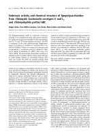
Fig. 1. (A) Scheme of extraction and purification of the
heterogalactan from fruiting bodies of G. frondosa. (B)
Elution profile of fraction EFP-Gf determined by HPSECMALLS using light scattering (—) and refractive index detectors (—).
bioactive, it is important to know the fine chemical structure of those
compounds in an attempt to determine the structure-activity relationship. Thus, at the present study the isolation and structural characterization of a different heteropolysaccharide, a partially methylated
fucose residues and α-terminal mannose substituting the O-2 position
(Wang et al., 2014).
Novel polysaccharides from G. frondosa have been frequently isolated, purified and evaluated. As most of them have been shown to be
111
Carbohydrate Polymers 187 (2018) 110–117
G.K.F. Oliveira et al.
Table 1
Partially O-methylated alditol acetates formed on methylation analysis of the EFP-Gf fraction obtained from the fruiting bodies of G. frondosa.
Partially O-methylated alditol acetatea
Linkage typeb
RT (min)c
Fraction (mol%)
Mass fragmentation (m/z)
2,3,4-Me3-Fuc
2,3,4,6-Me4-Man
2,4-Me2-Fuc
2,3,4-Me3-Gal
3,4-Me2-Gal
Fucp-(1→
Manp-(1→
→3)-Fucp-(1→
→6)-Galp-(1→
→2,6)-Galp-(1→
14.708
15.725
15.945
18.221
20.114
6.8
19.6
19.3
27.9
26.4
89,102,115,118,131,162,175
87,102,118,129,145,161,205
89,101,118,131,160,234
87,102,118,129,162,173,189,233
87,100,129,159,173,189,233
a
b
c
Analyzed by GC–MS after methylation, total acid hydrolysis, reduction (NaBD4) and acetylation.
Based on derived O-methylalditol acetates.
Retention time (minutes).
Fig. 2.
13
C NMR spectrum of mannofucogalactan (EFP-Gf fraction) from G. frondosa. EFP-Gf, analyzed in D2O at 50 °C (chemical shifts are expressed in δ ppm).
deionized with mixed ion-exchange resins. During the treatment with
ion-exchange resins, a part of these fractions became precipitated (pFPGf and pFS-Gf fractions, respectively), being separated by centrifugation (3000 rpm at 20 °C for 20 min). Fehling treatment was repeated
two more cycles under fraction sFP-Gf to ensure that no residue of the
supernatant was present in the precipitated fraction, giving the fraction
FP3-Gf.
FP3-Gf fraction was further purified by closed dialysis through a
membrane with a 500 kDa Mw cut-off (Spectra/Por® PVDF), giving rise
to a retained (RFP-Gf) and an eluted (EFP-Gf) material (Fig. 1A).
mannofucogalactan from the fruiting bodies of G. frondosa is described.
2. Material and methods
2.1. Biological material
Fresh basidiocarps (fruiting bodies) of Grifola frondosa (Dicks.) Gray
(1.03 kg) were provided by YURI Cogumelos (Owner: Iwao Akamatsu),
located in Sorocaba, State of São Paulo, Brazil, in May of 2010.
2.2. Extraction and purification of polysaccharides
The fresh fruiting bodies of G. frondosa (1.03 kg) were freeze-dried,
resulting in 209.6 g, which were pulverized and their polysaccharides
were extracted with water at 10 °C for 6 h (×6, 2000 mL). The combined aq. extracts were evaporated to a small volume and added to
excess ethanol (EtOH, 3:1; v/v) to precipitate polysaccharides, which
were collected after centrifugation at 3000 rpm at 20 °C for 20 min. The
precipitate was then dissolved in H2O, dialyzed against distilled water
for 20 h to remove low-molecular-weight carbohydrates, and freezedried (CW-Gf fraction). The fraction CW-Gf was then dissolved in distilled water and the solution submitted to a freeze-thawing process
furnishing a cold water-soluble (SCW-Gf) and an insoluble fraction
(ICW-Gf), which were separated under the same centrifugation conditions. The soluble portion (SCW-Gf) was treated with Fehling solution
(Jones & Stoodley, 1965) and the precipitated material (FPCW-Gf)
centrifuged off. Both fractions, FPCW-Gf (precipitate) and FSCW-Gf
(supernatant) were neutralized with HOAc, dialyzed against tap water,
2.3. Monosaccharide composition
Monosaccharide components of the polysaccharides were identified
and their ratios were determined following hydrolysis with 2 M trifluoroacetic acid (TFA) for 8 h at 100 °C, and conversion to alditol
acetates by successive NaBH4 and/or NaBD4 reduction, and acetylation
with Ac2O-pyridine (1:1, v/v) for 12 h at room temperature (Thompson,
1963a,1963b;). The resulting alditol acetates were analyzed by gas
chromatography–mass spectrometry (GC–MS) using a Varian model
3300 gas chromatograph linked to a Finnigan Ion-Trap, Model 810-R12
mass spectrometer. A DB-225 capillary column (30 m × 0.25 mm i.d.)
held at 50 °C during injection and later programmed to 220 °C (constant
temperature) at 40 °C min−1 was used for qualitative and quantitative
analysis of alditol acetates. The alditol acetates were identified by their
typical retention times and electron impact profiles.
112
Carbohydrate Polymers 187 (2018) 110–117
G.K.F. Oliveira et al.
Fig. 3. HSQC (A) spectrum of mannofucogalactan (EFP-Gf fraction)
from G. frondosa, with amplified inserts of the: C-6 region of Fucp (A1);
HSQC-DEPT C-6 region (B). EFP-Gf, analyzed in D2O at 50 °C (chemical shifts are expressed in δ ppm).
A = non reducing ends α-Man; B = α-Fucp substituted at O-3 by αManp; C = non reducing ends α-Fucp; D = 2,6-di-O- substituted αGalp units; E = 6-O- substituted α-Galp; F = 6-O- substituted 3-O-Meα-Galp units.
then held for 5 min), 150 °C (45 °C min−1, then held for 5 min), 200 °C
(55 °C min−1, then held for 15 min), 250 °C (65 °C min−1, then held for
10 min), and to 270 °C (50 °C min−1 and held for 10 min). Helium was
used as the carrier gas at a flow rate of 1.0 mL min−1. Partially O-methylated alditol acetates were identified from m/z of their positive ions,
by comparison with standards, the results being expressed as a relative
percentage of each component (Sassaki, Gorin, Souza, Czelusniak, &
Iacomini, 2005).
2.4. Determination of homogeneity of polysaccharides and their molar mass
(Mw)
The homogeneity and molar mass (Mw) of the fractions were determined using a Waters high-performance size-exclusion chromatography (HPSEC) apparatus coupled to a differential refractometer (RI)
and a Wyatt Technology Dawn-F Multi-Angle Laser Light Scattering
detector (MALLS). The eluent was 0.1 M NaNO3, containing 0.5 g L−1
NaN3. The polysaccharide solutions were filtered through a membrane
with 0.22 μm diameter pores (Millipore). The specific refractive index
increment (dn/dc) was determined using a Waters 2410 detector, the
samples being dissolved in the eluent, five increasing concentrations,
ranging from 0.2 to 1.0 mg mL−1 being used to determine the slope of
the increment.
2.6. Partial acid hydrolysis of heterogalactan
Fraction EFP-Gf (60 mg) was partially hydrolyzed with 0.2 M TFA
(2 mL) for 3 h at 100 °C. After neutralization with NaOH, the material
was dialyzed (2 kDa cut-off membrane) against distilled water. The retained fraction (HEFP-Gf) was lyophilized and analyzed by NMR spectroscopy.
2.5. Methylation analysis
2.7. Nuclear magnetic resonance (NMR) spectroscopy
Per-O-methylation of purified EFP-Gf fraction was carried out by the
method of Ciucanu and Kerek (1984). Briefly, the sample (10 mg) was
dissolved in dimethyl sulfoxide (Me2SO; 500 μL), powdered NaOH
(20 mg) and iodomethane (CH3I; 500 μL) were added. After 30 min at
25 °C with vigorous stirring, the mixture was maintained overnight at
25 °C. The reaction was interrupted by addition of water, neutralization
with HOAc, and the products were isolated by partition between CHCl3
and water. The per-O-methylated derivatives from the lower layer were
hydrolyzed with 1 M TFA (500 uL) for 4 h at 100 °C, followed by NaBD4
reduction and acetylation as above (item 2.3), to give a mixture of
partially O-methylated alditol acetates, which was analyzed by GC–MS
using an Agilent 7820A gas chromatograph interfaced to an Agilent
5975E quadrupole mass spectrometer, fitted with split/splitless capillary inlet system, an Agilent G4513A autosampler, and a capillary HP5MS column. The injector temperature was maintained at 250 °C, with
the oven increasing from 75 °C (hold 1 min) to 100 °C (35 °C min−1,
NMR spectra (1H, 13C, COSY, HSQC-DEPT, HSQC-TOCSY, HMBC,
HSQC-NOESY and coupled HSQC) were obtained using a 500 MHz
Bruker Avance spectrometer incorporating Fourier transform. Analyses
were performed at 50 °C on samples dissolved in D2O or Me2SO-d6.
Chemical shifts are expressed in δ relative to Me4Si (TMS; δ = 0) or
Me2SO-d6 (δ = 39.70 and 2.50 for 13C and 1H signals, respectively).
3. Results and discussion
G. frondosa was shown to contain 79.1% moisture on desiccation in
a freeze dryer, and the product was submitted to aqueous extraction at
10 °C.
The extracted polysaccharides were recovered by ethanol precipitation, dialyzed against tap water, and the solution freeze-dried,
113
Carbohydrate Polymers 187 (2018) 110–117
G.K.F. Oliveira et al.
giving CW-Gf fraction (8.0 g) (Fig. 1A), which showed to be composed
by glucose (Glc, 44%) as its main component, in addition to fucose
(Fuc, 10%), mannose (Man, 24%), and galactose (Gal, 22%), according
to GC–MS of derived alditol acetates. Fractionation of the CW-Gf by
freeze/thawing process furnished water-soluble (SCW-Gf, 3.7 g) and
insoluble (ICW-Gf, 2.8 g) polysaccharidic fractions, which were separated by centrifugation. SCW-Gf was composed of Fuc (7%), Man
(39%), 3-O-methyl-galactose (3-O-Me-Gal, 2%) (confirmed by GC–MS
ions at m/z 130 and 190 after reduction with NaBD4 and acetylation),
galactose (27%), and glucose (25%), and its HPSEC–MALLS analysis
showed heterogeneity.
In order to obtain a purified sample, the soluble fraction (SCW-Gf)
was treated with Fehling solution three times, sequentially, giving rise
to a precipitate (FP3-Gf; 151 mg), which was further fractionated by
dialysis (500 kDa Mw cut-off membrane).
The eluted fraction (EFP-Gf, 104 mg) was homogeneous on HPSECMALLS (Fig. 1B), had Mw 15.9 × 10³ g mol−1 (dn/dc = 0.147 mL g−1)
and contained fucose (25.5%), mannose (20.3%), 3-O-methyl-galactose
(10.8%) and galactose (43.4%) as monosaccharide components, suggesting the presence of a mannofucogalactan.
In order to characterize the glycosidic linkages of EFP-Gf, it was
submitted to methylation analysis, which showed a branched structure,
containing non-reducing end units of Fucp (2,3,4-Me3-Fuc; 6.8%), and
Manp (2,3,4,6-Me4-Man; 19.6%), in addition to 6-O-substituted (2,3,4Me3-Gal; 27.9%) and 2,6-di-O-substituted units (3,4-Me2-Gal; 26.4%) of
galactopyranose. The presence of the 2,4-Me2-Fucp (19.3%) derivative
indicates that Fucp was substituted at O-3 (Table 1).
Spectroscopic analysis [1H-, 13C- (Fig. 2), HSQC (Fig. 3A), HSQCDEPT (Fig. 3B), HSQC-TOCSY (Fig. 4) and coupled HSQC NMR] were
also helpful to elucidate the structure of EFP-Gf, since the coupling of
protons observed in COSY and TOCSY 2D-NMR spectra, made possible
the assignments of EFP-Gf respective units carbons using HSQC analysis
(Fig. 3; Table 2), which were confirmed by connectivities observed in
HSQC-TOCSY spectrum (Table 3).
The 1H NMR spectrum recorded in D2O at 50 °C showed the presence of mainly six signals in the anomeric region at δ 5.13, 5.12, 5.09,
5.05, 5.00, and 4.99. The sugar residues were designated as A–F according to their decreasing anomeric proton chemical shift values,
which were attributed to non-reducing end groups of Manp (δ 5.13) and
Fucp (δ 5.09), 3-O-substituted units of Fucp (δ 5.12), 6-O-substituted 3O-Me-Galp (δ 4.99), and 6-O- (δ 5.00) and 2,6-di-O-substituted Galp (δ
5.05), respectively.
HSQC spectrum (Fig. 3) showed signals (C-1/H-1) at δ 105.01/5.13
and 104.23/5.09, and 104.10/5.12 corresponding to non-reducing end
groups of Manp and Fucp, and 3-O-substituted Fucp residues, respectively. Anomeric signals (C1/H1) at δ 100.78/5.05 and 100.95/5.00,
were from 6-O- and 2,6-di-O-substituted Galp residues, respectively,
and that at δ 100.65/4.99 were from 6-O-substituted 3-O-Me-Galp
units. All units showed α-configurations due to the value of JC-1,H1
13
1 = 171.6 Hz found in H/ C coupled HSQC spectrum (Perlin & Casu,
1969).
The above methylation analysis indicated the presence of 3-O, 6-Oand 2-O-substituted linkages (Table 1), these being confirmed by NMR
spectroscopy. O-substituted C-3 signals for 3-O-substituted Fucp and C-2
signals from 2,6-di-O-substituted Galp units were at δ 80.35 and 80.50,
respectively (Figs. 2–4), and substituted eCH2 groups of the 6-O- and
2,6-di-O-substituted units of the main chain were at δ 69.59 (6-O-substituted Galp); 69.41 (6-O-substituted 3-O-Me-Galp) and δ 70.00 (2,6-diO-substituted Galp), respectively, giving rise to inverted signals in the
HSQC-DEPT spectrum (Fig. 3B).
The presence and position of O-methyl groups of the heteropolysaccharide were confirmed by δ 59.01/3.46 and δ 81.80/3.56 (C/
H) signals corresponding to eOCH3 and O-substituted C-3 substituted/
H-3, respectively (Figs. Figure 3A and Figure 4; Table 3).
The signals at δ 72.90/4.09, 73.28/3.92, 69.73/3.69, 76.19/3.80,
and 63.97/3.90;3.78 arose from C-2/H-2 to C-6/H-6 of Manp units,
Fig. 4. HSQC-TOCSY spectrum of mannofucogalactan (EFP-Gf fraction) from G. frondosa.
EFP-Gf, analyzed in D2O at 50 °C (chemical shifts are expressed in δ ppm).
respectively, while those at δ 71.12/3.83, 72.31/3.91, 74.62/3.85,
69.95/4.18, and 18.42/1.24 were from similar C-2/H-2 to C-6/H-6
correlations of Fucp residues.
In order to elucidate the core of the heterogalactan, a partial acid
hydrolysis was carried out. The product of partial hydrolisys gave a
HSQC-DEPT spectrum (Fig. 5) with signals characteristics of a linear
partially 3-O-methylated (1 → 6)-linked α-galactopyranan (Carbonero,
Gracher, Rosa et al., 2008), showing that side groups were removed
from main chain.
Interresidues correlations observed in the HSQC-NOESY and HMBC
experiments were important to confirm the glycosidic linkages between
monosaccharides, but due to the overlapping signals from substituted
eCH2 groups of Gal and 3-O-Me-Galp units of the main chain, it was not
possible to determine the sequence of all units of in this polymer. The
units of α-Manp (residue A) have an interresidue correlation with H-1 (δ
5.13) to C-3 (δ 80.35) of 3-O-substituted Fucp units (residue B). The Osubstituted C-2 signals (δ 80.50) from 2,6-di-O-Galp units of the main
chain (residue D) showed interresidue correlations with C-1/H-1 at δ
104.10/5.12 of 3-O-substituted Fucp units (residue B) and 104.23/5.09
of non-reducing ends of Fucp (residue C).
In summary, the results of monosaccharide composition, methylation and NMR spectroscopic analysis of EFP-Gf, showed it to be a
114
Carbohydrate Polymers 187 (2018) 110–117
G.K.F. Oliveira et al.
Table 2
The significant connectivities observed in an HSQC-TOCSY spectrum for the protons/carbons of the residues of the polysaccharide of G. frondosa.
Units
H/C δH/δC
Observed cross peaks δH/δC
α-D-Manp-(1→ (Residue A)
105.01(C1)
72.90 (C2)
73.28 (C3)
69.73 (C4)
76.19 (C5)
63.97 (C6)
104.10 (C1)
70.38 (C2) 80.35 (C3) 74.27 (C4)
69.95 (C5) 18.42 (C6)
104.23 (C1)
71.12 (C2)
72.31 (C3) 74.62 (C4)
69.95 (C5) 18.42 (C6)
100.78 (C1) 80.50 (C2) 71.27 (C3) 72.35 (C4)
71.89 (C5) 70.00 (C6)
100.95 (C1) 71.12 (C2) 72.40 (C3) 72.51 (C4)
71.63 (C5)
69.59 (C6)
100.65 (C1)
70.13 (C2)
81.80 (C3)
68.12 (C4)
71.56 (C5) 69.41 (C6)
5.13 (H1);
5.13 (H1);
4.09 (H2);
4.09 (H2);
4.09 (H2);
3.69 (H4);
5.12 (H1);
5.12 (H1);
4.18 (H5);
H1 (5.09);
3.82 (H2);
H1 (5.09);
4.18 (H5);
5.05 (H1);
4.14 (H5);
5.00 (H1);
4.20 (H5);
4.20 (H5);
4.99 (H1);
3.86 (H2);
4.99 (H1);
3.56 (H3);
4.24 (H5);
→3)-α-L-Fucp-(1→ (Residue B)
α-L-Fucp-(1→ (Residue C)
→2,6)-α-D-Galp-(1→ (Residue D)
→6)-α-D-Galp-(1→ (Residue E)
→6)-3-O-Me-α-D-Galp-(1→ (Residue F)
4.09
4.09
3.92
3.69
3.92
3.80
3.95
3.95
1.25
3.82
3.91
3.82
1.24
3.87
4.00
3.86
3.72
3.72
3.86
3.56
3.56
4.29
3.71
(H2)
(H2); 3.92 (H3)
(H3); 3.69 (H4); 3.80 (H5)
(H4)
(H3); 3.69 (H4); 3.80 (H5); 3.78 (H6a); 3.90 (H6b)
(H5); 3.78 (H6a); 3.90 (H6b)
(H2); 3.98 (H3)
(H2); 3.98 (H3); 3.99 (H4)
(H6)
(H2); 3.91 (H3); 3.85 (H4)
(H3); 3.85 (H4)
(H2); 3.91 (H3); 3.85 (H4)
(H6)
(H2); 4.08 (H3); 4.06 (H4)
(H6a); 3.71 (H6b)
(H2); 3.89 (H3); 4.04 (H4)
(H6a)
(H6a); 3.98 (H6b)
(H2); 3.56 (H3)
(H3)
(H3)
(H4)
(H6a); 3.92 (H6b)
Table 3
1
H and 13C NMR chemical shifts [expressed as δ (ppm)] of mannofucogalactan (EFP-Gf fraction) from G. frondosa.
Units
1
α-Manp-(1→ (Residue A)
13
C
H
13
C
1
H
13
C
1
H
13
C
1
H
13
C
1
H
13
C
1
H
1
→3)-α-Fucp-(1→ (Residue B)
α-Fucp-(1→ (Residue C)
→2,6)-α-Galp-(1→ (Residue D)
→6)-α-Galp-(1→ (Residue E)
→6)-3-O-Me-α-Galp-(1→ (Residue F)
(a)
Assignments are based on 1H,
13
105.01
5.13
104.10
5.12
104.23
5.09
100.78
5.05
100.95
5.00
100.65
4.99
2
3
72.90
4.09
70.38
3.95
71.12
3.83
80.50
3.87
71.12
3.86
70.13
3.86
C, HSQC-DEPT, HSQC-TOCSY, and COSY examination.
73.28
3.92
80.35
3.98
72.31
3.91
71.27
4.08
72.40
3.89
81.80
3.56
(b)
4
69.73
3.69
74.27
3.99
74.62
3.85
72.35
4.06
72.51
4.04
68.12
4.29
5
76.19
3.80
69.95
4.18
69.95
4.18
71.98
4.14
71.63
4.20
71.56
4.24
6
-O-CH3
6a
6b
63.97
3.78
18.42
1.25
18.42
1.24
70.00
3.71
69.59
3.72
69.41
3.71
–
3.90
–
–
–
–
–
4.00
–
3.98
–
3.92
–
–
–
–
–
–
–
–
–
–
59.01
3.46
The values of chemical shifts were recorded with reference to TMS as internal standard.
branched mannofucogalactan containing a (1 → 6)-linked main chain,
composed of 3-O-Me-α-D-galactopyranosyl (I), and α-D-galactopyranosyl units (II), partially substituted at O-2 by 3-O-α-D-mannopyranosyl-α-L-fucopyranosyl groups (III) and in a minor proportion with αL-Fucp single-unit side chains (IV). However, the presence of few percentage of α-D-Manp non-reducing end units were not completely ex-
compounds have a common structure consisting of a backbone of (1 →
6)-linked, α-D-Galp residues, and may present variations in the side
chains, being named fucogalactans, mannogalactans, mannofucogalactans or fucomannogalactans. Such structures are mainly substituted
at O-2 only by α-L-Fucp or by α-L-Fucp in addition to α- or β-Manp, βGalp single units, or 3-O-α/β-D-mannopyranosyl-α-L-fucopyranosyl side
cluded due to the possibility of signals overlapping on NMR analyzes.
chains.
Polysaccharides resembling the heterogalactan found at fraction
EFP-Gf have been previously described for G. frondosa (Wang et al.,
2014), Laetiporus sulphureus (Alquini et al., 2004), Fomitella fraxinea
There have been several other reports dealing with the isolation and
characterization of heterogalactans of basidiomycetes. Most of these
115
Carbohydrate Polymers 187 (2018) 110–117
G.K.F. Oliveira et al.
Bhavanandan, V. P., Bouveng, H. O., & Lindberg, B. (1964). Polysaccharides from
Polyporus giganteus. Acta Chemica Scandinavica, 18, 504–512.
Björnal, H., & Lindberg, B. (1969). Polysaccharides elaborated by Polyporus fomentarius
(Fr.) and Polyporus igniarius (Fr.). Part I. Water-soluble neutral polysaccharides from
the fruit bodies. Carbohydrate Research, 10, 79–85.
Bjorndal, H., & Wagstrom, B. (1969). A heterogalactan elaborated by Polyporus squamosus
(Huds.). Acta Chemica Scandinavica, 23, 3313–3320.
Borchers, A. T., Keen, C. L., & Gershwin, M. E. (2004). Mushrooms, tumors, and immunity: An update. Experimental Biology and Medicine, 229, 393–406.
Carbonero, E. R., Gracher, A. H. P., Komura, D. L., Marcon, R., Freitas, C. S., Baggio, C. H.,
et al. (2008). Lentinus edodes heterogalactan: Antinociceptive and anti-inflammatory
effects. Food Chemistry, 111, 531–537.
Carbonero, E. R., Gracher, A. H. P., Rosa, M. C. C., Torri, G., Sassaki, G. L., Gorin, P. A. J.,
et al. (2008). Unusual partially 3-O-methylated α-galactan from mushrooms of the
genus Pleurotus. Phytochemistry, 69, 252–257.
Cho, S. M., Koshino, H., Yu, S. H., & Yoo, I. D. (1998). A mannofucogalactan, fomitellan
A, with mitogenic effect from fruit bodies of Fomitella fraxinea (Imaz.). Carbohydrate
Polymers, 37, 13–18.
Cho, S. M., Yun, B. S., Yoo, I. D., & Koshino, H. (2011). Structure of fomitellan A, a
mannofucogalactan from the fruiting bodies of Fomitella fraxinea. Bioorganics &
Medicinal Chemistry Letters, 21, 204–206.
Ciucanu, I., & Kerek, F. (1984). A simple and rapid method for the permethylation of
carbohydrates. Carbohydrate Research, 131, 209–217.
Cui, F. J., Tao, W. Y., Xu, Z. H., Guo, W. J., Xu, H. Y., Ao, Z. H., et al. (2007). Structural
analysis of anti-tumor heteropolysaccharide GFPS1b from the cultured mycelia of
Grifola frondosa GF9801. Bioresource Technology, 98, 395–401.
Fan, J., Zhang, J., Tang, Q., Liu, Y., Zhang, A., & Pan, Y. (2006). Structural elucidation of
a neutral fucogalactan from the mycelium of Coprinus comatus. Carbohydrate Research,
341, 1130–1134.
Fang, J., Wang, Y., Lv, X., Shen, X., Ni, X., & Ding, K. (2012). Structure of a β-glucan from
Grifola frondosa and its antitumor effect by activating Dectin-1/Syk/NF-κB signaling.
Glycoconjugate Journal, 29, 365–377.
Fraser, R. N., Karacsonyi, S., & Lindberg, B. (1967). Polysaccharides elaborated by
Polyporus pinicola (Fr.). Acta Chemica Scandinavica, 21, 1783–1789.
Gu, C. Q., Li, J. W., Chao, F., Jin, M., Wang, X. W., & Shen, Z. Q. (2007). Isolation,
identification and function of a novel anti-HSV-1 protein from Grifola frondosa.
Antiviral Research, 75, 250–257.
He, X., Wang, X., Fang, J., Chang, Y., Ning, N., Guo, H., et al. (2017). Polysaccharides in
Grifola frondosa mushroom and their health promoting properties: A review.
International Journal of Biological Macromolecules, 101, 910–921.
Jakovlević, D., Miljković-Stojanović, J., Radulović, M., & Hranisavljević-Jakovlević, M.
(1998). On the mannogalactan from the fruit bodies of Pleurotus ostreatus (Fr.) Quél.
Journal of the Serbian Chemical Society, 63, 137–142.
Jones, J. K. N., & Stoodley, R. J. (1965). Fractionation using copper complexes. Methods in
Carbohydrate Chemistry, 5, 36–38.
Komura, D. L., Carbonero, E. R., Gracher, A. H., Baggio, C. H., Freitas, C. S., Marcon, R.,
et al. (2010). Structure of Agaricus spp. fucogalactans and their anti-inflammatory
and antinociceptive properties. Bioresource Technology, 101, 6192–6199.
Konno, S. (2007). Effect of various natural products on growth of bladder cancer cells:
Two promising mushroom extracts. Alternative Medicine Review, 12, 63–68.
Masuda, Y. M. A., Matsumoto, A., Toida, T., Oikawa, T., Ito, K., & Nanba, H. (2009).
Characterization and antitumor effect of a novel polysaccharide from Grifola frondosa. Journal of Agricultural and Food Chemistry, 57, 10143–10149.
Masuda, Y., Ito, K., Konishi, M., & Nanba, H. (2010). A polysaccharide extracted from
Grifola frondosa enhances the anti-tumor activity of bone marrow-derived dendritic
cell-based immunotherapy against murine colon cancer. Cancer Immunology,
Immunotherapy, 59, 1531–1541.
Mayell, M. (2001). Maitake extracts and their therapeutic potential. Alternative Medicine
Review, 6, 48–60.
Minamino, K., Nagasawa, Y., & Ohtsuru, M. (2008). A water-soluble extract from Grifola
frondosa, Maitake mushroom, decreases lipid droplets in brown adipocyte tissue cells.
Journal of Nutritional Science and Vitaminology, 54, 497–500.
Mizuno, T., Ohsawa, K., Hagiwara, N., & Kuboyama, R. (1986). Fractionation and characterization of antitumor polysaccharides from Maitake, Grifola frondosa. Agricultural
and Biological Chemistry, 50, 1679–1688.
Mukumoto, T., & Yamaguchi, H. (1977). The chemicall structure of a mannofucogalactan
from the fruit bodies of Flammulina velutipes (Fr.) Sing. Carbohydrate Research, 59,
614–621.
Ohno, N., Suzuki, I., Oikawa, S., Sato, K., Miyazaki, T., & Yadomae, T. (1984). Antitumor
activity and structural characterization of glucans extracted from cultured fruit
bodies of Grifola frondosa. Chemical and Pharmaceutical Bulletin (Tokyo), 32,
1142–1151.
Ohno, N., Iino, K., Takeyama, T., Suzuki, I., Sato, K., Oikawa, S., et al. (1985). Structural
characterization and antitumor activity of the extracts from matted mycelium of
cultured Grifola frondosa. Chemical and Pharmaceutical Bulletin (Tokyo), 33,
3395–3401.
Ohno, N., Adachi, Y., Suzuki, I., Sato, K., Oikawa, S., & Yadomae, T. (1986).
Characterization of the antitumor glucan obtained from liquid-cultured Grifola
frondosa. Chemical and Pharmaceutical Bulletin (Tokyo), 34, 1709–1715.
Perlin, A. S., & Casu, B. (1969). Carbon-13 and proton magnetic resonance spectra of Dglucose-13C. Tetrahedron Letters, 34, 2919–2924.
Rosado, F. R., Carbonero, E. R., Claudino, R. F., Tischer, C. A., Kemmelmeier, C., &
Iacomini, M. (2003). The presence of partially 3-O-methylated mannogalactan from
the fruit bodies of edible basidiomycetes Pleurotus ostreatus florida Berk. and Pleurotus
ostreatoroseus Sing. FEMS Microbiology Letters, 221, 119–124.
Ruthes, A. C., Rattmann, Y. D., Carbonero, E. R., Gorin, P. A. J., & Iacomini, M. (2012).
Fig. 5. HSQC-DEPT spectrum of partially degraded mannofucogalactan, in Me2SO-d6 at
50 °C, chemical shifts are expressed in ppm.
(Cho, Koshino, Yu, & Yoo, 1998; Cho, Yun, Yoo, & Koshino, 2011),
Flammulina velutipes (Mukumoto & Yamaguchi, 1977; Smiderle,
Carbonero, Sassaki, Gorin, & Iacomini, 2008), Polyporus pinicola
(Fraser, Karacsonyi, & Lindberg, 1967), Polyporus fomentarius (Björnal
& Lindberg, 1969), Polyporus giganteus (Bhavanandan, Bouveng, &
Lindberg, 1964), Polyporus squamosus (Bjorndal & Wagstrom, 1969).
However, none of these heterogalactans have 3-O-Me-Galp in their
structures, different from what was observed in the present study. The
presence of 3-O-Me-Galp units have only been described in fucogalactans, such as those from Agaricus bisporus var. hortensis (Komura
et al., 2010) and Agaricus bisporus (Ruthes, Rattmann, Carbonero,
Gorin, & Iacomini, 2012; Ruthes et al., 2013), and in mannogalactans,
all from Pleurotus species: P. pulmonarius (Smiderle, Olsen et al., 2008),
P. ostreatus (Jakovlević, Miljković-Stojanović, Radulović, &
Hranisavljević-Jakovlević, 1998), P. ostreatoroseus and P. ostreatus var.
florida (Rosado et al., 2003), and P. geesteranus (Zhang, Xu, Fu, & Sun,
2013).
In addition to presenting well-known chemical structures, heterogalactans are also recognized for their relevant biological activities,
whether antitumor (Cho et al., 1998), immunomodulatory (Fan et al.,
2006), or concerned to anti-inflammatory and antinociceptive effects
(Carbonero, Gracher, Komura et al., 2008; Fan et al., 2006; Komura
et al., 2010; Ruthes et al., 2012, 2013). Thus, the mannofucogalactan
(EFP-Gf) obtained from G. frondosa could present itself as a good candidate to be evaluated for its biological potential, taking into account
the results obtained for other heterogalactans.
Acknowledgements
The authors would like to thank the YURI Cogumelos Company
(Iwao Akamatsu) for supplying the biological material, the Prof. Dr.
Antonio Gilberto Ferreira (Universidade Federal de São Carlos), for
carrying out 2D NMR experiments, and the Brazilian funding agencies
CAPES (Coordenaỗóo de Aperfeiỗoamento de Pessoal de Nível
Superior), CNPq (Conselho Nacional de Desenvolvimento Científico e
Tecnolúgico), and FAPEG (Fundaỗóo de Amparo Pesquisa do Estado
de Goiás) for financial support.
References
Alquini, G., Carbonero, E. R., Claudino, R. F., Tischer, C. A., Kemmelmeier, C., &
Iacomini, M. (2004). Polysaccharides from the fruit bodies of the basidiomycete
Laetiporus sulphureus (Bull.: Fr.) Murr. FEMS Microbiology Letters, 230, 47–52.
116
Carbohydrate Polymers 187 (2018) 110–117
G.K.F. Oliveira et al.
heteropolysaccharide, L-fuco-D-manno-1, 6-α-D-galactan extracted from Grifola frondosa and antiangiogenic activity of its sulfated derivative. Carbohydrate Polymers,
101, 631–641. />Wasser, S. P. (2002). Medicinal mushrooms as a source of antitumor and immunomodulating polysaccharides. Applied Microbiology and Biotechnology, 60,
258–274.
Wolfrom, M. L., & Thompson, A. (1963a). Reduction with sodium borohydride. Methods in
Carbohydrate Chemistry, 2, 65–68.
Wolfrom, M. L., & Thompson, A. (1963b). Acetylation. Methods in Carbohydrate Chemistry,
2, 211–215.
Wu, M. J., Cheng, T. L., Cheng, S. Y., Lian, T. W., Wang, L., & Chiou, S. Y. (2006).
Immunomodulatory properties of Grifola frondosa in submerged culture. Journal of
Agricultural and Food Chemistry, 54, 2906–2914.
Xu, H., Liu, J., Shen, Z., Fei, Y., & Chen, X. D. (2010). Analysis of chemical composition,
structure of Grifola frondosa polysaccharides and its effect on skin TNF-α levels, IgG
content, T lymphocytes rate and caspase-3 mRNA. Carbohydrate Polymers, 82,
687–691.
Zhang, A. Q., Xu, M., Fu, L., & Sun, P. L. (2013). Structural elucidation of a novel mannogalactan isolated from the fruiting bodies of Pleurotus geesteranus. Carbohydrate
Polymers, 92, 236–240.
Structural characterization and protective effect against murine sepsis of fucogalactans from Agaricus bisporus and Lactarius rufus. Carbohydrate Polymers, 87,
1620–1627.
Ruthes, A. C., Rattmann, Y. D., Malquevicz-Paiva, S. M., Carbonero, E. R., Córdova, M. M.,
Baggio, C. H., et al. (2013). Agaricus bisporus fucogalactan: Structural characterization and pharmacological approaches. Carbohydrate Polymers, 92, 184–191.
Sassaki, G. L., Gorin, P. A. J., Souza, L. M., Czelusniak, P. A., & Iacomini, M. (2005). Rapid
synthesis of partially O-methylated alditol acetate standards for GC–MS: Some relative activities of hydroxyl groups of methyl glycopyranosides on Purdie methylation. Carbohydrate Research, 340, 731–739.
Smiderle, F. R., Carbonero, E. R., Sassaki, G. L., Gorin, P. A. J., & Iacomini, M. (2008).
Characterization of a heterogalactan: Some nutritional values of the edible mushroom
Flammulina velutipes. Food Chemistry, 108, 329–333.
Smiderle, F. R., Olsen, L. M., Carbonero, E. R., Marcon, R., Baggio, C. H., Freitas, C. S.,
et al. (2008). A 3-O-methylated mannogalactan from Pleurotus pulmonarius: Structure
and antinociceptive effect. Phytochemistry, 69, 2731–2736.
Talpur, N. A., Echard, B. W., Fan, A. Y., Jaffari, O., Bagchi, D., & Preuss, H. G. (2002).
Antihypertensive and metabolic effects of whole maitake mushroom powder and its
fractions in two rat strains. Molecular and Cellular Biochemistry, 237, 129–136.
Wang, Y., Shen, X., Liao, W., Fang, J., Chen, X., Dong, Q., & Ding, K. (2014). A
117