Chitosan/pvp-based mucoadhesive membranes as a promising delivery system of betamethasone-17-valerate for aphthous stomatitis
Bạn đang xem bản rút gọn của tài liệu. Xem và tải ngay bản đầy đủ của tài liệu tại đây (1.25 MB, 7 trang )
Carbohydrate Polymers 190 (2018) 339–345
Contents lists available at ScienceDirect
Carbohydrate Polymers
journal homepage: www.elsevier.com/locate/carbpol
Chitosan/pvp-based mucoadhesive membranes as a promising delivery
system of betamethasone-17-valerate for aphthous stomatitis
T
R.H. Sizílioa, J.G. Galvãoa, G.G.G. Trindadea, L.T.S. Pinaa, L.N. Andradeb, J.K.M.C. Gonsalvesa,
⁎
A.A.M. Liraa, M.V. Chaudc, T.F.R Alvesc, M.L.P.M. Arguelhod, R.S. Nunesa,
a
Pharmacy Department, Federal University of Sergipe, São Cristóvão, SE, Brazil
Instituto de Tecnologia e Pesquisa (ITP), Tiradentes University, Aracaju, SE, Brazil
c
Laboratory of Biomaterial and Nanotechnology, University of Sorocaba, Sorocaba, SP, Brazil
d
Chemistry Department, Federal University of Sergipe, São Cristóvão, SE, Brazil
b
A R T I C LE I N FO
A B S T R A C T
Keywords:
Betamethasone-17-valerate
Aphthous stomatitis
Chitosan
Polymeric blends
PVP
Mucoadhesive membranes were proposed in this study as drug delivery system for betamethasone-17-valerate
(BMV) in the treatment of recurrent aphthous stomatitis (RAS). The membranes were obtained by using the
polymers chitosan (CHI) in both presence and absence of polyvinilpyrrolidone (PVP), following the solvent
evaporation method. The presence of PVP in the membranes causes significant modifications in its thermal
properties. Changes in the thermal events at 114 and 193 °C (related to BMV melting point), and losses in mass
(39.38 and 30.68% for CH:PVP and CH:PVP-B, respectively), suggests the incorporation of BMV in these
membranes. However, the morphological aspects of the membranes do not change after adding PVP and BMV.
PVP causes changes in swelling ratios (> 80%) of the membranes, and it is suggested that the reorganization of
the polymer mesh was highlighted by the chemical interactions between the polymers leading to different
percentages of BMV released ∼40% and ∼80% from CH-B and CH:PVP-B. BMV release profile follows
Korsmeyer and Peppas model (n > 0.89) which suggests that the diffusion of the drug in the swollen matrix is
driven by polymer relaxation. In addition, the membranes containing PVP (higher swelling ability) present high
rates of tensile strength, and therefore, higher mucoadhesion. Moreover, given the results presented, the developed mucoadhesive membranes are a promising system to deliver BMV for the treatment of RAS.
1. Introduction
Recurrent aphthous stomatitis (RAS) is an oral disease that affects
one quarter of the world’s population, and it is common in the first
stage of human development (Kürklü-Gürleyen, Ưğüt-Erişen, Çakır,
Uysal, & Ak, 2016; Scully, 2006). This disease is characterized by oval
or round shaped lacerations in the oral mucosa and lips, and pain
ranging from mild to moderate in the first 24 h. Moreover, RAS are
small ulcers (2–10 mm of diameter) presenting well-defined edges and
white-yellowish color. Besides that, RAS affects the non-keratinazed
mucosa, and may occur isolated or associated with other diseases. The
mucous tissue is able to restructure spontaneously in 7–14 days after
the lesion first appears (Kürklü-Gürleyen et al., 2016; Scully, 2006;
Tappuni, Kovacevic, Shirlaw, & Challacombe, 2013). The mucosa lacerations may lead to impairment of upper digestive tract functions,
since these ulcers cause difficulties in speaking and swallowing food,
liquids and saliva (Kürklü-Gürleyen et al., 2016; Tappuni et al., 2013).
The treatment of RAS is focused on accelerating the healing, as well
as, easing the pain. In general, corticoids are the first choice in the
treatment of oral autoimmune diseases. According to Carrozzo and
Gandolfo (cited by (Rogulj, Brkic, Alajbeg, Džanić, & Alajbeg, 2014)),
steroidal anti-inflammatory drugs such as betamethasone-17-valerate
(BMV) are indicated to treat oral mucosa lesions, as they act reducing
inflammation and pain without leading to undesirable effects in short
term. However, the prolonged use of these drugs may cause adverse
effects that would result in non-adherence to treatment. Thus, polymeric systems have been investigated as an interesting viable option to
transport steroidal anti-inflammatory drugs, since they can deliver a
limited and continuous amount of drug, and can contribute to the tissue
healing process at the same time (Rogulj et al., 2014).
Mucoadhesive polymeric systems play a crucial role in the RAS
therapy as they are a suitable vehicle to deliver drugs as well as they
can cover the oral lesion for a long-term preventing the worsening of
the lesion and proliferation of bacteria (Kürklü-Gürleyen et al., 2016).
⁎
Corresponding author at: Pharmacy Department, Federal University of Sergipe, Av. Marechal Rondon, s/n, Prof José Aloísio de Campos City University, 49100-000, São Cristóvão,
Sergipe, Brazil.
E-mail address: (R.S. Nunes).
/>Received 28 November 2017; Received in revised form 17 January 2018; Accepted 23 February 2018
Available online 06 March 2018
0144-8617/ © 2018 Elsevier Ltd. All rights reserved.
Carbohydrate Polymers 190 (2018) 339–345
R.H. Sizílio et al.
Among polymers, chitosan (CHI) has been widely investigated for
biomedical application and drug delivery system.
CHI is a natural polysaccharide constituted of β-(1–4)-linked Dglucosamine and N-acetyl-D-glucosamine units and presents functional
groups such as amine and hydroxyl that have influence over its biological properties, especially in the cellular adherence and interaction
with mucosa proteins, particularly on α-2,3 and α-2,6 sialic acids. In
addition, CHI is considered non-toxic, biocompatible, mucoadhesive,
aids tissue healing, and is able to interact with human cells (Cai et al.,
2009; Liu et al., 2014; Swetha et al., 2010). However, it has been reported that one of the major drawbacks of CHI-based hydrogel membranes is their low mechanical stability because of their high water
content (especially in acidic solutions) and relatively loose three dimensional (3D) network formed by linear polyssacaride molecules
(Ostrowska-Czubenko, Pierõg, & Gierszewska-Druzyńska, 2013). According to Gierszewska & Ostrowska-Czubenko (2016), the modification of chitosan by crosslinking is an effective strategy to improve its
mechanical resistance. Therefore, in this work, a crosslinking agent
(TPP) was used for all membranes.
The combination of CHI and other polymers (mainly hydrophilic)
can also be used to improve its functional properties (e.g. mucoadhesive). The polyvinylpyrrolidone (PVP) is a synthetic copolymer, biocompatible and non-toxic (Elsabee & Abdou, 2013). This polymer has
been applied in formulations for controlled drug delivery, and wound
dressings by pharmaceutical and biomedical industries, respectively
(Archana, Singh, Dutta, & Dutta, 2013; Elsabee & Abdou, 2013).
The interaction between PVP and CHI occurs through the formation
of hydrogen bonds between the pyrrolidine rings of PVP and; amino
and hydroxyl groups of CHI, which can present high material miscibility with improved properties (Li, Zivanovic, Davidson, & Kit,
2010). Khoo, Frantzich, Rosinski, Sjöström, and Hoogstraate (2003)
evaluated the miscibility between CHI and hydrophilic polymers such
as PVP observing improved mechanical/physical and thermal properties when PVP was present.
Thus, the aim of this study is to develop BMV loaded CH-PVP mucoadhesive membranes as a potential drug delivery system for RAS
treatment. Furthermore, this work evaluates the influence of PVP in the
membranes regarding thermal properties, swelling capacity, drug release profile and mucoadhesive ability.
Table 1
CH-PVP blends and CH films composition.
SAMPLES
CHI (mL)
PVP (mL)
PPG (mL)
BMV (mg)
CH:PVP
CH:PVP-B
CH
CH-B
60
60
90
90
30
30
–
–
10
10
10
10
–
4
–
4
solution, poured into Petri dishes and maintained overnight in the oven
at 50 ± 2 °C, to allow the solvent evaporation. Subsequently, the
membranes were immersed in a 5% TPP solution (w/v, pH adjusted to
5.0), and kept at 4 °C for 1 h. Afterwards, the membranes were thoroughly washed several times with distilled water. When completely
dried, the membranes were kept in a desiccator to avoid humidity.
Following a similar procedure, membranes without PVP were prepared,
in order to evaluate its influence in membrane properties, as detailed in
Table 1. For the loaded BMV membranes, the addition of BMV
(1 mg mL−1), which was solubilized in PPG (10% of the membrane
composition as shown in Table 1), occurred soon after hydrogel preparation, by stirring continuously for 24 h. The other steps followed the
same procedures, as the inert membrane, previously described.
2.2.2. Thermal analysis
DSC curves were obtained using a DSC-TA Instruments (New Castle,
USA) under nitrogen dynamic atmosphere (20 mL min−1), heating rate
of 10 °C min−1, in the temperature range 25–300 °C. About 5 mg of
sample was sealed tightly in aluminum crucibles. DSC cell was calibrated with indium (m.p. 156.6 °C; ΔHmelt. = 28.54 J g−1) and zinc
(m.p. 419.6 °C). TG curves were carried out using a thermobalance,
model TGA-50 Shimadzu (Kyoto, Japan), in the temperature range of
25–800 °C, using alumina crucibles with approximately 5 mg of samples
under dynamic nitrogen atmosphere (50 mL min−1) and heating rate of
10 °C min−1. TG/DTG was calibrated using a CaC2O4·H2O standard in
conformity to ASTM.
2.2.3. X-ray diffaction
X-ray diffraction of CHI, TPP, PVP, BMV and membranes (CH and
CH-PVP) with or without the presence of BMV were performed in a
Rigaku Diffractometer, with CuKα (1.5406 Å) in the range of
3° < 2θ < 40° using 40 kV of voltage and 30 mA of current. The
measurements were carried out using steps at 0.02 and speed of 2°/min.
2. Material and methods
2.1. Material
2.2.4. Scanning electron microscopy (SEM)
The morphology of the membranes was analyzed by scanning
electron microscope (model JCM-5700, Tokyo, Japan) with LV acceleration voltage of 20 kV, and a magnitude of 500× and 1000×. The
samples were placed on copper strips, attached to a blade, and then
covered with gold film. SEM analysis was performed in the Northeast
Center for Strategic Tecnologies (CETENE, Pernambuco, Brazil).
Lower molecular weight chitosan (degree of deacetylation 95.25%
obtained experimentally) was acquired from Sigma-Aldrich® (St. Louis,
USA), betamethasone-17-valerate (BMV) was purchased from
Henrifarma® (São Paulo, Brazil), and polyvinylpyrrolidone (PVP), sodium tripolyphosphate (TPP) from SYNTH® (São Paulo, Brazil). Also
were used monobasic potassium phosphate USP-standard (KH2PO4)
(SYNTH®, São Paulo, Brazil), sodium hydroxide (NaOH) analytical
grade from SYNTH® (São Paulo, Brazil), ethanol analytical grade
(NEON®, São Paulo, Brazil), and propylene glycol (PPG) (VETEC®, Rio
de Janeiro, Brazil). Water used in this study was obtained from the
Milli-Q® purification water system (Millipore, Darmstadt, Germany).
2.2.5. Thickness and swelling studies
The thickness of the membranes were measured in five different
points of each sample using a manual micrometer Starrett® n° 436.2,
0–25 mm.
Swelling degree was evaluated through (%) hydration determination. The membranes of 2 cm2 were weighed and immersed in phosphate buffer pH 7.4 at 37 ± 2 °C. After immersion, the membranes
were taken out from the medium, excess fluid was removed with filter
paper and then the membranes were weighed at predetermined times
(10, 30, 60, 90, 120 min). All samples were performed in triplicate.
Swelling ratio was calculated based on the mass gain in relation to
dry membrane, according to Eq. (1). The results were expressed by the
average percentage and its standard deviation. In the equation, the
swollen membrane weight is represented by Pf and the dry membrane
2.2. Methods
2.2.1. Preparation of CH membranes
The membranes were obtained by using the casting/solvent evaporation technique (Liang, Liu, Huang, & Yam, 2009; Srinivasa,
Ramesh, Kumar, & Tharanathan, 2004). Firstly, CHI (1.5% w/v) was
solubilized in a 2% (v/v) acetic acid solution, and kept under stirring
for 24 h. For the membranes containing PVP, PVP solution (15%, w/v)
was added to the chitosan hydrogel, as described in Table 1. The obtained hydrogel had the pH adjusted to 5.0 using NaOH 1 mol L−1
340
Carbohydrate Polymers 190 (2018) 339–345
R.H. Sizílio et al.
by Pi.
One-way ANOVA followed by the Tukey’s post-test was carried out
using the statistical program Graph Pad Prism v 5.0 DEMO.
%W = 100 + (Pf − Pi)/(Pf)
(1)
2.2.6. In vitro release studies
In vitro release studies of BMV were conducted using suitable apparatus connected to thermostatically-controlled water bath at
37 ± 0.5 °C. Release medium was composed by phosphate buffer pH
7.4 and ethanol (7:3) which was kept under constant stirring (600 rpm).
The membranes were attached in proper holders and immersed in the
release medium that was appropriately sealed. At time intervals of
0–8 h, 5 mL of release medium was taken out and immediately replaced
by new medium solution, at each sample, in order to maintain sink
conditions. Drug released amount was measured by spectrophotometry
(UV/VIS FEMTO®, 800 XI, São Paulo, Brazil) at the wavelength of
240 nm (Rodrigues et al., 2009). In addition, BMV release data was
evaluated using kinetic models such as zero order, first order, Higuchi,
Korsmeyer & Peppas and Weibull by KinetDS Copyright (C) 2010
Aleksander Mendyk software.
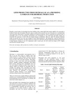
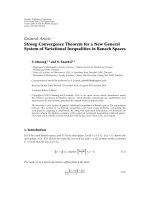
Fig. 1. TG/DTG obtained at 10 °C min−1 under dynamic nitrogen atmosphere
(50 mL min−1) for the blends (CH:PVP, CH:PVP-B) and membranes (CH, CH-B).
change significantly in relation to the inert membranes (Fig. 1). For the
CH-B, the first loss in mass was lower (1°Δm = 17% and
14% − DTGpeak = 53 °C; CH and CH-B, respectively) which indicates
that some water molecules were displaced in order to accommodate the
drug. The second event also occurred in the same range and identical
DTGpeak with higher loss in mass for CH, indicating that BMV demonstrated a better thermal stability for these temperature ranges
(2°Δm = 39% and 30% − DTGpeak = 231 °C; CH and CH-B, respectively). On the other hand, the presence of the drug was enough to
increase in 100% the last loss in mass step (11–32%). The higher the
amount of organic material, higher the loss in mass of carbonaceous
compounds that occurs exactly in this range of temperature. For the
membranes containing PVP was observed a strong possibility of favorable interaction between BMV and polymeric matrix due to the
disappearance of the last loss in mass in the membrane containing BMV.
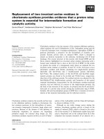
The membranes (CH:PVP-B, CH-B, CH:PVP and CH) also were
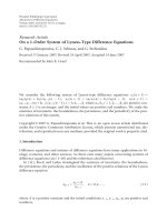
evaluated by DSC (Fig. 2(a)). The inert membranes (CH:PVP and CH)
exhibited endothermic events associated to water loss and hydroxyl
groups of the chitosan and PPG at 108 and 125 °C for CH and CH:PVP,
respectively (Abdelrazek et al., 2010; Li et al., 2010). CH membranes
presented lower temperature of this first event than CH:PVP membranes, probably due to the interaction between hydroxyl groups of
chitosan and carbonyl group of PVP suggesting the blend formation.
Moreover, CH:PVP showed other endothermic events at 188 and
319 °C, absent in CH, which also indicates an interaction between CHI
and PVP (Fig. 2(b)). Marsano, Vicini, Skopińska, Wisniewski, and
Sionkowska (2004) reported that the pyrrolidone rings in PVP contain a
proton accepting carbonyl moiety, while chitosan presents hydroxyl
and amino groups as side groups and, therefore, a hydrogen-bonding
interaction may take place between these two chemical moieties. They
also stated that the hydrogen bonds between two macromolecules
compete with the formation of hydrogen bonds between molecules of
the same polymer.
Another type of interaction that may occurs is associated with the
crosslinking of chitosan with TPP. The electrostatic interaction between
CHI and TPP occurs at molecular level with release of water molecules
and displacement of the main thermal events of the polymer (Hashad,
Ishak, Fahmy, Mansour, & Geneidi, 2016).
BMV interfered in thermal profile of the blend. The membranes
containing BMV exhibited an intensity reduction of the DSC peaks. The
first DSC event of the membranes shift to lower temperatures may be
associated with the presence of BMV in polymer matrix, since BMV
contributed with hydroxyl groups, indicating the incorporation of BMV
in the membranes. In absence of PVP (CH-B membrane), the DSC profile
2.2.7. Mucoadhesive property evaluation
The mucoadhesive property evaluation was determined by the relation of load (N) as a function of time (s) using texture analyzer (Stable
Micro Systems - TA-XT Plus Analyzer. Surrey, United Kingdom). The
texturometer was, previously, calibrated with 5 kg load cell and
equipped with 10 mm diameter analytical probe. To determine the
mucoadhesive property, a compact disc of the mucin from porcine
stomach was used (150 mg and 0.2 mm of thickness).
The discs were fixed with double-sided cohesive tape on the lower
base of the test piece (n.15347). The samples were transferred to mucoadhesion test apparatus (n.15467). Mucin discs were previously hydrated with ultrapure water. During the whole experiment, the temperature was kept constant at 37 °C. The method executed in this test
was adapted from (Fransén, Björk, & Edsman, 2008), and performed in
speed compression mode at 0.5 mm s−1, under a force of 5 g. After 60 s
of contact, the test piece was moved in opposite direction at 1.0 mm s−1
of speed. The maximum force required to separate the mucin disc on the
sample surface was detected and analyzed by Texture Expoente Lite
software. The measurements were performed in triplicate. One-way
ANOVA followed by the Tukey’s post-test was carried out using the
statistical program Graph Pad Prism v 5.0 DEMO.
3. Results and discussion
Thermal analysis is an important technique for the evaluation of
polymeric membranes regarding mass variations and thermal events
related to the blend formation (Abdelrazek, Elashmawi, & Labeeb,
2010; Rafique, Zia, Zuber, Tabasum, & Rheman, 2016). TG/DTG curves
of the membranes are shown in Fig. 1. All samples presents different
losses in mass possibly related to chemical changes that occurred after
the membrane formation. PVP as blend forming provides changes in the
thermal profile of the membranes. The first thermal event regarding
water loss (Nieto-Suárez, López-Quintela, & Lazzari, 2016) was
Δm = 17% and 12% (DTGpeak = 53 °C), with and without PVP respectively. Thermal decomposition and release of carbonaceous material are higher in the membrane containing PVP (Δm = 23%,
DTGpeak = 436 °C, and Δm = 4%, DTGpeak = 534 °C, against
Δm = 11%, above 343 °C of the membrane without PVP). After the
incorporation of PVP, the thermal stability of the membranes changed
exhibiting new losses in mass. Similar results were found by Bigucci
et al. (2015) in which blends composed of chitosan and hyaluronic acid
presented different losses in mass compared with pure chitosan.
The thermal profile of the membranes containing BMV did not
341
Carbohydrate Polymers 190 (2018) 339–345
R.H. Sizílio et al.
Fig. 2. (a) DSC curves for CH:PVP, CH, CH:PVP-B and CH-B; (b) DSC curves for CHI, TPP, PVP and BMV; obtained with a heating rate of 10 °C min−1 and dynamic atmosphere of nitrogen
(20 mL min−1).
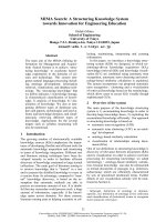
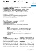
Croisier & Jérôme, 2013; Gonsalves, Ferro, Barreto, Nunes, & Valerio,
2016; Lewandowska, 2011). TPP presented several well-defined peaks,
which are related to its crystalline nature. The XRD pattern of BMV
showed main peaks in 14°, 17°, 28° and a wide peak with maximum
intensity between 11° and 12°.
Observing the membrane’s diffraction patterns, their similarity was
evident and both showed 4 diffraction peaks (two low intensity ones at
18 and 25°, and two high intensity ones at 14 and 16). The pattern shift
when comparing to the isolated components suggests a conformational
change when the membrane is formed. Conformational changes were
also observed by Abugoch, Tapia, Villamán, Yazdani-Pedram, and DíazDosque (2011) and by Lewandowska (2011), when they evaluated
chitosan/quinoa protein membranes, and chitosan acetate/PVP, respectively.
The XRD peaks related to the BMV were not observed in the
membranes containing the drug (CH-B and CH-PVP-B). According to
Subha, Mallikarjuna, Pallavi, Rao, and Rao (2015), this indicates that
the drug is dispersed at the molecular level in the membrane and
therefore, no drug peaks could be observed.
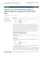
SEM analysis of the membranes is shown in Fig. 4. It is possible to
observe that the membranes present a smooth, compact and homogeneous surface. The absence of defects in the CH and CH-B indicates
that PPG contributed to CHI dispersion capacity during solvent casting
process showing a good compatibility between them. Usually, plasticizers acts on the polymer compatibility with CHI during membrane
formation (Van Den Broek, Knoop, Kappen, & Boeriu, 2015).
is similar to the raw materials with slightly shifts. No BMV melting
point is observed in the membranes containing PVP suggesting a good
miscibility of BMV in the blend.
Fig. 2(b) shows the DSC curves of chitosan, TPP, PVP and BMV.
Chitosan exhibited: i. an endothermic event at 97 °C, which corresponds
to water loss; ii. an endothermic event at 277 °C, related to decomposition of amino groups of chitosan (Abdelrazek et al., 2010; Santos,
Soares, Dockal, Campana Filho, & Cavalheiro, 2003); iii. a third event,
exothermic, close to 300 °C. TPP presents one main endothermic peak
at 114 °C related to melting. PVP exhibited an endothermic event at
113 °C regarding glass transition. Kadota, Otsu, Fujimori, Sato, and
Tozuka (2016) and Knopp et al. (2015) reported PVP glass transition at
168 and 160 °C respectively. This difference in glass transition temperature may be associated to changes in molecular weight, purity and
crystallinity degree of PVP obtained from different origins (Homayouni,
Sadeghi, Varshosaz, Garekani, & Nokhodchi, 2014; Knopp et al., 2015).
BMV is considered a crystalline drug, presenting melting point at
195 °C. There are three polymorphs of BMV commercially available,
and they differ in crystal lattice due to preparation process and crystallization. It was observed that the BMV polymorph studied in this
work is the polymorph II (Näther, Jess, Seyfarth, Bärwinkel, & Senker,
2015).
The XRD profile of the polymers, PVP and CHI are shown in Fig. 3. It
is possible to observe two main wide assymetric peaks at 11° and 21°;
12° and 20°, respectively, which are indicative of semi-crystalline materials, as previously described in the literature (Azevedo et al., 2011;
Fig. 3. X-ray diffraction pattern for (a) PVP, CHI, TPP and BMV; (b) CH, CH-B, CH:PVP and CH:PVP-B membranes.
342
Carbohydrate Polymers 190 (2018) 339–345
R.H. Sizílio et al.
Fig. 4. Photomicrographs of inert and drug loaded membranes through scanning electron microscopy.
The blend formation between CHI and PVP also was observed by
SEM, in which minor imperfections with circular shape were detected.
These imperfections may be related to casting process or an incompatibility between the polymers. Generally, polymeric blends surfaces are smooth and homogeneous with a certain degree of immiscibility. Yin, Luo, Chen, and Khutoryanskiy (2006) reported that
CHI/cellulose derivatives blends presented smooth surface, but in crosssection view they showed irregularities probably related to polymer
immiscibility. Nevertheless, DSC analysis did not demonstrate any
polymer immiscibility suggesting that the imperfections occurred due
to solvent casting process.
No changes were detected after drug incorporation (CH:PVP-B and
CH-B). No clusters were observed, suggesting BMV incorporation in the
polymeric matrix. These results are in agreement with previous characterizations.
The thickness of the inert chitosan membrane containing PVP
(48.66 ± 7.57 μm) was slightly thicker than the membrane without
PVP (39.33 ± 1.15 μm). On the other hand, almost no changes in
thickness were observed after BMV incorporation.
Swelling studies can be very useful to understand the drug delivery
mechanism, since the higher release can be attributed to the higher
extent of water uptake, resulting in increased wetting and penetration
of water into the film matrices, and hence, increased diffusion of the
drug (Koland, Charyulu, Vijayanarayana, & Prabhu, 2011). Several
parameters can affect the swelling ratio, hydrophilicity, stiffness and
pore structure of a matrix. The higher degree of swelling is, higher the
surface area/volume ratio. The hydrophilic nature of chitosan material
may be a major factor that influences the extent of swelling of these
matrices (Archana et al., 2013).
Fig. 5 presents the swelling profile of the CH membranes with PVP
(CH:PVP-B and CH: PVP) or without (CH-B and CH). It is possible to
observe that the presence of PVP in the membrane allowed higher
percentages of swelling (> 80%). Koland et al. (2011) found similar
Fig. 5. Swelling profile of the CH:PVP, CH:PVP-B, CH and CH-B performed at 37 °C using
phosphate buffer (pH = 7.4) as media.
results where the presence of PVP, a hydrophilic polymer, increased the
extent of swelling, and the maximum swelling was obtained in the
formulation that contained higher amounts of PVP. On the other hand,
the presence of the drug in the membranes slightly decreased their
swelling, which probably occurred due to the poor solubility of BMV in
water (Lucangioli et al., 2003), influencing the extent of swelling of
chitosan. In addition, as previously shown in the DSC analysis, the
presence of BMV in the membranes resulted in displacement of water
molecules, which may also reduce the chitosan swelling ability.
Fig. 6 shows the in vitro release of betamethasone-17-valerate for
chitosan films with (CH:PVP-B) or without PVP (CH-B), and in both
formulations the drug release occurred very quickly, and plateaus were
reached within 30 min and 1 h, respectively (Khoo et al., 2003). Thus,
since the BMV needs to reach the oral mucosa quickly, the developed
membranes were appropriate. As expected, the drug release followed
the trends for swelling ability; the chitosan films with PVP presented
the final total drug release of ∼80%, greater than that chitosan films
without PVP ∼40%.
343
Carbohydrate Polymers 190 (2018) 339–345
R.H. Sizílio et al.
organization, which promoted higher adhesivity. These results suggest
that there is two mechanism of mucoadhesion acting mutually. One by
electrostatic force between CHI- sialic acids and other by chain interpenetration of the PVP into the mucin layer. In this case, the first one is
more important than second.
In addition, the presence of BMV in the membranes causes decrease
in mucoadhesion, and probably occurred due to the rearrangement of
polymeric chain to accommodate the drug. As previously observed in
DSC, the incorporation of BMV resulted in displacement of water molecules in order to its incorporation. This displacement of water molecules led to a reduction in swelling ability (as previously shown in
swelling studies) and consequently in the decrease of mucoadhesion.
4. Conclusion
Fig. 6. In vitro release profiles of betamethasone-17-valerate from CH and CH:PVP
membranes.
This study proposed the preparation of mucoadhesive membranes
constituted of CHI and PVP as a potential drug delivery system for BMV
in the RAS treatment. The presence of PVP in the membranes possibly
provides chemical interactions with CHI which improves the thermal
stability as observed in thermal analysis. Moreover, PVP increased the
swelling ratio of the membranes, and therefore improved the BMV release rate (∼80% in less than 1 h) and promoted higher mucoadhesion.
On the other hand, BMV modifies the swelling ratio and the mucoadhesion, probably due to the displacement of water molecules originally found in the membranes by drug molecules. Thus, the results of
this study suggest that the developed system is appropriate to deliver
BMV aiming the RAS treatment. In addition, these systems may be
further evaluated using animal model.
Table 2
Mucoadhesive properties of CH, CH-B, CH:PVP, CH:PVP-B membranes.
Sample
Area to Positive Peak (N s)
Peak Positive Force
CH
CH-B
CH:PVP
CH:PVP-B
−1.167
−2.590
−3.783
−2.461
0.326
0.186
0.479
0.249
±
±
±
±
1.374
0.039
0.523
0.069
±
±
±
±
0.048
0.021
0.050
0.039
The betamethasone 17-valerate release profiles were fitted to the
Korsmeyer and Peppas model (Ritger & Peppas, 1987) to investigate
whether the release of the drug was related to both the polymer relaxation, in contact with the solvent, and/or the diffusion of the active,
through the hydrated matrix. This phenomenon has been reported to
occur in swellable polymers, such as chitosan (Talón, Trifkovic, Vargas,
Chiralt, & González-Martínez, 2017). The generalized expression of the
Korsmeyer and Peppas is described in Eq. (2).
Mt / M ∞ = kt n
Acknowledgments
The authors are grateful to CAPES (Coordenaỗóo de
Aperfeiỗoamento de Pessoal de Nớvel Superior) and FAPITEC/SE
(Fundaỗóo de Apoio Pesquisa e Inovaỗóo Tecnolúgica do Estado de
Sergipe) for financial support. CETENE-PE (Centro de Tecnologia do
Nordeste, Pernambuco, Brazil), Departments of Physics and Chemistry
of the Federal University of Sergipe (UFS) for carrying out the tests.
Rosangela H Sizílio is also grateful to CAPES for the Masters grant.
(2)
where Mt/M∞ corresponds to the fraction of the drug released at
time t, k is the rate constant of the membrane, related to the diffusion
process, and n is the diffusional exponent that is related to the mechanisms involved in the release process. Thus, for thin films a n value
of 0.5 means that the release obeys the Fickian diffusion model,
whereas if the n value is higher than 0.5, known as anomalous transport, the diffusion and the polymer relaxation are coupled (Serra,
Doménech, & Peppas, 2009; Siepmann & Peppas, 2012).
In this work the n value found was higher than 0.5, which corresponds to anomalous transport. According to de Souza, Goebel, and
Andreazza (2013), the anomalous transport suggests that the solvent
diffusion rate and polymer relaxation process occur in the same order of
magnitude, in other words, the transport consists in both drug diffusion
in the hydrated matrix and polymer relaxation.
Among several factors, the swelling ability is closely related to the
bioadhesive properties of polymers. The ability of certain polymers in
absorbing fluids, especially from human body, it becomes possible their
application in mucoadhesive formulations. The swelling ability is essential to enable the adherence of the formulation in the mucosa
(Carvalho, Chorilli, & Gremião, 2014). In order to adhere to the mucosa, the polymers should absorb a certain amount of fluid until the
polymeric structure reach the top of remodeling which is succeed by
permeation of mucin and other proteins. Only polymers with dissociated functional groups can interact electrostatically with mucin.
Table 2 shows that the blends (CH:PVP and CH:PVP-B), which
presented higher rates of water absorption (Fig. 5), also demonstrates
higher tensile strength rate, in other words, higher muco(bio)adhesivity. As related previously, the chemical interaction between the
functional groups of CHI and PVP provided a better structural
Appendix A. Supplementary data
Supplementary data associated with this article can be found, in the
online version, at />References
Abdelrazek, E. M., Elashmawi, I. S., & Labeeb, S. (2010). Chitosan filler effects on the
experimental characterization, spectroscopic investigation and thermal studies of
PVA/PVP blend films. Physica B: Condensed Matter, 405(8), 2021–2027. http://dx.
doi.org/10.1016/j.physb.2010.01.095.
Abugoch, L. E., Tapia, C., Villamán, M. C., Yazdani-Pedram, M., & Díaz-Dosque, M.
(2011). Characterization of quinoa proteine chitosan blend edible films. Food
Hydrocolloids, 25, 879–886. />Archana, D., Singh, B. K., Dutta, J., & Dutta, P. K. (2013). In vivo evaluation of chitosanPVP-titanium dioxide nanocomposite as wound dressing material. Carbohydrate
Polymers, 95(1), 530–539. />Azevedo, J. R., Sizilio, R. H., Brito, M. B., Costa, A. M. B., Serafini, M. R., Araújo, A. A. S.,
et al. (2011). Physical and chemical characterization insulin-loaded chitosan-TPP
nanoparticles. Journal of Thermal Analysis and Calorimetry, 106(3), 685–689. http://
dx.doi.org/10.1007/s10973-011-1429-5.
Bigucci, F., Abruzzo, A., Saladini, B., Gallucci, M. C., Cerchiara, T., & Luppi, B. (2015).
Development and characterization of chitosan/hyaluronan film for transdermal delivery of thiocolchicoside. Carbohydrate Polymers, 130, 32–40. />1016/j.carbpol.2015.04.067.
Cai, X., Tong, H., Shen, X., Chen, W., Yan, J., & Hu, J. (2009). Preparation and characterization of homogeneous chitosan-polylactic acid/hydroxyapatite nanocomposite
for bone tissue engineering and evaluation of its mechanical properties. Acta
Biomaterialia, 5(7), 2693–2703. />Carvalho, F. C., Chorilli, M., & Gremião, M. P. D. (2014). Plataformas bio (Muco) adesivas
poliméricas baseadas em nanotecnologia para liberaỗóo controlada de Fỏrmacos
propriedades, metodologias e aplicaỗừes. Polớmeros, 24(2), 203–213. .
344
Carbohydrate Polymers 190 (2018) 339–345
R.H. Sizílio et al.
phases for microemulsion and micellar electrokinetic chromatography of betamethasone and derivatives. Electrophoresis, 24(6), 984–991. />1002/elps.200390142.
Marsano, E., Vicini, S., Skopińska, J., Wisniewski, M., & Sionkowska, A. (2004). Chitosan
and poly(vinyl pyrrolidone): Compatibility and miscibility of blends. Macromolecular
Symposia, 218, 251–260. />Näther, C., Jess, I., Seyfarth, L., Bärwinkel, K., & Senker, J. (2015). Trimorphism of betamethasone valerate: Preparation, crystal structures, and thermodynamic relations.
Crystal Growth and Design, 15(1), 366–373. />Nieto-Suárez, M., López-Quintela, M. A., & Lazzari, M. (2016). Preparation and characterization of crosslinked chitosan/gelatin scaffolds by ice segregation induced selfassembly. Carbohydrate Polymers, 141, 175–183. />carbpol.2015.12.064.
Ostrowska-Czubenko, J., Pierõg, M., & Gierszewska-Druzyńska, M. (2013). Water state in
chemically and physically crosslinked chitosan membranes. Journal of Applied
Polymer Science, 130(3), 1707–1715. />Rafique, A., Zia, K. M., Zuber, M., Tabasum, S., & Rheman, S. (2016). Chitosan functionalized poly (vinyl alcohol) for prospects biomedical and industrial applications: A
review. International Journal of Biological Macromolecules, 87, 141–154. .
org/10.1016/j.ijbiomac.2016.02.035.
Ritger, P. L., & Peppas, N. A. (1987). A simple equation for description of solute release I.
Fickian and non-fickian release from non-swellable devices in the form of slabs,
spheres, cylinders or discs. Journal of Controlled Release, 5(1), 23–36. .
org/10.1016/0168-3659(87)90034-4.
Rodrigues, L. B., Leite, H. F., Yoshida, M. I., Saliba, J. B., Junior, A. S. C., & Faraco, A. A.
G. (2009). In vitro release and characterization of chitosan films as dexamethasone
carrier. International Journal of Pharmaceutics, 368(1–2), 1–6. />1016/j.ijpharm.2008.09.047.
Rogulj, A. A., Brkic, D., Alajbeg, I., Džanić, E., & Alajbeg, I. (2014). Nonaromatic naphthalan for the treatment of oral mucosal diseases. Acta Dermatovenerologica Croatica,
22(4), 250–258.
Santos, J. E. D., Soares, J. D. P., Dockal, E. R., Campana Filho, S. P., & Cavalheiro, ẫ. T. G.
(2003). Caracterizaỗóo de quitosanas comerciais de diferentes origens. Polímeros,
13(4), 242–249. />Scully, C. (2006). Aphthous ulceration. The New England Journal of Medicine, 355,
165–172. />Serra, L., Doménech, J., & Peppas, N. A. (2009). Engineering design and molecular dynamics of mucoadhesive drug delivery systems as targeting agents. European Journal
of Pharmaceutics and Biopharmaceutics, 71(3), 519–528. />ejpb.2008.09.022.
Siepmann, J., & Peppas, N. A. (2012). Modeling of drug release from delivery systems
based on hydroxypropyl methylcellulose (HPMC). Advanced Drug Delivery Reviews,
163–174. />Srinivasa, P. C., Ramesh, M. N., Kumar, K. R., & Tharanathan, R. N. (2004). Properties of
chitosan films prepared under different drying conditions. Journal of Food Engineering,
63, 79–85. />Subha, M. C. S., Mallikarjuna, B., Pallavi, K., Rao, K.c., & Rao, K.m. (2015). Biodegradable
interpenetrating polymer network hydrogel membranes for controlled release of
anticancer drug. Asian Journal of Pharmaceutics, 9(2), 129. />4103/0973-8398.154716.
Swetha, M., Sahithi, K., Moorthi, A., Srinivasan, N., Ramasamy, K., & Selvamurugan, N.
(2010). Biocomposites containing natural polymers and hydroxyapatite for bone
tissue engineering. International Journal of Biological Macromolecules, 47(1), 1–4.
/>Talón, E., Trifkovic, K. T., Vargas, M., Chiralt, A., & González-Martínez, C. (2017). Release
of polyphenols from starch-chitosan based films containing thyme extract.
Carbohydrate Polymers, 175, 122–130. />067.
Tappuni, A. R., Kovacevic, T., Shirlaw, P. J., & Challacombe, S. J. (2013). Clinical assessment of disease severity in recurrent aphthous stomatitis. Journal of Oral
Pathology and Medicine, 42, 635–641. />Van Den Broek, L. A. M., Knoop, R. J. I., Kappen, F. H. J., & Boeriu, C. G. (2015). Chitosan
films and blends for packaging material. Carbohydrate Polymers, 116, 237–242.
/>Yin, J., Luo, K., Chen, X., & Khutoryanskiy, V. V. (2006). Miscibility studies of the blends
of chitosan with some cellulose ethers. Carbohydrate Polymers, 63(2), 238–244.
/>
org/10.4322/polimeros.2014.043.
Croisier, F., & Jérôme, C. (2013). Chitosan-based biomaterials for tissue engineering.
European Polymer Journal, 49(4), 780–792. />2012.12.009.
de Souza, D. F., Goebel, K., & Andreazza, I. F. (2013). Development of enteric coated
sustained release minitablets containing mesalamine. Brazilian Journal of
Pharmaceutical Sciences, 49(3), 529–536. />Elsabee, M. Z., & Abdou, E. S. (2013). Chitosan based edible films and coatings: A review.
Materials Science and Engineering C, 33(4), 1819–1841. />msec.2013.01.010.
Fransén, N., Björk, E., & Edsman, K. (2008). Changes in the mucoadhesion of powder
formulations after drug application investigated with a simplified method. Journal of
Pharmaceutical Sciences, 97(9), 3855–3864. />Gierszewska, M., & Ostrowska-Czubenko, J. (2016). Chitosan-based membranes with
different ionic crosslinking density for pharmaceutical and industrial applications.
Carbohydrate Polymers, 153, 501–511. />126.
Gonsalves, J. K. M. C., Ferro, J. N. S., Barreto, E. O., Nunes, R. S., & Valerio, M. E. G.
(2016). Influence of concentration of hydroxyapatite surface modifier agent on
bioactive composite characteristics. Ceramics International, 42(15), 17023–17031.
/>Hashad, R. A., Ishak, R. A. H., Fahmy, S., Mansour, S., & Geneidi, A. S. (2016). Chitosantripolyphosphate nanoparticles: Optimization of formulation parameters for improving process yield at a novel pH using artificial neural networks. International
Journal of Biological Macromolecules, 86, 50–58. />ijbiomac.2016.01.042.
Homayouni, A., Sadeghi, F., Varshosaz, J., Garekani, H. A., & Nokhodchi, A. (2014).
Comparing various techniques to produce micro/nanoparticles for enhancing the
dissolution of celecoxib containing PVP. European Journal of Pharmaceutics and
Biopharmaceutics, 88(1), 261–274. />Kürklü-Gürleyen, E., Öğüt-Erişen, M., Çakır, O., Uysal, Ư., & Ak, G. (2016). Quality of life
in patients with recurrent aphthous stomatitis treated with a mucoadhesive patch
containing citrus essential oil. Patient Preference and Adherence, 10, 967–973. http://
dx.doi.org/10.2147/PPA.S106530.
Kadota, K., Otsu, S., Fujimori, M., Sato, H., & Tozuka, Y. (2016). Soluble hydrolysisresistant composite formulation of curcumin containing α-glucosyl hesperidin and
polyvinylpyrrolidone. Advanced Powder Technology, 27(2), 442–447. .
org/10.1016/j.apt.2016.01.012.
Khoo, C. G. L., Frantzich, S., Rosinski, A., Sjöström, M., & Hoogstraate, J. (2003). Oral
gingival delivery systems from chitosan blends with hydrophilic polymers. European
Journal of Pharmaceutics and Biopharmaceutics, 55(1), 47–56.
Knopp, M. M., Olesen, N. E., Holm, P., Langguth, P., Holm, R., & Rades, T. (2015).
Influence of polymer molecular weight on drug-polymer solubility: A comparison
between experimentally determined solubility in PVP and prediction derived from
solubility in monomer. Journal of Pharmaceutical Sciences, 104(9), 2905–2912. http://
dx.doi.org/10.1002/jps.24410.
Koland, M., Charyulu, R. N., Vijayanarayana, K., & Prabhu, P. (2011). In vitro and in vivo
evaluation of chitosan buccal films of ondansetron hydrochloride. International
Journal of Pharmaceutical Investigation, 1(3), 164–171. />2230-973X.85967.
Lewandowska, K. (2011). Miscibility and interactions in chitosan acetate/poly(N-vinylpyrrolidone) blends. Thermochimica Acta, 517(1–2), 90–97. />1016/J.TCA.2011.01.036.
Li, J., Zivanovic, S., Davidson, P. M., & Kit, K. (2010). Characterization and comparison of
chitosan/PVP and chitosan/PEO blend films. Carbohydrate Polymers, 79(3), 786–791.
/>Liang, S., Liu, L., Huang, Q., & Yam, K. L. (2009). Preparation of single or double-network
chitosan/poly(vinyl alcohol) gel films through selectively cross-linking method.
Carbohydrate Polymers, 77(4), 718–724. />02.007.
Liu, X., Chen, Y., Huang, Q., He, W., Feng, Q., & Yu, B. (2014). A novel thermo-sensitive
hydrogel based on thiolated chitosan/hydroxyapatite/beta-glycerophosphate.
Carbohydrate Polymers, 110, 62–69. />065.
Lucangioli, S. E., Carducci, C. N., Scioscia, S. L., Carlucci, A., Bregni, C., & Kenndler, E.
(2003). Comparison of the retention characteristics of different pseudostationary
345