Bioengineered carboxymethyl cellulose-doxorubicin prodrug hydrogels for topical chemotherapy of melanoma skin cancer
Bạn đang xem bản rút gọn của tài liệu. Xem và tải ngay bản đầy đủ của tài liệu tại đây (1.9 MB, 12 trang )
Carbohydrate Polymers 195 (2018) 401–412
Contents lists available at ScienceDirect
Carbohydrate Polymers
journal homepage: www.elsevier.com/locate/carbpol
Bioengineered carboxymethyl cellulose-doxorubicin prodrug hydrogels for
topical chemotherapy of melanoma skin cancer
T
Nádia S.V. Capanemaa, Alexandra A.P. Mansura, Sandhra M. Carvalhoa, Isadora C. Carvalhoa,
⁎
Poliane Chagasb, Luiz Carlos A. de Oliveirab, Herman S. Mansura,
a
Center of Nanoscience, Nanotechnology and Innovation—CeNano2I, Department of Metallurgical and Materials Engineering, Federal University of Minas Gerais/UFMG,
Brazil
b
Department of Chemistry—ICEX, Federal University of Minas Gerais/UFMG, Brazil
A R T I C LE I N FO
A B S T R A C T
Keywords:
Polysaccharide
Biopolymer
Hydrogels
Cellulose derivatives
Polymer-drug conjugates
Cancer therapy
Drug delivery
Melanoma is the most aggressive type of skin cancer with high rates of mortality. Despite encouraging advances
demonstrated by anticancer drug carriers in recent years, developing ideal drug delivery systems to target tumor
microenvironment by overcoming physiological barriers and chemotherapy side effects still remain intimidating
challenges. Herein, we designed and developed a novel carbohydrate-based prodrug composed of carboxymethylcellulose (CMC) polymer bioconjugated with anticancer drug doxorubicin hydrochloride (DOX) by
covalent amide bonds and crosslinked with citric acid for producing advanced hydrogels. The results demonstrated the effect of CMC hydrogel network structure with distinct degree of substitution of carboxymethyl
groups of the cellulose backbone regarding to the process of bioconjugation and on tailoring the DOX release
kinetics in vitro and the cytotoxicity towards melanoma cancer cells in vitro. To this end, an innovative platform
was developed based on polysaccharide-drug hydrogels offering promising perspectives for skin disease applications associated with topical chemotherapy of melanoma.
1. Introduction
Skin cancer represents one of the most commonly occurring carcinoma in human and it is growing at a rate of one million new cases
reported annually. Malignant melanoma is the most lethal form of skin
cancer and it is associated with poor prognosis causing deaths worldwide. Therefore, melanoma continues to remain an important health
threat, with death often occurring by metastasis. Although there are
several options for anti-melanoma therapy, it is resistant to some
therapies. The primary cutaneous melanoma can be managed by surgery at the early stage, but the advanced metastatic melanoma cannot
be properly treated by surgery alone. Therefore, it requires additional
therapeutic methods such as chemotherapy, biochemotherapy, immunotherapy, and adoptive cell therapy (Bharadwaj, Das, Paul, &
Mazumder, 2016; Vishnubhakthula, Elupula, & Durán-Lara, 2017). To
mitigate or avoid side effects commonly caused by oral administration
and intravenous injection of drugs (i.e., enteral or parenteral), topical
(i.e., local) transdermal drug delivery systems offer a promising alternative strategy as carriers of antineoplastic agents. Thus, the topical
administration of chemotherapeutics is considered an encouraging approach for effective therapy of skin cancer (Bharadwaj et al., 2016;
Vishnubhakthula et al., 2017). Polymeric-based drug delivery systems
are the most interesting vehicles in anti-cancer therapy. There are
several advantages of using polymer as carriers for antineoplastic
agents, including increased drug solubility, better bioavailability, high
stability, controlled drug release, selective organ or tissue distribution,
and reduction of the total dose required. Moreover, the association of
polymers with toxic anticancer drugs can significantly minimize the
adverse side effects (Ranjbari et al., 2017; Vishnubhakthula et al.,
2017). For that reason, polymeric (nano)carriers are the most extensively studied platforms for cancer treatment. Polymers are versatile
macromolecules that can be engineered to fulfill several properties required for sophisticated biomedical applications in oncology. The research at the interface of polymer chemistry and biomedical sciences
has given rise to the polymer-based pharmaceuticals, referred to as
‘polymer therapeutics’.
In this regard, polymer therapeutics has emerged as a new promising field of research, which encompasses rationally designed
⁎
Corresponding author at: Department of Metallurgical and Materials Engineering, Federal University of Minas Gerais, Av. Antônio Carlos, 6627 – Escola de Engenharia, Bloco 2 – Sala
2233, 31.270-901, Belo Horizonte, MG, Brazil.
E-mail addresses: (N.S.V. Capanema), (A.A.P. Mansur), (S.M. Carvalho),
(I.C. Carvalho), (P. Chagas), (L.C.A. de Oliveira), (H.S. Mansur).
/>Received 13 February 2018; Received in revised form 2 April 2018; Accepted 26 April 2018
Available online 30 April 2018
0144-8617/ © 2018 Elsevier Ltd. All rights reserved.
Carbohydrate Polymers 195 (2018) 401–412
N.S.V. Capanema et al.
amphiphilic anticancer drug, is the most clinically used anticancer drug
because of its high efficiency and a broad spectrum of activity against
diverse cancer types (e.g., breast, lung, skin, and brain cancers) but is
poorly soluble in water and physiological medium (solubility of hydrochloride salt < 2%). Therefore, the development of simply synthesized, economical, water-soluble, and biocompatible polymer-drug delivery systems with efficient DOX encapsulation is still highly needed
against skin cancers. Interestingly, despite intensive research in the
field of polymer-drug conjugates (He et al., 2015; Movagharnezhad &
Moghadam, 2016; Roy et al., 2014), no published study was found in
the literature of CMC-DOX crosslinked hydrogels for treating melanoma
cancer. We hypothesize that it may be possible to perform covalent
linkage between carboxymethyl cellulose (i.e., eCOO− groups) with
doxorubicin hydrochloride (i.e., eNH2 groups) forming polymer-drug
conjugates. In the sequence, they could be chemically crosslinked by
citric acid producing hydrogel matrices for active drug delivery against
skin cancer cells. Herein, we designed and synthesized CMC-DOX
conjugates via carbodiimide-mediated reactions for the formation of
amide bonds in aqueous medium. These polymer-drug conjugates were
used for producing hydrogel networks by chemical crosslinking with
eco-friendly citric acid. The results proved that hydrophilic hydrogel
membranes based on CMC-DOX conjugates were synthesized with
physicochemical characteristics and anticancer drug delivery profiles
effective for killing melanoma cells, which offer potential applications
as topical transdermal chemotherapy against skin cancer.
macromolecular drugs, such as polymer–protein conjugates, polymerdrug conjugates, polyplexes for encapsulating nucleotides (i.e., RNA,
DNA), and supramolecular drug-delivery systems (Duncan, 2003,
2013). Numerous polymer–based conjugates with improved chemical
and biological stability and pharmacokinetic properties have been developed by coupling low-molecular-weight anticancer drugs to highmolecular-weight polymers through cleavable covalent bonds, including N-(2-hydroxypropyl)methacrylamide conjugates of doxorubicin
hydrochloride (DOX) and paclitaxel (Duncan, 2003, 2013; Haag &
Kratz, 2006). Among different types of biocompatible polymers, carbohydrate-based polymers (or polysaccharides) are the most common
natural polymers with chemical structures consisting of long chains of
monosaccharide (or disaccharide) units bound by glycosidic linkages.
They possess properties such as biocompatibility, biodegradability,
non-toxicity, suitable reactivity for facile chemical modification associated with availability and low cost led to their widespread applications in pharmaceutical and biomedical fields including development of
nanocarriers for delivery of anticancer therapeutic agents. Generally,
polysaccharide-based polymer-drug systems can be used for reducing
systemic toxicity, increasing short half-lives and tumor localization of
agents for a successful cancer therapy. This approach can overcome the
most challenging factor in cancer therapy related to the toxicity of
anticancer therapeutic agents for normal cells and therefore, targeted
delivery of these drugs to the site of action can be considered as a very
promising therapeutic strategy (Duncan, 2003, 2013; Haag & Kratz,
2006; Ranjbari et al., 2017).
One option of rational design of innovative polymer-drug systems
for topical drug delivery systems is based on hydrogels. Hydrogels are
three-dimensional, hydrophilic polymeric networks that are capable of
absorbing large amounts of water, biological fluids, or molecules. These
systems possess unique properties to improve the efficacy of the therapeutic agents and minimize undesirable side effects. Hydrogels serve
as an in situ vehicle for localized delivery of antineoplastic agents by
topical application, allowing minimally or noninvasive delivery,
avoiding side effects of systemic chemotherapeutics and while reducing
infection risk associated with surgical procedures (Vishnubhakthula
et al., 2017). Moreover, some properties and important advantages of
these hydrogel-based polymer-drugs can be modulated by the chemical
crosslinking of the network and the interactions of the hydrogels with
the surrounding microenvironment, including drug release dynamics,
hydrogel degradation kinetics, drug levels at the cancer site, sustaining
duration of therapeutic concentrations, circumventing poor solubility
of anticancer drugs, which are critical to chemotherapeutic efficacy and
safety (Liu et al., 2016).
Among several alternatives of polysaccharides (e.g., hyaluronic acid,
chitosan, and cellulose) for producing polymer-drug conjugates, carboxymethyl cellulose (CMC), as a broadly available derivative of cellulose, finds widespread use in biology, medicine, nutrition and pharmaceutical formulations. It presents excellent characteristics such as
biocompatibility and water solubility combined with highly reactive
chemical groups including hydroxyl and carboxyl groups, which can
allow chemical biofunctionalization and the formation of hydrogels
with tailored crosslinked networks. Moreover, CMC is an inexpensive
compound with good compatibility to the skin and mucous membranes,
which has been approved by the United States Food and Drug
Administration (FDA) for parenteral use in drug products (Duncan,
2013; Haag & Kratz, 2006; He et al., 2015; Movagharnezhad &
Moghadam, 2016; Ranjbari et al., 2017; Roy et al., 2014). The degree of
substitution (or carboxymethylation, DS) of carboxymethyl cellulose
plays a pivotal role on all properties, including water solubility, pHsensitivity, chemical reactivity and stability, rheology and biodegradability, which can be tuned for several applications in biomedical, food,
and pharmaceutical fields (Ferro et al., 2017).
To this end, carbohydrate-based polymeric hydrogels have been
studied as anticancer drug carriers that are not soluble in water and
highly cytotoxic for chemotherapeutic applications. Doxorubicin, an
2. Material and methods
2.1. Materials and cell cultures
Sodium carboxymethyl cellulose with two degree of substitution
DS = 0.77 (CMC-0.77, Product Number: 419311, Batch Number:
MKBW1368V, average molar mass Mw = 250 kDa and, viscosity
735 cps, 2% in H2O at 25 °C) and DS = 1.22 (CMC-1.22, Product
Number: 419281, Batch Number: MKBV4486V, Mw = 250 kDa, viscosity 660 cps, 2% in H2O at 25 °C), 2-(N-Morpholino)ethanesulfonic
acid (MES, > 99%, low moisture content), 1-Ethyl-3-[3-dimethylaminopropyl]carbodiimide hydrochloride (EDC, ≥98%), doxorubicin hydrochloride (hydroxydaunorubicin hydrochloride, referred to as DOX,
≥98.0%,), ethalonamine hydrochloride (≥99.0%), and citric acid (CA,
≥99.5%,) were supplied by Sigma-Aldrich (USA).
Aforementioned chemicals were used without further purification,
deionized water (DI water, Millipore Simplicity™) with resistivity of
18 MΩ cm was used to prepare the solutions, and the procedures were
performed at room temperature (RT, 23 ± 2 °C), unless specified
otherwise.
Human embryonic kidney (HEK 293T, American Type Culture
Collection – ATCC® CRL-1573™) cells was provided by Federal
University of Minas Gerais. Human malignant melanoma (A375, ATCC®
CRL-1619™) was purchased from Brazilian Cell Repository (Banco de
Células do Rio de Janeiro: BCRJ, Brazil; cell line authentication molecular technique, Short Tandem Repeat (STR) DNA; quality assurance
based on the international standard NBR ISO/IEC 17025:2005).
2.2. Polymer-drug conjugation
The DOX anticancer drug was conjugated to the CMC polysaccharide backbone with two DS using 1-ethyl-3-[3-dimethylaminopropyl] carbodiimide hydrochloride (EDC) as a “zero-length” coupling
agent in MES buffer (0.25 M, pH 5.5 ± 0.1).
Polymer-drug bioconjugation of was performed as follows: 1.0 mL
of EDC solution (12.5 wt%) was added to the reaction flask with 10 mL
of CMC solution (2.0 wt.% in MES) and magnetically stirred for 15 min
at 6 ± 2 °C. Under continuous stirring, 400 μL of DOX solution
(0.145 wt.% in MES) was poured into the flask, and the system was
incubated at RT for 2 h in the dark. The polymer-drug systems were
402
Carbohydrate Polymers 195 (2018) 401–412
N.S.V. Capanema et al.
3.0 mg g−1 (5.2 μmol g−1). As references, hydrogels without DOX chemotherapeutic were also synthesized and referred to as “CMC-0.77_CA”
and “CMC-1.22_CA”.
referred to as “CMC-0.77_DOX” and “CMC-1.22_DOX” depending on the
DS of the polymer used for conjugation 0.77 or 1.22, respectively. The
molar ratio CMCunit:DOX was 1000:1.
All of the samples were kept in the dark at RT overnight and then,
ethanolamine hydrochloride was added to the reaction flasks and
magnetically stirred for 15 min at final concentration of 1.0 μM to
quench the reaction. These synthesized polymer-drug conjugates were
dialyzed for 24 h (with water changes after 2 h and 4 h) in the dark
against 2 L of distilled water using a Pur-A-Lyzer™ Mega Dialysis Kit
(Sigma, cellulose membrane with Mw cut-off of 12 kDa) under moderate
stirring at RT. After purification, the polymer-drug solutions were
stored at 6 ± 2 °C until further use.
2.4. Physicochemical characterization of CMC polymer, CMC-DOX
polymer-drug conjugates and hydrogels
For CMC polymers, infrared spectroscopy and 1H nuclear resonance
spectroscopy (1H NMR) analyses were performed with details described
in Supplementary Material. CMC is available as regular commercial
product by worldwide reliable supplier and USA FDA approved.
Therefore, this study characterized the most relevant aspects by FTIR
and 1H NMR spectroscopy techniques.
Fourier-transform infrared spectroscopy (FTIR) spectra were obtained using attenuated total reflectance method (ATR,
4000–675 cm−1, 32 scans, and 4 cm−1 resolution, Nicolet 6700,
Thermo Fischer) with background subtraction (replicates, n = 3).
Ultraviolet-visible (UV-vis) spectroscopy measurements were performed (Lambda EZ-210, Perkin-Elmer) in transmission mode over the
wavelength range between 600 and 350 nm (n = 3).
Photoluminescence spectroscopy (PL) was performed at RT using a
violet diode laser at 405 nm excitation wavelength (λexc) (150-mW,
Roithner LaserTechnik) coupled to a USB4000 VIS-NIR (visible-near
infrared) spectrophotometer (Ocean Optics, Inc.) (n = 3).
2.3. Synthesis of prodrug hydrogels
Crosslinking agent, citric acid (CA), was added under stirring at
concentration of 15% m/m of polymer-drug solutions (CMC-0.77_DOX
and CMC-1.22_DOX) and homogenized for 20 min. Then, 10 mL of the
solutions were poured into plastic molds (polystyrene petri dish, diameter = 60 mm) and were allowed to dry at 40 ± 2 °C for 24 h to remove water. In the sequence, the samples were kept at 80 ± 2 °C for
24 h for the crosslinking reaction (slow evaporation method). The
prodrug hydrogels (Fig. 1) were referred to as “CMC-0.77_DOX_CA” and
“CMC-1.22_DOX_CA”. The concentration of DOX in both hydrogels was
Fig. 1. Schematic representation of the precursors, reaction and CMC_DOX conjugate product.
403
Carbohydrate Polymers 195 (2018) 401–412
N.S.V. Capanema et al.
Zeta potential (ZP or ζ-potential) analysis was performed using
ZetaPlus instrument (Brookhaven Instruments, 35 mW red diode laser
light, wavelength λ = 660 nm) at 25 ± 2 °C under the Smoluchowski
approximation method (n ≥ 10).
For evaluating the loading efficiency, the polymer-drug conjugates
were centrifuged (15 min at 14,000 rpm and 4 ± 1 °C, Hettich Mikro
200R) using an ultracentrifuge filter with a 50 kDa cut-off cellulose
membrane (Amicon filter, Millipore). The filtrate was collected and
analyzed by UV–vis spectroscopy (Lambda EZ-210, Perkin Elmer) to
determine the DOX concentration based on the Beer–Lambert correlation curve (Fig. 1S). The loading efficiency (LE, %) was calculated using
Eq. (1).
LE = ((A − B)/A) × 100
3. Results and discussion
3.1. Characterization of CMC polymer
To avoid redundancy, the characterization of carboxymethylcellulose with two degree of substitution (DS) was performed
using FTIR (Fig. 2S) and 1H NMR (Fig. 3S) techniques and the results
are presented in Supplementary Material. In brief, the FTIR analysis
showed the most important chemical functionalities of CMC polymer
(e.g., carboxylic, carboxylates and hydroxyls) and CMC with higher DS
values (i.e., DS = 1.22 > 0.77) presented more prominent spectroscopy intensities associated with the carboxymethyl groups grafted in
the cellulose polymer backbone. In 1H NMR spectra, resonance signals
associated with unsubstituted and substituted hydroxyls were detected.
The integration of the unsubstituted protons signals for CMC polymers
for both DS values indicated the reduction of the relative intensity of
hydroxyls for DS = 1.22 due to the higher carboxymethylation content
of the polymer (i.e., cellulose-O-H → cellulose-O-CM).
(1)
−1
where A (mg mL ) is the initial concentration of DOX in solution and
B (mg L−1) is the concentration of DOX at filtrate.
For fluid uptake measurement and solvation assessments, the hydrogels were cut into 10 × 10 mm2 samples, dried at 40 ± 2 °C for
stabilization of mass, and weighted (W0, initial mass). Then, the hydrogel samples (n = 3) were placed in 70 mL sample pots with 10.0 mL
PBS (phosphate buffered saline, pH 7.4) at 37 ± 1 °C. After 60 min, the
hydrogel was removed from solution, gently wiped with filter paper to
remove excess of liquid of the sample surface and weighted (Ws,
swollen mass). In the sequence, samples were dried at 40 ± 2 °C until
mass stabilization and the final weight was recorded (Wf, final mass).
The measurements of weight obtained in each step of the process
were used to calculate the swelling degree (SD) and gel fraction (GF) of
the hydrogels using Eqs. (2) and (3) (Dumont et al., 2016; Fekete,
Borsa, Takács, & Wojnárovits, 2017).
SD (%) = ((Ws − W0)/W0) × 100%
(2)
GF (%) = (Wf/W0) × 100%
(3)
3.2. Physicochemical characterization of polymer-drug conjugates
CMC polymer has no electronic transitions in the visible range
(Fig. 2A(d and e)) due to the absence of unsaturated bonds of conjugated π-electrons for this absorption. Conversely, DOX is an anthracycline antibiotic with a chromophore anthraquinone nucleus linked to
daunosamine amino sugar through a glycosidic bond, which presents a
characteristic resonance peak at 484 nm assigned to π→π* energy state
transitions of quinonoid (Fig. 2A(a)) (Mohan & Rapoport, 2010). For
that reason, both polymer-drug conjugates made of CMC with
DS = 0.77 and 1.22 (Fig. 2A(b and c)) presented the same transition
due to activity of DOX as chromophores.
In vitro drug release from hydrogels was performed in triplicate at
37.0 ± 0.1 °C in PBS buffer (pH 7.4). Prodrug hydrogels (6 cm2) were
placed inside a plastic basket immersed into 15 mL of PBS under
magnetic stirring and drug release monitored for 24 h (n = 3). At determinated time intervals, 1 mL of PBS medium was collected and
analyzed by UV–vis to determine the DOX concentration based on the
Beer–Lambert correlation curve (λ = 484 nm).
2.5. Biological characterization of hydrogels
2.5.1. Cell viability in vitro – Mitochondrial activity (MTT) assay
MTT (3-(4,5-dimethylthiazol-2yl) 2,5-diphenyl tetrazolium bromide) experiments were performed to evaluate the toxicity of free DOX
and prodrug conjugates at final concentration of 25 μM after incubation
with HEK 293T and A375 cells for 6 h, 24 h and 48 h (detailed protocol
in Supplementary Material). The percentage of cell viability was calculated after blank corrections, according to Eq. (4), where the values
of the control group were set to 100% cell viability.
Cell viability = (Absorbance of sample and cells)/(Absorbance of control) × 100%
(4)
2.5.2. Cellular uptake of polymer-drug bioconjugates – confocal laser
scanning microscopy (CLSM)
DOX distribution inside the cells was monitored using CLSM after
treatment of HEK 293T and A375 cells for 30 min, 1 h, 2 h and 6 h,
using the inherent fluorescence imaging capability of DOX and TOPRO®-3 (Invitrogen™, USA) to selective staining the nuclei of the cells
(detailed protocol in Supplementary Material).
Fig. 2. (A) Absorbance (visible) and (B) Emission (PL) spectra of (a) DOX, (b)
CMC-0.77_DOX, (c) CMC-1.22_DOX, (d) CMC-0.77 and (e) CMC-1.22 solutions.
404
Carbohydrate Polymers 195 (2018) 401–412
N.S.V. Capanema et al.
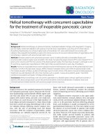
Fig. 3. (A) FTIR spectra of CMC-0.77 before (a) and after (b) conjugation with DOX. (B) Detail of FTIR spectra region associated with the range of Amide I, Amide II
and νasCOO− vibrations. (C) Evolution of Amide I and carboxylate bands due to chemical conjugation reactions.
(1645 cm−1) and decrease of νasCOO− (1582 cm−1) bands using β1-4
vibration at 896 cm−1 as the internal reference band, caused by the
formation of amide bonds between carboxylic groups of CMC and
amino groups of DOX in agreement with the literature (Cao et al., 2017;
Li et al., 2017; Mansur & Mansur, 2012; Mansur, Mansur, SorianoAraújo, & Lobato, 2014). Thus, these FTIR results provided additional
strong evidence of the effective covalent conjugation of the CMC to
anticancer chemotherapeutic drug via chemical amide bonds, which
validated the hypothesis of this research.
Zeta potential analyzes are crucial for investigating conjugated
systems based on polymer-drugs for biomedical and pharmaceutical
applications. The presence of local and global surface charges on the
systems can drastically affect the responses when in contact with biological microenvironment in vitro and in vivo. The results of ZP measurements showed the relative reduction of the average negative values
of CMC (absolute values, standard deviation, SD = ± 2 mV) before and
after conjugation with DOX, from −39.4 mV to −36.9 mV, and from
−50.6 mV to −43.1 mV, for CMC-0.77 and CMC-1.22 systems, respectively. These values are coherent with the designed chemical reaction developing amide bonds between negative groups of CMC
(eCOO−) with positive groups of protonated DOX (eNH3+) under
mildly acidic or physiological conditions. In addition, it was observed
Fluorescence spectrum of DOX (Fig. 2B(a)) shows an orange emission at λ = 594 nm with a characteristic Stokes shift of 110 nm from the
absorption peak at λ = 484 nm (Motlagh, Parvin, Ghasemi, & Atyabi,
2016). This inherent fluorescence can be used as biomarker providing
information on drug distribution inside cells and tissues (Mohan &
Rapoport, 2010) providing therapeutic and bioimaging capabilities
combined in the same molecule. CMC_DOX prodrugs (Fig. 2B(b and c))
presented similar spectra with lower intensity, which was attributed to
fluorescence quenching caused by molecular interactions after the
conjugation reaction. Therefore, based on the optical absorption and
emission spectra it is affirmed that DOX was effectively coupled to CMC
polymer and retained its chemical stability as chromophore after the
EDC-mediated reaction.
FTIR spectroscopy (Figs. 3A (DS = 0.77) and 4S(A) (DS = 1.22))
was used to monitor changes in the polysaccharide polymer chains
caused by chemical conjugation. After the coupling reaction with EDC
zero-length linker, two new bands appeared associated with Amide I
(C]O) and Amide II (NH and CN) at 1645 cm−1 and 1565 cm−1, respectively (detailed in Figs. 3B and 4S(B)). In addition, the presence of
vibration bands related to DOX at 1725 cm−1 (δNeH), 1710 cm−1
(nC]O, CeH2, OeH) and 1582 cm−1 (phenyl ring) was detected (Das
et al., 2010). Fig. 3C shows the relative increase of Amide I
405
Carbohydrate Polymers 195 (2018) 401–412
N.S.V. Capanema et al.
Fig. 4. (A) FTIR spectra of CMC-0.77_DOX polymer not crosslinked (a) and crosslinked with CA. (B) Schematic representation of CMC_DOX_CA crosslinked structure
and precursors (CMC_DOX and CA).
Loading efficiency (LE) measured after synthesis (pH 5.5, MES
buffer) was more than 98% for both hydrogels (DS = 0.77 and 1.22).
Based on FTIR results, the formation of polymer-drug covalent bonded
conjugates by the presence of the amide linkage between the COO−
groups of CMC and amino groups (NH2) of the sugar moiety of DOX
anticancer drug was confirmed. However, due to the presence of electrostatic interactions between protonated DOX molecules (i.e.,
higher values of ZP for CMC polymer with superior degree of substitution (CMC DS = 1.22, ZP = −50.6 mV > CMC DS = 0.77,
ZP = −39.4 mV), which was assigned to the higher concentration of
negatively charged carboxylate groups inserted in the polysaccharide
chain. These results endorsed the findings of previous sections and
validated the mechanism of covalent coupling of CMC with DOX producing macromolecular structures.
406
Carbohydrate Polymers 195 (2018) 401–412
N.S.V. Capanema et al.
in water) and also caused repulsion between adjacent negatively
charged carboxylate groups, restricting the formation of crosslinking
bonds inter-intra cellulose polymer chains. The results for hydrogels
made with polymer-drug conjugates (187 ± 7.5% for CMC0.77_DOX_CA and 254.0 ± 22% for CMC-1.22_DOX_CA) showed important reduction of the swelling behavior indicating that the covalent
coupling with DOX reduced the polarity of the system for solvation with
water molecules. This trend was assigned to the consumption of carboxylate groups of CMC during the initial formation of amide bonds
with DOX combined with intra- and intermolecular interactions between chemical functionalities of CMC and DOX and the presence of
hydrophobic regions in the aromatic structure of the drug.
Similarly, the gel fraction results are presented in Fig. 5B. For CMC
with higher degree of substitution (DS = 1.22), the hydrogel changed
from practically soluble (i.e., GF = 0%) to approximately 50% of gel
fraction due to the presence of DOX. For lower DS = 0.77, the observed
trend was the same, i.e. decrease of GF for drug loaded hydrogel,
consistent with the swelling behavior. It is important to mention that
the gel fraction measurements are associated with the combination of
effects. The fraction from the polymer chains that were effectively
crosslinked by CA forming the hydrogel network and remained stable
after solvation combined with the release of physically entangled CMC
chains. No degradation of polymer chain backbone is expected to occur
under these mild experimental conditions. Regarding to the application
as polymer-drug therapeutics, this means that part of the loaded DOX
drug will be more readily available because it is conjugated to uncrosslinked CMC (i.e., loose chains) or due to the DOX absorbed to CMC
by electrostatic interactions (i.e., not covalently bonded). Conversely,
the DOX bonded to CMC crosslinked polymer forming the hydrogel
structure will be available only after biochemical degradation processes
at the site of application, which occurs at much slower kinetics. It
should be stressed that, in order to effectively alter the drug release
profile, the cleavage of the amide bonds between DOX with CMC
polymer chains is expected to occur mostly at lysosomes after cellular
uptake (catalyzed by proteases, etc.) (Zhang, Li, You, & Zhang, 2017).
Therefore, this profile is very appropriate for potential topical transdermal application for polymer-drug chemotherapy of skin cancer as it
can promote tuned release of the drug for longer periods of time.
Moreover, recent studies revealed that topical transdermal administration is being considered for the systematic drug circulation. Transdermal delivery (e.g., hydrogel, gel cream, ointment and paste) offers
several clinical benefits over conventional oral, nasal, intramuscular,
and intravenous administration because it requires a lower daily dose,
it has direct access to the target site and minimize the pain and prevent
systemic side-effects (Alkilani, McCrudden, & Donnelly, 2015; Mandal
et al., 2017). Hence, the hydrogels developed in this study based on
CMC-DOX conjugates forming crosslinked matrices were designed to be
potentially utilized as transdermal “patches” favoring skin penetration
due to their physicochemical properties such as hydrophilicity, high
swelling degree at physiological conditions, chemical stability, and nontoxicity. They offer prospective capability to continuously release low
water-soluble anticancer therapeutic agents across the skin via diffusion
transport for longer period of time in a more convenient manner than
enteral or parenteral administration. As a result, this controlled, sustained, and release profile of DOX from the swollen hydrogel network at
the melanoma site would be maintained until the concentration gradient ceases to exist, which could reduce the risk of high-level spikes of
therapeutics in the systemic circulation.
In vitro test of drug release performed in aqueous medium aims at
preliminary accessing the profiles associated with the changes on the
hydrogel bioconjugates, which are a combination of events: (a) DOX
adsorbed (“DOXads”) entrapped in the hydrogel matrix; (b) DOX covalently conjugated to uncrosslinked CMC chains (solvated in water,
“DOXSol”); (c) CMC-DOX conjugates immobilized in the crosslinked
hydrogel network (“DOXCross”). The release profile of both CMC-DOX
conjugates with DS 0.77 and 1.22 (Fig. 6A) indicated the initial burst in
Fig. 5. Histograms of (A) Swelling degree and (B) Gel fraction obtained from
hydrogels.
positively charged, pKa = 8.2 (He et al., 2015)) and negatively charged
CMC chains forming water soluble complexes cannot be ruled out.
3.3. Physicochemical characterization of prodrug hydrogels
CMC-DOX bioconjugates were crosslinked with eco-friendly citric
acid (CA) for producing 2D hydrogel membranes as prodrug carriers
against skin cancer cells. These hydrogels were characterized and the
UV–vis spectra are presented in Fig. 5S. The characteristic initial absorbance bands of DOX were also observed in the polymer-drug hydrogels indicating the chemical stability of DOX after the crosslinking
reaction of CMC-based conjugates with citric acid.
FTIR spectroscopy was used to monitor the crosslinking of
CMC_DOX
macromolecules
by
CA.
Crosslinked
hydrogels
(CMC_DOX_CA, Figs. 4A and 6S) showed a significant decrease of OH
intensity peak at approximately 3400–3200 cm−1 due to the formation
of ester bonds by the consumption of hydroxyls from CMC in the reaction with CA, as reported in literature (Capanema et al., 2017). In
addition, as expected, amide bonds were not affected by this crosslinking reaction. The schematic representation of the crosslinking
chemical reaction is depicted in Fig. 4B.
Fig. 5A shows the swelling degree of the hydrogels with DS 0.77 and
1.22 crosslinked with 15% of CA using CMC and CMC-DOX conjugates.
For hydrogels without DOX, the CMC-1.22_CA system was fully dissolved in aqueous medium (i.e., absence of stable crosslinked network)
but the swelling value for CMC-0.77_CA indicated the formation of
covalent bonds bridging the functional groups of the polymer chains
causing an increase of the rigidity of the hydrogel. This behavior may
be explained by considering the higher concentration of COO− groups
in the CMC with DS 1.22 that increased its hydrophilicity (i.e., solvation
407
Carbohydrate Polymers 195 (2018) 401–412
N.S.V. Capanema et al.
Fig. 6. (A) In vitro release of DOX by solvation (a) CMC-0.77_DOX_CA and (b) CMC-1.22_DOX_CA prodrug hydrogels. (B) Contributions of different “types” of DOX
(DOXads, DOXsol, DOXCross) after 24 h of release. (C) Schematic representation of hydrogel structure.
the first hour and then a sustained release up to 24 h (1440 min). The
cumulative release of “DOX” from prodrug hydrogels CMC0.77_DOX_CA and CMC-1.22_DOX_CA was 18 ± 2% and 25 ± 2%,
respectively. In order to evaluate the amount of DOXads and DOXSol,
aliquots of the media were collected after 24 h and centrifuged (Amicon
filter, Millipore, cut-off 50,000 kDa, 15 min at 14,000 rpm and
4 ± 1 °C). The amount of DOX in the filtrate was quantified using BeerLambert correlation curve. The results (Fig. 6B) indicated that “DOXads”
content was 5 ± 1% (CMC-0.77_DOX_CA) and 7 ± 1% (CMC1.22_DOX_CA), indicating the majority of covalent conjugation
(> 90%) in the prodrugs. As expected, these results evidenced the effective release of DOX at distinct kinetics rates. Initially driven by the
drug readily available DOXads (i.e., unbound to CMC and “free” in the
hydrogel network), followed by DOXsol (conjugated to CMC but uncrosslinked, solvated in water medium) at slower rate. However, DOX
of conjugates immobilized in the crosslinked hydrogel network
(“DOXCross”) are not expected to be released in this in vitro assay as the
mild conditions used do not favor the cleavage of the amide bonds of
CMC_DOX, but only inside cellular vesicles where enzyme catalyzed
reactions occur.
3.4. Biological characterization of hydrogels
3.4.1. Cell viability in vitro – Mitochondrial activity (MTT) assay
Cell viability assays are of pivotal importance for preliminary evaluation of new materials and devices for potential biomedical applications in order to verify possible cytotoxicity of the system. Thus, MTT in
vitro bioassay was performed with A375 cancer cells and HEK 293T
normal cells at concentration of 25 μM of free DOX and CMC_DOX
hydrogels and three incubation times (6 h, 24 h and 48 h) as shown in
Fig. 7. For free DOX, after 6 h of incubation, cell viability responses
indicate a high lethality of the chemotherapeutic drug, with a reduction
to approximately 35–40% for both cell types. On contrary, only a small
reduction of cell viability (65–75%) was observed for novel CMC-DOX
hydrogels, independent of the cell type and degree of substitution of
CMC. At 24 h of incubation, free DOX showed even a higher toxicity
(cell viability < 20%) for both cell lines, comparable to positive control, but for all of the polymer-drug hydrogels the cell viability remained above 50%. At the higher incubation time (48 h) of anticancer
compound, the cytotoxicity was similar for both normal and cancer
cells and for free DOX and conjugates. No statistical difference (Bonferroni Multiple Analysis Test, one way analysis of variance, α: 0.05)
was verified for cell viability of normal and tumor cells at the same
condition of assay. Although the cell viability by MTT assay indicated
slightly higher responses for CMC with DS = 0.77 compared to 1.22,
408
Carbohydrate Polymers 195 (2018) 401–412
N.S.V. Capanema et al.
Fig. 7. Evaluation of in vitro cytotoxicity of (A) HEK 293T and (B) A375 cell lines after incubation with DOX, DOX-loaded prodrug hydrogels and CMC hydrogels after
6 h, 24 h and 48 h of incubation ((a) DOX, (b) CMC-0.77_DOX_CA and (c) CMC-1.22_DOX_CA). (C) t50% results.
CMC-DOX conjugate hydrogels was attributed to the combination of
physicochemical features related to the presence of amide bonds between CMC and DOX that requires the cleavage inside the cell for releasing the active drug with the specific biochemical behavior of each
cell type (i.e., A375 cancer cells and HEK 293T). The differences on the
behaviors of CMC-DOX bioconjugate hydrogels with DS 0.77 and 1.22
are in agreement with the physicochemical properties of swelling, gel
fraction and in vitro release presented in Section 3.3. Moreover, the
dependence on cell type is related to the metabolism of cancer cells,
which are much more active than normal cells combined with the more
the results were statistically equivalent. However, more importantly,
these MTT results evidenced the modulation of drug cytotoxicity with
time by conjugation strategy and its dependence on cell type and carboxylate content of CMC as quantified by the time of 50% (t50%) cell
viability response (Fig. 7B). As summarized in Fig. 7C, there is no difference between t50% for free DOX effect in cancer or normal cells.
However, t50% was 3.3-fold and 4.5-fold delayed for CMC-DOX with DS
1.22 and 0.77, respectively, for A375 melanoma cells, and 5.0-fold
(CMC-1.22_DOX_AC) and 6.9-fold delayed (CMC-0.77_DOX_AC) for
HEK 293T cells. This important delayed effect of toxicity observed for
409
Carbohydrate Polymers 195 (2018) 401–412
N.S.V. Capanema et al.
Fig. 8. CLSM images of cellular uptake of (A) free DOX and (B) CMC-1.22_DOX_CA by HEK 293T cells after incubation for (a) 0 min, (b) 30 min, (c) 60 min, (d) 2 h
and (e) 6 h (scale bar = 10 μm).
staining. Confocal laser scanning microscopy (CLSM) images were obtained after 0 min (control), 30 min, 60 min, 2 h and 6 h of incubation of
HEK 293T (Fig. 8) and A375 (Fig. 9) cell lines with free DOX and CMC1.22_DOX_CA prodrug hydrogel.
For CMC_DOX_CA anticancer hydrogels (Figs. 8A and 9A) and free
DOX samples (Figs. 8B and 9B), from 30 min up to 6 h, fluorescence was
mostly concentrated at the nucleus and increasing with time for both
cell types. To evaluate the kinetics of DOX accumulation in the nucleus,
the Mean Fluorescence Intensity – MFI of DOX emission was calculated
by image processing software (ImageJ, v.1.5+) for normal and melanoma cells and the results are presented in Fig. 10. These profiles endorsed our previous findings indicating the initial burst, a smaller
concentration of DOX localized at the nucleus for prodrug hydrogel in
comparison to free DOX in agreement with the delayed toxicity observed by MTT assays. Additionally, this is consistent with the slower
process of cleavage of the amide bonds to release DOX from CMC
conjugates before reaching the nucleus. When comparing free DOX
(uptake mostly by passive diffusion) in normal and cancer cells, a relative faster accumulation rate (or higher slope of the curve) of DOX at
nucleus for HEK 293T than for A375 cells and a higher MFI was observed. This behavior is related to the permeability of HEK 293T cell
membranes, which usually present higher transfection efficiency (e.g.,
virus and nucleotides) than other cell lines. In addition, the slower rate
and MFI intensity verified for HEK 293T incubated with prodrug
acidic pH of cytosol favoring the kinetics of DOX release from conjugates (Wang, Bhattacharyya, Mastria, & Chilkoti, 2017). Based on
these results, these novel polymer-drug prodrugs made of CMC-DOX
hydrogels emerge as new tools for tailoring drug delivery with killing
activity against cancer cells and reducing acute effects in normal cells,
which validated the hypothesis of this study.
3.4.2. Cellular uptake of polymer-drug bioconjugates – Confocal laser
scanning microscopy (CLSM)
According to the literature (Dai et al., 2008), DOX acts in the cancer
cells by intercalation into DNA and disruption of topoisomerase-IImediated DNA repair. To reach the nucleus, DOX in free form or loaded
into prodrug hydrogels has to undergo cellular uptake. Free DOX is
known to be taken up by cells mostly through passive diffusion. Conversely, endocytosis is the most common mechanism for internalizing
macromolecules and nanoparticles across the cellular membrane
(Speelmans, Staffhorst, de Kruijff, & de Wolf, 1994) followed by endolysosomal trafficking and lysosomal degradation amide bonds that release DOX at cytosol. After reaching the cytosol, DOX migrates to nucleus due to its high affinity for DNA where triggers cell death.
To evaluate DOX distribution pattern inside the cell, the inherent
fluorescence of DOX was explored combined with cellular staining with
TO-PRO®-3 that has a very strong binding affinity for double strand
DNA and therefore, a high selectivity for nuclear over cytoplasmic
410
Carbohydrate Polymers 195 (2018) 401–412
N.S.V. Capanema et al.
Fig. 9. CLSM images of cellular uptake of (A) free DOX and (B) CMC-1.22_DOX_CA by A375 cells after incubation for (a) 0 min, (b) 30 min, (c) 60 min, (d) 2 h and (e)
6 h (scale bar = 10 μm).
Funding sources
hydrogel evidenced the lower t50% previously observed and probably
associated with the reduced metabolism of normal cells in comparison
to melanoma cancer cells.
The authors acknowledge the financial support from the following
Brazilian
research
agencies:
CAPES
(PROEX-433/2010;
PNPD;PROINFRA2010-2014), FAPEMIG (PPM-00760-16; BCN-TEC
30030/12), CNPq (PQ1B-306306/2014-0; UNIVERSAL-457537/20140; PIBIC-2014/2015), and FINEP (CTINFRA-PROINFRA 2008/2010/
2011).
4. Conclusions
In this study, we designed and synthesized novel CMC_DOX
polymer-drug bioconjugates using carboxymethylcellulose derivative
with two degree of substitution DS = 0.77 and 1.22. These systems
were characterized by several spectroscopy methods, which proved the
hypothesis of effective conjugation of the carboxylic groups of the
biopolymer with amino groups of DOX via the formation of covalent
amide bonds. In the sequence, these bioconjugates were crosslinked
with citric acid using a “green” processing route for producing polymerdrug hydrogels with swelling and gel fraction behaviors affected by the
degree of carboxymethylation of CMC (DS). Moreover, the results demonstrated the effect of CMC-DOX hydrogel matrices with distinct DS
values on tailoring the DOX release kinetics in vitro and the cytotoxicity
response towards melanoma cancer cells. Hence, new polysaccharidebased polymer-drug hydrogels were developed for prospective applications in topical anticancer chemotherapy against highly lethal skin
melanoma cells.
Conflicts of interest
The authors declare that they have no competing interests.
Acknowledgments
The authors thank the staff at the Center of Nanoscience,
Nanotechnology and Innovation-CeNano2I/CEMUCASI/UFMG for the
spectroscopy analyses.
Appendix A. Supplementary data
Supplementary data associated with this article can be found, in the
online version, at />411
Carbohydrate Polymers 195 (2018) 401–412
N.S.V. Capanema et al.
Das, G., Nicastri, A., Coluccio, M. L., Gentile, F., Candeloro, P., Cojoc, G., et al. (2010). FTIR, Raman, RRS measurements and DFT calculation for doxorubicin. Microscopy
Reseach and Technique, 73, 991–995.
Dumont, V. C., Mansur, A. A. P., Carvalho, S. M., Borsagli, F. G. L. M., Pereira, M. M., &
Mansur, H. S. (2016). Chitosan and carboxymethyl-chitosan capping ligands: Effects
on the nucleation and growth of hydroxyapatite nanoparticles for producing biocomposite membranes. Materials Science and Engineering C, 59, 265–277.
Duncan, R. (2003). The dawning era of polymer therapeutics. Nature Reviews Drug
Discovery, 2.
Duncan, R. (2013). Polymer therapeutics-prospects for 21 st century: The end of the beginning. Advanced Drug Delivery Reviews, 65, 60–70.
Fekete, T., Borsa, J., Takács, E., & Wojnárovits, L. (2017). Synthesis of carboxymethylcellulose/starch superabsorbent hydrogels by gamma-irradiation. Chemistry
Central Journal, 11, 46.
Ferro, M., Castiglione, F., Panzeri, W., Dispenza, R., Santini, L., Karlsson, H. J., et al.
(2017). Non-destructive and direct determination of the degree of substitution of
carboxymethyl cellulose by HR-MAS 13C NMR spectroscopy. Carbohydrate Polymers,
169, 16–22.
Haag, R., & Kratz, F. (2006). Polymer therapeutics: Concepts and applications.
Angewandte Chemie International Edition, 45, 1198–1215.
He, L., Liang, H., Lin, L., Shah, B. R., Li, Y., Chen, Y., et al. (2015). Green-step assembly of
low density lipoprotein/sodiumcarboxymethyl cellulose nanogels for facile loading
and pH-dependent release of doxorubicin. Colloids and Surfaces B: Biointerfaces, 126,
288–296.
Li, N., Chen, G., Chen, W., Huang, J., Tian, J., Wan, X., et al. (2017). Multivalent cationstriggered rapid shape memory sodium carboxymethyl cellulose/polyacrylamide hydrogels with tunable mechanical strength. Carbohydrate Polymers, 178, 159–165.
Liu, J., Qi, C., Tao, K., Zhang, J., Zhang, J., Xu, L., et al. (2016). Sericin/dextran injectable
hydrogel as an optically trackable drug delivery system for malignant melanoma
treatment. ACS Applied Materials & Interfaces, 8, 6411–6422.
Mandal, B., Rameshbabu, A. P., Soni, S. R., Ghosh, A., Dhara, S., & Pal, S. (2017). In situ
silver nanowire deposited cross-linked carboxymethyl cellulose: A potential transdermal anticancer drug carrier. ACS Applied Materials & Interfaces, 9, 36583–36595.
Mansur, H. S., & Mansur, A. A. P. (2012). Fluorescent nanohybrids: Quantum dots coupled to polymer recombinant protein conjugates for the recognition of biological
hazards. Journal of Materials Chemistry, 22, 9006–9018.
Mansur, A. A., Mansur, H. S., Soriano-Araújo, A., & Lobato, Z. I. (2014). Fluorescent
nanohybrids based on quantum dot-chitosan-antibody as potential cancer biomarkers. ACS Applied Materials & Interfaces, 6, 11403–11412.
Mohan, P., & Rapoport, N. (2010). Doxorubicin as a molecular nanotheranostic agent:
Effect of doxorubicin encapsulation in micelles or nanoemulsions on the ultrasoundmediated intracellular delivery and nuclear trafficking. Molecular Pharmaceutics, 7,
1959–1973.
Motlagh, N. S. H., Parvin, P., Ghasemi, F., & Atyabi, F. (2016). Fluorescence properties of
several chemotherapy drugs: Doxorubicin, paclitaxel and bleomycin. Biomedical
Optics Express, 7, 2400–2406.
Movagharnezhad, N., & Moghadam, P. N. (2016). Folate-decorated carboxymethyl cellulose for controlled doxorubicin delivery. Colloid and Polymer Science, 294, 199–206.
Ranjbari, J., Mokhtarzadeh, A., Alibakhshi, A., Tabarzad, M., Hejazi, M., & Ramezani, M.
(2017). Anti-cancer drug delivery using carbohydrate-based polymers. Current
Pharmaceutical Design, 23, 6019–6032.
Roy, A., Murakami, M., Ernsting, M. J., Hoang, B., Undzys, E., & Li, S. D. (2014).
Carboxymethylcellulose-based and docetaxel-loaded nanoparticles circumvent pglycoprotein-mediated multidrug resistance. Molecular Pharmaceutics, 11,
2592–2599.
Speelmans, G., Staffhorst, R. W. H. M., de Kruijff, B., & de Wolf, F. A. (1994). Transport
studies of doxorubicin in model membranes indicate a difference in passive diffusion
across and binding at the outer and inner leaflets of the plasma membrane.
Biochemistry, 33, 13761–13768.
Vishnubhakthula, S., Elupula, R., & Durán-Lara, E. F. (2017). Recent advances in hydrogel-based drug delivery for melanoma cancer therapy: A mini review. Journal of
Drug Delivery, 2017, 7275985.
Wang, J., Bhattacharyya, J., Mastria, E., & Chilkoti, A. A. (2017). Quantitative study of
the intracellular fate of pH-responsive doxorubicin-polypeptide nanoparticles.
Journal of Controlled Release, 260, 100–110.
Zhang, X., Li, X., You, Q., & Zhang, X. (2017). Prodrug strategy for cancer cell-specific
targeting: A recent overview. European Journal of Medicinal Chemistry, 139, 542–563.
Fig. 10. MFI profiles at nucleus: (A) HEK 293T and (B) A375 cells after incubation with (a) DOX and (b) CMC-1.22_DOX_CA and (c) control samples.
References
Alkilani, A. Z., McCrudden, M. T. C., & Donnelly, R. F. (2015). Transdermal drug delivery:
Innovative pharmaceutical developments based on disruption of the barrier properties of the stratum corneum. Pharmaceutics, 7, 438–470.
Bharadwaj, R., Das, P. J., Paul, P., & Mazumder, B. (2016). Topical delivery of paclitaxel
for treatment of skin cancer. Drug Development and Industrial Pharmacy, 42,
1482–1494.
Cao, Z., Shen, Z., Luo, X., Zhang, H., Liu, Y., Cai, N., et al. (2017). Citrate-modified
maghemite enhanced binding of chitosan coating on cellulose porous membranes for
potential application as wound dressing. Carbohydrate Polymers, 166, 320–328.
Capanema, N. S. V., Mansur, A. A. P., de Jesus, A. C., Carvalho, S. M., de Oliveira, L. C., &
Mansur, H. S. (2017). Superabsorbent crosslinked carboxymethyl cellulose-PEG hydrogels for potential wound dressing applications. International Journal of Biological
Macromolecules, 106, 1218–1234.
Dai, X., Yue, Z., Eccleston, M. E., Swartling, J., Slater, N. K., & Kaminski, C. F. (2008).
Fluorescence intensity and lifetime imaging of free and micellar-encapsulated doxorubicin in living cells. Nanomedicine, 4, 49–56.
412