Chemical characterization of fructooligosaccharides, inulin and structurally diverse polysaccharides from chamomile tea
Bạn đang xem bản rút gọn của tài liệu. Xem và tải ngay bản đầy đủ của tài liệu tại đây (887.13 KB, 7 trang )
Carbohydrate Polymers 214 (2019) 269–275
Contents lists available at ScienceDirect
Carbohydrate Polymers
journal homepage: www.elsevier.com/locate/carbpol
Chemical characterization of fructooligosaccharides, inulin and structurally
diverse polysaccharides from chamomile tea
Pedro Felipe P. Chaves, Marcello Iacomini, Lucimara M.C. Cordeiro
T
⁎
Department of Biochemistry and Molecular Biology, Federal University of Paraná, CP 19.046, CEP 81.531-980, Curitiba, PR, Brazil
A R T I C LE I N FO
A B S T R A C T
Keywords:
Chamomile tea
Inulin
Fructooligosaccharides
Homogalacturonan
Arabinogalactan
Chamomile is one of most known species of medicinal plants. It has valuable pharmacological properties that
produce positive effects in many therapeutical uses. Some of these properties are attributed to the presence of
secondary metabolites but is already known that primary metabolites can also produce positive effects. In this
study we elucidate the fine chemical structure of polysaccharides present in the infusion of chamomile flower
chapters. After ethanolic precipitation, polysaccharides were obtained from the tea (fraction MRW, 3.2% yield),
purified and characterized as an inulin type fructan, a highly methyl esterified and acetylated homogalacturonan
(DE = 87% and DA = 19%), and a type II arabinogalactan. From ethanolic supernatant (20.2% yield), fructooligosaccharides (FOS) ranging from GF2 (m/z 543) to GF10 (m/z 1839) were detected. Inulin and FOS are
well-established prebiotics, as well as the pectic polysaccharides. Thus, chamomile could be a source of structurally diverse dietary fibers with potential prebiotic, gastrointestinal and immunological functions.
1. Introduction
Medicinal plants have a fundamental role in the world health, they
can be used as sources of direct therapeutic agents, can serve as a raw
material for the elaboration of semi-synthetic pharmaceuticals or the
discovery of new compounds (Akerele, 1993). Hence, every year more
species have their chemical components described, their therapeutic
effectiveness are proven and also the discovery of new therapeutic uses
occurs (Halberstein, 2005).
Numberless species are explored for their pharmacological effects,
among them are the chamomile. Chamomilla recutita [L.] Rauschert,
commonly called German chamomile, is one of most known medicinal
species and is included in the pharmacopoeia of almost all countries
(Franke & Schilcher, 2005). It is consumed in infusion or decoction
form from its floral chapters, to obtain the positive effects as improver
of digestion, to facilitate the elimination of gases, to stimulate the appetite, to relief anxiety, to treat colic, wounds or diseases of the skin, as
healing agent and mainly as an anti-inflammatory medicine (Lorenzi &
de A.Matos, 2008; Sousa, Matos, Matos, Machado, & Craveiro, 1991).
Moreover, the chamomile oil is extensively used in perfumery, cosmetics, aromatherapy and in pharmaceutical and food industries. Thus,
there is a great demand for chamomile in the market and it is the fifth
top selling herb in the world (Singh, Khanam, Misra, & Srivastava,
2011).
⁎
The pharmacological properties exhibit by medicinal plants are
usually attributed to the presence of specific secondary metabolites,
however it is already known that some primary metabolites, such as
polysaccharides, can work together to produce these properties and also
can exhibit strong biological effects per se (Halberstein, 2005; Liu,
Willför, & Xu, 2015).
Polysaccharides can also have prebiotic effect (Roberfroid, 2007a).
Their capacity of escaping the digestion in the upper gastrointestinal
tract and become available for fermentation by microbiota is already
known and can be linked to their structural characteristics, such as
monosaccharide composition, glycosidic bond configuration, amount
and size of branches and molar mass (Roberfroid, 2007a; CantuJungles, Cipriani, Iacomini, Hamaker, & Cordeiro, 2017, 2007b). Thus,
in the present study we described the purification process of polysaccharides obtained from chamomile infusion, its structural characterization and with the results we suggested a new therapeutical use
to the species, as a source of prebiotic polysaccharides.
2. Material and methods
2.1. Plant material
Dried floral C. recutita chapters were kindly provided by Chamel®
Produtos Naturais Industry. The plant material was stored in a sealed
Corresponding author.
E-mail address: (L.M.C. Cordeiro).
/>Received 7 February 2019; Received in revised form 11 March 2019; Accepted 14 March 2019
Available online 16 March 2019
0144-8617/ © 2019 Elsevier Ltd. All rights reserved.
Carbohydrate Polymers 214 (2019) 269–275
P.F.P. Chaves, et al.
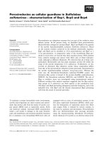
Fig. 1. Scheme of extraction and purification of polysaccharides from infusion of Chamomilla recutita floral chapters.
2.3. Determination of monosaccharide composition
plastic container at room temperature until use. In addition, a voucher
specimen of industry’s crop was collected (Campo Largo - PR, Brazil,
25º24.58’’S 49º27.64’’W, in 2013 September) to confirm the botanical
identity and deposited in the Museu Botânico Municipal de Curitiba,
under registration number 382674.
All fractions (except MRW-30E) were hydrolyzed in 500 μL 2 M TFA
at 100 °C for 8 h. MRW-30E was hydrolyzed with 500 μL 0.2 M TFA at
80 °C for 30 min. The TFA was evaporated and the samples were converted to alditol acetates by NaBH4 reduction at 100 °C for 10 min
followed by acetylation with Ac2O-pyridine (1:1, v/v, 1 mL) at 100 °C
for 30 min. The resulting alditol acetates were then extracted with
CHCl3 and analyzed by GC-MS using a Varian 3800 gas chromatograph
coupled to a Varian Ion-Trap 2000R mass spectrometer (Varian, Palo
Alto, CA). The column was DB-225 MS (30 m 0.25 mm i.d.; Agilent
Santa Clara, CA) programmed from 50 to 220 °C at 40 °C/min, with
helium as carrier gas, at a flow rate of 1 mL/min. The inlet temperature
was 250 °C, and the MS transfer line was set at 250 °C. MS acquisition
parameters included scanning from m/z 50–550 in electron ionization
mode (EI) at 70 eV. Components were identified by their retention
times and EI spectra. Fructose upon reduction and acetylation gives
glucitol and mannitol acetates on GC–MS analysis. The amounts of both
derivatives have been summed up to give the amount of fructose present in the sample.
Uronic acid contents were determined using the modified m-hydroxybiphenyl method (Filisetti-Cozzi & Carpita, 1991).
2.2. Extraction of polysaccharides
The floral chapters were reserved in a beaker and boiling distilled
water was added (40 g/L), the beaker was closed and let rest for about
30 min. The extract (tea) was filtered, concentrated under reduced
pressure and the polysaccharides precipitated with 95% ethanol
(3 vol.). The polysaccharides were recovered by filtration, dialyzed in
semipermeable membrane (Cellulose Spectrumlabs 6–8 kDa cut-off)
and freeze-dried (MRW fraction) (Fig. 1). These procedures were repeated several times to enable the extraction of 628 g of floral chapters.
MRW was further fractionated by ultrafiltration on 100 kDa cutoff
membrane (Fig. 1), giving MRW-100R (retained on the membrane) and
MRW-100E (eluted). This latter was ultrafiltrated on 30 kDa membrane.
The retained fraction (MRW-30R) was treated with Fehling solution
(Jones & Stoodley, 1965), and the resulting insoluble Cu2+ complex
isolated by centrifugation. Both (Fehling supernatant and precipitated
fractions, SF and PF, respectively) were neutralized with acetic acid,
dialyzed, and deionized with H+ form cation-exchange resin. SF was
then treated with endo-inulinase enzyme (316 U/mg, Megazyme) in
acetic acid/sodium acetate buffer (pH 4.6) for 16 h at 45 °C and then
dialyzed (Cellulose Spectrumlabs 6–8 kDa cut-off), giving SF-EN fraction. Finally, it was submitted to anion exchange chromatography on
DEAE Sepharose Fast Flow (GE Healthcare) and eluted with water, to
give fraction SF-EN-AG. All the fractionation steps are summarized in
Fig. 1. Yields of polysaccharide fractions were expressed as percent
based on the weight of dried floral chapters that were submitted to
extraction (628 g).
2.4. Determination of homogeneity and relative molecular weight
The homogeneity and relative molecular weight (Mw) of water-soluble polysaccharides were evaluated by high performance steric exclusion chromatography (HPSEC), with a Waters 2410 differential refractometer as equipment for detection. A series of four columns, with
exclusion sizes of 7 × 10⁶ Da (Ultrahydrogel 2000, Waters), 4 × 10⁵ Da
(Ultrahydrogel 500, Waters), 8 × 10⁴ Da (Ultrahydrogel 250, Waters)
and 5 × 10³ Da (Ultrahydrogel 120, Waters) was used. The eluent was
0.1 M aq. NaNO2 containing 200 ppm aq. NaN3 at 0.6 mL/min. The
sample, previously filtered through a membrane (0.22 μm, Millipore),
was injected (250 μl loop) at a concentration of 1 mg/mL. To obtain the
270
Carbohydrate Polymers 214 (2019) 269–275
P.F.P. Chaves, et al.
relative Mw, standard dextrans (487 kDa, 266 kDa, 124 kDa, 72.2 kDa,
40.2 kDa, 17.2 kDa and 9.4 kDa, from Sigma) were employed to obtain
the calibration curve. The relative Mw of the sample was calculated
according to the calibration curve.
Table 1
Monosaccharide composition of fractions obtained from chamomile (C. recutita)
tea.
Fraction
2.5. Methylation analysis
MRW
MRW-100R
MRW-30E
MRW-30R
SF-EN
SF-EN-AG
Fraction SF-EN-AG was carboxyl reduced by the carbodiimide
method, using NaBH4 as the reducing agent, giving products with the
eCOOH groups of its uronic acid residues reduced to eCH2OH (Taylor
& Conrad, 1972). The carboxyl reduced sample was O-methylated according to Ciucanu and Kerek (1984) method, using powdered NaOH in
DMSO-MeI. The per-O-methylated polysaccharide was then submitted
to methanolysis in 3% HCl–MeOH (80 °C, 2 h) followed by hydrolysis
with H2SO4 (0.5 M, 12 h) and neutralization with BaCO3. The material
was then submitted to reduction and acetylation as described above for
sugar composition, except that the reduction was performed using
NaBD4. The products (partially O-methylated alditol acetates) were
examined by capillary GC–MS. A capillary column (30 m × 0.25 mm
i.d.) of DB-225, held at 50 °C during injection for 1 min and then programmed at 40 °C/min to 210 °C and held at this temperature for
31 min, was used for separation. The partially O-methylated alditol
acetates were identified by their typical electron impact breakdown
profiles and retention times (Sassaki, Gorin, Souza, Czelusniak, &
Iacomini, 2005).
Neutral sugarsa
Uronic acidb
Rha
Ara
Xyl
Gal
Fruc
3.0
1.1
–
2.6
3.0
–
24.6
4.0
4.7
18.5
40.6
37.4
8.6
1.9
–
6.9
18.4
–
12.0
2.2
–
11.6
24.0
58.0
12.6
–
95.3d
16.3
tr
–
39.2
91.0
nde
44.1
14.0
4.6
a
% of peak area relative to total peak area, determined by GC–MS.
Determined using the m-hydroxybiphenyl method (Filisetti-Cozzi &
Carpita, 1991).
c
The amounts of glucitol and mannitol acetates on GC–MS analysis have
been summed up to give the amount of fructose present in the sample.
d
Hydrolysis with 0.2 M TFA at 80 °C followed by GC–MS analysis.
e
Not determined.
b
2.6. Nuclear magnetic resonance spectroscopy
The 1H, 13C and heteronuclear single quantum coherence (HSQCDEPT 135) spectra were obtained from samples dissolved in D2O, at
70 °C using a 400 MHz Bruker model DRX Avance III spectrometer,
operating at 9.5 T, observing 1H at 400.13 MHz and 13C at 100.61 MHz,
equipped with a 5-mm multinuclear inverse detection probe with zgradient. The chemical shifts are expressed in ppm relative to CH3
signal from internal reference acetone (δ 30.2/2.22). All pulse programs
were supplied by Bruker.
2.7. Electrospray ionization mass spectroscopy analysis
A syringe pump was used at a flow rate of 5 μL/min to infuse
fraction MRW-ET (at 200 μg/mL) directly into the mass spectrometer.
The positive high-resolution mass spectroscopy analysis was carried out
with electrospray ionization (ESI) at atmospheric pressure ionization
(API) in an LTQ-OrbiTrap-XL (Thermo-Scientific), using N2 for sample
desolvation with sheath gas at a flow rate of 8 UA and auxiliary gas at 2
UA with a source temperature of 300 °C. The ionization was performed
following the operational parameters: electrospray voltage at 4 kV,
capillary voltage 25 V, tube lens offset 125 V. The spectra were processed and analysed with Thermo Xcalibur 1.0.0.42 software.
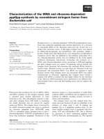
Fig. 2. HSQC correlation map of MRW fraction in D2O at 70 °C, the chemical
shifts are expressed as δ ppm. Ara = arabinose, GalA = galacturonic acid,
GalA’= methyl esterified galacturonic acid, Fru = fructose.
δ 81.1/3.86 (C5-H5) and δ 62.0/3.76 and 3.83 (C6-H6) (CorrêaFerreira, Noleto, & Oliveira Petkowicz, 2014; de Oliveira et al., 2011;
Perrone et al., 2002; Popov et al., 2011; Vriesmann & de Oliveira
Petkowicz, 2009). Small amounts of an arabinogalactan may also be
present by the observed anomeric signals of β-D-Galp units at δ 102.9/
4.47 and that of α-L-Araf at δ 107.6/5.07 and δ 109.0/5.25 (do
Nascimento, Iacomini, & Cordeiro, 2017; de Oliveira, do Nascimento,
Iacomini, Cor deiro, & Cipriani, 2017).
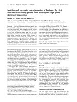
These two main polysaccharide types were also observed in homogeneity analysis, where a heterogeneous profile with two evident peaks
(I and II) (Fig. 3) were present. To isolate them, the fraction was submitted to ultrafiltration using a 100 kDa cutoff membrane. The process
was highly efficient, once peak II was concentrated in the eluted fraction (MRW-100E, 1.4% yield), while peak I remained retained on the
membrane (MRW-100R, 1.0% yield). This latter contained the pectic
homogalacturonan. It had mainly uronic acid (Table 1) on sugar analysis, identified as galacturonic acid by GC–MS of carboxyl-reduced
sample, and a relative Mw of 500 kDa. 13C NMR spectrum (Fig. 4A)
showed typical signals of the methyl esterified HG (as cited above). The
3. Results and discussion
The process of extraction by infusion of C. recutita floral chapters
produced a crude polysaccharide fraction named MRW with 3.2% yield
from the dry weight and an ethanolic supernatant (MRW-ET, 20%
yield). The sugar composition, which showed uronic acids, arabinose,
galactose, xylose, rhamnose and fructose (Table 1) together with HSQC
correlation map analysis of MRW (Fig. 2) allowed a preliminary identification of two main polysaccharide types present in chamomile tea:
(1) a methyl esterified homogalacturonan (HG) could be detected due
to the signals at δ 100.0/4.97 (C1-H1 from methyl esterified GalpA), δ
99.3/5.18 (C1-H1 from GalpA), δ 68.0/3.75 (C2), δ 68.3/3.98 (C3), δ
78.6/4.46 (C4), δ 70.5/5.05 (C5 from methyl esterified GalpA) and δ
52.8/3.82 (eCOOCH3); and (2) a fructan of inulin-type, due to the
signals at δ 61.0/3.73 (C1-H1), δ 103.2 (C2, visible only in the 13C
spectrum, data not shown), δ 77.2/4.23 (C3-H3), δ 74.6/4.09 (C4-H4),
271
Carbohydrate Polymers 214 (2019) 269–275
P.F.P. Chaves, et al.
Mw < 9.4 kDa) also showed a third small peak in HPSEC analysis (with
relative Mw of 60 kDa) (Fig. 3) and thus was submitted to a new ultrafiltration procedure using a 30 kDa cutoff membrane. Peak II was
eluted in the membrane (MRW-30E fraction) and had fructose on sugar
analysis as the major constituent (Table 1). 13C NMR analysis (Fig. 4B)
indicated the presence of the inulin-type fructan, with six typical signals
of →1)-β-D-Fruf-(2→ at δ 61.2 (C1), δ 103.2 (C2), δ 77.5 (C3), δ 74.8
(C4), δ 81.3 (C5) and δ 62.3 (C6) (Corrêa-Ferreira et al., 2014; de
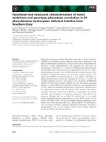
Oliveira et al., 2011). Looking for the presence of fructooligosaccharides (FOS) in chamomile tea, we analyzed fraction MRW-ET, which was
obtained in high yield (20%), using the LTQ Orbitrap-XL Hybrid Ion
Trap-Orbitrap Mass Spectrometer. The MS spectra (Fig. 5) showed besides sucrose, FOS ranging from GF2 (m/z 543) to GF10 (m/z 1839).
Thus, the results showed that chamomile tea contains as main
polysaccharides a highly methyl esterified and acetylated homogalacturonan and inulin, besides high amounts of fructooligosaccharides. A previous study about C. recutita polysaccharides pointed out the
existence of a polysaccharide containing (1→4)-linked α-D-GalpA residues (Yakovlev & Gorin, 1977), but the structural characterization of
the polymer has not been performed by the authors. Later, Füller and
Franz (1993) observed the presence of a fructan of the inulin type in
their C. recutita extracts, but the presence of FOS in chamomile tea has
not been reported in the literature yet. Fructans are commonly found in
species from the Asteraceae family, to which C. recutita belongs. These
can be found as reserve polymers in the tuberous roots of Jerusalem
artichoke (Helianthus tuberosus) (Saengthongpinit & Sajjaanantakul,
2005), chicory (Cichorium intybus) (Toneli, Park, Ramalho, Murr, &
Fabbro, 2008) and yacon (Smallanthus sonchifolius) (Paredes et al.,
2018). In the aerial parts, fructans have already been found in artemisia
Fig. 3. HPSEC elution profile of fractions MRW, MRW-100E and MRW-100R.
Refractive index detector. Elution volume of dextran standards of molecular
weight 487 kDa, 266 kDa, 124 kDa, 72.2 kDa, 40.2 kDa, 17.2 kDa and 9.4 kDa
(left to right) were employed to construct the calibration curve.
degree of methyl esterification was determined by 1H NMR following
the method of Grasdalen, Einar Bakøy, and Larsen, (1988) giving a
value of 87%, characterizing the chamomile pectin as a HM pectin
(Silva et al., 2006). Due to the presence of acetyl signals at δ 20.3 in the
13
C NMR spectrum, the degree of acetylation was also determined by 1H
NMR following the method of An et al. (2011) and spectrophotometrically by Hestrin (1949) methodology, giving a value of 19%.
Fraction MRW-100E containing the peak II of MRW (with relative
Fig. 4.
13
C NMR spectra of fractions MRW-100R (A), MRW-100E (B) and SF-EN (C) in D2O at 70 °C, the chemical shifts are expressed as δ ppm.
272
Carbohydrate Polymers 214 (2019) 269–275
P.F.P. Chaves, et al.
Fig. 5. MS spectra (+ve mode) of MRW-ET fraction obtained in LTQ Orbitrap-XL Hybrid Ion Trap-Orbitrap Mass Spectrometer.
103.4 (anomeric carbon of β-D-Galp) and at δ 107.6 and δ 109.0
(anomeric carbons of α-L-Araf units), probably from an arabinogalactan
(Nascimento et al., 2017; Oliveira et al., 2017). Finally, fraction SF-EN
was further purified by anion exchange chromatography in DEAE Sepharose Fast Flow. The column was eluted with water, giving a fraction
(SF-EN-AG) composed mainly of galactose and arabinose (Table 1).
Methylation analysis of carboxyl reduced sample (Table 2) confirmed
the presence of an arabinogalactan. The main methylated derivative
was 2,4-Me2-Gal-ol acetate, from 3,6-di-O-substituted Galp units. Other
Gal derivatives were 2,3,4,6-Me4-Gal-ol, 2,3,4-Me3-Gal-ol, 2,4,6-Me3Gal-ol and 4-Me-Gal-ol acetates, from terminal, 6-O-, 3-O- and 2,3,6-triO-substituted Galp units, respectively. Arabinose was present as terminal, 5-O- and 3,5-di-O-substituted Araf units. Terminal Glcp units were
also observed, from GlcpA units. Its HSQC-DEPT correlation map
(Fig. 6) showed anomeric cross peaks at δ 109.0/5.24 and δ 107.3/5.07
from terminal and →5)-α-L-Araf-(1→ units, at δ 103.8/4.69, δ 103.2/
4.46 and δ 103.0/4.51 from terminal, →3)-β-D-Galp-(1→/→3,6)-β-DGalp-(1→ and →6)-β-D-Galp-(1→, respectively. Inverted DEPT signals
were at δ 69.2/3.92–4.04 from 6-O-linked β-D-Galp units and at δ 66.6/
3.80–3.87 from 5-O-linked α-L-Araf units. Other inverted signals were
at δ 61.2/3.80, δ 61.1/3.73 and δ 60.9/3.77 from unsubstituted C-6/H-
(Artemisia vulgaris) (Corrêa-Ferreira et al., 2014), stevia (Stevia rebaudiana) (de de Oliveira et al., 2011) and another Matricaria species
(M. maritima (Cérantola et al., 2004). They were also extracted from the
monocotyledon agave plant (Agave tequilana var. azul) (Praznik,
Löppert, Cruz Rubio, Zangger, & Huber, 2013).
It is well stablished in the literature that inulin is a versatile substance with numerous health benefits. Inulin and FOS are the most
studied and well-established prebiotics. They escape digestion in the
upper gastrointestinal tract and reach the large intestine virtually intact, where they modulate the composition and activities of the gut
microbiota (Roberfroid, 2007a). Moreover, it has been demonstrated
that pectic polymers from different sources can also be prebiotics, being
extensively fermented in the colon and are able to modulated the gut
microbiota (Cantu-Jungles et al., 2017; Gulfi, Arrigoni, & Amadò, 2005;
Jonathan et al., 2012; Licht et al., 2010; Min et al., 2015; Titgemeyer,
Bourquin, Fahey, & Garleb, 1991). It is worth noting that inulin, FOS
and pectins can also specifically affect several other gastrointestinal
functions (for example, mucosal functions, endocrine activities and
mineral absorption) as well as systemic functions (especially glucose
and lipid homeostasis and immune functions) (Lunn & Buttriss, 2007;
Popov & Ovodov, 2013; Roberfroid, 2007a; Vogt et al., 2015).
To a comprehensive identification of chamomile polysaccharides,
the low-yield fraction MRW-30R which corresponded to the peak III
(Fig. 3) was also chemically characterized. It had a very complex
monosaccharide composition, composed of rhamnose, arabinose, xylose, fructose, galactose and uronic acid (Table 1). Galacturonic acid
and fructose came from HG and inulin, that were still present in this
fraction (observed in its 13C NMR spectrum, data not shown). To further
purification and characterization of other polysaccharides, MRW-30R
was treated with Fehling reagent once homogalacturonans interact with
copper and precipitate. Thus, due to alkaline pH of Fehling reagent,
deesterified and deacetylated HG remained in PF fraction, as could be
observed in its 13C NMR spectrum (Suppl. Fig. 1). Fraction SF was also
treated with endo-inulinase, due to the presence of some amounts of
contaminating inulin. On sugar analysis, fraction SF-EN presented
rhamnose, arabinose, galactose, xylose and uronic acids (Table 1). Its
13
C NMR spectrum (Fig. 4C) showed signals at δ 101.1 and δ 101.7
assigned to anomeric β-D-Xylp units, and at δ 97.6 (C1) and 59.4
(eOCH3) assign to 4-O-Me-α-D-GlcpA units, probably from an acid
xylan (Dinand & Vignon, 2001; Vignon & Gey, 1998), and signals at δ
Table 2
Linkage types based on analysis of partially O-methyl alditol acetates obtained
from methylated type II arabinogalactan (fraction SF-EN-AG) from chamomile
(C. recutita) tea.
Partially O-methylalditol acetate
SF-EN-AGa
Linkage typeb
2,3,5-Me3-Arafc
2,3,4,6-Me4-Glcp
2,3,4,6-Me4-Galp
2,3-Me2-Araf
2-Me-Araf
2,4,6-Me3-Galp
2,3,4-Me3-Galp
2,4-Me2-Galp
4-Me-Galp
11.5
7.2
14.9
8.8
1.1
2.3
4.1
45.2
4.9
Araf-(1→
Glcp-(1→
Galp-(1→
→5)-Araf-(1→
→3,5)-Araf-(1→
→3)-Galp-(1→
→6)-Galp-(1→
→3,6)-Galp-(1→
→2,3,6)-Galp-(1→
a
Fraction was carboxyl reduced by Taylor and Conrad (1972) method. % of
peak area of O-methyl alditol acetates relative to total area, determined by
GC–MS.
b
Based on derived O-methyl alditol acetates.
c
2,3,5-Me3-Ara = 2,3,5-tri-O-Methylarabinitolacetate, etc.
273
Carbohydrate Polymers 214 (2019) 269–275
P.F.P. Chaves, et al.
Appendix A. Supplementary data
Supplementary material related to this article can be found, in the
online version, at doi: />References
Akerele, O. (1993). Summary of WHO guidelines for the assessment of herbal medicines.
Pharmacology & Pharmacy, 28, 13.
An, T. N., Thien, D. T., Dong, N. T., Dung, P., Le, Du, N., ... Van (2011). Isolation and
characteristics of polysaccharide from Amorphophallus corrugatus in Vietnam.
Carbohydrate Polymers, 84(1), 64–68. />074.
Brecker, L., Wicklein, D., Moll, H., Fuchs, E. C., Becker, W. M., & Petersen, A. (2005).
Structural and immunological properties of arabinogalactan polysaccharides from
pollen of timothy grass (Phleum pratense L.). Carbohydrate Research, 340(4), 657–663.
/>Cantu-Jungles, T. M., Cipriani, T. R., Iacomini, M., Hamaker, B. R., & Cordeiro, L. M. C.
(2017). A pectic polysaccharide from peach palm fruits (Bactris gasipaes) and its
fermentation profile by the human gut microbiota in vitro. Bioactive Carbohydrates
and Dietary Fibre, 9(August 2016), 1–6. />Capek, P., Matulová, M., Navarini, L., & Suggi-Liverani, F. (2010). Structural features of
an arabinogalactan-protein isolated from instant coffee powder of Coffea arabica
beans. Carbohydrate Polymers, 80(1), 180–185. />2009.11.016.
Cérantola, S., Kervarec, N., Pichon, R., Magné, C., Bessieres, M.-A., & Deslandes, E.
(2004). NMR characterisation of inulin-type fructooligosaccharides as the major
water-soluble carbohydrates from Matricaria maritima (L.). Carbohydrate Research,
339(14), 2445–2449. />Ciucanu, I., & Kerek, F. (1984). A simple and rapid method for the permethylation of
carbohydrates. Carbohydrate Research, 131(2), 209–217. />0008-6215(84)85242-8.
Corrêa-Ferreira, M. L., Noleto, G. R., & Oliveira Petkowicz, C. L. (2014). Artemisia absinthium and Artemisia vulgaris: A comparative study of infusion polysaccharides.
Carbohydrate Polymers, 102(1), 738745. />096.
de Oliveira, A. J. B., Gonỗalves, R. A. C., Chierrito, T. P. C., Dos Santos, M. M., De Souza,
L. M., Gorin, P. A. J., ... Iacomini, M. (2011). Structure and degree of polymerisation
of fructooligosacchari des present in roots and leaves of Stevia rebaudiana (Bert.)
Bertoni. Food Chemistry, 129(2), 305–311. />2011.04.057.
Dinand, E., & Vignon, M. R. (2001). Isolation and NMR characterisation of a (4-O-methylD-glucurono)-D-xylan from sugar beet pulp. Carbohydrate Research, 330(2), 285–288.
/>Dong, Q., & Fang, J. N. (2001). Structural elucidation of a new arabinogalactan from the
leaves of Nerium indicum. Carbohydrate Research, 332(1), 109–114. />10.1016/S0008-6215(01)00073-8.
Filisetti-Cozzi, T.m.c.c., & Carpita, N.c. (1991). Measurement of uranic acids without from
neutral sugars. Analytical Biochemistry, 162, 157–162. />Franke, R., & Schilcher, H. (2005). Chamomile industrial profiles. Boca Raton: CRC Press
Taylor & Francis Group.
Füller, V. E., & Franz, G. (1993). Neues von den kamillenpolysacchariden. Deutsche
Apotheker Zeitung, 133, 4224–4227.
Goellner, E. M., Utermoehlen, J., Kramer, R., & Classen, B. (2011). Structure of arabinogalactan from Larix laricina and its reactivity with antibodies directed against typeII-arabinogalactans. Carbohydrate Polymers, 86(4), 1739–1744. />1016/j.carbpol.2011.07.006.
Grasdalen, H., Einar Bakøy, O., & Larsen, B. (1988). Determination of the degree of esterification and the distribution of methylated and free carboxyl groups in pectins by
1H-n.m.r. spectroscopy. Carbohydrate Research, 184, 183–191. />1016/0008-6215(88)80016-8.
Gulfi, M., Arrigoni, E., & Amadò, R. (2005). Influence of structure on in vitro fermentability of commercial pectins and partially hydrolysed pectin preparations.
Carbohydrate Polymers, 59(2), 247–255. />018.
Halberstein, R. A. (2005). Medicinal plants: Historical and cross-cultural usage patterns.
Annals of Epidemiology, 15(9), 686–699. />02.004.
Hestrin, S. (1949). The reaction of acetylcholine and other carboxylic acid derivatives
with hydroxylamine, and its analytical application. The Journal of Biological
Chemistry, 180(1), 249–261.
Jonathan, M. C., Van Den Borne, J. J. G. C., Van Wiechen, P., Souza Da Silva, C., Schols,
H. A., & Gruppen, H. (2012). In vitro fermentation of 12 dietary fibres by faecal
inoculum from pigs and humans. Food Chemistry, 133(3), 889–897. />10.1016/j.foodchem.2012.01.110.
Jones, J. K. N., & Stoodley, R. J. (1965). Fractionation using copper complexes. Methods in
Carbohydrate Chemistry, 5, 36–38.
Liang, F., Hu, C., He, Z., & Pan, Y. (2014). An arabinogalactan from flowers of
Chrysanthemum morifolium: Structural and bioactivity studies. Carbohydrate Research,
387(1), 37–41. />Licht, T. R., Hansen, M., Bergström, A., Poulsen, M., Krath, B. N., Markowski, J., ...
Wilcks, A. (2010). Effects of apples and specific apple components on the cecal environment of conventional rats: Role of apple pectin. BMC Microbiology, 10(13),
Fig. 6. HSQC-DEPT correlation map of SF-EN-AG fraction in D2O at 50 °C, the
chemical shifts are expressed as δ ppm. Inverted signals in DEPT experiment are
shown in blue color (For interpretation of the references to colour in this figure
legend, the reader is referred to the web version of this article).
6 or C-5/H-5 from α-L-Araf-(1→, β-D-Galp-(1→ and →3)-β-D-Galp-(1→
units. The assignments are in agreement with published literature data
and methylation analysis described above (2015, Brecker et al., 2005;
Capek, Matulová, Navarini, & Suggi-Liverani, 2010; Dong & Fang,
2001; Goellner, Utermoehlen, Kramer, & Classen, 2011; Liang, Hu, He,
& Pan, 2014; Wang, Shi, Bao, Li, & Wang, 2015) and shows the presence
of a type II arabinogalactan in SF-EN-AG fraction. In their preliminary
characterization of C. recutita polysaccharides, Füller and Franz (1993)
also suggested the presence of a rhamnogalacturonan with type II
arabinogalactan and a glucuronoxylan in the aqueous chamomile extracts. However, the fine chemical structure of these polysaccharides
had not been determined.
Matricaria chamomilla belongs to a major group of cultivated medicinal plants, often referred to as the “star among medicinal species”.
More than 120 chemical constituents have been identified in chamomile flower as secondary metabolites, which gives to chamomile its
multitherapeutic, cosmetic, and nutritional values, that have been established through years of traditional and scientific use and research
(Singh et al., 2011). The presence of inulin, FOS, highly methyl esterified homogalacturonan, type II arabinogalactan and acid xylan in
chamomile tea shows that not only can the secondary metabolites be
the responsible molecules by the health benefits of chamomile consumption and adds to chamomile a new property, as a source of
structurally diverse dietary fibers with potential prebiotic, gastrointestinal and immunological functions.
Acknowledgements
This research was supported by CAPES (Process 1264763),
Fundaỗóo Araucỏria and by Universal Project (Process 404717/2016-0)
provided by CNPq foundation (Brazil). The authors are grateful to
Chamel® Produtos Naturais Industry who kindly provided the dried
floral C. recutita chapters, to the NMR Center of UFPR for recording the
NMR spectra and to Dr. Lauro M. de Souza for the mass spectroscopy
analysis.
274
Carbohydrate Polymers 214 (2019) 269–275
P.F.P. Chaves, et al.
1016/j.postharvbio.2005.03.004.
Sassaki, G. L., Gorin, P. A. J., Souza, L. M., Czelusniak, P. A., & Iacomini, M. (2005). Rapid
synthesis of partially O-methylated alditol acetate standards for GC-MS: Some relative activities of hydroxyl groups of methyl glycopyranosides on Purdie methylation. Carbohydrate Research, 340(4), 731–739. />2005.01.020.
da Silva, J. A. L., & Rao, M. A. (2006). Pectins: Structure, functionality, and uses. In A. M.
Stephen, G. O. Phillips, & P. A. Williams (Eds.). Food polysaccharides and their applications (pp. 353–412). (2nd ed.). Boca Raton: CRC Press.
Singh, O., Khanam, Z., Misra, N., & Srivastava, M. (2011). Chamomile (Matricaria chamomilla L.): An overview. Pharmacognosy Reviews, 5(9), 82–95. />4103/0973-7847.79103.
Sousa, M. P., Matos, M. E. O., Matos, F. J. A., Machado, M. I. L., & Craveiro, A. A. (1991).
Constituintes químicos ativos de plantas medicinais brasileiras. Fortaleza: EUFC.
Laboratório de Produtos Naturais.
Taylor, R. L., & Conrad, H. E. (1972). Stoichiometric depolymerization of polyuronides
and glycosaminoglycuronans to monosaccharides following reduction of their carbodiimide-activated carboxyl group. Biochemistry, 11(8), 1383–1388. https://doi.
org/10.1021/bi00758a009.
Titgemeyer, E. C., Bourquin, L. D., Fahey, G. C., & Garleb, K. A. (1991). Fermentability of
various fiber sources by human fecal bacteria in vitro. The American Journal of Clinical
Nutrition, 53(6), 1418–1424. />Toneli, J. T. C. L., Park, K. J., Ramalho, J. R. P., Murr, F. E. X., & Fabbro, I. M. D. (2008).
Rheological characterization of chicory root (Cichorium intybus L.) inulin solution.
Brazilian Journal of Chemical Engineering, 25(3), 461–471. />S0104-66322008000300004.
Vignon, M. R., & Gey, C. (1998). Isolation, 1H and 13C NMR studies of (4-O-methyl-dglucurono)-d-xylans from luffa fruit fibres, jute bast fibres and mucilage of quince
tree seeds. Carbohydrate Research, 307(1–2), 107–111. />S0008-6215(98)00002-0.
Vogt, L., Meyer, D., Pullens, G., Faas, M., Smelt, M., Venema, K., ... De Vos, P. (2015).
Immunological properties of inulin-type fructans. Critical Reviews in Food Science and
Nutrition, 55(3), 414–436. />Vriesmann, L. C., & de Oliveira Petkowicz, C. L. (2009). Polysaccharides from the pulp of
cupuassu (Theobroma grandiflorum): Structural characterization of a pectic fraction.
Carbohydrate Polymers, 77(1), 72–79. />007.
Wang, H., Shi, S., Bao, B., Li, X., & Wang, S. (2015). Structure characterization of an
arabinogalactan from green tea and its anti-diabetic effect. Carbohydrate Polymers,
124, 98–108. />Wang, P., Zhang, L., Yao, J., Shi, Y., Li, P., & Ding, K. (2015). An arabinogalactan from
flowers of Panax notoginseng inhibits angiogenesis by BMP2/Smad/Id1 signaling.
Carbohydrate Polymers, 121, 328–335. />073.
Yakovlev, A. I., & Gorin, A. C. (1977). Structure of the petic acid of Matricaria chamomilla.
Khimiya Prirodnykh Soedinenlii, 2, 186–189.
/>Liu, J., Willför, S., & Xu, C. (2015). A review of bioactive plant polysaccharides: Biological
activities, functionalization, and biomedical applications. Bioactive Carbohydrates and
Dietary Fibre, 5(1), 31–61. />Lorenzi, H., & de A.Matos, F. J. (2008). Plantas Medicinais Do Brasil Nativas E Exóticas (2nd
ed.). Nova Odessa. São Paulo: Instituto Plantarum.
Lunn, J., & Buttriss, J. L. (2007). Carbohydrates and dietary fibre. Nutrition Bulletin, 32(1),
21–64. />Min, B., Kyung Koo, O., Park, S. H., Jarvis, N., Ricke, S. C., Crandall, P. G., ... Lee, S.-O.
(2015). Fermentation patterns of various pectin sources by human fecal microbiota.
Food and Nutrition Sciences, 06(12), 1103–1114. />612115.
do Nascimento, G. E., Iacomini, M., & Cordeiro, L. M. C. (2017). New findings on green
sweet pepper (Capsicum annum) pectins: Rhamnogalacturonan and type I and II
arabinogalactans. Carbohydrate Polymers, 171, 292–299. />carbpol.2017.05.029.
de Oliveira, A. F., do Nascimento, G. E., Iacomini, M., Cor deiro, L. M. C., ... Cipriani, T. R.
(2017). Chemical structure and anti-inflammatory effect of polysacchari des obtained
from infusion of Sedum dendroideum leaves. International Journal of Biological
Macromolecules, 105, 940–946. />Paredes, L. L. R., Smiderle, F. R., Santana-Filho, A. P., Kimura, A., Iacomini, M., & Sassaki,
G. L. (2018). Yacon fructans (Smallanthus sonchifolius) extraction, characterization
and activation of macrophages to phagocyte yeast cells. International Journal of
Biological Macromolecules, 108, 1074–1081. />2017.11.034.
Perrone, P., Hewage, C. M., Thomson, A. R., Bailey, K., Sadler, I. H., & Fry, S. C. (2002).
Patterns of methyl and O-acetyl esterification in spinach pectins: New complexity.
Phytochemistry, 60(1), 67–77. />Popov, S. V., & Ovodov, Y. S. (2013). Polypotency of the immunomodulatory effect of
pectins. Biochemistry Biokhimiia, 78(7), 823–835. />s0006297913070134.
Popov, S. V., Ovodova, R. G., Golovchenko, V. V., Popova, G. Y., Viatyasev, F. V.,
Shashkov, A. S., ... Ovodov, Y. S. (2011). Chemical composition and anti-inflammatory activity of a pectic polysaccharide isolated from sweet pepper using a
simulated gastric medium. Food Chemistry, 124(1), 309–315. />1016/j.foodchem.2010.06.038.
Praznik, W., Löppert, R., Cruz Rubio, J. M., Zangger, K., & Huber, A. (2013). Structure of
fructo-oligosaccharides from leaves and stem of Agave tequilana Weber, var. azul.
Carbohydrate Research, 381, 64–73. />Roberfroid, M. B. (2007a). Inulin-type fructans: Functional food ingredients. The Journal
of Nutrition, 137(11), 2493S–2502S. />Roberfroid, M. B. (2007b). Prebiotics: The concept revisited. The Journal of Nutrition,
137(3), 830S–837S. />Saengthongpinit, W., & Sajjaanantakul, T. (2005). Influence of harvest time and storage
temperature on characteristics of inulin from Jerusalem artichoke (Helianthus tuberosus L.) tubers. Postharvest Biology and Technology, 37(1), 93–100. />
275