Biomechanical Walking Pattern Changes in the Fitand and Healthy Elderly docx
Bạn đang xem bản rút gọn của tài liệu. Xem và tải ngay bản đầy đủ của tài liệu tại đây (893.09 KB, 10 trang )
1990; 70:340-347.PHYS THER.
Sharon E Walt
David A Winter, Aftab E Patla, James S Frank and
and Healthy Elderly
Biomechanical Walking Pattern Changes in the Fit
at:
The online version of this article, along with updated information and services, can be found
Collections
Kinesiology/Biomechanics
Geriatrics: Other
Falls and Falls Prevention
in the following collection(s):
This article, along with others on similar topics, appears
e-Letters
"Responses" in the online version of this article.
"Submit a response" in the right-hand menu under
or click onhere To submit an e-Letter on this article, click
E-mail alerts to receive free e-mail alerts hereSign up
by guest on December 24, 2012 from
Research
Report
Biomechanical Whng Pattern Changes in the Fit and
Healthy Elderly
A
descriptive study of the biomechanical variables of the walking patterns of the fit
and
healtky elderly compared with those of young adults revealed several signzfi-
cant dzfferences. The walking patterns of 15 elderly subjects, selected for their
active
life style and screened for any gait- or balance-related pathological condi-
tions, were analyzed. Kinematic and kinetic data for a minimum of 10 repeat
walking
t~ials were collected using a video digitizing system and a force platform.
Basic kinematic analyses and an inverse dynamics model yielded data based on
the following variables: temporal and cadence measures, heal and toe trajectories,
joint kinematics, joint moments of force, and joint mechanical power generation
and absorption.
Signzjicant dzfferences between these elderly subjects and a data-
base of young adults revealed the following: the same cadence but a shorter step
length, an increased double-support stance period,
decreasedpush-offpower, a
more flat-footed landing, and a reduction in their "index of dynamic balance."
All of these
dzfferences, except reduction in index of dynamic balance, indicate
adaptation by the elderly toward a safer, more stable gait pattern. The reduction
in index of dynamic balance suggests deterioration in the
eficiency of the bal-
ance control system during gait. Because of these
sign@cant dzfferences attribut-
able to age alone, it is apparent that a separate gait database is needed in order
to pinpoint falling disorders of the elderly.
/Winter
DA,
Patla
AE,
Frank
JS,
et al.
Biornechanical waking pattern changes in the fit and healthy elderly. Phys Ther
1990; 70:340-3471
Key Words:
Equilibrium; Geriatrics;
Kinesiologylbiomechanics,
gait analysis;
Posture, tests and measurements.
David
A
Winter
Aftab
E
Patla
James S Frank
Sharon
E
Walt
The reduction of frequency of falls Research has focused on epidemio- terizing the changes in the standing
among the elderly is the goal of many logical studies to provide a better balance control system that occur
researchers addressing the resultant description and assessment of the with age. The epidemiological data
injuries, death, and loss of mobility.'
extent of the problem and on charac-
have implicated some aspects of loco-
motion (ie, initiation of walking, turn-
ing, walking over uneven surfaces,
stopping) in almost all incidences of
D Winter, PhD, PEng, is Professor, Department of Kinesiology, University of Waterloo, Waterloo,
falls.2-5
Ontario, Canada
N2L
3G1. Address all correspondence to Dr Winter.
A
Patla,
P~D,
is Associate Professor, Department of Kinesiology, University of Waterloo.
Despite this strong evidence linking
Frank, PhD,
is
Assistant Professor, Department of Kinesiology, University of Waterloo.
locomotion to falls, studies of changes
in the balance control system have
S
Walt, MASc, is Research Assistant, Department of Kinesiology, University of Waterloo.
been limited mainly to tests that
Financial support For this study was provided by the Medical Research Council of Canada (Grant
probe the integrity of the system dur-
MT4343) and Health and Welfare Canada (Grant 6606-3675-R).
ing quiet standing. Performance on
This study was approved by the University of Waterloo's Office of Human Research.
these tests does not correlate with
incidence of falls and is a poor pre-
This arlicle
was
submiffed July
19, 1989,
and was accepted January
19, 1990.
Physical Therapyflolume 70, Number 6/June 1990
340
/
15
by guest on December 24, 2012 from
dictor of fallers.6 Even during per-
turbed standing tests,' the predictions
have been no better than 30% (60%
of fallers predicted, and 30%
of
non-
fallers are false positives). This finding
is hardly surprising because the bal-
ance challenges during walking are
quite different from those involved in
maintaining upright posture.
During standing, the goal is to main-
tain the body's center of gravity (CG)
within the base of support. The initia-
tion of gait, however, is an
unstabiliz-
ing event whereby the body's CG is
made to fall forward and outside of
the stance
foot.8 By the time the
selected cadence is achieved, the only
stabilizing period is double-support
stance, and even during that time
period the one limb is pushing off
with considerable force while the
other limb is accepting the full weight
of the
body.9 During natural cadence,
80% of the stride period is single-
support stance, when the CG
of
the
body has been shown to be outside
the
footlo; the closest
it
gets to the
base of support is when
it
passes for-
ward along the medial border of the
foot. Even during the two 10%
double-support stance periods, both
feet are not flat on the ground. Dur-
ing the first half of double-support
stance, or heel contact (HC), the
weight-accepting foot is being low-
ered to the ground; during the latter
half
of
double-support stance, the
final stage of push-off has weight only
under the toes. Thus, the body is in
an inherent state of instability. Most of
the findings from balance studies dur-
ing standing, therefore, have very lim-
ited relevance to gait. The dynamic
balance of the head, arms, and trunk
(HAT)
and the safe transit of the foot
during the swing phase of gait (safe
toe clearance and a gentle foot land-
ing) present a challenge to the central
nervous system during walking. The
HAT constitutes two thirds of the
body mass, and the
HAT'S center of
mass (CM) is located about two thirds
of the body height above ground
level. The CM is the point where all
the mass of the HAT can be consid-
ered to act in all three axes as com-
pared with the CG, which is its loca-
tion in the gravitational axis. In the
sagittal plane, even in slow walking,
the horizontal momentum of the HAT
results in inherent instability. The role
of the ankle muscles in standing bal-
ance is paramount, but in walking the
role of the ankle plantar-flexor and
dorsiflexor muscles for balance has
not been seen to be important." The
moment of inertia of the HAT about
the ankle is about eight times what
it
is about the hip." Thus, during the
first half of stance, for example, when
a posterior acceleration at the hip is
attempting to collapse the HAT in the
forward direction, the ankle muscles
do not act to intervene. If they did,
they would require a plantar-flexor
moment of about 300
N-m to control
the huge inertial load. Instead, the
ankle muscles produce a small dorsi-
flexor moment to lower the foot to
the ground, followed by a small
plantar-flexor moment to control the
forward leg rotation. The hip extensor
muscles, however, intervene to con-
trol the lesser inertial load in conjunc-
tion with a tight coupling with the
knee
muscles.llJVhe tight coupling
of these two motor patterns has been
labeled an "index of dynamic
balance."'* This balance control of the
large inertial load of the HAT acts pri-
marily during single-support stance
with a transfer of responsibility
between limbs taking place during
double-support stance.
The swing phase of gait has been
shown to be executed with consider-
able
precision15 with average toe
clearances of about
1
cm, and this
clearance occurs while the horizontal
velocity is maximal
(3.64.5 mlsec).
The heel velocity is also reduced dras-
tically in both horizontal and vertical
directions immediately prior to HC.
Thus, any degeneration in this fine
motor control of the foot may result
in problems of stumbling during
swing and in rebalancing immediately
after HC.
Numerous studies have addressed the
changes in the gait patterns of the
elderly compared with those of the
younger adult. The majority of these
studies1"-" have concentrated on
basic outcome measures (ie, stride
length, cadence, velocity) and the vari-
ability of those measures. Several of
these studies have related these gait
changes to
falls,l8 rnobility,l9 and post-
fall
anxiety.20 All
of
these studies have
made inferences about the reasons
for the observed changes: lower
cadence, shorter and more variable
step length, increased head and torso
flexion, and increased knee and
elbow flexion. The suggested reasons
imply a degeneration
of
balance con-
trol combined with a general loss of
muscle strength. The measures
reported, however, were outcome
measures, which provide limited
insight into the changes in the motor
system for balance control and limit
our ability to identify the mechanisms
behind the observed changes.
With this background in mind, there
is a need to document the motor pat-
tern changes that occur in the gait of
the elderly and to determine whether
those changes are related to balance.
Fit and healthy elderly individuals
were chosen for this initial study to
eliminate effects of a sedentary life
style or pathological conditions on
walking patterns. Of interest was the
normal biological degeneration
that
takes place with age prior to the
advent of any identifiable neural, mus-
cular, or skeletal disorder. All kine-
matic and kinetic patterns were exam-
ined in detail in order to pinpoint
major or subtle changes that would
point to the degeneration or to com-
pensations that reduce the chance of
stumbling or losing balance. Simulta-
neously, a second major goal was
achieved, that of developing a full
database of kinematic and kinetic pro-
files against which to compare indi-
vidual elderly patients with known or
suspected balance or tripping
disorders.
Subjects
Fifteen elderly subjects were screened
based on a life-style and medical
questionnaire and examined by a ger-
iatrician to eliminate any volunteers
who had any pathological condition
related to the human locomotor sys-
tem. Informed consent forms were
16
/
341
Physical Therapyflolume 70, Number 6/June 1990
by guest on December 24, 2012 from
signed by each subject prior to the
walking trials. These
fit
and healthy
elderly individuals (10 men, 5
women) ranged in age from 62 to 78
years
@
=
68 years).
Procedure
The protocol for the biomechanical
gait analyses was identical to that
reported
pre~iously"~~~~~3J~ and is
summarized as follows. Each subject
was instrumented with reflective
markers to define the following joint
centers and segments: toe, fifth meta-
tarsal, heel, lateral malleolus (ankle),
head of the fibula, lateral epicondyle
of the femur (knee), and greater
tro-
chanter (hip). Additional markers, not
part of this link-segment analysis,
were also attached to the trunk and
head to define upper body kinemat-
ics: L4-L5, sternum,
C1-C2, ear canal,
and forehead.
A
standard link-segment
model of the lower limb was devel-
oped for
he
foot, leg, and thigh seg-
ments in order to calculate the
moments of force at the ankle, knee,
and
hip.12,21 Each subject walked at
his or her natural cadence on a level
walkway a minimum
of
10 times; the
repeat trials were conducted over a
period
of
about one hour (one trial
every 5 or 6 minutes). Each subject
walked
over a force platform* while a
Charge-Coupled Device (CCD) video
camerat located 6 m
to
the side of the
walkway recorded the marker trajec-
tories over the stride period. The
CCD camera was electronically shut-
tered at 1 msec with a field rate of 60
Hz. The video signal was stored on a
Sony Motion
halyzers and subse-
quently digitized using a specially
designed video interface into an IBM
PC-AP computer.bhe precision of
the marker centroids was calculated
to within
1
mm. The raw coordinate
data were digitally filtered with a
fourth-order zero-lag Butterworth
filter with a cutoff at 6
Hz.
The
smoothed coordinates then became
inputs
to
the standard link-segment
model.
In addition
to
the joint moments of
force, the mechanical power gener-
ated and absorbed at each joint was
~alculated~~ and the area under each
power burst was integrated
to
deter-
mine the mechanical work performed
during each of the generating and
absorbing phases. The support
moment, as defined a decade
ago,9
was calculated and is equal to the
sum of the moments at the ankle,
knee, and hip (extensor moments
were set positive, and flexor moments
were set negative). The support
moment is the total motor pattern of
the lower limb, which has been seen
to
be positive (extensor) during most
of stance, negative (flexor) during late
double-support and early swing, and
positive (extensor) during late
swing.14 The ensemble average
of
the
moment-of-force patterns over all the
strides yielded a mean variance mea-
sure for the ankle, knee, and hip pro-
files, from which the hip-knee and
knee-ankle covariances were readily
calculated.13 The kinematics of toe
markers over the stride period
yielded the toe clearance during
mid-
swing.
Toe clearance
was defined as
the difference in the vertical displace-
ment of the toe marker at its lowest
point in stance (just before toe-off)
and its lowest point in mid-swing.
Data Analysis
Identical measures were taken from
our database on 12 young adults
(7
men, 5 women), ranging in age from
21 to 28 years
O[
=
24.6 years).
Because the population variances
were not identical, a modified
t
test23
was used
to
determine any significant
differences between selected kine-
matic and kinetic variables that had
potential impact on balance and
fall-
'Advanced Medical Technology Inc, 141 California St, Newton,
MA
02158.
'~odel TI-SOES, NEC America, 1255 Michael Dr, Wood Dale, IL 60191.
*Model SVM-1010, Sony of Canada,
88
Horner Ave, Toronto, Ontario, Canada K2B 8K1
"nternational Business Machines Corp, PO Box 1328-S, Boca Raton
FL
33432.
ing during walking. These variables
are presented in the Table.
Results
and
Discussion
The kinematic and kinetic patterns of
one elderly subject are used in this
section
to
illustrate the nature and
format of the data. The mean cadence
for this subject was 105
steps/min
(s
=
1.8), and the following
ensemble-averaged waveforms were
plotted at 2% intervals over the stride
period
(HC
=
0%, next HC
=
100%).
The average toe-off for this subject
was
65.7%, so it was set
to
the nearest
2% interval (66%). The following pro-
files are presented: ankle, knee, and
hip angles (Fig 1); toe vertical dis-
placement, vertical velocity, and hori-
zontal velocity (Fig 2); ankle, knee,
hip, and support moments (Fig
3);
and ankle, knee, and hip powers
(Fig 4). In all of these diagrams, the
mean of the repeat trials is plotted as
a solid line with one standard devia-
tion plotted at each 2% interval over
the stride period. The mean coeffi-
cient
of
variation
(CV)
is reported and
represents the average variability over
the stride period expressed as a per-
centage
of
the mean signal ampli-
tude.13
The
CV
measure is a single
score that allows comparison of the
percentage of variability of any wave-
form over any group of repeat walk-
ing trials.
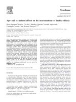
Figure
1
shows the variability of this
subject's ankle, knee, and hip joint
angles
to
be quite low. The CV for the
ankle, knee, and hip joints was
2196,
8%, and 8%, respectively. Similar low
variabilities have been reported for
intrasubject repeat trials performed
across days as well as minutes apart
on young
adults." These consistent
results caution against any inferences
about similar invariance in the motor
patterns. The indeterminacy of the
human motor system during stance is
such that many combinations
of
moments of force at the ankle, knee,
and hip can still result in the same
lower limb kinematics, especially at
the hip and knee, and this finding is
supported by the data for this subject.
Physical
TherapyNolume 70, Number 6June 1990
by guest on December 24, 2012 from
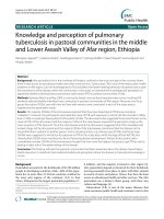
The toe trajectory data (Fig 2) show
the vertical displacement (upper
trace), the vertical velocity (middle
trace), and the horizontal velocity
(lower trace). These trajectory plots
all have low
CVs, indicating a highly
consistent control of the distal seg-
ment of the limb, the toe. The aver-
age toe clearance of 1.5 cm
(s
=
0.5)
for this subject occurred at 80% of
stride as the toe reached its peak hor-
izontal velocity of 4.3
m/sec. The com-
plex nature of this end-point control
task needs to be recognized. The
length of the link-segment chain is
over 2 m, starting with the stance
phase foot and continuing up to the
hip, across the pelvis, and down the
swing limb, and the chain involves at
least 12 degrees of freedom at the
joints and scores of muscles. The gen-
eration and execution of such a con-
sistent toe trajectory is evidence of
fine motor control.
The moment-of-force curves for this
elderly subject are presented in
Fig-
urc 3 with extensor moments plotted
as positive, along with the suppon
moment? which is the algebraic sum
of the three joint moments. The inter-
pretation of the support-moment pat-
tern has been discussed in detail
previously.7J3 In summary, the sup-
port moment quantifies the total limb
synergy, which is extensor during
most of stance, becomes flexor during
late double-support and early swing,
and returns to extensor during late
swing. We have identified this suppon
synergy in over 50 assessments on a
wide variety of gait pathologies in
healthy young (n
=
200) and elderly
(n
=
15) subjects.
The variability of these moment pat-
terns varies with the joint. This sub-
ject's CV was 9% at the ankle,
31%
at
the knee, 19% at the hip, and only 9%
in the support moment. Because CV is
a ratio of mean variance and mean
signal, the low CV for suppon
moment was partially due to
increased mean signal as well as
decreased mean variance. It has been
shown that the variance in these
motor patterns is not random, espe-
cially in the highly variable hip and
knee
patterns.l3 There is
a
tight neu-
+-I
STD.DEV
-10-
-20
-
TOa61.
SUBJECT:
K84
%
OF
STRIDE
Fig
1.
Ensemble-averaged joint angles for
I1
repeat walking trials of one elderly
subject. Stride period is normalized to
100%
from heel contact (HC) to HC, and for this
subject the average toe-off (TO) was
66%.
Solid lines plot average joint angle, and dot-
ted lines represent one standard deviation at each
2%
interval of stride period.
As
dem-
onstrated by low
coe8cient of variation (m scores, lower limb kinematics remained
very consistent. (DORSI
=
dorsijlexion;
FLEX
=
flexion.)
ral and anatomical coupling between
the knee and hip motor patterns. The
covariance between the hip and knee
moments can reach 89% in repeat
strides assessed days
apart and ranges
from 60% to 70% for repeat assess-
ments performed minutes
apart.14
This covariance is expressed
as
a per-
centage of the maximum possible and
would reach 100% if the covariance
were equal to the sum of the knee
and hip variances. This coupling
between the joint moments is
revealed in the small CV for the sum-
mation of hip and knee moments,
which was 14% for this set of repeat
trials. The reason for these trade-offs
between the hip and knee moments
is related to a second limb synergy,
that of dynamic
balance.11J4 This bal-
ance synergy is described as follows:
On a stride-to-stride basis, the
anterior-posterior balance of the HAT
is controlled by the hip flexors and
extensors during stance (mainly
single-support stance). Each stride is
somewhat different, and the regula-
tion of this large mass (two thirds of
body mass) requires a modified hip
motor pattern on each stride. Thus,
the high variance in the hip moment
during stance is directly due to a con-
tinuously changing balance control
task. The hip moment, however, is
also pan of the support synergy. To
keep the suppon pattern nearly con-
stant, there must be an opposite
change in the knee moment, which is
almost as variable, but in the opposite
direction. Such a trade-off between
Physical
Therapyflolume 70, Number 6/June 1990
by guest on December 24, 2012 from
.IS-
KRTICR DISPLACMMT
loo
-
KRTICR \nKlPI
.5-
CV=25.I%
N
t
m
0
CI
%
OF
STRIDE
Fig
2.
Ensemble-averaged toe trajectory plots for same subject as in Figure
I
over
11
repeat walking trials. Vertical displacement of toe (top trace) shows a minimum (set
to
0)
just prior to toe-off
(TO)
and minimum toe clearance during swing at about
80%
of stride period when horizontal velocity (bottom trace) is rzear its maximum.
(CV
=
coeficient of variation.)
the hip and knee moment patterns is
almost one-for-one and is the reason
for the low variance in the summation
of the hip and knee
moments."J4 The
covariance between the hip and knee
moments is a measure of this syner-
gistic trade-off and has been labeled
an
"index of dynamic balance."l4 For
this subject, it was 59.3%.
The comparison between the young
adults' gait and that of the elderly sub-
jects is presented in the Table. Nine-
teen gait variables, ranging from basic
outcome measures (temporal,
cadence), a key swing-phase kine-
matic variable (toe clearance), per-
centage covariances between the hip
and knee and the knee and ankle, and
key energetic variables (work per-
formed during each power phase) are
listed. Five of these gait variables
showed significant differences
(p
<
.01) between the two groups,
and two were borderline (p
<
.On.
The natural cadence of these
fit
and
healthy elderly adults was no different
than that of the young adults, but the
stride length was significantly shorter,
independent of whether it was docu-
mented in meters or as a fraction of
body height (statures). Previous stud-
ies of elderly gait all showed a reduc-
tion in both cadence and stride
length.lG18 The major possible expla-
nation is that our subjects were
screened
carehlly to eliminate the
unfit and those with any gait-related
pathological condition. All of our sub-
jects were enrolled in a fitness pro-
gram and had a generally active life
style, and these factors appear to have
kept their cadence up to normal.
Associated with this shorter stride
length was an increase in the stance
time (elderly subjects, 65.5%; young
adults,
62.3%), which was also statisti-
cally significant
(p
<
.01). Although
this increase appears small, it did
result in a somewhat larger percent-
age of change in total double-support
stance (elderly subjects, 31.0%; young
adults, 24.6%). Toe clearance for the
elderly subjects was not statistically
different from that of the younger
adults. This low toe clearance was
achieved with less variability in the
elderly subjects, despite the large
number of degrees of freedom in the
link chain (made up of stance and
swing limb). This reduced variability
appears to be a consequence of the
shorter step lengths adopted by the
elderly subjects.
The knee-hip covariance
(%
COV hip-
knee) was marginally less for the
elderly subjects (elderly subjects,
57.7%; young adults, 67.0%;
p
<
.On.
The interpretation of this score as an
index of dynamic balance suggests
that the elderly are less able to make
the anterior-posterior shifts in the
moment patterns on a stride-to-stride
basis to dynamically control the bal-
ance of the HAT in the sagittal plane
and at the same time maintain the
extensor support moment. Currently,
it is not possible to speculate whether
the covariance reduction is
hnction-
ally significant. Only after a large
number of balance-impaired patients
are analyzed will the safety threshold
of this synergy be evident. Because of
the somewhat higher variability in the
hip-knee covariance score for the
elderly subjects, these individual
scores were examined and revealed
that the elderly subjects had a
bio-
modal distribution, with 10 of them
falling within the same range as the
young adults and 5 of them with quite
low covariances. Our cautious inter-
pretation of this finding is that some
of our healthy elderly subjects may
have a balance impairment that has
not yet been detected by the current
simple clinical tests.
Physical
TherapyNolume 70, Number 6/June 1990
by guest on December 24, 2012 from
The last three significant differences
were seen in the mechanical power
profiles at the three joints. The work
performed (absorbed or generated)
during each of these concentric and
eccentric bursts is illustrated by the
power curves shown in Figure 4 and
is described in the Table. Figure 4
shows the average power plots for the
11 repeat trials for the same subject
discussed previously. The time inte-
gral of each of these power phases
(in watts per kilogram) yields the
mechanical work (in joules per kilo-
gram) performed by the muscles. The
push-off generation (A4 work) by the
elderly subjects was considerably
reduced (elderly subjects, 0.191;
young adults,
.296 Jkg;
p
<
.01) at the
same time as the absorbed energy
(K3
work) was increased (elderly sub-
jects, -0.087; young adults, -0.047
Jkg;
p
<
.01). Thus, the vigor of push-
off by the elderly individual is drasti-
cally reduced.
As
stated previously,
push-off normally starts at about 40%
to 44% of the walking cycle, when the
push-off leg is about 30 degrees for-
ward of vertical and the contralateral
limb has not yet reached
HC.22 Thus,
a normal push-off is a "piston-like"
thrust from the ankle, which acts
upward and forward, and is destabiliz-
ing. The elderly subjects in this study
appear to have recognized this fact
and are reducing that potential for
instability. Another possibility is that
their plantar flexors may have
reduced in strength, and, because of
the overpowering gravitational load
associated with push-off, a small
reduction in strength resulted in a
significant reduction in power genera-
tion. By-products of this weaker push-
off were a shorter step length and the
increased double-support time
already discussed. Finally, because of
the shorter step length, the angle of
the foot relative to the ground at HC
was reduced in the elderly subjects;
thus, the need for absorption of
energy by the dorsiflexors
(A1 work)
in lowering the foot to the ground
would be reduced. This difference
was borderline significant (elderly
subjects, -0.0028; young adults,
-0.0074
Jkg;
p
<
.08).
SUPPORT
cv.9
%
-
HIP+KNfE
CV=14%
5
CV=3 12
5
-
1
MW
+-I
STD. EV.
C
SUBJECT:
KO9
I
I YLLII' I , ,
0 0
0 0 0 0
N
*
UI
0
0
-
%
of
STRIDE
Fig
3.
Ensemble-averaged moment-offorce profles for same subject as in Figure 1
over 11 repeat walking trials. Extensor moments at each joint are shown as positive.
Variability of ankle moment for these repeat trials was low (9%)
but considerably
higher at the knee (31%) and hip (19%)).
(PLANTAR
=
plantarJexion;
EXT
=
extension;
CV
=
coeficient of uariation;
TO
=
toe-off)
Note that all the remaining variables
that showed a significant difference
were related and reflect functional
changes in the gait pattern of the
elderly subjects, as represented in the
"circular" interrelationships presented
in Figure
5.
Three possible causes
could equally account for all of the
observed changes. First, the elderly
subjects may have increased their
double-support time and reduced the
foot angle at HC to improve their
restabilizing time. This adaptation
would be accomplished with a
shorter step length, which could be
achieved at the motor level by a less
vigorous push-off. A second cause
could be that they felt more stable
with a shorter step length or a lower
velocity, with the associated more flat-
footed landing achieved by a weaker
push-off and with the longer double-
support stance time being a natural
consequence. Finally, the primary
adaptation may have been a reduced
push-off, caused either by muscle
weakness or the inherent instability
involved in that task, the consequence
being a shorter step length and
increased double-support stance time.
With these three equally acceptable
explanations, the exact primary cause
of the adaptations may never be
known. However, these age-related
adaptations by the healthy elderly are
important to recognize when
researchers and therapists assess
elderly individuals with balance disor-
ders. This recognition will enable
researchers and therapists to pinpoint
Physical
Therapy/Volume 70, Number 6lJune 1990
by guest on December 24, 2012 from
KNEE
CV=42%
. .
-1.
,
K1 K3
K4
-2
.
-
:!
OF
STRIDE
Fig
4.
Ensemble-averaged mechanical power curues for same subject as in Figure
I
over
11
repeat walking trials. Major focus is on reduced ankle push-offpower (A4) by
ankle plantar
Jlexors and increased energy absorption by quadriceps femoris muscle
(K4) duriqq late stance and early swing. (See Table for definitions of work phase abbre-
viations.)
(CV
=
coeficient of variation; TO
=
toe-ox
GEN
=
generation.)
\oo.a
A.
2
$2
0,
:z
5
U
0
GAIT ADAPTIONS
)a
.C
L
m
IN
-4
FITIHEALTHY ELDERLY
$'
50
5
changes attributable to the disorder
and not to age.
Based on previous findings with
young adults where no gait-related
sex differences were evidenced, this
study assumed that the mix of sexes
in our elderly group would not alter
our findings. In future work, we plan
to expand the elderly subject pool to
determine whether that assumption
was correct.
1
Summary and
Conclusions
reduced; this reduction was not
due to a decrease in cadence, but
rather to a reduction in stride
length. Accompanying this decrease
was an increased double-support
stance time.
\
C
%"Q~
ot'
a4.n~d
2.
Toe clearance in the elderly sub-
jects was not significantly different
from that of the younger adults.
This biomechanical study of the gait of
3.
The covariance between the hip
and knee moments of force pat-
terns, which has been identified as
an "index of dynamic balance," was
reduced slightly in the elderly
subjects.
young adult and fit and healthy elderly
Fig
5.
Schematic "circular" algu-
subjects revealed the following:
ment showing possible explanations for
major gait adaptations by the
fir
and
1. The natural walking velocity of the
healthy elderly group.
elderly subjects was significantly
4.
Significant differences, which were
related to a less vigorous push-off
and a more flat-footed landing,
were noted in the mechanical
power patterns.
5.
The significant differences noted
above are all attributable to an
adaptation related to a safer (less
destabilizing) gait stride.
6. Because of the significant differ-
ences attributable to age alone, it
appears that a separate database is
necessary in order to pinpoint fall-
ing disorders of the elderly.
Acknowledgment
We acknowledge the technical
research assistance of Paul Guy.
References
1
Baker PS, Harvey H. Fall injuries in the
elderly. In: Radebough TS, et
al, eds.
Clinics in
Geriatric Medicine.
Philadelphia, Pa:
W
Saun-
ders Co;
1985:501-508
2
Gabell
A,
Simons
MA,
Mayak USL. Falls in
the healthy elderly: predisposing causes.
Eqo-
nomics.
1986;28:965-975
3
Gryfe CI, Arnes
A,
Askley MJ.
A
longitudinal
study of falls in an elderly population,
1:
incidence and morbidity.
Age Ageing.
1977;
6:201-211
4
Prudham
D,
Evans JG. Factors associated
with falls in the elderly: a community study.
Age Ageing,
1981;10:141-146
5
Sheldon JH. On the natural history of falls
in old age.
Br
MedJ
1960;2:1685-1690
6
Overstall PW, Exton-Smith
AN,
Imms FI, et
al. Falls in the elderly related to postural
imbalance.
Br
MedJ
1977;1:261-264
Physical TherapyNolume
70,
Number 6/June 1990
by guest on December 24, 2012 from
-
Table.
Comparison of Young Adults and Elderly Subjects
Young Adult (n
=
12)
Elderly (n
=
15)
-
X
s
x
s
P
Age (yr)
Weight (kg)
Height (m)
Cadence
(stepslmin)
Stride length (m)
Stride length (statures)
Stance time
(%)
Toe clearance (cm)
Toe clearance variance (cm)
%
COVb (hip-knee)
%
COV (knee-ankle)
A1 work (Jlkg)
A2 work (Jlkg)
A3 work (Jlkg)
A4 work (Jlkg)
K1 work (Jlkg)
K2 work (Jlkg)
K3 work (Jlkg)
K4 work
(Jlkg)
HI
work (Jlkg)
H2 work (Jlkg)
H3 work (Jlkg)
"Work phase: A1
=
absorption by dorsiflexors after heel contact;
A2
=
generation by dorsiflexors to pull the leg forward over foot;
A3
=
absorption
by plantar flexors as leg rotates forward over foot; A4
=
generation of energy by plantar flexors at push-of; K1
=
energy absorbed at knee by quadri-
ceps femoris muscle during weight acceptance;
K2
=
energy generated by quadriceps femoris muscle as knee extends during mid-stance;
K3
=
energy
absorbed by quadriceps femoris muscle as knee flexes during late stance and early swing; K4
=
energy absorbed by knee flexors (hamstring muscles)
as knee extends late in swing;
H1
=
energy generated by hip extensors as hip extends (hip flexion reduces) during weight acceptance; H2
=
energy
absorbed by hip flexors in mid-stance as backward-rotating thigh is decelerated; H3
=
energy generated by hip during late stance and early swing to
accelerate to lower limb upward and forward.
"5%
COV
=
percentage of covariance.
7
Wolfson LI, Whipple R, Amerman
RN,
et al:
Stressing the postural response: a quantitative
method for testing balance.
J Am Geriatr Soc.
1986;34:845-850
8
Mann
RA,
Hagy JL, White V, et al. The initia-
[ion of gait.
J Bone Joint Sulg Am.
1979;
61:232-239
9
Winter DA. Overall principle of lower
limb support during stance phase of gait.
./
Hiomech.
1980;13:923-927
10
Shimba T.
An
estimation of center of grav-
ity from force platform data.
J Biomech.
1984; 17:53-60
11
Winter DA. Balance and posture in human
walking.
Engineering in Medicine and Biol-
ogy.
1987;6:8-11
12
Winter DA.
Biomechanics of Humari
Movement.
New York,
NY:
John Wiley
8i
Sons
Inc; 1979
13
Winter DA. Kinematic and kinetic patterns
in
human gait: variability and compensating
effects.
fiuman Movement Science.
1984;
351-76
14
Winter DA. Biomechanics of normal and
pathological gait: implications for understand-
ing human motor control.
Journal of Motor
Behavior.
1989;21:337-356
15
Winter DA.
Biomechanics and Motor Con-
trol of Human Gait.
Waterloo, Ontario, Can-
ada: University of Waterloo Press;
1987:18
16
Finley FR, Cody
KA,
Finizie
RV.
Locomotion
patterns in elderly women.
Arch
Phys
Med
Rehabil
1969;50:140-146
17
Murray MP, Kory RC, Clarkson
BH.
Walking
patterns in healthy old men.
J Geroritol.
1969;24: 169-178
18
Azar GJ. Lawton
AH.
Gait and stepping as
factors in the frequent falls in elderly women.
Gerontologist.
1964;4:8344
19
Imms FJ, Edholdm OG. Studies of gait and
mobility in the elderly.
Age Ageing.
1981;
10:147-156
20
Guimeres
RM,
Isaacs B. Characteristics of
the gait of old people who fall.
international
Rehabilitation Medicine.
19802: 177-180
21
Bresler B, Frankel JP. The forces and
moments in the leg during level walking.
Transactions American Society of Mechanical
engineer.^.
1950;72:27-36
22
Winter DA. Energy generation and absorp-
tion at the ankle and knee during fast, natural
and slow cadences
Clin Orthop.
1983,197
147-154
23
Cochran WG. Approximate significance
levels of the Behrens-Fisher test.
Biometrics.
1964;20:191-195
22
/
347
Physical Therapyllrolume 70, Number 6/June 1990
by guest on December 24, 2012 from
1990; 70:340-347.PHYS THER.
Sharon E Walt
David A Winter, Aftab E Patla, James S Frank and
and Healthy Elderly
Biomechanical Walking Pattern Changes in the Fit
Cited by
/>articles:
This article has been cited by 50 HighWire-hosted
Information
Subscription
/>Permissions and Reprints />Information for Authors /> by guest on December 24, 2012 from