Báo cáo khoa học: The association of heavy and light chain variable domains in antibodies: implications for antigen specificity pot
Bạn đang xem bản rút gọn của tài liệu. Xem và tải ngay bản đầy đủ của tài liệu tại đây (376.95 KB, 9 trang )
The association of heavy and light chain variable domains
in antibodies: implications for antigen specificity
Anna Chailyan
1,
*, Paolo Marcatili
1,
* and Anna Tramontano
1,2
1 Department of Physics, Sapienza University of Rome, Italy
2 Istituto Pasteur Fondazione Cenci Bolognetti, Sapienza University of Rome, Italy
Keywords
antigen binding; immunoglobulins; interface;
structure analysis; variable domain packing
Correspondence
P. Marcatili or A. Tramontano, Department
of Physics, Sapienza University of Rome,
P. le A. Moro, 5, 00185 Rome, Italy
Fax: +39 06 4957697
Tel: +39 06 49914550
E-mail: or
*These authors contributed equally to this
work
(Received 14 April 2011, revised 2 June
2011, accepted 6 June 2011)
doi:10.1111/j.1742-4658.2011.08207.x
The antigen-binding site of immunoglobulins is formed by six regions,
three from the light and three from the heavy chain variable domains,
which, on association of the two chains, form the conventional antigen-
binding site of the antibody. The mode of interaction between the heavy
and light chain variable domains affects the relative position of the anti-
gen-binding loops and therefore has an effect on the overall conformation
of the binding site. In this article, we analyze the structure of the interface
between the heavy and light chain variable domains and show that there
are essentially two different modes for their interaction that can be identi-
fied by the presence of key amino acids in specific positions of the antibody
sequences. We also show that the different packing modes are related to
the type of recognized antigen.
Introduction
Immunoglobulins are multi-chain proteins usually con-
sisting of two pairs of light chains and two pairs of
heavy chains (with the remarkable exception of ‘heavy
chain antibodies’, which are found in camelids [1] and
in a number of fishes [2,3], and are devoid of light
chains).
In higher vertebrates, there are two types of light
chain – j and k – whereas heavy chains can be of five
types: l, d, c, e and a. The type of heavy chain defines
the class of immunoglobulin: IgM, IgD, IgG, IgE and
IgA, respectively. Each chain contains four (heavy
chains) or two (light chains) intrachain disulfide bonds
and is composed of multiple variants of a basic
domain (two for the light and usually four for the
heavy chain) assuming the characteristic immunoglob-
ulin fold, in which two b-sheets are packed face to face
and linked together by conserved interchain disulfide
bridges and by interstrand loops.
On the basis of the sequence analysis of several anti-
bodies, Wu and Kabat [4] correctly predicted that six
loop regions (three from the light and three from the
heavy variable domains) are involved in antigen bind-
ing, and called them ‘complementarity determining
regions’ or CDRs. This sequence-based definition
largely overlaps with the structural definition of the
‘hypervariable loops’ subsequently provided by Chothia
et al. [5].
The regions of the variable domains outside these
loops are called the framework, and are highly con-
served in both sequence and main-chain conformation,
Abbreviations
CDR, complementarity determining region; F(ab)2, two connected Fabs; Fab, antigen-binding fragment; Fv, variable fragment; GDT_HA,
global distance test–high accuracy; PDB, Protein Data Bank; RMSD, root-mean-square deviation; VH, heavy chain variable domain; VL, light
chain variable domain.
2858 FEBS Journal 278 (2011) 2858–2866 ª 2011 The Authors Journal compilation ª 2011 FEBS
whereas the six loops of the antigen-binding site, pri-
marily responsible for recognizing and binding the
antigen, are more variable in sequence and structure.
Antibody fragments obtained by limited proteolytic
digestion, which contain only a subset of the domains
of a complete antibody, maintain either the antigen-
binding ability [antigen-binding fragment (Fab), two
connected Fabs (F(ab)2), variable fragment (Fv)] or
the effector functions (Fc, hinge) [6].
There is great interest in correctly predicting the
structure and specificity of these molecules, given their
essential role in the physiological immune response, as
well as in relevant disease processes. Furthermore,
their modular nature and the conservation of their
scaffold structure make antibody molecules particu-
larly suitable candidates for protein engineering. It is
possible to ‘transplant’ the antigen-binding property
from a ‘donor’ to an ‘acceptor’ antibody by exchang-
ing either fragments or antigen-binding regions. In this
way, the specificity of an antibody against a given anti-
gen, obtained for example in the mouse, can, in princi-
ple, be transferred to a human antibody, thereby
obtaining a molecule with the desired specificity and
less likely to elicit an immune response. Several strate-
gies have been devised to reach this goal, such as
antibody chimerization [7], humanization [8,9], super-
humanization [10,11], resurfacing [12] and human
string content optimization [13]. All of these methods
rely on a correct understanding of the relationship
between sequence and structure in this class of mole-
cule.
We and others have contributed to the development
of the canonical structure method to predict the struc-
ture of the hypervariable loops [5,14–16]. This method
is based on the observation that, in spite of their high
sequence variability, five of the six loops of the anti-
gen-binding site, and part of the sixth, can assume a
small repertoire of main-chain conformations, called
‘canonical structures’, determined by the length of the
loops and by the presence of key residues at specific
positions, inside and outside of the loops themselves.
The other loop residues are free to vary to modify the
topography and physicochemical properties of the anti-
gen-binding site. Most of the hypervariable regions of
known structures have conformations very close to the
described canonical structures [5,14]. The method is
implemented in the publicly available web server PIGS
[17] and has been extended recently to allow the pre-
diction of the structure of loops from immunoglobulin
k chains [15].
Previous studies [18–21] have shown that changes in
the heavy chain variable domain–light chain variable
domain (VH–VL) association can modify the relative
positions of the hypervariable loops, which, in turn,
can alter the general shape of the antigen-binding site,
as well as the disposition of side-chains that interact
directly with the antigen [22–25].
In 1985, Chothia et al. [26] proposed a model for
the association of VH and VL, taking into account the
interface geometry and the packing of residues
involved in the interaction. However, the study was
based on only three crystallographic structures. More
recently, attempts to study and predict the VH–VL
packing geometry [27–29] have led to the conclusion
that a large number of residues from both the frame-
work and the hypervariable loops contribute to the
tuning of the interface geometry.
In this article, we present a comprehensive analysis
of the VH–VL interface of several experimental struc-
tures of immunoglobulins currently available. We show
that there are two fundamentally different modes of
interaction between the domains. Notably, we also
identify the specific sequence features associated with
the two geometries and highlight the effect of the dif-
ferent packing modes on the size of the recognized
antigen.
Results
A nonredundant dataset of immunoglobulins of known
structure taken from the Protein Data Bank (PDB)
[30], balanced in terms of light chain type, was con-
structed, as described in the Materials and methods
section, and contains 101 immunoglobulin structures
(56 antibodies with j- and 45 antibodies with k-type
light chains). We applied several clustering methods to
the immunoglobulins of this dataset, all based on the
structural distance among the residues contributing to
the interface. The diana divisive clustering method
(M. Maechler, P. Rousseeuw, A. Struyf and M. Hubert,
unpublished results) was selected as the best performing
technique on the basis of the corresponding silhouette
value [31] (see Materials and methods section for
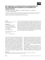
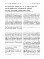
details), and produced three clusters (Fig. 1).
The first cluster (hereafter referred to as cluster A)
contains 69 immunoglobulin structures, the second
(cluster B) contains 31 immunoglobulin structures and
the third (cluster C) is formed by a single antibody
structure (PDB code:
1Q1J).
The interface of 1Q1J does not resemble any other
structure in our dataset. Its residues have a root-mean-
square deviation (RMSD) of about 1.4 A
˚
from the
residues contributing to the interface of a cluster A rep-
resentative structure (PDB code:
2ORB) and about
1.4 A
˚
from those of a cluster B representative structure
(PDB code:
2A6I).
A. Chailyan et al. Analysis of VH–VL interface in antibodies
FEBS Journal 278 (2011) 2858–2866 ª 2011 The Authors Journal compilation ª 2011 FEBS 2859
1Q1J is the structure of the human monoclonal anti-
body 447-52D complexed with a peptide derived from
the V3 region of the HIV-1 gp120 protein. Another
structure (PDB code:
3C2A) for the same antibody,
bound to a variant of the same peptide, is available
and has an interface essentially identical to that of
1Q1J. This is the only antibody in our set that uses
the heavy chain V gene IGHV3-15. Its uniqueness did
not allow us to analyze it further.
There is no strong correlation between the structural
clustering and the type of light chain. k and j chains
contribute to both clusters, and therefore the structural
difference in the interface cannot be attributed to the
type of light chain (Fig. 1).
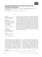
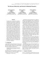
Cluster A is formed by immunoglobulins from both
mouse and human, whereas cluster B is only populated
by immunoglobulins from Mus musculus (28 immuno-
globulins) and by chimeric antibodies with a mouse
variable domain and a human constant domain (three
immunoglobulins) (Fig. 2). This implies, as discussed
later, that some packing modes observed in mouse
antibodies cannot be found in human antibodies, with
obvious implications for humanization experiments.
We observed a bias in the usage of light chain V
germline genes, whereas this was not the case for the
heavy chain V genes. There is no intersection between
the light chain germlines used in cluster A and those
used in cluster B. The latter set of germlines is
enriched in k-type light chains [IGLV1 (23 ⁄ 31)], even
though a number of j-type light chains [IGKV10-94
(2 ⁄ 31), IGKV10-96 (4 ⁄ 31), IGKV9-124 (1 ⁄ 31),
IGKV14-100 (1 ⁄ 31)] are found in the cluster. In cluster
A, the numbers of immunoglobulins of k and j type
are 21 and 48, respectively. In other words, there is a
mode of interaction between the two chains character-
istic of the immunoglobulins of cluster B, specific for a
subset of mouse immunoglobulins and never observed
in humans (Table S1).
Fig. 1. Results of the cluster analysis. Dendrogram based on the difference between the positions of residues at the interface in the light
and heavy chain variable domains. The red line indicates the clustering with the highest silhouette value (0.47). In the bottom panel, red,
green and blue indicate the A, B and C clusters, respectively. The type of light chain is shown in the bottom panel.
Fig. 2. Antibody source. Frequency of mouse, human, chimeric
and humanized antibodies in clusters A (red bars) and B (green
bars).
Analysis of VH–VL interface in antibodies A. Chailyan et al.
2860 FEBS Journal 278 (2011) 2858–2866 ª 2011 The Authors Journal compilation ª 2011 FEBS
Our next step involved the investigation of whether
the structural difference in the packing of the two
domains could be ascribed to the presence of specific
amino acids. To this end, we used the Random Forest
technique [32] (see also Materials and methods section)
to evaluate the relative ability of each residue to iden-
tify the structural cluster to which the immunoglobulin
belongs. The Gini index [33], a measure of the impor-
tance of the sequence positions, was used to select the
most significant. The eight sequence positions with the
largest Gini index, described and analyzed in detail
below, are able to discriminate between the two clus-
ters with a classification error lower than 10%. These
positions (listed here in order of their relevance) are
L44, L43, L41, L42, L8, L28, L66 and L36.
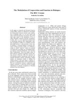
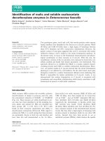
The sequence logo for all eight positions [34] (Fig. 3)
clearly shows that immunoglobulins belonging to dif-
ferent clusters have different preferences for specific
amino acids in these positions. It should be mentioned
that cluster B is formed by a large fraction (23 of 31)
of mouse immunoglobulins with a k chain from the
IGLV1 germline, and three of the positions highlighted
by the Random Forest analysis (L8, L28 and L66) are
completely conserved in all sequences of this type. Fur-
thermore, none of them is in contact with the heavy
chain. This strongly suggests that they discriminate this
particular type of k chain from all the others and are
not specific for the type of interface.
The remaining five positions (L41–L44 and L36) are
instead located at the interface between the two chains,
and the difference in the amino acids occupying them
is likely to be related to the packing of the domains.
In particular, position L44 is always occupied by a
proline in immunoglobulins belonging to cluster A,
whereas a medium ⁄ large hydrophobic amino acid is
preferred in the equivalent position in cluster B
(Table 1). Proline L44 in cluster A adopts a trans
conformation and interrupts the b-strand regularity
preserved in cluster B. This affects the type of turn
observed in the two clusters: the region L41–L43 forms
a tight turn (typically a 3 : 3 class hairpin confor-
mation) connecting the two proximal b-strands in
immunoglobulins belonging to cluster B. Conversely, a
7 : 7-type hairpin is present between residue L38 and
residue L44 in cluster A.
In all immunoglobulins, residue L44 interacts with
the amino acid at position L36, which is a large amino
acid in most of the members of cluster A, and usually
smaller, typically a valine, in those belonging to cluster
B (Table 1).
The side-chain of residue L36 packs against the last
insertion before residue H101 (which has a different
numbering according to the specific structure and is
called H100X here for clarity), which is, in most cases,
a phenylalanine or a methionine. A different frequency
of residues in position H100X is observed in clusters A
and B (Table 1).
The packing between residues L36 and H100X is dif-
ferent in the two clusters. We computed the distribu-
tion of the distances between the residue 36 Ca of the
light chain and that of residue 100X of the heavy
chain. In cluster A, the average is 9.79 A
˚
with a stan-
dard deviation of 1.36 A
˚
, whereas the corresponding
values for cluster B are 8.22 and 1.17 A
˚
, respectively.
The two distributions are statistically significantly dif-
ferent (P = 1.3 · 10
)6
).
The presence of a proline in position L44 is the best
predictor of the presence of a type A interface. We
computed the distance between the Ca of the residues
Fig. 3. Logo of discriminative positions. Sequence logos [34] for
the positions highlighted as discriminative for clusters A (left side)
and B (right side) by the Gini index analysis in the structure dataset.
The height of the letters is proportional to the frequency of the cor-
responding amino acid in the position indicated on the x axis. The
letters are colored according to the scheme used in Lesk [35].
Orange: small nonpolar G, A, S, T; green: hydrophobic C, V, I, L, P,
F, Y, M, W; magenta: polar N, Q, H; red: negatively charged D, E;
blue: positively charged K, R.
Table 1. Amino acid occurrence at positions L36, H100X and L44
in immunoglobulins belonging to clusters A and B.
Cluster A Cluster B
Position Amino acid: occurrences Amino acid: occurrences
L36 Y: 58
F: 8
L: 2
N: 1
V: 22
Y: 5
L: 2
F: 1
I: 1
H100X F: 28
M: 21
V: 5
S: 4
P: 4
G: 3
L: 3
I: 1
F: 14
M: 7
G: 5
L: 4
S: 1
L44 P: 69 F: 24
V: 5
I: 2
A. Chailyan et al. Analysis of VH–VL interface in antibodies
FEBS Journal 278 (2011) 2858–2866 ª 2011 The Authors Journal compilation ª 2011 FEBS 2861
contributing to the interface and the corresponding
residues of the centroid of clusters A (PDB code:
2ORB) and B (PDB code: 2A6I) for all the immuno-
globulins of known structure that were left in our ini-
tial nonredundant dataset (584 antibodies), and plotted
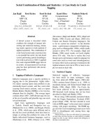
one against the other (Fig. 4). Almost all of the immu-
noglobulins that contain a proline in position L44 are
more similar to those of cluster A (515 ⁄ 533). A few
immunoglobulins have an interface that is different
from those observed in both clusters. Fourteen are
expected to adopt a type A interface because they have
a proline at position L44 (PDB codes:
1BGX, 1AY1 ,
1FL3, 3CFC, 3CFB, 1UB5, 1UB6, 1RUL, 1RU9 ,
1RUA, 3DGG, 1A0Q, 2D7T and 3GKW) but do not,
and only one (PDB code:
2GFB) does not have the
expected type B interface, although the proline in posi-
tion L44 is not present. In the first seven cases, the
structures are either not well resolved or have a high B
factor.
1RUL, 1RU9 and 1RUA are solved structures
of the same antibody after UV irradiation. The same
nonirradiated antibodies (PDB codes:
1NCW and
1ND0) display the normal interface and are properly
classified in cluster A. In
3DGG, a magnesium ion
coordinates several residues in the region L39–L46 dis-
torting the loop.
1A0Q is a catalytic antibody with
esterase activity that contains a ligand (S-norleucine
phenyl phosphonate) deeply buried in the binding site.
The last three cases (PDB codes:
2D7T, 3GKW and
2GFB) seem to be genuine outliers.
Two more structures of antibodies containing a pro-
line in position L44, (corresponding to entries
1PZ5
and
1N0X) are more similar to cluster B. However,
there are different determinations of their structures
with different ligands and in these cases the interface
packing follows the rules outlined here. In
1AE6, the
proline is present, but in a cis conformation, and the
region has a very high B factor. A high B factor is also
observed for the whole
2QSC molecule.
The next question we asked is whether the difference
in the packing geometry observed in the two clusters
has an impact on the conformation of the antigen-
binding site. We selected two pairs of residues on
opposite sides of the binding site (L55 and H57; L24
and H25, Fig. 5) and computed the distribution of the
distances between their Ca atoms in immunoglobulins
belonging to clusters A and B.
The average distance between L55 and H57 is
26.49 ± 0.98 A
˚
in cluster A and 24.82 ± 1.39 A
˚
in
cluster B. The corresponding values for L24 and H25
are 35.87 ± 0.65 A
˚
and 34.95 ± 0.58 A
˚
for clusters
A and B, respectively, corresponding to a difference
of about 10% in the area of the rhomboid defined by
the four Ca atoms. The two distributions are statisti-
cally significantly different (P = 1.9 · 10
)7
and P =
Fig. 4. Interface distance plot of antibodies not included in the
original dataset. Plot of the distance (1 – GDT_HA) between the Ca
of the 20 residues at the VH–VL interface of the immunoglobulins
not originally included in the nonredundant structure dataset and
the corresponding atoms of the centroids of clusters A and B. Red
dots indicate immunoglobulins in which position L44 is occupied by
a proline. Outliers are labeled and discussed in the text.
Fig. 5. Antigen-binding site dimensions. Positions of the residues
used to estimate the width of the antigen-binding site in the two
clusters. The Ca moieties of the selected residues (L55, H57, L24
and H25) are indicated by spheres. Broken lines indicate the mea-
sured distances. The structure shown is the PDB entry
2FL5.
Analysis of VH–VL interface in antibodies A. Chailyan et al.
2862 FEBS Journal 278 (2011) 2858–2866 ª 2011 The Authors Journal compilation ª 2011 FEBS
2.9 · 10
)3
for the first and second pair, respectively).
In some cases, the antibodies included in our dataset
were solved in a complex with their antigen (71 of 101
cases). To exclude the possibility that the presence of
the antigen is responsible for the observed differences
in the distance distributions, we recalculated them by
considering bound and unbound antibodies separately
(Table 2). The observed differences are still present
and still statistically significant. This implies that, on
average, the binding site of the type A immunoglobu-
lins is wider than that of the type B immunoglobulins.
In 71 cases in our dataset, the structure of the
immunoglobulin has been determined in a complex
with an antigen. We computed the volume of these
antigens and classified them into two groups as
described in the Materials and methods section. Clus-
ters A and B contain 46 and 25 immunoglobulins com-
plexed with an antigen, respectively. Among the 17
that are bound to a small antigen (volume < 505 A
˚
3
),
14 belong to cluster B and only three to cluster A.
Such a difference is statistically meaningful (P =
6.9 · 10
)6
; see Materials and methods section for
details). It is therefore evident that antibodies belong-
ing to cluster B generally bind smaller antigens,
whereas those in cluster A are more promiscuous. For
comparison, the p-nitrophenyl-phosphocholine mole-
cule (molecular formula: C
11
H
18
N
2
O
6
P; PDB code:
1DL7) is a simple hapten and has a volume of 451 A
˚
3
,
whereas the nine-residue rhodopsin epitope mimetic
peptide (sequence TGALQERSK; PDB code:
1XGY)
has a volume of 809 A
˚
3
. In practice, this threshold dis-
criminates small hapten-like antigens from peptide and
protein antigens.
In summary, the results of the analysis described
here clearly indicate that there are at least two differ-
ent packing modes for the association between the
light and heavy domains in immunoglobulins, and
these can be specifically associated with key residues in
their sequence.
Importantly, the two different packing modes have a
significant effect on the geometry of the binding site,
as illustrated by the statistically significantly different
distribution of distances between residues at the
periphery of the binding site, and we have shown that
these differences are related to the size of the recog-
nized antigen. Furthermore, visual analysis indicates
the presence of a narrow pocket in the middle of the
binding site in the majority of the immunoglobulins of
cluster B (Fig. 6).
Discussion
The results presented here are clearly relevant for anti-
body and antibody library design, but also for human-
ization experiments. The type B interface is only found
in the mouse, and therefore grafting the antigen-bind-
ing site of a type B murine antibody into a human
antibody will be ineffective if the recipient molecule
has a type A interface. One instructive example can be
found in the work by Worn et al. [37]. These authors
produced two single-chain Fv humanized intrabody
versions of a murine anti-GCN4 immunoglobulin
molecule (with a k chain) using, as recipient, two
human antibodies that differed in the type of light
chain (k in one case and j in the other) and in only
seven residues (including residues L36, L43 and L44).
The k-graft variant had an activity comparable with
the wild-type antibody, whereas the j-graft variant,
although extraordinarily stable in vitro, had a five order
of magnitude decreased antigen affinity, presumably,
Table 2. Average distances between residues L55–H57 and
between residues L24–H25 in all immunoglobulins belonging to
clusters A and B. The table also shows the values for bound (holo-
form) and unbound (apo-form) cases separately.
L55–H57
distance (A
˚
)
L24–H25
distance (A
˚
)
Total dataset (100) Cluster A (69) 26.49 ± 0.98 35.87 ± 0.65
Cluster B (31) 24.82 ± 1.39 34.95 ± 0.58
Holo-form (70) Cluster A (45) 26.51 ± 0.94 35.87 ± 0.57
Cluster B (25) 24.62 ± 1.34 34.96 ± 0.63
Apo-form (30) Cluster A (24) 26.45 ± 1.08 35.89 ± 0.8
Cluster B (6) 25.62 ± 1.45 34.95 ± 0.34
Fig. 6. Antigen-binding site of type B antibody. Molecular surface
of the antigen-binding site of the CHA255 antibody (PDB code:
1IND). The presence of a rather narrow pocket is clearly visible.
The surface is colored according to the atom depth (using the DPX
web server [36]); the ligand (indium chelate) is depicted in red using
a ball and stick representation.
A. Chailyan et al. Analysis of VH–VL interface in antibodies
FEBS Journal 278 (2011) 2858–2866 ª 2011 The Authors Journal compilation ª 2011 FEBS 2863
as the authors suggest, caused by differences in the
mutual orientation of the two domains.
Finally, we would like to mention that the ability of
type B antibodies to bind smaller antigens, and the
presence of the pocket described, might open up the
possibility of using them as potential drug delivery vec-
tors. Indeed, this has been proposed already in the
case of the
1IND antibody [38], a type B immunoglob-
ulin with an exceptionally high affinity binding for an
indium-chelate hapten.
The ability to use sequence data to predict the mode
of association of the variable domains of antibodies
also has implications for methods to predict their
structure. Indeed, the information obtained through
the analysis described here is being used to implement
a better prediction protocol in our immunoglobulin
structure prediction server [17].
Materials and methods
Throughout this article, we have used the Kabat–Chothia
numbering scheme [39] with the additional insertion at posi-
tion L68 proposed by Abhinandan and Martin [40]. The
letters L and H preceding a residue number indicate light
and heavy chain residues, respectively.
We constructed a dataset of immunoglobulins of known
structure containing both k and j chains. Starting from 120
structures with k-type light chains, downloaded from the
PDB database [30], version 21st February 2010, we
removed single-chain immunoglobulins (34), single-chain
variable fragments (5), redundant structures (i.e. structures
for which both the light and heavy chain variable regions,
if present, are identical in sequence) (26) and the ten struc-
tures with resolution worse (higher) than 3 A
˚
(using the
PISCES web server [41]). The final set contained 45 immu-
noglobulins of known structure with a k light chain. The
number of known structures of immunoglobulins with a
j-type light chain stored in PDB is much higher (930).
We removed all single-chain immunoglobulins and light chain
dimers, and subsequently only retained those with a resolu-
tion better than 3 A
˚
(using the PISCES web server [41]).
This resulted in a set of 640 structures with j light chains.
In order to obtain a balanced dataset for j and k light
chains, whilst, at the same time, preserving diversity among
the j light chains, we grouped together immunoglobulins
with j light chains with similar residues in positions con-
tributing to the interface. This was achieved using cd-hit
[42]. The residues used in clustering were defined according
to Chothia et al. [28]: L34, L36, L38, L43, L44, L46, L87,
L89, L98, L100, H35, H37, H39, H44, H45, H47, H91,
H93, H103 and H105. Using a similarity threshold of
80%, we obtained 93 clusters, 37 of which contained less
than three elements and were discarded to avoid the inclu-
sion of immunoglobulins with unusual interfaces in our
analysis. The immunoglobulins representing the centroid of
each of the remaining 56 clusters were added to the 45
selected k-type immunoglobulin structures to obtain the
final dataset.
The structural similarity of the residues contributing to
the interfaces and listed above was measured using lga
software [43] in sequence-dependent mode with a 10 A
˚
dis-
tance cut-off. The distances computed by lga were used to
calculate the global distance test–high accuracy (GDT_HA)
parameter:
GDT
HA ¼ (GDT P0.5 + GDT P1
+ GDT
P2 + GDT P4)/4
where GDT_Pn denotes the percentage of residues that can
be superimposed within a distance cut-off of n A
˚
or less.
The GDT_HA values were employed to cluster the struc-
tures using the R package ‘cluster’ routine (M. Maechler
et al., unpublished results) with both diana (divisive) and
hclust (agglomerative) methods. For agglomerative cluster-
ing, we used the ‘average’, ‘complete’, ‘ward’ and ‘single’
joining functions. For each clustering method, the optimal
number of clusters was identified with the silhouette valida-
tion technique [31], which provides an estimate of the clus-
ter tightness and separation, as implemented in the R
package. The highest silhouette value (0.47) was obtained
using the diana divisive clustering method with three clus-
ters, one of which was formed by only one structure that
was not included in the analysis (see Results section).
We used the automatic feature selection procedure already
described in ref. [15] to select the sequence positions that
have a significantly different residue distribution in anti-
bodies belonging to different clusters, i.e. specific for a given
type of interface. Each immunoglobulin was labeled accord-
ing to the cluster it belonged to, and the Gini Impurity Index
(as implemented in the Random Forest package [32,44]) was
computed for each light and heavy chain residue. This index
provides a relative ranking of the sequence positions on the
basis of their ability to correctly discriminate the structural
cluster to which an immunoglobulin belongs. The eight
sequence positions with the highest Gini index are able to
discriminate between the clusters with a classification error
lower than 10%, and were manually analyzed.
In order to verify whether the difference in the packing
geometry of immunoglobulins in the two clusters is
reflected in a different geometry of their binding site, we
measured the distances between the Ca of residues L55 and
H57 and of residues L24 and H25 (which are the furthest
structurally conserved residues in the antigen-binding site)
and between the Ca of residue 36 of the light chain and of
the last insertion before residue 101 of the heavy chain (this
residue has a different Kabat–Chothia number according to
the length of the H3 loop, and is called H100X here) for
each immunoglobulin in our dataset. We used Pearson’s
chi-squared test (as implemented in the R package) to
Analysis of VH–VL interface in antibodies A. Chailyan et al.
2864 FEBS Journal 278 (2011) 2858–2866 ª 2011 The Authors Journal compilation ª 2011 FEBS
verify whether they were statistically significantly different
in immunoglobulins belonging to the two clusters.
We measured the volumes of the antigens bound to the
immunoglobulin structures of our dataset, where present,
using the Voronoi procedure, as implemented in the calc-
volume program [45], with default parameters, and classified
them into two groups according to whether their volume was
smaller or larger than 505 A
˚
3
. This value corresponds to the
first quartile of the antigen size distribution in our dataset.
We calculated the P value for the hypothesis that immuno-
globulins in a given cluster bind to smaller antigens by means
of the hypergeometric cumulative distribution function,
which measures the probability of finding at least as many
antibodies binding to a small antigen in a cluster of similar
size randomly extracted from the whole set of antibodies.
Acknowledgements
This work was partially supported by Award No.
KUK-I1-012-43 made by the King Abdullah Univer-
sity of Science and Technology (KAUST), by Fondazi-
one Roma and by the Italian Ministry of Health,
contract no. onc_ord 25 ⁄ 07, FIRB ITALBIONET and
PROTEOMICA.
References
1 Hamers-Casterman C, Atarhouch T, Muyldermans S,
Robinson G, Hamers C, Songa EB, Bendahman N &
Hamers R (1993) Naturally occurring antibodies devoid
of light chains. Nature 363, 446–448.
2 Greenberg AS, Avila D, Hughes M, Hughes A,
McKinney EC & Flajnik MF (1995) A new antigen
receptor gene family that undergoes rearrangement and
extensive somatic diversification in sharks. Nature 374,
168–173.
3 Rast JP, Amemiya CT, Litman RT, Strong SJ & Lit-
man GW (1998) Distinct patterns of IgH structure and
organization in a divergent lineage of chrondrichthyan
fishes. Immunogenetics 47, 234–245.
4 Wu TT & Kabat EA (1970) An analysis of the
sequences of the variable regions of Bence Jones pro-
teins and myeloma light chains and their implications
for antibody complementarity. J Exp Med 132, 211–
250.
5 Chothia C, Lesk AM, Tramontano A, Levitt M, Smith-
Gill SJ, Air G, Sheriff S, Padlan EA, Davies D, Tulip
WR et al. (1989) Conformations of immunoglobulin
hypervariable regions. Nature 342, 877–883.
6 Padiolleau-Lefevre S, Alexandrenne C, Dkhissi F,
Clement G, Essono S, Blache C, Couraud JY, Wijkhu-
isen A & Boquet D (2007) Expression and detection
strategies for an scFv fragment retaining the same high
affinity than Fab and whole antibody: implications for
therapeutic use in prion diseases. Mol Immunol 44,
1888–1896.
7 Krauss J, Forster HH, Uchanska-Ziegler B & Ziegler A
(2003) Chimerization of a monoclonal antibody for
treating Hodgkin’s lymphoma. Methods Mol Biol 207,
63–79.
8 Verhoeyen M & Riechmann L (1988) Engineering of
antibodies. Bioessays 8, 74–78.
9 Riechmann L, Clark M, Waldmann H & Winter G
(1988) Reshaping human antibodies for therapy. Nature
332, 323–327.
10 Hwang WYK, Almagro JC, Buss TN, Tan P & Foote J
(2005) Use of human germline genes in a CDR homol-
ogy-based approach to antibody humanization. Meth-
ods 36, 35–42.
11 Tan P, Mitchell DA, Buss TN, Holmes MA, Anasetti C
& Foote J (2002) ‘Superhumanized’ antibodies: reduc-
tion of immunogenic potential by complementarity-
determining region grafting with human germline
sequences: application to an anti-CD28. J Immunol 169,
1119–1125.
12 Delagrave S, Catalan J, Sweet C, Drabik G, Henry A,
Rees A, Monath TP & Guirakhoo F (1999) Effects of
humanization by variable domain resurfacing on the
antiviral activity of a single-chain antibody against
respiratory syncytial virus. Protein Eng 12, 357–362.
13 Lazar GA, Desjarlais JR, Jacinto J, Karki S & Ham-
mond PW (2007) A molecular immunology approach to
antibody humanization and functional optimization.
Mol Immunol 44, 1986–1998.
14 Al-Lazikani B, Lesk AM & Chothia C (1997) Standard
conformations for the canonical structures of immuno-
globulins. J Mol Biol 273, 927–948.
15 Chailyan A, Marcatili P, Cirillo D & Tramontano A
(2011) Structural repertoire of immunoglobulin lambda
light chains. Proteins 79, 1513–1524.
16 Tramontano A, Chothia C & Lesk AM (1990) Frame-
work residue 71 is a major determinant of the position
and conformation of the second hypervariable region in
the VH domains of immunoglobulins. J Mol Biol 215,
175–182.
17 Marcatili P, Rosi A & Tramontano A (2008) PIGS:
automatic prediction of antibody structures. Bioinfor-
matics 24, 1953–1954.
18 Davies DR & Metzger H (1983) Structural basis of
antibody function. Annu Rev Immunol 1, 87–117.
19 Mariuzza RA, Phillips SE & Poljak RJ (1987) The
structural basis of antigen–antibody recognition. Annu
Rev Biophys Biophys Chem 16, 139–159.
20 Novotny J, Bruccoleri R, Newell J, Murphy D, Haber
E & Karplus M (1983) Molecular anatomy of the anti-
body binding site. J Biol Chem 258, 14433–14437.
21 Narayanan A, Sellers BD & Jacobson MP (2009)
Energy-based analysis and prediction of the orientation
A. Chailyan et al. Analysis of VH–VL interface in antibodies
FEBS Journal 278 (2011) 2858–2866 ª 2011 The Authors Journal compilation ª 2011 FEBS 2865
between light- and heavy-chain antibody variable
domains. J Mol Biol 388, 941–953.
22 Banfield MJ, King DJ, Mountain A & Brady RL (1997)
V-L:V-H domain rotations in engineered antibodies:
crystal structures of the Fab fragments from two mur-
ine antitumor antibodies and their engineered human
constructs. Proteins Struct Funct Bioinformatics 29,
161–171.
23 Nakanishi T, Tsumoto K, Yokota A, Kondo H &
Kumagai I (2008) Critical contribution of VH–VL inter-
action to reshaping of an antibody: the case of human-
ization of anti-lysozyme antibody, HyHEL-10. Protein
Sci 17, 261–270.
24 Stanfield RL, Takimoto-Kamimura M, Rini JM, Profy
AT & Wilson IA (1993) Major antigen-induced domain
rearrangements in an antibody. Structure 1, 83–93.
25 Tan PH, Sandmaier BM & Stayton PS (1998) Contribu-
tions of a highly conserved VH ⁄ VL hydrogen bonding
interaction to scFv folding stability and refolding effi-
ciency. Biophys J 75, 1473–1482.
26 Chothia C, Novotny J, Bruccoleri R & Karplus M
(1985) Domain association in immunoglobulin mole-
cules. The packing of variable domains. J Mol Biol 186,
651–663.
27 Abhinandan KR & Martin AC (2010) Analysis and pre-
diction of VH ⁄ VL packing in antibodies. Protein Eng
Des Sel 23, 689–697.
28 Chothia C, Gelfand I & Kister A (1998) Structural
determinants in the sequences of immunoglobulin vari-
able domain. J Mol Biol 278, 457–479.
29 Vargas-Madrazo E & Paz-Garcia E (2003) An improved
model of association for VH–VL immunoglobulin
domains: asymmetries between VH and VL in the pack-
ing of some interface residues. J Mol Recognit 16, 113–
120.
30 Dutta S, Burkhardt K, Young J, Swaminathan GJ,
Matsuura T, Henrick K, Nakamura H & Berman HM
(2009) Data deposition and annotation at the World-
wide Protein Data Bank. Mol Biotechnol 42, 1–13.
31 Rousseeuw PJ (1987) Silhouettes – a graphical aid to
the interpretation and validation of cluster-analysis.
J Comput Appl Math 20, 53–65.
32 Breiman L (2001) Random forests. Mach Learn 45,
5–32.
33 Archer KJ & Kimes RV (2008) Empirical characteriza-
tion of random forest variable importance measures.
Comp Stat Data Anal 52, 2249–2260.
34 Crooks GE, Hon G, Chandonia JM & Brenner SE
(2004) WebLogo: a sequence logo generator. Genome
Res 14, 1188–1190.
35 Lesk AM (2002) Introduction to Bioinformatics. Oxford
University Press, Oxford, New York.
36 Pintar A, Carugo O & Pongor S (2003) DPX: for the
analysis of the protein core. Bioinformatics 19, 313–314.
37 Worn A, der Maur AA, Escher D, Honegger A,
Barberis A & Pluckthun A (2000) Correlation between
in vitro stability and in vivo performance of anti-GCN4
intrabodies as cytoplasmic inhibitors. J Biol Chem 275,
2795–2803.
38 Love RA, Villafranca JE, Aust RM, Nakamura KK,
Jue RA, Major JG, Radhakrishnan R & Butler WF
(1993) How the anti-(metal chelate) antibody Cha255 is
specific for the metal-ion of its antigen – X-ray struc-
tures for 2 Fab’ hapten complexes with different metals
in the chelate. Biochemistry
32, 10950–10959.
39 Chothia C & Lesk AM (1987) Canonical structures for
the hypervariable regions of immunoglobulins. J Mol
Biol 196, 901–917.
40 Abhinandan KR & Martin AC (2008) Analysis and
improvements to Kabat and structurally correct num-
bering of antibody variable domains. Mol Immunol 45,
3832–3839.
41 Wang G & Dunbrack RL Jr (2003) PISCES: a protein
sequence culling server. Bioinformatics 19, 1589–1591.
42 Li W & Godzik A (2006) Cd-hit: a fast program for
clustering and comparing large sets of protein or nucle-
otide sequences. Bioinformatics 22, 1658–1659.
43 Zemla A (2003) LGA: a method for finding 3D similari-
ties in protein structures. Nucleic Acids Res 31, 3370–
3374.
44 Liaw A & Wiener M (2002) Classification and regres-
sion by Random Forest. R News 2, 18–22.
45 Voss NR & Gerstein M (2005) Calculation of standard
atomic volumes for RNA and comparison with pro-
teins: RNA is packed more tightly. J Mol Biol 346,
477–492.
Supporting information
The following supplementary material is available:
Table S1. Antibody germline usage. Usage of IGLV ⁄
IGKV germline genes in immunoglobulins belonging
to clusters A and B.
This supplementary material can be found in the
online version of this article.
Please note: As a service to our authors and readers,
this journal provides supporting information supplied
by the authors. Such materials are peer-reviewed and
may be re-organized for online delivery, but are not
copy-edited or typeset. Technical support issues arising
from supporting information (other than missing files)
should be addressed to the authors.
Analysis of VH–VL interface in antibodies A. Chailyan et al.
2866 FEBS Journal 278 (2011) 2858–2866 ª 2011 The Authors Journal compilation ª 2011 FEBS